Slide Review and You

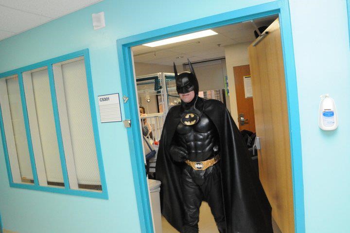
Welcome back everybody!

Last month, I wrote about some projects I did while rotating through the pathology program at Danbury Hospital in Connecticut. This month I’m in a more clinical setting with a hematology/oncology clerkship at Northwell’s Staten Island University Hospital. But, over the past few months of rotations (and arguably a lot longer before medical school) I’ve been noticing a part of laboratory medicine which often intersects with our clinical colleagues at the bedside. I’ve told you about the pitfalls and successes in the relationships between surgeons and anatomic pathologists before, where frozen sections are critical and time is of the essence. And we’ve all seen collaboration between the bench and bedside before—think microbiology and infectious disease, blood bank and literally everyone, etc. Still, one collaborative effort sort of happens behind the shadows, behind phone calls and lab reports, and sometimes with no communication at all! So, what kind of vigilante medicine am I talking about? Who is this Batman of medicine? It’s just our friends in hematology.

When you’re working the hematology bench in the lab, it’s pretty commonplace for a physician on a hematology service to call and ask for a peripheral smear to review. Many times, it’s for the purpose of teaching residents, fellows, or medical students but more often than not it’s a confirmatory exercise. See, when that hematologist asks to review a slide, she’s probably coming down to the lab to look at the morphology of red cells and white cells to help in their differential diagnosis. They might have a patient with a suspected thalassemia or hemoglobinopathy and, before starting the full work up of lab tests, just want to see if there are any RBC morphology traits or target cells that stand out. Thrombocytopenia? Let’s make sure there’s no platelet clumping. Maybe they’ve got a patient with some kind of liver or kidney pathology and are on the hunt for acantho- or echinocytes. Or better yet, someone went hiking, there’s an infectious etiology on their differential—let’s go hunting for babesia, malaria, oh or even erlichia!

Image 1. Here’s a few examples of three parts of a patient’s smear that are contributory to a particular pathology in vivo. Think you know what it is? I bet you’d be surprised…not all that hyper-segments is a B12/Folate deficiency. But technically it is; read about cobalamin and homocysteine pathology in a neonatal patient here: http://www.bloodjournal.org/content/128/21/2584 (Source: Blood 2016)

I know what you’re thinking. Wait—that’s our job as medical laboratory scientists; our literal job. Our instruments, that we validate, and correlate, and make sure work fantastically give us flags. We investigate those flags and look at smears ourselves! We collaborate with other lab techs, and with our pathologist colleagues and send out final lab results with all kinds of helpful information: including platelet clumping, microorganisms, RBC and WBC morphology, and loads more. What gives?

Hold on to your lab coats. I’ll get there in a minute.

Slide review and differential training in medical school and residency

[This section intentionally left blank]

Image 2. There is nothing wrong with your television set. Do not attempt to adjust the picture. You are about to experience the awe and mystery which reaches from the inner mind to… the bench tech working in hematology. The one who went to school for this? Medical school and residency are starkly devoid of any in-depth, comprehensive learning for differentials.

A Differential, Differential

So let me address the issue I brought up: why do hematologists come down to the lab to look at the slides themselves, when perfectly capable BOC certified, degree-holding medical laboratory scientists and pathologists sign out validated differentials? It might not happen this way at all hospitals, but I think the answer is a simple two-part problem.

First, as with the many things I’ve learned in medical school, one of the lab-centric pieces of information that is well understood is that, well, no one really knows what the lab does and how it operates. Virtually nobody knows the depth and breadth of the testing that pathologists manage, let alone the scientific precision and accuracy that instrument validation requires. Learning that MLS techs are certified, can hold graduate degrees, and even do their own research is often surprising to most of our clinical colleagues. And—I will tell you for a fact—that pathology and laboratory testing methodology is not covered in medical school the way you might think. Pathology is more of a class of distinguishing the identifying details of a disease, not understanding the interdisciplinary diagnostic teamwork that goes into those CBC index results on a computer screen on the clinical floors.

Second, hematologists are specialists just like any other practicing clinician. They know their stuff! They manage patient diagnosis, treatment, and follow-up with the most up to date literature, national cancer guidelines, and anything else available to better their patients’ outcomes. Despite the notes in the CBC results that there are numerous macrocytes with hypersegmented neutrophils, or 3+ schistocytes reported in a manual differential—seeing is believing. It helps to see the slide yourself and get a feel for the disease “state” with your own eyes. Moreso, it could be a learning opportunity. It’s well within a clinicians’ scope to come down and look at a peripheral smear, I actually encourage it. But it should come with a few caveats…I’ll get to those too…

I-CARE

One of the places I was proud to hang my lab coat was actually my first job as an assistant lab technician in the blood bank at Rush University Medical Center in Chicago. Before I got my MLS and way before grad school or med school, I was a blood bank “expediter.” Super fancy title, but all I did was make sure specimens were logged in and blood products were up to par with labels on their way out. Clerical but critical! (Let me have this, please…haha) Anyway, part of the culture at that hospital has stayed with me all these years. I’ve talked before about culture and the way it permeates an institution’s practice like at the Mayo Clinic, but for my first foray into clinical work their acronym was clutch: I CARE.

  • I for innovation
  • C for collaboration
  • A for accountability
  • R for respect and
  • E for excellence

Why am I telling you this? No, there are no royalties. I just think it’s an easy way to remind ourselves about the meaning of interdisciplinary medicine and they way we should work together across specialties, and from bench to bedside. When we incorporate those values into our work for the purpose of improving patient care and outcomes, everyone wins. In this case, effective utilization of resources tells us that peripheral slide review means different things to different people. In the setting of hematologic work-ups, flags and review at the bench can signal something to the clinician which could spark a conversation with the pathologist. All parts contributing to a whole of patient care. Vigilante medicine is bad news. Collaboration is key.

One place I was lucky enough to be a part of this interdisciplinary collaboration was Swedish Covenant Hospital. One of the hematology physicians would routinely call me and ask to look as peripheral smears down in the lab, often as a group with med students, residents, and fellows. I’d throw the image of his patients’ slides on a large flat screen and go over what certain traits meant with regard to morphology and identification from the lab setting. Dr. Cilley would add what this all meant clinically and discuss treatment algorithms and next steps. That was collaboration at it’s finest: lab tech working with pathologists, clinicians working with the lab, and patient’s benefiting from all of it.

Video 1. ASCP’s 2015 Membership Video. I was super thrilled to be part of this video back in 2015 after winning the Midwest regional ASCP member of the year. If you’re bored enough to make it about 40 seconds into the video, that was my actual desk where Dr. Cilley and his residents would come to discuss patient slides. I would talk to them about morphology and hematologic clues with digital hardware and software to make it clear in group settings, rather than taking turns at the scope. Good times. (Source: https://www.youtube.com/watch?v=86fBRXGrZFo)
Video 2. Dr. Jeffrey Cilley talks about treating cancer as a “team approach” and he’s right. Hematology/Oncology to patient. Lab to clinician. Bench to bedside. (Source: https://www.youtube.com/watch?v=q0waKLyT1Dg)

Teamwork makes the dream work

About those caveats for collaboration I mentioned earlier… Let me put it briefly: it’s well within the scope of a clinician to come over to the laboratory and get some information on their patient’s lab results/testing. But why not consider the following:

  • If a physician calls to review a smear, offer to go over it with them. Likewise, to our clinical friends: if you go to the lab for a slide don’t be batman—ask the tech what they think!

Experienced techs are one of the hospital’s most valuable resources. Some folks I’ve worked with have been looking at slides longer than I’ve been using my eyes at all! They’ll save you and your residents the time when those terrifying intracellular microorganisms are really just overlying platelets. I mean, they’ve got a cute halo.

  • If you need help, just ask. This applies to everyone.

Talking with the tech about the slide is great start, but there’s more resources in the lab than most people know what to do with! Clinical physicians: check the shelves around the hematology microscope. Stuck on something? Find a CAP atlas or a proficiency survey booklet guide. Easy to read. Techs and pathologists: have someone who constantly comes down for slide review despite your immaculate and detailed SOPs on CBC results reporting? Have a quick chat about the work that goes into resulting those diffs—you might even improve your heme TAT, who knows?

  • If it’s well within the right of a physician to leave the unit and see a patient’s slide, logic says that maybe, just maybe, it should be okay for a pathologist to leave the lab and see a patient at the bedside!

Hospitals are full of never-ending rounding white coats, all asking patients questions, and all contributing specialty notes to their charts. But its not only to prevent patients from getting a decent nap. We’re all parts of a large interdisciplinary patient team. A recent Medscape survey found that somewhere around 3% of pathologists see patients, routinely! Got an interesting case in the lab, someone who’s part of lots of tumor boards, someone with an interesting case to write up, or even someone who nobody knows exactly what’s going on with? Try walking over to 4 south and have a conversation with Mr. Jones; it might help. At least he’ll know how many people are working on his care team!

The bottom line: we’re in this together, and like the flag on the ASCP ship says, we’re Stronger Together. Innovation, collaboration, accountability, respect, and excellence are—and should be—simple cornerstones of clinical medicine that translate across every discipline. When we share information and expertise, everyone gets better at what they do.

Bonus Image. This was a hard picture to take. Usually, a quick hematologist just comes down to see if there are any real schistocytes. But, after reading a draft of this post, BatDoc’s cool with chatting about red cell indices and automated flow cytometry methods in auto-diff validation. That’s the hero we deserve, and the one healthcare needs! (Source: https://gunaxin.com/batman-doesnt-police-stop-visiting-children-hospital)

Thanks for reading!

See you next time!

–Constantine E. Kanakis MSc, MLS (ASCP)CM graduated from Loyola University Chicago with a BS in Molecular Biology and Bioethics and then Rush University with an MS in Medical Laboratory Science. He is currently a medical student actively involved in public health and laboratory medicine, conducting clinicals at Bronx-Care Hospital Center in New York City.

Tips for Performing Internal Lab Audits

In previous blog posts we discussed some hints and tips for how to survive when your lab is being inspected. Today we get to flip things around and let you be the inspector. Whether it’s an internal audit of your own laboratory, or an external inspection of a peer laboratory, we’ll discuss some ways to help keep you on track to cover the most important aspects of the overall testing process in a limited amount of time.

For external audit preparation, the CAP has a wonderful training program that all volunteer inspectors are required to take prior to participating in an inspection. For labs that are not CAP accredited, they still have helpful information on their website that is free and open to all: https://www.cap.org/laboratory-improvement/accreditation/inspector-training. CLSI document QMS15-A (Assessments: Laboratory Internal Audit Program; Approved Guideline) is another great resource to use when planning your audit.

The primary role of an auditor is to review policies, processes, and procedures to identify any inconsistencies (does your SOP match the manufacturer recommendations, and is staff following the SOP as written). Audits should focus on collecting objective evidence and facts, rather than subjective opinions. For example, staff failing to document required weekly maintenance tasks, as opposed to an auditor simply not liking the particular form the tasks are being documented on.

Define the Objective of the Audit

Laboratory leadership should be involved in the planning process to help define the scope and expected goal of performing the audit. This can range from an overall assessment of general laboratory quality and safety, to a more directed and focused audit on either a single department, instrument/test, or test process (specimen collection, physician notification of critical values, etc). The format for the audit findings should also be discussed – will the site require a formal, written report outlining all observations detected, or will a simple informal summation discussion be sufficient?

Draft a Schedule for the Audit

Once the scope of the audit is defined, a tentative schedule should be created so all staff involved in the audit process are aware and available to participate. If the audit will encompass multiple departments and all phases of testing (pre-analytic, analytic, post-analytic), it may be necessary to split the audit up over multiple days, or to recruit multiple auditors. The frequency of audits will depend on the perceived risk to quality based on previous findings or complaints received, but at a minimum should be completed annually.

Prepare for the Audit

Reach out to the local management team of the site being audited for help in gathering the information you’ll need to prepare. This can include things such as a testing activity menu, list of new instrumentation or new test validation studies, employee roster if personnel and competency records will be reviewed, and copies of previous audit/inspection results to check for corrective action implementation and sustainability. Review the information provided, and use it as a guide for where you feel your efforts should be focused on based on highest risk.

Utilize a Patient Tracer

Ask the site to pull all related records and reports for a particular patient sample by choosing a date, and specifying any particular characteristics for the specimen that you want to follow (such as age of the patient, sex, or focusing on abnormal/critical results). By asking the sites to prepare a patient tracer ahead of time, this will reduce the amount of time spent waiting and digging for specific files or log sheets as they are already organized and ready when you walk in for the audit. Tracers should adhere to the defined scope/objective of the audit, and will help you follow the path of a specimen through the entire process from pre-analytical, analytical, and finally post-analytical phases.

Pre-analytical: Include any specimen collection instructions or a printout/photocopy from the test directory for each test requested. This information should be compared to the information within the applicable SOPs to ensure they match and are both current and accurate. Physician orders can be included to confirm that the correct test was ordered and performed based on what was requested by the clinician.

Analytical: Copies of the related SOPs for the test being reviewed should be included. Ensure the SOPs have all required elements, including a current, valid signature of approval from the medical director. Instrument QC and maintenance logs for the day of testing, calibration records, and patient correlation studies should also be reviewed, along with the reagent lot-lot validation performed. When available, copies of the actual instrument printouts should be included to check for accuracy in result transcription. Training and competency records for the staff who performed any handling or testing of the specimens in question may also be reviewed.

Post-analytical: Check for supervisory review of patient log sheets and QC records, along with appropriate corrective actions documented as applicable. Review the patient results in the same format that is seen by the physician: confirm reference ranges and units of measure are accurate, interpretive notes are valid and appropriate, test methodology is stated when applicable, abnormal values are flagged, and confirm result transcription accuracy from the original instrument printout. Proficiency testing results should be reviewed for any unsuccessful events to confirm sustainability of corrective actions.

Conduct the Audit

Perform an objective review of the documents provided, along with any affiliated records and logs based on the scope of the audit (temperature logs, reagent inventory records, decontamination records, etc). As with an official inspection, be transparent with the staff as issues are identified so they can have an opportunity to clarify any confusion, or locate additional records that may be missing or incomplete. Document any discrepancies or possible issues noted, as well as any good lab practices observed that should be celebrated. When logging your findings, be specific and provide as much details as possible so the staff can quickly identify what was found and make the needed corrections (SOP numbers, dates, instrument serial numbers, etc).

In addition to reviewing documentation, perform a direct observation of the staff doing specific tasks. Are they following the steps outlined in their procedures, or are deviations noted? Rather than a formal interview, ask the staff to explain what they are doing, or why they are performing certain steps in a particular order. Again, the audit is not meant to be punitive or to ‘catch someone in the act’, but rather to help identify areas for improvement or clarification so that testing processes can be improved and standardized among all staff members. Asking open ended questions will provide more information than directed ones. For example, “Show me how you would access testing instructions if your computer network was down” as opposed to “Where are the paper versions of your SOPs?”

Prepare an Audit Report

The audit findings should be summarized for the site based on the format agreed upon during the initial planning stage (written report, verbal discussion). Whenever possible, similar findings should be grouped together so the location can identify systemic problems that need to be addressed on a more global level (expired reagents found in multiple departments, staff failing to utilize appropriate PPE in multiple departments, etc). Depending on the number and severity of the issues identified, sites may prefer to have the observations grouped by department as well for easy assignment of follow-up action items to the department leaders. Issues should also be ranked by risk severity so that the site knows where to focus their improvement efforts first: 1) Patient care and employee safety issues; 2) Regulatory compliance gaps; 3) Recommendations for improved overall good laboratory practice.

Implement Corrective Actions

Any issues identified during the audit should be assigned to a specific person for follow-up, along with an anticipated date of completion. Perform a proper root cause analysis to identify why the issue happened, and then decide how to correct it and prevent it from happening again. Depending on the scope of the audit, the audit team members may be involved with these tasks, or this may fall to the sole responsibility of the management team being inspected.

Evaluate the Effectiveness of the Audit

The utility of the audits will depend greatly on the commitment of laboratory leadership to both implement, and sustain, effective corrective actions based on the quality gaps identified. This can be assessed by the overall level of compliance with the regulations being checked, and comparing the results of this audit to previous and subsequent ones to hopefully show a downward trend in potential citations detected. The audit team should obtain feedback on the audit process to assess the inspected lab’s overall satisfaction with the program, the amount of support offered to the inspected laboratory, effectiveness of communication between the teams, and any potential areas for improvement in the process.

Performing internal audits is a great way to meet regulatory, accreditation, and customer requirements. It allows you an opportunity to identify non-conformances and risks that can affect both quality, and patient/employee safety. By performing regularly scheduled internal audits, not only will staff members become more experienced and better prepared for the official external inspections from regulatory and accrediting agencies, but the laboratory will move from a culture of reactive, corrective actions to that of a proactive model of continual improvements.


-Kyle Nevins, MS, MLS(ASCP)CM is one of ASCP’s 2018 Top 5 in the 40 Under Forty recognition program. She has worked in the medical laboratory profession for over 18 years. In her current position, she transitions between performing laboratory audits across the entire Northwell Health System on Long Island, NY, consulting for at-risk laboratories outside of Northwell Health, bringing laboratories up to regulatory standards, and acting as supervisor and mentor in labs with management gaps.

Surgical Pathology Case Study: A 3 Year Old Male with a Suspicious Lesion on Imaging Following an Injury

Case History

The patient is a 3 year old male with no significant past medical history who presented to the ED with left lower extremity pain for 24 hours after falling while playing with family members. The patient’s mother was present at bedside providing the history, but was not present at the time of the fall. It is unclear how the patient injured his ankle, but family members noticed the child grabbing his ankle and suspected that he may have twisted it. After the fall, the patient was unable/unwilling to ambulate on the ankle. There is no history of fractures or cancer.

An x-ray and subsequent MRI were ordered of the ankle which demonstrated an expansile lytic lesion involving the metaphysis of the distal tibia measuring approximately 3.4 x 2.2 cm (Figure 1 and 2). The margins of this lesion are indistinct, and there is cortical irregularity at the anterior and lateral aspect of the distal metaphysis of the tibia, likely representing a pathologic fracture. The differential diagnosis includes infection, aneurysmal bone cyst, nonossifying fibroma, osteoblastoma and histiocytosis.

The patient and family then followed up with Orthopedics, who proceeded to perform a biopsy of the lytic lesion in order to determine the nature of the lesion. The results are below.

Figure 1. Xray of the distal tibia demonstrating the lesion.
Figure 2. MRI demonstrating the lytic lesion involving the metaphysis of the distal tibia.

Diagnosis

Received fresh for intraoperative consultation is a 1.1 x 0.6 x 0.5 cm aggregate of white-tan soft tissue fragments. Half of the tissue fragments are frozen and read out as “spindle cell proliferation. Consideration of low-grade vasoformative lesion. Defer to permanent,” with 3 pathologists consulting on the diagnosis. The remainder of the tissue not submitted for frozen section, as well as the entirety of a second container from the same lesion, is submitted for routine processing.

On microscopy, the biopsies demonstrate a moderately cellular proliferation of fasciculated spindle cells in a collagenous to myxoid stroma. Nuclei are predominantly oval with variably fine to granular chromatin. Many cells have moderate amounts of tapering eosinophilic cytoplasm, resembling strap cells. Inflammatory cells and osteoclast-like giant cells are admixed (Figure 3 and 4). Immunohistochemical stains demonstrate lesional spindle cells to be positive for CD31, ERG, and FLI1. AE1/AE3 and CAM5.2 highlight rare lesional spindle cells. SMA stains some stellate spindle cells, favored to represent associated myofibroblasts. Desmin, MDM2, CDK4, ALK, and S100 are negative in plump lesional cells (Figure 5 and 6). Overall, the features are consistent with pseudomyogenic hemangioendothelioma, a rare vascular tumor. Although more commonly present in soft tissue, primary bone cases have been reported. These neoplasms have some risk for local recurrence, but only rarely distant metastasis. A portion of tissue was sent to the University of Nebraska Medical Center to evaluate for a characteristic gene rearrangement (SERPINE1-FOSB) that is present in at least a subset of pseudomyogenic hemangioendotheliomas. This was negative.

The lesion was then curettaged by the surgical team.The patient and his family had two follow up office visits with the Orthopedics department. The first one, a week after surgery, was unremarkable. The second visit, two weeks after surgery, was notable for the patient developing a cutaneous rash on both arms and chest. Due to literature citing that these tumors generally arise in the soft tissue, the clinician suggested that the patient and family follow up with pediatric dermatology to ensure that this new rash is not related to the pseudomyogenic hemangioendothelioma. Unfortunately due to insurance, the patient and family had to see a dermatologist at a different institution, and no further visits have taken place.

Figure 3. Photomicrograph of the strap-like cells with tapering eosinophilic cytoplasm , and osteoclast-like giant cells.
Figure 4. Higher power photomicrograph demonstrating the appearance of the strap-like cells with tapering eosinophilic cytoplasmFigure 4.

Discussion

Pseudomyogenic hemangioendothelioma (PHE) is a rare vascular tumor that most commonly arises in the skin and soft tissues of the extremities. It is usually multifocal, appearing in multiple tissue planes, such as the mucosa, dermis, subcutis and skeletal muscle, in a variety of different anatomic sites. Although even less common, PHE can also involve bone (such as this case). PHE has a male predilection, typically appearing in the second to fourth decades of life. Of the most common symptoms that the patient presents with, pain appears to top the list, although it should be stated that only about half of the patients experience pain.

Grossly, skin and soft tissue PHE tumors appear firm, ill-defined and gray-white. When they involve bone, they appear as multiple discrete, pink-tan to dark brown hemorrhagic tumors with surrounding sclerosis, ranging from 0.1 to 6.5 cm in greatest dimension.

Histologically, PHE demonstrates plump spindle and rhabdomyoblast-like cells with densely eosinophilic cytoplasm that grows in sheets and fascicles. The cells can be mistaken as rhabdomyoblasts because of the eosinophilic cytoplasm that pushes the nucleus to the periphery of the cell. Immunohistochemical studies are very helpful in order to determine a diagnosis of PHE. AE1/AE3, ERG, FLI-1 and CD31 are positive, whereas CD34, desmin and S100 are negative. Karyotyping has revealed a fusion of genes SERPINE1-FOSB that corresponds to the recurrent translocation t(7;19)(q22;q13). In this case, the SERPINE1-FOSBgene rearrangement was negative, but could possibly be due to a variant fusion gene.

Making a histologic diagnosis can be difficult for a Pathologist, due to the wide variety of differential diagnoses that will need to be excluded first.

The differential diagnosis for a cutaneous tumor includes:

  • Cellular benign fibrous histiocytoma (lacks rhabdomyoblast-like cells and neutrophilic infiltrates, contains mitotic figures, and is negative for cytokeratin and CD31)
  • Spindle cell squamous cell carcinoma (usually in sun-damaged skin, with nuclear atypia and negative endothelial markers)
  • Epithelioid sarcoma (negative INI1, positive EMA and CD34, and a nodular architecture with central necrosis and more nuclear atypia)

The differential diagnosis for soft tissue tumors include:

  • Epithelioid sarcoma (see above)
  • Epithelioid hemangioendothelioma (usually intracytoplasmic vacuoles, positive CD34 and CAMTA1, and a t(1;3)(p36.3;q25) translocation resulting in WWTR1-CAMTA1 gene fusion)
  • Epithelioid angiosarcoma (vasoformative architecture with sheet-like pattern, nuclear atypia, high nuclear grade, frequent mitosis and irregular vascular channels)

 The differential diagnosis for bone tumors includes:

  • Epithelioid hemangioma (lacks rhabdomyoblast-like cells)
  • Giant cell tumor (lacks rhabdomyoblast-like cells and fascicles of spindle cells)
  • Osteoblastoma (lacks rhabdomyoblast-like cells and fascicles of spindle cells)

In a study by Inyang et al, when PHE involved bone, imaging would demonstrate multiple to innumerable discontinuous tumors throughout the affected bone, involving the cortex and/or medullary cavity of the epiphysis, metaphysis, or diaphysis. On x-ray and computed tomography, the lesions appeared as well circumscribed, lobulated and lytic, with a sclerotic rim on some of the lesions. On magnetic resonance imaging, T1-weighted images would appear dark, and T2-weighted images would appear hyperintense.

PHE has a tendency to recur locally, but rarely develops distant metastases. Since PHE presents as a multifocal disease and can be easily confused for a distant metastasis, care needs to be taken to ensure that a diagnosis of PHE is not overlooked.

Surgical ablation and excision is the standard treatment for a patient with PHE, with a few cases noted of patients being treated with radiotherapy and/or adjuvant chemotherapy, in addition to surgery. Everolimus and sirolimus have recently been found to be effective in cases of patient with PHE that had metastatic and relapsing multifocal PHE.

Figure 5. Immunohistochemical stains (part 1 of 2)
Figure 6. Immunohistochemical stains (part 2 of 2)

References

  1. Hornick JL, Fletcher CDM. “Pseudomyogenic Hemangioendothelioma: A Distinctive, Often Multicentric Tumor With Indolent Behavior.” Am J Surg Pathol. 2011; 35: 190201.
  2. Inyang A, et al. “Primary Pseudomyogenic Hemangioendothelioma of Bone.” Am J Surg Pathol. 2016; 40: 587598.
  3. Pradhan D. “Pseudomyogenic hemangioendothelioma of skin, bone and soft tissue; a clinicopathological, immunohistochemical, and fluorescence in situ hybridization study.” Hum Pathol. 2018; 71: 126134.
  4. Sugita S, Hirano H, Kikuchi N, et al. Diagnostic utility of FOSB immunohistochemistry in pseudomyogenic hemangioendothelioma and its histological mimics. Diagn Pathol. 2016;11(1):75. Published 2016 Aug 11. doi:10.1186/s13000-016-0530-2

-Cory Nash is a board certified Pathologists’ Assistant, specializing in surgical and gross pathology. He currently works as a Pathologists’ Assistant at the University of Chicago Medical Center. His job involves the macroscopic examination, dissection and tissue submission of surgical specimens, ranging from biopsies to multi-organ resections. Cory has a special interest in head and neck pathology, as well as bone and soft tissue pathology. Cory can be followed on twitter at @iplaywithorgans.

Regulating Your Lab Medical Waste

In general, there are two reasons employees in the laboratory should care about proper waste disposal. Improper disposal is expensive. Laboratorians like raises, bonuses, and updated equipment, but there is less money for those things when paper items are tossed into sharps containers or when used gloves go into red bag trash containers. Labs in many states also risk large fines if items with biohazard symbols are disposed of into regular trash containers. The other reason to care about trash disposal involves the environment. Regulated Medical Waste (red bag trash and sharps) has to be treated, and some of it is incinerated while some ends up in special biohazard landfills. Both of those are things we want less of in our environment.

As a lab safety professional, you may know of several other reasons to implement and maintain proper lab waste segregation, but in my years of safety training, money and the environment are the two that tend to hit home with staff. There are multiple waste streams generated in the lab setting, and while management in some departments may choose to offer only biohazard waste receptacles for everything, the safety savvy professional knows this is wasteful and perhaps a bit lazy. With proper education and training, laboratorians are capable of goo trash segregation that meets the regulations and meets best practice standards.

Appropriate trash segregation in the lab requires knowledge about what waste goes into what type of container, and it requires availability and proper placement of those containers. If a processing department only uses red bag trash cans, for example, then much of the non-hazardous waste will end up there. Assess the laboratory areas for proper placement of all necessary types of waste receptacles.   

In one lab, it was discovered that staff was throwing out urine containers with embedded needles into red bag trash containers. Why? There simply were no sharps containers in the area. It was a simple fix to move containers nearby, but no one was paying attention, and there could have been an unnecessary needle stick exposure. In another lab staff emptied urine sample cups into the sink and tossed them into regular trash bins. From a waste standpoint, that was fine, but because there was patient information on the container labels, HIPAA violations occurred.

Many venipuncture sample tubes used today are plastic, and they cannot be broken to create sharp edges. Given that, those items could be disposed of into biohazard trash bags. That can save a lab some money by reducing the volume of sharps containers used (they are more expensive to handle). However, glass specimen tubes are still available for purchase. Be sure to check for these in your racks before throwing out all lab tubes into a plastic bag. A broken tube can cause a very unfortunate exposure event.

Place patient information and extra labels into bins for shredding if available. Teach staff that in most cases it is acceptable to place used disposable lab coats and gloves into regular trash receptacles provided they are not visibly bloody. Other items can go into the regular waste stream such as plastic transfer pipettes, gauze pads, and paper towels (again, provided there is no blood visible on them).

If items can be broken to create a sharp edge, they should be disposed of into a sharps container. That includes specimen cups made of hard plastic, sharp pipette tips, and any glass item. Agar plates and wooden applicator sticks should also go into a sharps container. Remember, if the item breaks while a trash bag is handled, an employee may become exposed, and the incident would need to be treated as an unknown source exposure, something that should always be avoided.

Make sure staff know the proper disposal of chemical waste as well. Never pour chemical waste down the drain unless your facility has a permit to do so. Place chemical waste containers in appropriate locations and label them according to EPA regulations. Provide proper training for employees who sign waste manifests when hazardous waste is hauled away from the lab. If you take the easy route and combine all of your laboratory waste, you would be responsible for both increased departmental expenses and for unnecessarily adding bio-waste to the environment. Talk regularly to your group of trained lab scientists about proper waste segregation, use signage as reminders, and assess their lab waste knowledge regularly. Proper waste management takes work. Mistakes can be made easily, and some of them can cause injury and invoke heavy fines. Invest in a robust laboratory waste management program to avoid those issues and to create a safety savvy example for others.

Dan Scungio, MT(ASCP), SLS, CQA (ASQ) has over 25 years experience as a certified medical technologist. Today he is the Laboratory Safety Officer for Sentara Healthcare, a system of seven hospitals and over 20 laboratories and draw sites in the Tidewater area of Virginia. He is also known as Dan the Lab Safety Man, a lab safety consultant, educator, and trainer.

Hormone Blockers = Blood Letting for Female Athlete with high T?

Caster Semenya celebrates as she wins gold in the women’s 800 meters in the Commonwealth Games on April 13, 2018, on Australia’s Gold Coast (1). Jason O’Brien/Getty Images

I will continue this month along the thread of last month’s post, which addressed the controversy surrounding South African female mid-distance runner Caster Semenya. Caster has won many international mid-distance races (400-800m), but she has been suspected of naturally producing higher levels of testosterone.

Since last month, I’ve learned the reason for the higher testosterone is uncertain: it could be due to natural production (hyperandrogenism) or rumors of her being intersex1. Regardless, what I will discuss here is how the proposed actions of the International Olympic Committee would be expected to affect Semenya’s performance. Specifically, how would lowering testosterone levels affect her athletic performance?

Last month, we saw that muscle mass might be expected to decrease, but this may not affect athletic performance significantly.

Another important effect of testosterone is on red blood cell levels including hemoglobin, which by carrying oxygen to muscle is a central part of calculating VO2max. VO2max is maximal oxygen consumption. This is strongly linked to performance in cardiovascular athletic events.

Mid-distance running requires a large cardiovascular capacity. Maybe not the same level of Tour-de-France long distance bikers in the Alps, but still substantial. As a runner that feels pretty proud at having run a sub-3 minute 800m, I can say Caster’s feat of running it in less than 2 minutes is incomprehensible. From the burning feeling in my lungs and thudding, maximum heart rate at the end of the half-mile, I can attest that this event requires substantial cardiovascular efficiency.

Maximal oxygen consumption (VO2max) by exercising skeletal muscle is principally limited most by cardiac output and oxygen-carrying hemoglobin levels. This has been shown quite convincingly in a series of experiments in the 1950’s-70’s2,3 that probably wouldn’t be approved by the IRBs of today charged to protect research subject rights.

First, transfusing blood increased hemoglobin concentration and similarly the VO2max and exercise endurance of participants.  (This practice was exploited most notably later on in the Tour de France).  In other studies3, blood was removed from participants before assessing their exercise tolerance (10% loss of hemoglobin à 13% reduction in VO2max). Another study removed 400mL, 800mL and 1,200mL over several days, which decreased hemoglobin by 10%, 15%, and 18% respectively. There was a concomitant decrease in endurance time (-13%, -21%, -30%) and VO2max as well (-6%, -10%, -16%)3.  A summary of blood transfusion and hemodilution studies is shown in Figure 1 from Otto JM et al4.

Figure 1. Reproduced from Otto JM et al (4)

In transgender women (gender incongruent with sex assigned male at birth), hormone therapy to increase estrogen levels (oral estradiol) and block testosterone (anti-androgen: spironolactone) reduces hemoglobin by 9% on average (from 15.2 g/dL to 13.9 g/dL)5. I would expect a smaller decrease for Semenya as she will likely not get a full dose hormone regimen used for transgender transition and because her testosterone levels wouldn’t be as high as biologic males’.  However, she would still be expected to have lower hemoglobin- similar to donating a half or whole unit of blood. If hemoglobin decreased even just 5%, that could affect her performance substantially when the difference between competitors boils down to seconds in mid-distance races.

Arguably, forced blood donation could produce the same effects as testosterone-lowering therapy. But it would be far too dramatic to suggest something like bloodletting by the International Olympic Committee.

In the end, I don’t feel qualified to say what should be done in this case. All I can say is that I don’t think lowering Caster Semanya’s testosterone levels will have the intended effect of decreasing muscle mass. On the other hand, it would decrease hemoglobin levels tempering her performance. But who should determine the point where her hormone levels should be? There is such a strong biologic connection between hormone levels and physiology that manipulating them for athletic fairness could be akin to playing puppeteer.

References

  1. North, Anna. ““I am a woman and I am fast”: what Caster Semenya’s story says about gender and race in sports” Vox. May 3, 2019
  2. BALKE B, GRILLO GP, KONECCI EB, LUFT UC. Work capacity after blood donation. J Appl Physiol. 1954 Nov; 7(3):231-8.
  3. Ekblom B, Goldbarg AN, Gullbring B. Response to exercise after blood loss and reinfusion. J Appl Physiol. 1972 Aug; 33(2):175-80.
  4. Otto JM, Montgomery HE, Richards T. Haemoglobin concentration and mass as determinants of exercise performance and of surgical outcome. Extrem Physiol Med. 2013; 2: 33.
  5. SoRelle JA, Jiao R, Gao E et al. Impact of Hormone Therapy on Laboratory Values in Transgender Patients. Clin Chem. 2019; 65(1): 170-179.

-Jeff SoRelle, MD is a Molecular Genetic Pathology fellow at the University of Texas Southwestern Medical Center in Dallas, TX. His clinical research interests include understanding how the lab intersects with transgender healthcare and advancing quality in molecular diagnostics.

Hematopathology Case Study: A 77 Year Old Man with Rash

Case History

The patient is a 77 year old man with a longstanding history of increased white blood cell (WBC) count who presented with a new rash and increasing absolute lymphocytosis.

Labs

Peripheral Blood Smear

Peripheral blood smear shows small to medium-sized lymphocytes with basophilic cytoplasm, cytoplasmic protrusions or blebs, round to oval nuclei with indented nuclear contours and some cells with prominent nucleoli.

Bone Marrow Biopsy

Bone marrow aspirate (top left) shows increased lymphocytes with similar features to those seen in the peripheral blood. The core biopsy (top right) shows an abnormal lymphocytic infiltrate. By immunohistochemistry, CD3 highlights markedly increased interstitial T-lymphocytes (30-40%) that predominantly express CD4. CD8 highlights only few scattered T-cells.

Flow Cytometry

Concurrent flow cytometry identifies an expanded population of lymphocytes comprising 73% of the total cellularity. Of the lymphocytes, 98% are T-cells. The T-cell population is almost entirely composed of CD4 positive cells (CD4/8 ratio = 301). The T-cells show expression of TCR (a/b), normal T-cell antigens CD3, CD2, CD5 and CD7 and express CD52 (bright).

Cytogenetics

Concurrent chromosome analysis shows that 90% of the metaphase bone marrow cells examined have a complex abnormal karyotype with a paracentric inversion of chromosome 14 that results in the TRA/D/TCL1 gene rearrangement. There is also a rearrangement resulting in three copies of 8q with partial loss of 8p as well as other chromosome aberrations.

Diagnosis

Altogether, the presence of an abnormal CD4 positive and CD52 (bright) lymphocyte population with the characteristic cytogenetic finding of inv(14), is diagnostic of T-cell prolymphocytic leukemia (T-PLL). This patient’s course is unusual in that he initially presented with indolent disease that ultimately progressed. The lymphocyte morphology was also somewhat atypical in that only occasional cells had prominent nucleoli. This is consistent with the “small cell variant” of T-PLL.

Discussion

T-PLL is generally an aggressive disorder characterized by small to medium sized mature T-cells that are found in the peripheral blood, bone marrow, lymph nodes, spleen, liver and sometimes skin. T-PLL is rare and occurs in adults usually over 30 years old. The clinical presentation includes a lymphocytosis, often >100 x 10^9/L, hepatosplenomegaly and lymphadenopathy. Serous effusions and skin infiltration can be seen in a subset of cases. On microscopy, the cells are usually small to medium in size with basophilic cytoplasm, round to irregular nuclei and visible nucleoli. Characteristic cytoplasmic blebs or protrusions are a common feature. The immunophenotype is of a mature T-cell and cells are positive for CD2, CD3, CD5 and CD7. They are negative for TdT and CD1a. Another characteristic feature is bright expression of CD52. Sixty percent of cases are positive for CD4, while 25% show double expression of CD4 and CD8. The most frequent chromosome abnormality is inversion of chromosome 14 at q11 and q32, which is seen in 80% of patients. Translocations involving chromosome X and 14 are also seen, as well as abnormalities of chromosome 8. The overall prognosis is generally poor with a median survival of 1-2 years. Patients with expression of CD52 may respond well to the monoclonal anti-CD52 antibody alemtuzumab, but other treatment options are limited.1

The small cell variant (SV) of T-cell prolymphocytic leukemia was once referred to as T-cell chronic lymphocytic leukemia due to a predominant population of small lymphocytes with condensed chromatin and lack of conspicuous nucleoli. In addition, unlike the aggressive course seen in most patients with T-PLL, patients with this morphology tended to have an indolent or more chronic disease course. Eventually, it became clear that this was merely a variant of T-PLL due to similar immunophenotypic and cytogenetic findings. Ultimately, the term T-cell CLL was retired from use.2

In a comparison of patients with SV T-PLL to three large studies of classic T-PLL patients, the SV patients were found to have a higher frequency of a normal karyotype and increased double negative (CD4-/CD8) immunophenotype. Interestingly, 38% of the SV patients did not receive treatment for the entire duration of follow-up, while 19% required treatment after initially just being observed. This time period ranged between 2 months to 3 years. The remaining patients were treated at diagnosis. Most of the patients ultimately progressed and the cause of death was disease progression in 86% of the patients who died during follow-up. Overall, SV T-PLL tended to show less aggressive clinical behavior than classic T-PLL, however many aggressive cases of patients with the small cell variant have been seen. Likewise, more indolent cases of classic T-PLL featuring cells with larger nuclei with prominent nucleoli have also been described.2

While cases of SV T-PLL may initially present with more indolent disease, they almost always progress to a similarly aggressive disease course as seen in classic T-PLL. T-PLL is generally resistant to most conventional chemotherapies. As mentioned earlier, cases of T-PLL tend to express bright CD52, which is a glycoprotein present on the surface of mature lymphocytes. CAMPATH-1H is an anti-CD52 monoclonal antibody that may result in complement-mediated lysis and antibody-dependent cellular cytotoxicity. In a study by Dearden et. al., thirty-nine patients with T-PLL received CAMPATH-1H treatments. The overall response rate was 76% with 60% achieving complete remission. These rates are significantly higher than those reported for conventional therapies like CHOP. Unfortunately, almost all of the patients ultimately progressed and all but 2 had relapsed following 1 year of therapy. This indicates that CAMPATH-1H is good for first line therapy, but is not a curative treatment for this aggressive and most often deadly disease. 3

References

  1. Swerdlow SH, Campo E, Harris NL, et al. WHO Classification of Tumours of Haematopoetic and Lymphoid Tissues (Revised 4th edition). IARC: Lyon 2017.
  2. A. Rashidi and S. Fisher. T-cell chronic lymphocytic leukemia or small-cell variant of T-cell prolymphocytic leukemia: a historical perspective and search for consensus. European Journal of Haematology. 2015(Vol 95).
  3. C. Dearden, E. Matutes and B. Cazin, et. al. High remission rate in T-cell prolymphocytic leukemia with CAMPATH-1H. Blood. 2001(98)1721-1726.

Chelsea Marcus, MD is a Hematopathology Fellow at Beth Israel Deaconess Medical Center in Boston, MA. She has a particular interest in High-grade B-Cell lymphomas and the genetic alterations of these lymphomas.

New at the Multiplex: The Syphilis Shuffle

Hello again everyone and welcome back!

If you caught my previous post it was a rather long twofer for my March rotation at the Mayo Clinic’s Department of Pathology and Laboratory Medicine as well as a great case study of a patient’s therapy-related AML in the setting of Li-Fraumeni Syndrome. Now, as I inch closer and closer to my last months of medical school, I’m doing another pathology clerkship at Danbury Hospital in southwestern Connecticut. It’s an excellent community-based pathology program with a great staff. As you’ve read in my posts before, community hospitals are no stranger to the leading edge of laboratory innovation. A fellow medical scientist in a recent ASCP membership video on social media said, “Laboratory medicine is at the precipice of change.” As a beacon of translational medicine, labs turn routine medical unknowns into answers. Often, they act as leaders in the lab medicine community because of certain population-specific testing and reporting that goes on at their institutions. You might recall my discussion of Bronx-Care Hospital leading the charge in New York City with the newest, 5th generation troponin testing or my experiences at Swedish Covenant Hospital in Chicago with lab automation, software innovation, and CQI.

But back to medical school: the coordinators of this rotation asked be about my interests in pathology and we discussed my past as a medical laboratory scientist. As such, they offered some special projects for me to be a part of in their lab! Specifically, and in addition to my subspecialty objectives and observership, I’ve been helping them with three small projects. First, I assisted in calibrating a freshly validated second Bio-Rad Bioplex2200 analyzer to correlate to a second instrument for some very interesting testing. Second, I’m helping gather ongoing inter-instrument data for a Sebia serum protein electrophoresis instrument. And finally, I’ve been assisting the histopathology section with cross-instrument validation of immunohistochemical stains as well as gathering data for the validation of a great new IHC that would replace PSA.  All of these have both used skills from my MLS foundational experience but taken that one step further into the scope of a pathologist by going over the clinical implications and testing outcomes provided by these analyses.

The Syphilis Shuffle

I already mentioned that Danbury’s lab uses the Bioplex2200, so let me tell you a bit about the analyzer and a bit more about an interesting way to do syphilis testing. It’s an interesting immunoassay instrument that uses something called “multiplex” technology. I won’t go into the details about footprint, throughput, and timing because, well, I’m not exactly expecting any Bio-Rad checks over here. But what I do want to talk to you about is the testing methodology. So, what exactly is “multiplex” testing. Basically, by using magnetic beads with fluorescent dyes associated with various tests you can create bead-set assays. Those beads are coated with detection proteins for each assay and exist in a single reagent pack. That allows numerous analytes to be detected at the same time from a single aspirated specimen. A laser in the instrument detects these immunologic events and reports the values. Now, in my experience with immunoassays for tests like syphilis, hepatitis, EBV, or HIV, there are usually honorable mentions of Abbott’s ARCHITECT and Siemens ADVIA Centaur alongside the Bioplex2200. I’m much more familiar with the former two, so getting a chance to work with the latter was great. But what did I learn?

Figure 1. Immunoassay guests-of-honor from left-to-right: Siemens ADVIA Centaur, Abbott ARCHITECT, and Bio-Rad BioPlex2200. I guess clinical immuno is all about the swivel workstation screen placement. But all three instruments are excellent at what they do; an aspect of laboratory medicine is analyzing your analyzers. Depending on the needs of the institution, your lab might have different demands of what limits of detection, turn-around, and sensitivity/specificity statistics are required for populations of patients.
Figure 2. Final reaction mixture with coated, tagged beads passing through the detector simultaneously. (Source: Bio-Rad)

First of all, other analyzers like the ADVIA Centaur use chemiluminescent immunoassay technology and the Abbott ARCHITECH use photometric, potentiometric, or turbidimetric detection for immuno and chemistry tests. There are published demonstrations of how the three instruments compare regarding various metrics and detection statistics. One of those papers from 2017 demonstrated the sensitivity and specificity for HIV testing across the analyzers. Overall, it said the ARCHITECT was the best performing instrument (spoilers: this study was funded by Abbott), though each had their strengths—read the paper here. The multiplex method, although similar in principle, is unique to the BioPlex. And one of the tests I find interesting is their syphilis assay. On the BioPlex, its tested as a total immunoassay with RPR that improves accuracy and precision in real-time. It’s a dual treponemal/non-treponemal test. Those bead packs contain two types of beads that qualitatively detect IgG and IgM antibodies to Treponema pallidum while also qualitatively detecting RPR antibodies in serum or plasma. It comes with its own internal control and verification beads for internal QC. Fast, simple, easy, unique and just as accurate as most syphilis testing—that’s what I’m talking about.

Figure 3. Multiplex dual treponemal/non-treponemal immunoassay acts as a one-step test with less steps and less requirments for confirmatory testing. Just set it and forget it! Unless your test comes back positive, then call the local health department and get that penicillin injection, STAT! (Source: Bio-Rad)

(Immuno) Fixing Everyone’s Problems

I remember learning a lot about gel electrophoresis, serum/urine protein electrophoresis, and immunofixation in MLS school but never really spent too much time with gels. I didn’t get to do much work with electrophoresis because I spent my time in labs either on a microscope doing diffs, a blood bank shaking tubes, or monitoring chemistry outputs and validating reports. Despite spending years with an ID badge that often said “chemistry” on it, gels were a less common test and not just for me. The primary care clinician usually only orders SPEP and subsequent gels in the investigation of multiple myeloma or other paraproteinemia from a vast array of disease process. As such, the results are often more challenging to interpret and require reporting and education from the pathology department. There are, however, a myriad of interpretable patterns and information within the gels of an SPEP. The principle is standard—proteins separate in media based on charge and mass—and particular patterns tell us more information than one might realize. For example, chronic inflammatory processes might increase production of acute phase reactants like alpha-1-antitrypsin or haptoglobin. Where do the peptide building blocks for those new proteins come from? Wherever the most protein is: Albumin, obviously for those playing at home. So your friendly normal neighborhood SPEP in a chronic inflammatory process might morph into something with a fainter albumin band and some extra attenuation in the alphas 1 and 2. If you see that, you might correlate with an ESR or CRP (or hs-CRP, you fancy laboratorian) …

Figure 4. We’ve all seen one of these before, but there are so many patterns to interpret which translate to real pathology and correlate well with concomitant serum values implicated in anything from myeloma, to infection, inflammation, or hepato-renal disorders. (Source: Univ. of Washington)

NKX3.1, the Lexus of Prostate IHCs

Now for something different. Let’s talk about prostates. Fun! More specifically, let’s talk about prostate-related immunohistochemical stains. The first one you’ll think off right away is probably …drumroll… PSA, and you’d be absolutely correct. That’s a standard IHC for detecting prostatic adenocarcinoma and is especially useful in finding metastases from a prostate primary. Though not brand new, there is another stain for prostatic IHC detection that, in some recent studies, has been shown to be more sensitive for prostate malignancy than PSA by about 5%. It’s called NKX3.1 and it has PSA’s sensitivity of 94.2% beat at around 98.6%–that could translate to plenty of earlier diagnoses and better outcomes for patients.

Image 1. Immunohistochemical staining of prostatic tissue. In IHC stains, brown is positive for expression. The immunologic technique of marking a specific antigen with a detectable antibody can be translated to a long list of tissue typing and can identify nuclear, cytoplasmic, or membranous patterns. For something like prostate tissue (seen here) sometimes PSA and/or NKX3.1 can identify prostatic malignancy or distant, suspicious metastases. (Source: BioCare medical)

What’s that got to do with my current pathology rotation? Well, I’ve gotten a lot of anatomical pathology exposure in my time here and I even helped correlate IHC stain quality across two instruments. With that done, I’m currently collecting specimens of saved tissue blocks that were both positive and negative for the lab’s current prostate IHC, PSA, and retesting them all with NKX3.1 in order to switch protocols to the new, more sensitive test. At the very least, the addition of a secondary validated prostatic stain would be useful. What’s important in gathering specimens for this kind of correlation is understanding when and where this new stain would be positive and negative and making sure it behaves in your patient population the way you would expect. NKX3.1 is supposed to be positive in nearly 99% of prostatic adenocarcinomas whether they’re primary or metastatic. It is a Chromosome 8 protein which is expressed in the prostate and testis and can even be found in the salivary glands, bronchial submucosal glands, and regions of the ureters. It can be positive in 27% of invasive lobular carcinoma, 25% of metastatic lobular carcinoma, 2-9% of invasive ductal carcinoma, and 5% of metastatic ductal carcinoma. (Source: Pathology Outlines) While I’m looking over specimens with historical orders for PSA IHCs, not all are positive, and not all are prostatic tissue. Conducting validation studies like these in pathology really require a good understanding of how to clinically correlate data with useful decision-making and tailor it to your patient population.

I wrote about a lot of topics this month, I know, but I think there’s a common theme. As a medical laboratory scientist, like many of you, I’ve worked out countless QC problems and instrument validations per protocol. Now that I’m making the transition to medicine in pathology, there’s a lot of forethought and planning that goes into validating or calibrating any test. In chemistry you need to get your limits of detection just right and match your throughput with the test volume your population needs. In hematology, you better know exactly how cells get detected by your analyzers and have a solid algorithm for working up and understanding aberrant flags. When it comes to anatomic pathology, speaking a common language of morphology and pattern-recognition is vital to reporting reliable and critically important data. Laboratory medicine always exists at the forefront of medical testing and methodology, and what that translates to on a day-to-day basis is being able to know how to find, make, or confirm a good, reliable test. As for me, medical school is full of unique experiences and rewarding opportunities to learn. This month, I couldn’t be happier to use my skills in the lab to connect my time at the bench to my work learning, calibrating, and validating for the next step.

And, after all, aren’t we all looking for a little validation now and then?

Thanks to Danbury Hospital’s Department of Pathology and Laboratory Medicine for having me this month and thank you all for reading.

See you next time!

–Constantine E. Kanakis MSc, MLS (ASCP)CM graduated from Loyola University Chicago with a BS in Molecular Biology and Bioethics and then Rush University with an MS in Medical Laboratory Science. He is currently a medical student actively involved in public health and laboratory medicine, conducting clinicals at Bronx-Care Hospital Center in New York City.

Surgical Pathology Case Study: A 2.5 Year Old Male Who Presents with Jaundice and Pruritus

Case History

The patient is a 2.5 year old male who is being evaluated for a liver transplant versus biliary diversion surgery. The patient was born at 2 kilograms and went home with mom one week after birth. The patient was readmitted back to the hospital for evaluation of jaundice and since then the patient has been intermittently hospitalized for episodes of worsening jaundice, acholic stools, scleral icterus, and pruritus. At 5 months of age, the patient was diagnosed with progressive familial intrahepatic cholestasis, type 2, and was placed on the liver transplant list. As a result of the liver failure, the patient has developed coagulopathy, hypocalcemia resulting in seizures, and pruritus. The family history is significant for no known congenital liver diseases.

Table 1. Pertinent lab findings.

The father was worked up for living donation and was found to be a suitable donor, and is donating the left lateral segment of his liver.

Diagnosis

Received in the Surgical Pathology laboratory is a 700 gm, 23.5 x 14.5 x 3.5 cm explanted liver with an attached 4.5 x 1.2 x 0.4 cm gallbladder. The liver specimen has a smooth, green-red liver capsule without any grossly identifiable nodules or lesions (Image 1). The gallbladder has a yellow-pink external surface and is opened to reveal a 1.5 x 0.7 x 0.4 cm dark brown stone with a small amount of brown-yellow bile fluid. The liver is sectioned to reveal a smooth green-red cut surface (Image 2). No lesions are identified and minimal hilar structures are included with the specimen. Portions of the specimen have been taken for electron microscopy and frozen for future diagnostic purposes. Submitted sections include:

Cassette 1 and 2:   Hilar structures

Cassettes 3-15:   Representative sections of liver parenchyma

Cassette 16:   representative section of gallbladder

Image 1. Posterior aspect of green-tinged liver
Image 2. Cut section of liver

On microscopy, the trichrome stain highlights the presence of portal and centrilobular fibrosis, with focal bridging. However, regenerative nodule formation is not evident. The portal tracts contain sparse mononuclear cell infiltrates. Significant bile ductular proliferation is also evident, as confirmed by a CK7 immunostain. However, the native bile ducts appear unremarkable. There is also considerable hepatocellular and canalicular cholestasis in the centrilobular regions. Occasional multinucleated hepatocytes are also seen within the centrolobular zones. No steatosis is evident.

This constellation of histologic features is consistent with the clinical history of progressive familial intrahepatic cholestasis, type II.

Discussion

Progressive familial intrahepatic cholestasis (PFIC) is a group of autosomal recessive disorders that affects bile formation and results in cholestasis of the liver, usually beginning in infancy and childhood. There are three types of PFIC, each related to a mutation in the liver transport system genes that are involved in bile formation. PFIC type 1 (PFIC1), which is also referred to as Byler disease, is due to impaired bile salt secretion related to a ATP8B1 gene that encodes the FIC1 protein. PFIC type 2 (PFIC2), which is referred to as Byler syndrome, is due to impaired bile salt secretion (similar to type 1), but is related to the ABCB11 gene that encodes the bile salt export pump, or BSEP. PFIC type 3 (PFIC3) is due to impaired biliary phospholipid secretion that is related to a defect in the ABCB4 gene that encodes the multi-drug resistant 3 protein, or MDR3.

PFIC is suspected to be the cause of cholestasis in 10-15% of children, and is also the underlying cause of liver transplants in 10-15% of children. The exact prevalence remains unknown, but is estimated to be between 1 in every 50,000-100,000 births. PFIC1 and PFIC2 account for 2/3 of all PFIC cases, with PFIC3 making up the other 1/3. PFIC is present worldwide, and there does not appear to be a gender predilection.

The main clinical manifestation in all forms of PFIC, hence the name, is cholestasis, and will usually appear in the first few months of life with PFIC1 and PFIC2. Recurring episodes of jaundice are also present in PFIC1, whereas permanent jaundice and a rapid evolution to liver failure are characteristic of PFIC2. In PFIC3, cholestasis is noted within the first year of life in 1/3 of all cases, but rarely will be present in the neonatal period. PFIC3 can also present later in infancy, childhood or even early adulthood, with gastrointestinal bleeding due to portal hypertension and cirrhosis being the main symptoms that the patient would present with. Pruritus is severe in PFIC 1 and 2, but has a more mild presentation in PFIC3. There have been multiple cases reported of hepatocellular carcinoma that are associated with PFIC2, but there so far have not been any cases of hepatocellular carcinoma reported that are associated with PFIC3. Other signs and symptoms that may be present in PFIC1 include short stature, deafness, diarrhea, pancreatitis and liver steatosis. When examining clinical laboratory results, patients with PFIC1 and PFIC 2 will have normal serum gamma-glutamyltransferase (GGT) levels, but patients with PFIC3 will have elevated GGT levels. PFIC1 and PFIC2 can be differentiated from each other by the higher transaminase and alpha-fetoprotein levels that are found in PFIC2. When analyzing the biliary bile salt concentrations, PFIC1 will have mildly decreased levels (3-8 mM), PFIC2 will have drastically decreased levels (<1 mM), and PFIC3 will have normal levels. In addition, the biliary bile salt:phospholipid ratio and the cholesterol:phospholipid ratio will be approximately 5 times higher in PFIC3 than in normal bile, due to the biliary phospholipid levels being dramatically decreased (normal phospholipid range = 19-24%, PFIC phospholipid range = 1-15%).

Histologically, PFIC1 and PFIC 2 will have canalicular cholestasis, an absence of true ductular proliferation, and periportal biliary metaplasia of the hepatocytes. In PFIC2, these manifestations are much more worrisome with more marked lobular and portal fibrosis, and inflammation, as well as having much more pronounced necrosis and giant cell transformation (Images 3 and 4). PFIC3 will show portal fibrosis and true ductal proliferation, with a mixed inflammatory infiltrate. In addition, cholestasis can be present in the lobule and in some of the ductules that contain bile plugs. Cytokeratin staining can help confirm the ductular proliferation within the portal tract. Mild or absent canalicular staining with BSEP and MDR3 antibodies will help to diagnose PFIC2 and PFIC3, respectively.

Image 3. Photomicrograph demonstrating cholestasis, centrilobular necrosis, lobular inflammation, and giant cells (H&E)
Image 4. Photomicrograph demonstrating portal, centrilobular and bridging fibrosis (Trichrome)

A diagnosis of PFIC is based on the clinical manifestations, liver ultrasonography, cholangiography and liver histology, as well as on specific tests for excluding other causes of childhood cholestasis (such as biliary atresia, Alagille syndrome, cystic fibrosis and alpha-1 antitrypsine deficiency). Ultrasonography of the liver will be normal with the exception of a possible dilated gallbladder. At the time of the liver biopsy, a portion of tissue can be submitted for electron microscopy, which in the case of PFIC, can show canalicular dilatation, microvilli loss, abnormal mitochondrial internal structures, and varying intra-canalicular accumulations of bile. PFIC1 will have coarsely, granular bile on electron microscopy, whereas PFIC2 will have a more amorphous appearance. If biliary obstruction is noted on the liver biopsy, a cholangiography will need to be performed to exclude sclerosing cholangitis. If a normal biliary tree is observed, as in PFIC, bile can be collected for biliary bile salt analysis (which was discussed earlier in the laboratory results section). Differentiating between PFIC1, PFIC2 and PFIC3 can be quite troublesome, but luckily Davit-Spraul, Gonzales, Baussan and Jacquemin proposed a fantastic schematic for the clinical diagnosis of PFIC, which is presented as Figure 1.

Figure 1. Schematic proposed for the clinical diagnosis of progressive familial intrahepatic cholestasis

Ursodeoxycholic acid (UDCA) therapy should be considered in all patients with PFIC to prevent liver damage and provide relief from pruritus. Rifampicin and Cholestyramine can help in cases of PFIC3, but have been found to provide no improvement in PFIC1 or PFIC2. In some PFIC1 or PFIC2 patients, biliary diversion can also relieve pruritus and slow disease progression. The total caloric intake should be around 125% of the recommended daily allowance. Dietary fats should come in the form of medium chain triglycerides, and care should be taken to check the patient’s vitamin levels to look for signs of vitamin deficiency. Patients with PFIC2 should be monitored for hepatocellular carcinoma, beginning from the first year of life. Ultimately, most PFIC patients develop fibrosis and end-stage liver disease before adulthood, and are candidates for liver transplantation. Diarrhea, steatosis and short stature may not improve after liver transplantation, and could become aggravated from the procedure. Hepatocyte transplantation, gene therapy or specific targeted pharmacotherapy are possible alternative therapies for PFIC, but will require more research and studies to determine whether they are viable options.

References

  1. Davit-Spraul A, Gonzales E, Baussan C, Jacquemin E. Progressive familial intrahepatic cholestasis. Orphanet J Rare Dis. 2009;4(1). doi:10.1186/1750-1172-4-1
  2. Evason K, Bove KE, Finegold MJ, et al. Morphologic findings in progressive familial intrahepatic cholestasis 2 (PFIC2): correlation with genetic and immunohistochemical studies. Am J Surg Pathol. 2011;35(5):687–696. doi:10.1097/PAS.0b013e318212ec87
  3. Srivastava A. Progressive Familial Intrahepatic Cholestasis. J Clin Exp Hepatol. 2013;4(1):25-36. doi: 10.1016/j.jceh.2013.10.005

-Cory Nash is a board certified Pathologists’ Assistant, specializing in surgical and gross pathology. He currently works as a Pathologists’ Assistant at the University of Chicago Medical Center. His job involves the macroscopic examination, dissection and tissue submission of surgical specimens, ranging from biopsies to multi-organ resections. Cory has a special interest in head and neck pathology, as well as bone and soft tissue pathology. Cory can be followed on twitter at @iplaywithorgans.

Sex Hormones in Competitive Athletics

Image 1. Photo from NBC News.

Given my previous work in lab value changes in transgender individuals on hormone therapy, I was recommended to consider discussing the case of Olympic mid-distance runner, Caster Semenya. Although she is not transgender, this professional runner from South Africa has won her last 30 races and been scrutinized for her muscular build as having potentially higher levels of testosterone, a condition called hyperandrogenism. The International Olympic Committee’s (IOC) regulations require testosterone levels to be below a certain threshold for female athletes. 

While no competitor can achieve great victories without hard work and practice, there are certainly examples of outliers whose genetics give them an advantage. However, I don’t think we would endorse shortening Michael Phelps’ arms or lobotomizing chess master Bobby Fisher to decrease their inborn advantages for a level playing field.

But this gets into an area of ethics that I’m not an expert on, so instead I will stick to my area of science and examine what evidence may exist to support the IOC’s policy. Then I will extrapolate the results from our study of transgender individuals to see if hormone regulation may impact contributions to athleticism. The most strongly shifted lab values in hormone therapy for transgender individuals are red blood cells (including oxygen-carrying hemoglobin) and creatinine (byproduct of muscle used to monitor kidney function, but also reflects total muscle mass).

Once looking more closely at this topic, I realized there is a lot to say about the contributions of 1) muscle mass and 2) red blood cells to athleticism. So, I will discuss muscle mass this month and wait until next month to discuss hemoglobin levels (including athletic performance by blood removal/ doping).

Mid-distance running, which is Caster Semenya’s sport, is a mix of anaerobic and aerobic activity. This means having more muscle would be advantageous. This is supported by a study that was commissioned by the IAAF (International Association of Athletics Federation), which shows a 1.8-2.6% increased competitive advantage in short distance track events (400m, 800m and, 400m hurdles)1. However, this study had several limitations. First, the sample size was quite low with only 22 female athletes. Next, they use a p-value of 0.05 for significance without correction for multiple hypothesis testing (21 hypotheses tested representing each event), which increases the likelihood of a false positive result by chance.

What makes me curious is whether following the International Olympic Committee’s recommendations of lowering testosterone levels would even have a meaningful impact and improve competitiveness?

From my research, I know that adding testosterone to individuals assigned female at birth to transition to transgender males (TM ) does substantially increase creatinine (p<0.005, Figure 1)2 to male levels (baseline TW). This is likely not due to changes in kidney function (although this has not yet been proven), but rather due to increased muscle mass.

Figure 1.

However, the inverse is not quite true for transgender women who take combinations of estrogen for feminization and spironolactone to block the effects of testosterone. In these patients, we see a slight decrease in the creatinine (TW). While this decrease is statistically significant, the range is not clinically different from male creatinine levels. This concurs with the observations that musculature in transgender women does not change substantially upon taking hormone altering medication.

A more rigorous examination of muscle mass, performed by MRI measurement, determined that after 1 year of hormone therapy testosterone increased muscle mass in transgender men to biological male levels3, similar to our observations of creatinine. Further, they saw a significant reduction in muscle mass from baseline of transgender women on hormone therapy for 12 months, but it was still much higher than the muscle mass of biologic females4.

Therefore, were Casten Semenya to take testosterone blocking medication, I suspect there would be little impact on her overall muscle mass. Which is one of, if not the explicit purpose of taking testosterone lowering medicine. The strength of my conclusions is limited by the fact that we don’t know Casten Semenya’s testosterone levels, and furthermore a hyperadrogenic female is not the same as a male-to-female transgender woman.

As mentioned above, I will continue this discussion next month with an exploration of how testosterone lowering therapy could affect red blood cell levels, which would affect athletic performance differently.

References

  1. Bermon S and Garnier P. Serum androgen levels and their relation to performance in track and field: mass spectrometry results from 2127 observations in male and female elite athletes. British Journal of Sports Medicine. 2017; 51(17): 1309-1314.
  2. SoRelle JA, Jiao R, Gao E et al. Impact of Hormone Therapy on Laboratory Values in Transgender Patients. Clin Chem. 2019; 65(1): 170-179.
  3. Gooren LJ, Bunck MC. Transsexuals and competitive sports. Eur J Endocrinol. 2004; 151(4): 425-9.
  4. Jones BA, Arcelus J, Bouman WP, Haycraft E. Sport and Transgender People: A Systematic Review of the Literature Relating to Sport Participation and Competitive Sport Policies. Sports Med. 2017;47(4):701-716.

-Jeff SoRelle, MD is a Molecular Genetic Pathology fellow at the University of Texas Southwestern Medical Center in Dallas, TX. His clinical research interests include understanding how the lab intersects with transgender healthcare and advancing quality in molecular diagnostics.

Hematopathology Case Study: A 71 Year Old Man with a History of Multiple Myeloma

Case History

A 71 year old man with a history of multiple myeloma presented with urinary incontinence and confusion and was found to have hyperkalemia with renal failure. Imaging showed extensive inguinal lymphadenopathy with concern for new lymphoma.

Excisional Lymph Node Biopsy

H&E 40x

Diagnosis

Sections show an enlarged lymph node with complete effacement of the normal lymph node architecture by sheets of medium and large plasmablastic cells. The cells have round nuclear contours, large prominent nucleoli and moderate amounts of amphophilic cytoplasm. Frequent apoptotic cells and scattered mitoses are seen.

Immunohistochemical stains show that the neoplastic cells are immunoreactive for CD138, CD38, CD19 (dim) and MUM1. They are negative for CD20, which highlights only small admixed B-cells. The cells are kappa restricted by kappa and lambda immunostain. The Ki-67 proliferation index is greater than 90%.

Taken together, the morphologic and immunophenotypic features are of a high grade plasmablastic neoplasm. The differential diagnosis includes plasmablastic myeloma and a plasmablastic lymphoma. Given the patient’s history of a kappa restricted plasma cell dyscrasia, plasmablastic myeloma is favored.

Discussion

Multiple myeloma is a neoplasm of clonal plasma cells that accounts for 10% of all hematologic malignancies. It is most commonly seen in adult and elderly patients with a male predominance. Plasma cells are generally characterized by the presence of a “clockface” nuclei and distinct perinuclear Hof or clearing of the cytoplasm containing a large number of Golgi bodies. The morphology of plasma cell tumors can range from small mature plasma cells to anaplastic or plasmablastic morphology. In this case, the cells showed plasmablastic (PB) morphology, which is characterized by a large nucleus, large nucleolus, fine reticular nuclear chromatin pattern, lack of nuclear Hof and less abundant cytoplasm than typical plasma cells.1

The differential diagnosis for cases with this morphology primarily includes PB lymphoma and PB myeloma with extramedullary involvement. PB lymphoma is seen more commonly in HIV positive patients or patients with other causes of immunodeficiency. It typically occurs in adults and has a male predominance. The tumor generally presents outside of nodes and is most frequently seen in the oral cavity/jaw. Patients tend to present with advanced stage and bone marrow involvement. While PB lymphoma is categorized as a distinct subtype of diffuse large B-cell lymphoma, PB myeloma is considered an atypical morphologic variant of multiple myeloma and is treated with therapy geared towards plasma cell neoplasms. 2

Making the distinction between these entities is difficult due to similarities in morphology and immunophenotype. Ultimately, the diagnosis is generally made based on the clinical context. In one series of “plasmablastic” neoplasms by Ahn, et. al., 6 out of 11 cases were called PB lymphoma, 2 out of 11 were called multiple myeloma and 3 were called indeterminate. Among the PB lymphoma patients, 4 were either HIV positive or had a history of immunosuppression. All 6 cases were positive for CD138 and negative for CD20 with EBV in situ hybridization positivity in 3 out of 6 cases. The multiple myeloma cases had evidence of end organ damage without lymphadenopathy. One indeterminate case had peritoneal nodules, lytic lesions and an EBV positive neoplasm in the bone marrow, which precluded a definitive diagnosis. 3

The immunophenotypic pattern seen in this case is typical of these neoplasms and is characterized by the expression of plasma cell antigens (CD138, CD38, MUM1) with either weak or negative expression of B-cell antigens (CD20). A study by Vega et. al. looked at the immunophenotypic profiles in nine cases of PB lymphoma and seven cases of PB myeloma. They found that the profiles were nearly identical.  All cases were positive for MUM1/IRF4, CD138 and CD38 and negative for CD20, consistent with a plasma cell immunophenotype. PAX5 and BCL6 were weakly positive in 2/9 and 1/5 PB lymphomas and were negative in all PB myelomas. A high Ki-67, overexpression of P53 and loss of p16 and p27 were present in both tumors. There was no evidence of HHV8 detected in either neoplasm. The presence of EBV-encoded RNA, was seen in all PB lymphoma cases tested and negative in all plasma cell myeloma cases. This was found to be statistically significant. 4

Unfortunately, both PB lymphoma and PB myeloma are aggressive high grade neoplasms with a poor prognosis. A study conducted by Greipp et. al. assessed the prognostic significance of plasmablastic morphology in a cohort of patients from the Eastern Cooperative Oncology Group Myeloma Trial E9486. They looked at bone marrow aspirates from 453 newly diagnosed multiple myeloma cases in a 5 year period. Of the 453 aspirates, 8.2% were classified as PB morphology.  The overall survival of patients with PB morphology was significantly shorter than patients with non-PB morphology with a median of 1.9 years compared to 3.7 years. There did not appear to be a relationship between PB morphology to other clinical or laboratory features such as age, sex, bone lesions or type of M-protein. 5

References

  1. M Srija, P Zachariah, V Unni, et. al. Plasmablastic myeloma presenting as rapidly progressive renal failure in a young adult, Indian Journal of Nephrology, Volume 24(1): 2014, Page 41-44.
  2. JJ Castillo, M Bibas, RN Miranda, The biology and treatment of plasmablastic lymphoma, Blood, Volume 125, 2015, Page 2323-2330.
  3. J Ahn, R Okal, J Vos, et. al. Plasmablastic Lymphoma vs Myeloma With Plasmablastic Morphology: An Ongoing Diagnostic Dilemma, American Journal of Clinical Pathology, Volume 144(2): 2015, Page A125.
  4. F Vega, CC Chang, LJ Medeiros, et. al. Plasmablastic lymphomas and plasmablastic plasma cell myelomas have nearly identical immunophenotypic profiles. Modern Pathology, Volume 18: 2005, Page 806-815.
  5. PR Greipp, T Leong, J Bennett, et. al. Plasmablastic Morphology – An Independent Prognostic Factor With Clinical and Laboratory Correlates: Eastern Cooperative Oncology Group (ECOG) Myeloma Trial 39486 Report by the ECOG Myeloma Laboratory Group, Blood, Volume 91: 1998, Page 2501-2507.

Chelsea Marcus, MD is a Hematopathology Fellow at Beth Israel Deaconess Medical Center in Boston, MA. She has a particular interest in High-grade B-Cell lymphomas and the genetic alterations of these lymphomas.