Hematology Case Study: Brrrr, It’s Cold Outside!

A CBC was received on a 70-year-old surgical inpatient at our facility and was run in automated mode on our Sysmex XN analyzer. On the first run, the analyzer gave flags for RBC agglutination and MCHC >37.5. These flags require evaluation of the high MCHC with investigation of a cold agglutinin, lipemia or icterus. A smear review for RBC agglutination was also indicated. The sample was incubated at 37°C for 30 minutes and the CBC was repeated. Results are shown below in Table 1.
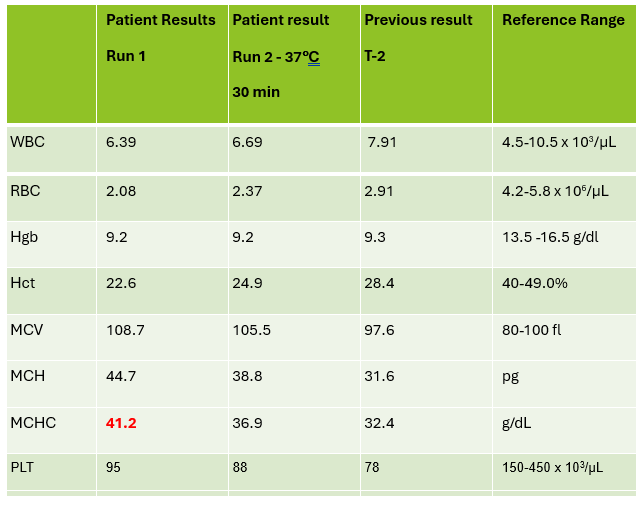
Table 1. CBC results before warming at 37°C and after 15 minutes incubation.

On Run 2, the MCHC is now below 37.5, so there was no operator alert to incubate further or to investigate the high MCHC. This result was validated with the comment “37°C results, possible cold agglutinin.” However, there was still an RBC agglutination flag, a high MCV, and the hemoglobin and hematocrit still don’t look great, i.e. they don’t follow ‘the rules of 3’. We know that these rules are only valid for normal samples, but if we look at the previous results from 2 days ago, the Hgb 9.3 and Hct 28.4 did follow the ‘rules of 3’ and additionally, today’s results look like the patient’s hemoglobin has not changed but the hematocrit has dropped. The RBC did go up on the second run, but it is considerably lower than 2 days ago. The indicies are also inconsistent with the previous sample. “Hmmm…What could cause this?” Instead of validating, this is what you should be asking yourself.

Cold agglutinins are known for causing pre-analytical and analytical spurious results with CBCs. With cold agglutinins, IgM antibodies bind to the RBCs after exposure to cold, causing RBC agglutination which leads to a classic pattern of false results. There is an increased MCV because the RBCs are clumped and sticking to one another, making the analyzer think these larger clumps are individual RBCs. This, in turn, will make the RBC count appear deceased because large clumps of RBCs going through the RBC aperture are counted as one RBC. The hematocrit is lowered because the volume of a clump is less than those cells individually. Hemoglobin concentration, on the other hand, is not affected by cold agglutinins, but because the hematocrit and RBC are falsely lowered, this makes the MCH and MCHC spuriously markedly increased. Cold agglutinins may be subtle, like this one, but some have extremely high MCHC and MCV, and extremely low RBC and Hct. Clues that you have a cold agglutinin are not only the high MCHC, but also flags from your analyzer such as “RBC agglutination”. The hematocrit will likely seem too low for the hemoglobin, and you may even see a hemoglobin that is higher than the hematocrit (yes it happens!) and RBC values so low as to be incompatible with life.

The first thing is to do if you get these spurious results is to compare the parameters and instrument flags and decide if the results are consistent with a cold agglutinin. Other factors that may cause a high MCHC include lipemia, icterus, low sodium, abnormal proteins, hemolyzed samples and samples from patients with hemoglobinopathies. The patterns of result with these samples will show a high MCHC, but with a normal or low MCV, and you won’t usually see an RBC agglutination flag. A sample with cold agglutinins may also appear grainy or clumpy to the naked eye. With a suspected cold agglutinin, warming the sample for 15-30 minutes will allow the RBCs to disperse and improve the results. However, these results must be reviewed, and if there are still instrument flags and/or if there is still clumping, the results may not yet be corrected or ‘correct’. Be sure to review a smear from the tube before warming and after. The first smear confirms the presence of the cold agglutinins and clumping, and the second smear should confirm the resolution of the clumping after warming.

This sample did have the characteristic grainy appearance of a cold agglutinin, and the post warming results did look a bit better, but the RBC agglutination flag was still present, and a review of the smear showed that the sample still had RBC clumping. After warming for another 30 min, the sample was quickly mixed and placed back on the analyzer. The results of this 3rd run are shown below, in Table 2.
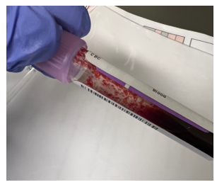
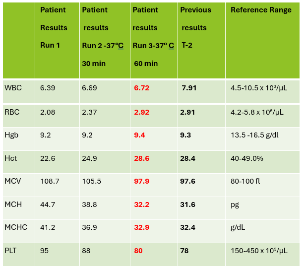
Figure 1. EDTA tube with RBC agglutination
Table 2. CBC results on 3 runs. Run 3, after 60 min of incubation.

Notice that hemoglobin has not changed as it is not affected by the cold agglutinins, but after warming for 60 minutes, the RBC, hematocrit and indicies all now look consistent with the previous sample drawn 2 days ago, and a review of the smear showed no RBC agglutination. These results from the 3rd run are ready to validate.

Cold agglutinins are IgM autoantibodies that react best at 4°C but may also react at room temperature. They are generally not clinically significant and may be found in many healthy individuals. These natural cold autoantibodies occur at low titers, less than 1:64, and have no activity at higher temperatures. However, because they react at room temperature, they are notorious as a pre-analytical and analytical factor that causes spurious CBC results. They can also cause difficulties in Blood Banking during ABO/Rh typing and antibody detection.

Cold agglutinins have various clinical manifestations. Benign cold agglutinins generally do not cause hemolytic anemia and need no treatment. Most benign cold autoantibodies have anti-I specificity, are polyclonal, low titer, and do not react above 30°C. Cold agglutinins associated with Mycoplasma pneumoniae, and infectious mononucleosis are usually clinically insignificant. In cases where they do cause hemolytic anemia, the antibodies are polyclonal IgM with normal κ and λ light chains. The anemia is acute and generally spontaneously resolves in several weeks without treatment.

Though most cold agglutinins are benign and do not cause RBC destruction, when they do, they can cause hemolytic anemia that varies in severity from mild to life-threatening. This chronic cold agglutinin disease (CAD) is now known to be a form of autoimmune hemolytic anemia caused by a bone marrow lymphoproliferative disorder. Chronic CAD is a cold-autoantibody autoimmune hemolytic anemia (cAIHA) that is caused by an autoantibody produced by the clonal B cell lymphocytes. This antibody is usually monoclonal IgM with κ light chains and “I” or “i” specificity. These pathological cold agglutinins are high titer and usually react at 28°C to 32°C, and even up to 37°C. The highest temperature at which the antibodies continue to be activated is called the thermal amplitude. Because these can act at higher thermal amplitude, they may lead to CAD. In CAD the IgM autoantibodies bind to red cell antigens at 30-32°C, typically in the cooler extremities. IgM’s structure, a large immunoglobin pentamer, makes it an effective activator of the classical complement system. As the blood circulates to the central parts of the body, the RBCs warm up and the IgM antibodies dissociate from the RBC membranes, but the complement activation will continue, leading to RBC hemolysis and a cAIHA.

Chronic CAD occurs most often in adults over 50, is more common in women, and produces anemia with varying severity. Patients may be seasonally affected. In the winter, the temperature of blood may fall below 30°C in the extremities, activating the cold agglutinins. Patients may experience acrocyanosis of the hands, feet, ears, and nose with exposure to cold. They may also experience other cold related symptoms such as numbness and Raynaud’s. Patients with chronic CAD and mild anemia are therefore monitored with a ‘wait and see’ plan and advised to avoid cold temperatures. In patients with more severe anemia, it is found that targeting the underlying lymphoproliferative disorder provides the best treatment. Rituximab has been used to achieve partial remission. Therapeutic plasma exchange is also used in severe cases to rapidly remove cold agglutinins.

I have been thinking about cold agglutinins recently because of the number I have seen come into our lab this winter. As I am writing this, watching the temperature outside drop in anticipation of more snow coming in tonight, cold agglutinins came to my mind again. I used to live in the cold northern Northeast, but after moving further south, we see fewer cold agglutinins in hematology than I used to see. This winter we have had some cold spells, and, interestingly, I’ve seen more cold agglutinins. That led me to ask myself if cold agglutinin disease is really more common when patients are exposed to cold temperatures. I remember learning myself, and telling my students that that the treatment for mild to moderate CAD was to advise patients to move to someplace warmer. It has been assumed for many years that CAD worsens in colder climates or seasons. Interestingly, there have been a number of studies done since the 1950’s that examined the relationship of cold temperatures and CAD. The studies used hemoglobin, bilirubin and LDH for monitoring. Early case reports had findings that supported the theory of more anemia and higher LDH in the winter. (Dacie, Lyckholm)One recent article in 2022 found a 4-fold difference in the incidence of CAD between cold (Norway) and warm (Italy) climates. (Berentsen). However, atthe same time another study found that there was no statistically significant seasonal variation in hemoglobin, but that LDH levels were higher in winter. It concluded that these conditions should be monitored through all seasons because of the risk of hemolysis and thrombotic episodes. It was also demonstrated that though there may not be obvious statistical difference in CAD between cold and warm months, that there is a large variability of disease severity across patients and even with an individual patient. (Roth).

In conclusion, when working in any department of the laboratory, quality results are important. Results on the patient chart that vary considerably from day to day because sometimes a cold agglutinin has been effectively resolved in lab testing and other days results after 15-30 minutes of warming are just reported without a good review of the smear and the parameters, are confusing, and could affect patient care. If one tech reports the results after 30 min incubation with values that are still spurious, and the next tech resolves the agglutination with further warming, the lab will be reporting out inconsistent results. A patient who actually has stable CBC results may have deltas and what appear to be erratic results. Cold agglutinins do take time to resolve, but with over 80% of samples autoverifying with the use of auto verification, we have time to work on these problem samples. If something doesn’t look or feel right about a sample, look at all the parameters, check the instrument flags and operator alerts, check the previous results and investigate any changes. It is important to review results carefully, because we want to report out the best results possible.

References

S Berentsen, W Barcellini, S D’Sa, U Randen, THA Tvedt, B Fattizzo, E Haukås, M Kell…

Blood, The Journal of the American Society of Hematology, 2020•ashpublications.org

Climent F, Cid J, Sureda A. Cold Agglutinin Disease: A Distinct Clonal B-Cell Lymphoproliferative Disorder of the Bone Marrow. Hemato. 2022; 3(1):163-173. https://doi.org/10.3390/hemato3010014

Nikousefat Z, Javdani M, Hashemnia M, Haratyan A, Jalili A. Cold Agglutinin Disease; A Laboratory Challenge. Iran Red Crescent Med J. 2015 Oct 17;17(10):e18954. doi: 10.5812/ircmj.18954. PMID: 26566452; PMCID: PMC4636857.

Patriquin, C.J. and Pavenski, K. (2022), O, wind, if winter comes … will symptoms be far behind?. Transfusion, 62: 2-10. https://doi.org/10.1111/trf.16765

Rodak, Bernadette F., et al. Hematology: Clinical Principles and Applications. 5th ed. St. Louis, Mo., Elsevier Saunders, 2016

Röth A, Fryzek J, Jiang X, Reichert H, Patel P, Su J, et al. Complement-mediated hemolysis persists year round in patients with cold agglutinin disease. Transfusion. 2022; 62: 51–59. https://doi.org/10.1111/trf.16745

Socha-small

-Becky Socha, MS, MLS(ASCP)CMBBCM graduated from Merrimack College in N. Andover, Massachusetts with a BS in Medical Technology and completed her MS in Clinical Laboratory Sciences at the University of Massachusetts, Lowell. She has worked as a Medical Technologist for over 40 years and has taught as an adjunct faculty member at Merrimack College, UMass Lowell and Stevenson University for over 20 years.  She has worked in all areas of the clinical laboratory, but has a special interest in Hematology and Blood Banking. She currently works at Mercy Medical Center in Baltimore, Md. When she’s not busy being a mad scientist, she can be found outside riding her bicycle.

Hematology Case Study: Spurious CBC results on a Chronic Lymphocytic Leukemia patient

Working in Hematology, I have learned that things aren’t always black and white. With about 80-85% of our CBC’s autovalidating, it’s those other “problem child” specimens that can give us a challenge. When we get one of these tricky specimens, it’s time to put on our detective hats and investigate what is going on.

This patient was a 65-year-old female with Chronic Lymphocytic Leukemia (CLL). WBC and RBCs are counted on our analyzer by impedance, which sorts cells by size. When we ran this sample, we noticed a few things right away. See results below in Figure 1.

Figure 1. Original CBC results with Instrument Flags

The first thing I notice on this specimen is the @ next to the WBC. This indicates that the count is over linearity and was confirmed by dilution. The corrected WBC was 577 x 103/mL. See Figure 2.

Figure 2. 1:3 dilution results for WBC. 192.47 x 3 = 577.41 x 10³/μL

Extreme leukocytosis may interfere with the RBC, HGB, HCT and MCV determinations. The degree of RBC interference depends on the number and size of WBCs present. WBC and RBC are counted using impedance technology. In impedance counting, the RBC count is done first by passing the sample though the aperture in the RBC/platelet channel. This count is actually the sum of both RBC and WBC counts. Then, the RBCS are lysed and the WBCs are counted in the WBC channel. Normally, the WBC count has very little, if any, effect on the reported RBC count. Normal RBC counts are 4-6 million/μL. Normal WBC counts are a fraction of this, at about 5-10,000/μL. If the the RBC count is 3.50 x 10⁶/μL and the analyzer includes 10,000 WBCs in the count, this only changes the RBC to 3.51 x 10⁶/μL. (3,500,000 + 10,000 = 3,510,000). Because WBC counts are so much lower than RBC counts, even if a WBC count is 100,000, the effect on the RBC count is clinically insignificant. (3,500,000 + 100,000= 3,600,000 = 3.60 x 10⁶/μL) However, in this patient, the WBC count was 577,000/μL. After reviewing the smear and confirming the WBC count with a WBC estimate, we corrected the RBC count, by subtracting the WBC from the RBC. As you can see in figure 3 below, the extreme leukocytosis did affect the RBC count.

Figure 3. Corrected RBC count. Subtract the WBC count from the RBC count. Corrected RBC (cRBC) = RBC (x 106/μL) – WBC (x 103/μL)

So, how does this affect the hematocrit? The next thing noticed right away is that the Hgb and Hct don’t follow the “rules of 3”. Now, we know that these rules really only hold true for normocytic, normochromic RBCS, but extreme leukocytosis can interfere with Hct determination. A Hgb of 9.2 g/dL and Hct 39.4% doesn’t look ‘right’. We have just corrected the RBC count, and now we need to ask ourselves how this can affect the Hct. The hematocrit is the packed cell volume, or the % of red blood cells per total volume of the sample. Since we now know that the RBC count is 2.92 x 10⁶/μL, not 3.50 x 10⁶/μL, we can correct the hematocrit. If you have a hematocrit centrifuge in your laboratory, a spun hematocrit can be used to determine the corrected hematocrit. After correcting the Hct, you must also correct the MCV using the following formula.

Corrected MCV (cMCV) = HCT(%) x 10/cRBC

Another option for resolving interferences and correcting the Hct for extreme leukocytosis is using the clues that the RBC histogram gives us. We know the RBC count needed correcting, and we subtracted the WBC to get the corrected RBC. This sample had multiple flags. One of them was “Dimorphic population”. This indicates two populations of RBCs in the sample. Since we know a considerable number of WBCs were counted in the RBC chamber, this would account for the dimorphic population. A dimorphic population on histogram looks like what I call a ‘double humped camel’. In this case, the patient’s RBCs are the first, smaller population and the lymphocytes of this CLL patient are the 2nd larger population. See Figure 4.

Figure 4. Example of a dimorphic RBC histogram.

Another option for recalculating the MCV is using the information in the service tab of your analyzer. Note that the results from the service tab are not FDA approved, and therefore not directly reportable, so must be confirmed first. If using values from the service tab, the spun hematocrit and calculations can be used as a check. The service tab displays the MCV of these 2 populations. These are listed as the MCV of the small population, S-MCV, and the MCV of the large cell population, L-MCV. Using the small MCV (sMCV) value and the corrected RBC (cRBC), we can back calculate the Hct using the following formula.

Corrected Hct (cHct) (%) = (sMCV x cRBC)/10

For this sample:

S-MCV = 105.1

L-MCV= 215.4

(cHct) (%) = 105.1 x 2.92/10 = 30.7 %

Hgb is another parameter that may be affected by extreme leukocytosis. Turbidity may be present in the diluted and lysed sample when reading the Hgb. This sample did not give us a Hgb turbidity flag, but because of the high WBC, the Hgb was confirmed using a diluted sample. The sample was diluted 1:3 with the analyzer diluent. Results were multiplied by the dilution factor. Lastly, when performing Hgb corrections (and in this case, also the RBC corrections) you must also recalculate the MCH and MCHC using the corrected values. Figure 5 shows these corrected values.

MCH (pg)= (cHgb/cRBC) x 10

MCHC (g/dL) =(cHgb/cHct) x 100

Figure 5. Corrected CBC results

We can breathe a sigh of relief that we finally have accurate and reliable results for the CBC. But what about the differential? This big ugly grey mess seen in Figure 6 on the differential scattergram indicates a very abnormal scattergram. This is telling us that there is no separation between the types of cells. There were multiple flags for WBC abnormal scattergram, leukocytosis, lymphocytosis, and a flag for dimorphic RBC populations. These flags are all telling us not to accept the instrument results. In these cases, we want to review the smear, do a WBC estimate, and perform a manual differential, examining the differential carefully to look for any abnormalities. The differential had many lymphocytes and smudge cells. An albumin smear was made to resolve the smudge cells and a manual differential was performed.

Figure 6. WDF Scattergram

I’ll admit that this type of specimen is not something we encounter every day (thankfully). But I thought it a very interesting example of a spurious results on many levels. These challenges are some of my favorite things about working in Hematology. Using autovalidation is a great tool in the laboratory to help workflow. With about 85% of specimens autovalidating, this allows us to spend time on these tricky specimens. And this tricky specimen was an epic one! We had CBCs on this patient several days in a row. Unfortunately, some of her results were simply repeated and reported. Some WBC results over linearity were reported without dilution. Other parameters were not corrected. This gives inconsistent and confusing results to the physicians and is not beneficial to the patient. Because of the inconsistencies, we issued a couple corrected reports which can be very time consuming. Sometimes we may not have the answers and can’t resolve a problem. If a specimen cannot be resolved, it is always better to report what you can and use ‘not reported’ or ‘not measured’ for any results that are not available. It’s better to report the good results that you have than to report junk that physicians can’t rely on. I often say that simply repeating a sample and reporting results if they match is not sufficient. We need to investigate spurious results so that we may report the best quality results possible for every patient.

References

  1. Gulati G, Uppal G, Gong J. Unreliable Automated Complete Blood Count Results: Causes, Recognition, and Resolution. Ann Lab Med. 2022 Sep 1;42(5):515-530. doi: 10.3343/alm.2022.42.5.515. PMID: 35470271; PMCID: PMC9057813.
  2. Sysmex USA. XN-Series Flagging Interpretation Guide. Document Number: 1166-LSS, Rev. 6, March 2021
  3. Zandcki, M. et al. Spurious counts and spurious results on haematology analysers: a review. Part II: white blood cells, red blood cells, haemoglobin, red cell indicies and reticulocytes. International Journal of Laboratory Hematology. 09January 2007.
Socha-small

-Becky Socha, MS, MLS(ASCP)CMBBCM graduated from Merrimack College in N. Andover, Massachusetts with a BS in Medical Technology and completed her MS in Clinical Laboratory Sciences at the University of Massachusetts, Lowell. She has worked as a Medical Technologist for over 40 years and has taught as an adjunct faculty member at Merrimack College, UMass Lowell and Stevenson University for over 20 years.  She has worked in all areas of the clinical laboratory, but has a special interest in Hematology and Blood Banking. She currently works at Mercy Medical Center in Baltimore, Md. When she’s not busy being a mad scientist, she can be found outside riding her bicycle.

Case Studies in Hematology: Hemoglobin S Beta Thalassemia Compound Heterozygosity in a 61 Year Old Female

A 61 year old Black woman with a diagnosis of sickle cell beta thalassemia presented to the ER with fatigue, dyspnea, and back and leg pain with some swelling in the hands and feet. Splenomegaly was noted on exam. The patient has a history of moderate to severe symptomatic anemia and is being followed by a hematologist. Her baseline Hgb is 9-10 g/dL. Her treatment plan includes Hydroxyurea, 500 mg daily, and transfusions, as needed. Her last sickle cell crisis was 2 years ago. CBC was ordered. Hgb on admission was 6.1 g/dL. Her RBC morphology showed polychromasia, target cells, sickle cells, anisocytosis, and numerous nucleated RBC forms.

The patient was admitted to the hospital. Type and crossmatch for 2 units of packed red blood cells was ordered. CT imaging was performed and revealed severe osteopenia and vertebrae deformities consistent with her history of sickle cell disease. Chest CT showed hypoinflated lungs and areas of consolidation in the lower lobes consistent with acute chest syndrome due to sickle cell beta thalassemia. She was transfused with 2 units of pRBCs and treated for sickle cell crisis. The patient remained stable and was discharged 3 days later.

Table 1. CBC results on ER admission
Figure 1. Peripheral blood smear on admission. Patient with sickle cell/beta thalassemia shows sickle cells, target cells, nucleated red cells, anisocytosis, poikilocytosis, polychromasia. (Mercy Medical Center, Baltimore, Md)

Sickle cell anemia (HbSS) and thalassemia are the world’s most common single gene disorders. Both are inherited in an autosomal recessive manner, and result in hemolytic anemias. But what happens when you inherit one gene for sickle cell and one gene for beta-Thalassemia (β-thalassemia)?

Sickle cell disease is caused by mutations in the HBB gene that provide instructions for making beta-globin. Sickle cell anemia is a hemoglobinopathy, a qualitative defect in the structure of globin chains, resulting in the production of abnormal hemoglobin. Normal adult Hemoglobin A has 2 α chains and 2 β chains (α2β2). Hb S results from the substitution of valine for glutamic acid at position 6 of the β globin chain. The resultant Hb S has reduced solubility at low oxygen tensions. Patients with sickle cell anemia have a moderate to severe chronic hemolytic anemia with recurrent painful sickle cell crisis.

Sickle cell disease is inherited in an autosomal recessive pattern from parents who have at least one mutated gene. Anyone with a sickle cell gene can pass this gene on to their children. Sickle cell anemia (HbSS) is the homozygous expression of a sickle gene from both parents and is the world’s most common inherited hematological disease. A heterozygote inherits a sickle gene from only one parent. This person is a carrier of sickle cell (HbSs), often referred to as sickle cell trait. HbSs persons do not generally exhibit symptoms or may exhibit only a mild anemia. However, under stressful conditions, such as at high altitudes, they may experience vaso-occlusive sickle crisis.

While hemoglobinopathies are a qualitative defect due to structural changes in the normal amino acid sequence of globin, thalassemias result from an imbalance in the synthesis of the globin chains that make up the hemoglobin molecule. Thalassemias involve the rate of globin chain synthesis leading to a quantitative defect. Thalassemia is divided into α-thalassemias and β-thalassemia. α-thalassemias involve genes for the α chains on chromosome 16. In α-thalassemia, the deletions involve the α1 and/or the α2 globin genes and result in decreased production of α chains. β-thalassemias mainly affect β chain production. They are disorders of reduced globin chain production from the globin chain cluster on chromosome 11.

(Since this case involves a known diagnosis with a compound heterozygous state involving a β-thalassemia gene mutation, the discussion of α-thalassemia has been limited here. Watch for a case involving α-thalassemia in a future blog!)

Beta thalassemia occurs when the beta globin chains are either produced inadequately or not at all. There are many mutations in and around the β globin gene that result in decreased β chain production. Mutations that result in the complete absence of β chain production are designated as β0. In the most severe form of β-thalassemia the patient is homozygous β00 and does not produce any β chains. Without β chains there is no Hb A (α2β2). β+ is used as the designation for any mutations of the β globin gene that cause a partial deficiency of β chains (5-30% decrease) and therefore result in a decrease in production of Hb A. The βsilent designation is used for carrier state gene mutations that result in only a mildly decreased β chain production. The degree of decrease in the β chain production is related to the degree of anemia and the severity of clinical disease.

Thalassemia, like sickle cell anemia, is a hereditary anemia inherited in the autosomal recessive manner. β-thalassemia is divided into categories based on clinical severity of disease. In β-thalassemia major a child inherits a copy of a β-thalassemia gene mutation from both parents. There are various mutations that cause genes with these mutations and different variants may be inherited from each parent. A person with thalassemia major may be homozygous β+/ β+, homozygous β0/ β0, or the compound heterozygous state β+/ β0. Hb A is only produced in patients with the β+ mutation. β-thalassemia major patients have the most severe hemolytic anemia and symptoms. β-thalassemia intermedia is characterized as homozygous βsilent or heterozygous βsilent with β+ or β0 and mild to moderate disease. β-thalassemia minor, also called β-thalassemia trait or carrier state, presents with mild but asymptomatic hemolytic anemia. These patients are heterozygous with normal β globin and have slightly decreased Hb A.

In people with sickle cell disease, at least one of the beta globin is replaced with hemoglobin S. In homozygous sickle cell anemia, both beta globin subunits in hemoglobin are replaced with hemoglobin S. In compound sickle cell diseases, one beta globin is replaced with hemoglobin S and the other beta globin is replaced with a different abnormal variant. Examples of this are Hb SC disease, and Hb SD syndrome. Compound heterozygosity is the inheritance of two different mutated genes that share the same locus. If mutations that produce hemoglobin S and beta thalassemia occur together, individuals have hemoglobin S-beta thalassemia disease. (sickle cell beta-thalassemia, Hb S β thal or sickle-β-thal). Sickle cell beta thalassemia patients have hemoglobin S (α2β26Glu→Val) and either β0 or β+.

When a qualitative hemoglobinopathy is inherited with a quantitative disorder of hemoglobin synthesis, the severity of the compound disorder is dependent on the β gene mutation. Patients with β0 produce no Hb A and have moderate to severe symptoms comparable to that of Hb SS patients. β+ patients will produce some β chains and therefore have some Hb A and milder or no symptoms.

Figure 2. Peripheral Blood smear on day 3. Sickle cell forms, polychromasia, target cells. nucleated RBCs. (Mercy Medical Center, Baltimore, Md)

Newborn screening can diagnose β0-thalassemia at birth by detecting a complete absence of hemoglobin A. However, it is not possible to make a definitive diagnosis of β+-thalassemia in the newborn because newborns have Hb F, and the reduced amount of hemoglobin A overlaps the range for normal babies. In adults with Hb S – β thal the amount of Hb S is variable. There is some Hb A in β+ patients but no Hb A detected in β0. Hb A2 and Hb F are increased. In addition to hemoglobin electrophoresis, molecular testing may also aid in the diagnosis by identifying genetic mutations. Beta globin gene sequencing can identify beta thalassemia alleles that are caused by point mutations in the beta globin gene. As well, structural variants of the beta globin gene such as Hb S can be identified with this technique. This can lead to a better understanding and clinical management of the disease.

Case Study, continued: This patient inherited a Hb S gene from one parent and a β-thal gene from the other, resulting in sickle cell beta thalassemia. This compound heterozygosity affects red blood cells both by the production of structurally abnormal hemoglobin, and by the decreased synthesis of beta globin chains. Clinical manifestations depend on the amount of beta globin chain production. Symptoms may include anemia, vascular occlusion, acute episodes of pain, acute chest syndrome, pulmonary hypertension, sepsis, ischemic brain injury, splenic sequestration crisis and splenomegaly.

Hemoglobin electrophoresis was sent out to a reference lab and results are shown in Table 2. Based on the Hemoglobin electrophoresis, is this patient Hb S- β0-thal or Hb S- β+-thal?

Table 2. Hemoglobin pattern and concentrations of a S/betathalassemia patient

Hemoglobin electrophoresis results for Hb S beta thalassemia patients are expected to show 60-90% Hb S and 10-30% Hg F. This patient’s Hgb S at 74% is within this range. This result reflexed a sickle solubility test, which was positive. As well, the elevated Hb F and Hb A2 are consistent with this diagnosis. It was noted in the discussion above that Hb S- β+-thal mutations cause a decrease of 5-30% in beta chains and therefore a decrease in Hb A. This patient’s Hb S is greater than the Hb A and her Hb A concentration is 14.8%, which is consistent with this diagnosis. Hb S- β0 mutations produce no Hb A. In this case there is some Hb A on electrophoresis but not as much as would be expected in a β+-thal mutation. Also, of note it that this patient was recently transfused with 2 units of pRBCs. Interpretations of hemoglobin electrophoresis assume that the patient has not been transfused in the last 3 months. The Hb A in this patient can be explained by these recent transfusions. Therefore, it can be concluded that the hemoglobin pattern and concentrations are consistent with transfusion of a Hb S beta0 thalassemia patient. The β0 mutation is also consistent with this patient’s moderately severe symptomatic anemia.

References

  • Keohane, Elaine, et al. Rodak’s Hematology, Clinical Principles and Application, 5th ed, Elsevier, 2016
  • McKenzie, Shirlyn. Clinical laboratory Hematology. Pearson Prentice Hall, 2004.
  • McGann PT, Nero AC, Ware RE. Clinical Features of β-Thalassemia and Sickle Cell Disease. Adv Exp Med Biol. 2017;1013:1-26. doi: 10.1007/978-1-4939-7299-9_1. PMID: 29127675.
  • Origa R. Beta-Thalassemia. 2000 Sep 28 [Updated 2021 Feb 4]. In: Adam MP, Mirzaa GM, Pagon RA, et al.,editors. GeneReviews® [Internet]. Seattle (WA): University of Washington, Seattle; 1993-2023. Available from: https://www.ncbi.nlm.nih.gov/books/NBK1426/
  • Turgeon, Mary Louis. Clinical Hematology Theory and Procedures, 6th ed, Jones and Bartlett Learning, 2017.
Socha-small

-Becky Socha, MS, MLS(ASCP)CMBBCM graduated from Merrimack College in N. Andover, Massachusetts with a BS in Medical Technology and completed her MS in Clinical Laboratory Sciences at the University of Massachusetts, Lowell. She has worked as a Medical Technologist for over 40 years and has taught as an adjunct faculty member at Merrimack College, UMass Lowell and Stevenson University for over 20 years.  She has worked in all areas of the clinical laboratory, but has a special interest in Hematology and Blood Banking. She currently works at Mercy Medical Center in Baltimore, Md. When she’s not busy being a mad scientist, she can be found outside riding her bicycle.

Bandemia: What is it and Why is it Important?

Recently our lab was asked by physicians to start reporting band counts of ≧10% as critical values. While we have always reported bands when a manual differential is performed, we have also heard for years about other labs that have stopped reporting bands. Mayo Clinic stopped reporting bands in the 1990’s- other nationally and internationally known hospitals and community hospitals alike have followed suit. CAP proficiency surveys for cell ID do not separate segmented neutrophils and band neutrophils for cell IDs. Our hematology analyzer reports Immature granulocytes (IG) and we have learned about the benefits of IG over the band count. Thus, when our pathologists asked us to add a critical value for bandemia, we wondered if we were moving ‘backwards’.

Bandemia is defined as elevated band neutrophils in the peripheral blood. Neutrophils are produced to help fight infection. With infection, there is an increase in WBCs being released by the bone marrow into the peripheral blood. Bands are considered mature neutrophils and can fight infection, and in an effort to keep up with demand, some of these infection fighting cells which are released are bands. However, we are reminded that this is a nonspecific clinical finding. Bands can be elevated in many situations including inflammation, autoimmune disease, metabolic abnormalities, pregnancy, and treatment with granulocyte colony stimulating factors. Band counts of ≧10% have been used clinically as an indicator of serious bacterial illness. A finding of bandemia can help providers decide what steps need to be taken in evaluation of a patient, and in conjunction with clinical findings, can help in making a differential diagnosis.

The trouble with bands is that they are notoriously difficult to measure accurately and precisely. Bands are somewhat controversial and there are conflicting opinions on the utility of bands. Should bands be reported or included with neutrophils? A left shift reflects bone marrow response to bacterial infection, and this has been quantified as band count or immature granulocyte count. Is the IG a better parameter than the band count?

If your Hematology analyzer reports the absolute neutrophil count (ANC) with an automated differential, this includes all mature neutrophils. The theory that supports this is that neutrophilic bands and segmented neutrophils are both mature cells, and both fight infection. A true left shift would therefore be the presence of immature granulocytes (metamyelocytes, myelocytes and promyelocytes). Yet, we also now have analyzers that offer a 6-part differential that includes the immature granulocyte (IG) count. And we know that an automated differential is based on counting thousands of cells, and a manual differential is based on counting 100 cells. So, which is better? We can theorize that the IG is a better indicator of left shift because the automated diff count looks at more cells than a 100-cell manual diff.

Let’s say that we have a patient with a WBC of 20 x 103/μL. Left shift is defined as an absolute IG count >0.1 x 10³/μL. Automated diffs can count >30,000 cells depending on the WBC count. If the auto diff counts 32,000 cells and finds 1% IG, this means that the analyzer identified 320 IG. Absolute counts are calculated as % x the WBC, in this case 0.01 x 20,000=.2 x 103/μL. This meets the definition of a left shift. If we do a manual diff and count 100 cells and see 1 meta, the absolute IG would be .2 x 103/μL, again meeting the criteria for a left shift. However, if we did not see any metas or other IG on the manual diff, the absolute IG would be 0. Thus, performing a manual diff, the difference in seeing 1 cell or 0 would make the difference of reporting a suspected infection versus no suspicion of infection.

Table 1: Left shift? comparison of absolute IG when 1 or no IGs are seen on manual diff

With the advent of the IG count on automated differentials, labs have moved away from reporting bands. I have attended conferences and heard presentations about “banning” bands. A few years ago, I wrote a blog called “Beyond Bands: The Immature Granulocyte Count”, describing the benefits of using the IG count over bands in manual diffs. The above example would support “Ban the bands” arguments. Using the IG count from analyzers can take advantage of analyzing many more cells and give us statistically more precise values.

Results from auto diff can get to patients’ chart faster than a manual diff result, leading to faster treatment. To report bands, a manual differential must be done. We must wait for a slide to be made, dry, and a diff to be analyzed either under the microscope or on CellaVision. Bands are subjective, relying on technologist interpretation.

So, why are we suddenly being asked to make bandemia a critical value?

Physicians asked for this change and have cited cases where patients were seen in the emergency department (ED) and subsequently released, later to return, or experiencing negative outcomes. These patients had bands reported on differentials. Other area hospitals are reporting bands and have critical values for bandemia. Because patients often are seen at more than one area hospital, and doctors may have privileges at more than one of these, for consistency, this makes sense. But is also makes sense clinically.

Recent data has drawn renewed attention to bands as a reliable predictor of severity of patient condition. A number of research papers have been published that indicate that bands may indeed be important for patient care. A 2012 study investigated bandemia in patients with normal white blood cell counts. This cohort study found that patients with normal WBC counts with moderate (11%-18%) or high (>20%) band counts had increased odds of having positive blood cultures and in-hospital mortality. (Drees, 2012)

In 2019 a study showed that there was an “increasing risk for death with increasing bandemia, irrespective of leukocyte count. (Davis, 2019) A 2021 study done at Rhode Island Hospital showed a strong correlation between increasing percentages of bands on an initial emergency room CBC and the likelihood of significant positive blood cultures and in-hospital mortality. This was noted even at band levels below 10%. (Hseuh, 2021) S. Davis, MD, from the Department of Emergency Medicine, George Washington University School of Medicine and Health Sciences, in Washington, DC wrote that “While emergency physicians may find reassurance in a normal leukocyte count, the balance of evidence strongly suggests a more prudent approach would be to wait for the bands.” (S. Davis, 2021) In other words, wait for that manual differential. He stated that emergency room physicians get results from automated CBCs before the manual diff and do not see or are aware of any internal laboratory flags on these specimens. Physicians should be aware of reporting processes to avoid early discharge of otherwise well-appearing patients before band counts are reported. Last year, trends in bandemia and clinical trajectory among patients was reviewed in a retrospective chart review at George Washington University Hospital. They noted that “Bandemia clearance and trending, in conjunction with other existing clinical tools, may be of use as a marker of improvement in sepsis. Conversely, worsening bandemia may be predictive of a deteriorating clinical status and possibly a higher mortality.” They also noted that following trends of band levels in patients with sepsis or septic shock may help to predict a clinical course and overall prognosis. (Prasanna, 2022) Additionally, a band count greater than 10% is one of the American College of Chest Physicians/Society of Critical Care Medicine’s systemic inflammatory response syndrome (SIRS) criteria used to diagnose sepsis. (Chakraborty, 2022) These are just a few of the many articles that support a clinical utility of reporting the band count.

When we first learned that we would be reporting bandemia as a critical value, we realized that we would need to get everyone on the same page. We do most of our diffs on CellaVision, so, in theory, that should be easy, but, and a big but, is that bands are notoriously subjective. Different technologists may have been taught or have used their own definitions of bands. Variation can occur depending on slide quality, tech training, definition of bands used, and number of cells counted. So, how do we make this work? We need to be sure that all our technologists are reporting bands using the same criteria, so we are not reporting differing or confusing information to physicians. The concern lies in a physician making a significant clinical decision based on apparent changes in band counts that are not real but only reflect predictable statistical factors and unpredictable technologist variability.

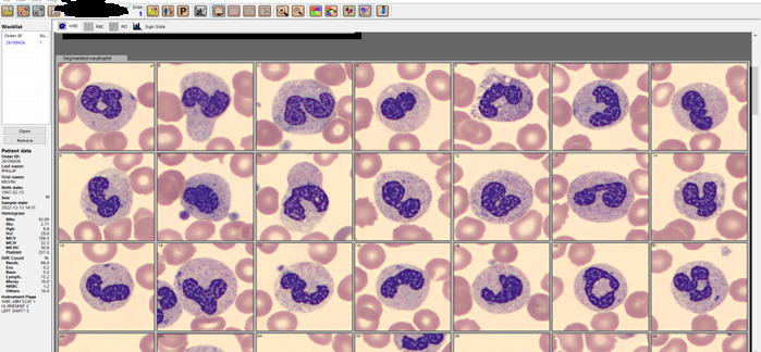
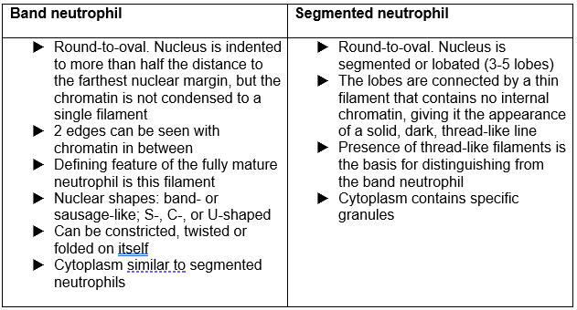
In our laboratory, we approached the implementation of this new critical value as an educational opportunity. Differentials that had been performed on our CellaVision were reviewed, and it was apparent that bands were not being categorized consistently among techs. (See Figure1) We started with reviewing the definition of bands with all technologists and writing updated detailed procedures that includes these definitions. We use CAP’s definition of bands, which is the definition used in most textbooks and references for over 60 years. (See Table 1) Many examples of both bands and segmented neutrophils were added to our reference library on CellaVision. These included textbook perfect bands and some that may be more subjective. These reference cells can be used by techs to compare cells when making decisions as to in which category they belong. It was also stressed to techs that they need to look at cells carefully, and in a view that is large enough to see both detail and differentiation.

Figure1. Cells reported as segmented neutrophils on Differential. How many bands do you see?
Table 2. CAP Definition of bands vs segmented neutrophils
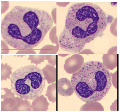
Figure 2. Are these bands or segmented neutrophils?

Patient samples that had bands reported were located on CellaVision and multiple slides were made from these samples to be used as competency slides. In developing the differential evaluation tool, Rumke’s data showed that for a differential with a reported 12% bands, a second differential would have to have greater than 23% bands or fewer than 3% bands before the difference could be considered statistically significant. But this can be significant to our patients and patient care. In this example, diffs with <10% bands would not indicate bandemia, and diffs over 10% would initiate a critical bandemia call. And this could happen on the same slide depending on who did the diff, or on sequential samples on the same patient over a short period of time. These competency slides were assigned to techs to collect statistics on the mean and SD of the bands reported on each slide. Retraining and coaching will be provided as necessary. Follow up competency slides will be assigned, and statistics will be recalculated. The goal is to decrease variability in our band counts and to show that we have done so. This ongoing quality project has involved writing procedures, offering continuing education, assigning and reviewing competency slides, coaching technologists and reviewing slides with them and calculating statistics. Our goal is consistency and reporting meaningful results to our physicians. 

As we saw in the studies cited above, % bands and trends are both important when evaluating clinical correlations. The chart below shows examples of how this might affect patient care. If a patient on presenting at the ED had a 19% band count, this would be called as a critical, and the patient would be further evaluated, and depending on clinical symptoms and medical history would likely have blood cultures drawn and be admitted to the hospital. If a second tech did the diff and reported 5% bands, the patient may be sent home without further evaluation. On subsequent CBCs, we could be giving confusing results to the physician if we are not consistent in our reporting. With multiple techs doing differentials on different shifts, it could look like this patient is getting better,

getting worse, or it could look like the patient is responding to therapy, and then the next day they had a setback. While we understand that bands will probably always be somewhat subjective, we need to narrow this down. By adhering to one definition, our goal is to report consistent and accurate results.

Table 3. Various results on differentials on the same patient over the course of 3 days, showing technologist dependent results.

ED physicians are looking for an early marker that can be used to identify septic patients as early as possible. Bandemia may be used as this marker. We therefore need to be as objective as possible when reporting bands. “Ultimately the band count is only one factor amongst several others which will be used in assessing the patient’s clinical state and in determining any subsequent medical management. Yet, identifying bands is important, and emphasizes the key role that our laboratory professionals play in identifying causes for concern” Dr Edgar Alonsozana, Mercy Medical Center, Baltimore, Md.

I welcome your comments about how your laboratories report bands and if bandemia is a critical value in your facilities!

References

P.Joanne Combleet, Clinical utility of the band count, Clinics in Laboratory Medicine, 2002;22:101-136

Al-Gwaiz, Layla A. and H H Babay. “The Diagnostic Value of Absolute Neutrophil Count, Band Count and Morphologic Changes of Neutrophils in Predicting Bacterial Infections.” Medical Principles and Practice 16 (2007)

S. Davis, R. Shesser, K. Authelet, A. Pourmand. “Bandemia” without leukocytosis: A potential Emergency Department diagnostic pitfall. The American Journal of Emergency Medicine,Volume 37, Issue 10, 2019

Harada T, Harada Y, Morinaga K, Hirosawa T, Shimizu T. Bandemia as an Early Predictive Marker of Bacteremia: A Retrospective Cohort Study. Int J Environ Res Public Health. 2022 Feb 17;19(4):2275.

Christine DeFranco DO and Terrance McGovern DO MPH
St. Joseph’s Regional Medical Center, Paterson, NJ. Isolated Bandemia: What Should We Do with It? Critical Care, Oct. 2016,

Harmening, Denise. Clinical hematology and Fundamentals of Hemostasis, 4th ed. 1997

Prasanna N, DelPrete B, Ho G, et al. The utility of bandemia in prognostication and prediction of mortality in sepsis. Journal of the Intensive Care Society. 2022;0(0).

https://www.mlo-online.com/home/article/13007276/answering-your-questions

Takayuki Honda, Takeshi Uehara, Go Matsumoto, Shinpei Arai, Mitsutoshi Sugano, Neutrophil left shift and white blood    cell count as markers of bacterial infection,Clinica Chimica Acta, Volume 457, 2016

Drees M, Kanapathippillai N, Zubrow MT. Bandemia with normal white blood cell counts associated with infection. Am J Med. 2012 Nov;125(11):1124.e9-1124.e15.

Leon Hsueh, Janine Molino, Leonard Mermel,

Elevated bands as a predictor of bloodstream infection and in-hospital mortality,The American Journal of Emergency Medicine,2021

https://www.ncbi.nlm.nih.gov/books/NBK547669/

Socha-small

-Becky Socha, MS, MLS(ASCP)CMBBCM graduated from Merrimack College in N. Andover, Massachusetts with a BS in Medical Technology and completed her MS in Clinical Laboratory Sciences at the University of Massachusetts, Lowell. She has worked as a Medical Technologist for over 40 years and has taught as an adjunct faculty member at Merrimack College, UMass Lowell and Stevenson University for over 20 years.  She has worked in all areas of the clinical laboratory, but has a special interest in Hematology and Blood Banking. She currently works at Mercy Medical Center in Baltimore, Md. When she’s not busy being a mad scientist, she can be found outside riding her bicycle.

Lymphocyte Subset Panels (AKA T4T8 Assays)

While writing my last blog, I asked “What is your least favorite test to do in Hematology?” (I’m not ignoring our favorite tests! I will get to those in another blog.) And then, I started thinking about why we may not like certain testing. Is it because they are time consuming, or repetitive? Is it because they hurt our eyes, or necks, or fingers? Or is it because it’s a test that we perform but we may not be sure what the test is for, or we don’t understand the theory behind it? I started thinking about my coworkers and other tests that could be on those lists and I immediately decided that a good candidate in our lab is the T4T8 panel. Probably the primary reason is that the instrument we do these on has given us many problems over the last year. The instrument has spent most of the year with an “instrument out of service” sign on it. Service has been here many times, but the instrument just appears to have exceeded its life expectancy. In normal times, when the instrument was in its prime, setting up and running a T4T8 panel does require a number of steps, and some time. In the last year we have had to add lots of coaxing, even more time, and some luck to get the test to run. This can be frustrating in any lab situation, but is particularly frustrating when we are short staffed, trying to train new staff, and very busy. So, I don’t think it’s the test itself that techs dislike, it’s the time it takes, not being comfortable with setting up the test, juggling our other work while struggling with another instrument, and the fact that even after we get results, a percentage of the samples have results that don’t meet our criteria and still need to be sent out to the reference lab. 

Another reason why this test may be a little intimidating is its unfamiliarity. It’s not a test that is done in every lab. I have worked as a Medical Laboratory Scientist for many years. I’ve worked in 6 labs since the mid 1980’s and the introduction of CD4 testing for human immunodeficiency virus (HIV) patients, yet my current place of work is the first place that we have done these in house. Before this job, if you asked me or any of my coworkers what a T4T8 panel was, we probably would have answered “a send out test”. A few weeks ago, we had a call from a doctor asking questions about his patient’s T4T8 assay results. The tech answering the phone got a blank look on their face and quickly handed the phone to me. This told me that techs, and even doctors, may not really understand what this test is testing and what the results mean. This further confirmed to me that the lack of knowledge about these tests may be another reason why these don’t win any popularity contests in our lab.

So, what exactly is a T4T8 panel?

Some other names for the test are a Lymphocyte subset panel, an Immuno T-cell (CD3/4/8) assay, T-Cell subsets Percent and Absolute panel or T-Lymphocyte Helper/Suppressor Panel. As a quick review, we know that lymphocytes are either B-lymphocytes or T-lymphocytes. Immunotyping lymphocytes can provide information for disease diagnosis and monitoring. All T-lymphocytes express CD3 antigens on their surfaces, which can be used to differentiate B-cell disorders from T-cell disorders. T-lymphocyte subsets include T-helper/inducer cells which express both CD3 and CD4, and T-cytotoxic/suppressor cells, which express CD3 and CD8. In a T4T8 panel we are concerned with identifying T-lymphocytes, and the percentage of each subset both individually, and compared to one another.

The test we perform uses monoclonal antibodies, anti CD3, anti CD4 and anti CD8, which recognize specific human lymphocyte subsets. Our reagents come as antibody containing tubes and are run on the Cell-Dyn Sapphire. After performing a CBC on the sample, the instrument is programmed to add an aliquot of the sample to the CD3 +CD4 reagent tube and a second aliquot to the CD3 + CD8 reagent tube. Immunophenotyping is performed by flow cytometry on these 2 aliquot tubes. The CD3 antibody in both tubes separates out all T-lymphocytes, and the addition of the CD4 in the first tube identifies the cells which are also CD4 positive, the T4 or helper cells. The CD3 + CD8 tubes identifies the percentage of T cells that are T8 or suppressor cells. The assay uses the CBC results and the immunophenotyping runs to calculate the helper/suppressor ratio, also known as T4/T8 ratio or CD4/CD8 ratio.

Why is this test performed?

After the discovery of lymphocyte subset abnormalities in human immunodeficiency virus (HIV) patients in the 1980s, lymphocyte immunophenotyping has become widely used in this patient population for the evaluation of their prognosis, immune deficiency status, response to therapy, and diagnosis of AIDS. The test is most often done to assess HIV infection status but may also be useful in the diagnosis and monitoring of other diseases or after organ transplantation. Some examples of conditions in which this assay may be useful include other viral and bacterial infections, severe combined immunodeficiency, Hodgkin disease, certain leukemias, multiple sclerosis, and myasthenia gravis. A newer application of CD4/CD8 ratios are as potential biomarkers of cancer progression. The most interesting new use of T-cell subset testing that I have read about has been with the recent COVID-19 pandemic. Several studies have shown that CD4 and CD8 T- cell counts reflected disease severity and can predict clinical outcomes of COVID-19 infection. These studies have concluded that COVID-19 patients presenting with relatively low CD4 and CD8 T-cell counts are more severely infected and may have a worse prognosis. The Abbott test we use was designed to be used to monitor immune status in (HIV)-infected individuals. It is not intended for screening for leukemic cells or for phenotyping samples in leukemia patients.

What do the results mean?

The absolute CD4 count and CD4/CD8 Ratio can be used as a snapshot of immune system health. Normal absolute CD4 counts are 600 to 1200 /mm3. In immune suppression, values drop below 500/mm3 and in advanced infection, values of less than 200/mm3 are consistent with a definition of acquired immunodeficiency syndrome (AIDS). In advanced disease, some patients may have a normal CD4 count but experience a weakening immune system. Or the immune system can become exhausted and unable to produce sufficient T-cells. The CD4/CD8 ratio is useful for judging the strength of the immune system. A normal CD4/CD8 ratio is between 1.0 and about 3.0-4.0.

 T-helper cells start the defensive immune response by signaling other cells that infectious pathogens are present. At initial infection with HIV, T-suppressor cells increase in an effort to destroy infected cells. We see an increase in CD8 cells as the CD4 cells are destroyed. These events result in a low CD4/CD8 ratio. When HIV antiretroviral therapy (ART) is initiated, the ratio will usually, gradually return to normal. However, if ART is not started or if the immune system is severely affected, the body may not be able to make adequate new CD4 cells and the ratio may never return to normal.

With the availability of very effective therapies available for the treatment of HIV, the CD4/CD8 ratio has become more important in patients with long term HIV infection. Recent studies have suggested that people with a low CD4/CD8 ratio who have been on treatment for years are at an increased risk from non-HIV illnesses such as cardiovascular and renal disease.

CD4 counts are important in HIV management and used to guide treatment including the decision to initiate prophylactic treatment against opportunistic infections. It is recommended that CD4 counts be performed every 3-6 months after initiation of ART. After the first 2 years on ART, CD4 monitoring can be decreased in frequency to every 12 months for people whose CD4 count is between 300 and 500 and may be considered optional for those with CD4 counts over 500. Table 1 and 2 shown below are examples of patient reports for the T4T8 assay.

Table 1. Patient with AIDS, CD4 count 200, T4/T8 ratio 0.16*
Table 2. Patient with absolute CD4 within normal range, but CD4/CD8 below 1.0*

*There are times when the absolute or % CD3T may be less than the sum of the CD4T and CD8T. This is due to averaging of CD3T counts from the 2 monoclonal tubes

In our lab, these tests are performed daily, as they are received, from 7am to 7PM, 7 days a week. There are no commercial quality control materials available for the test, so we must choose negative and positive QC from our patient population. For the QC we choose patients with CBC and WBC differential values within normal ranges, with no flags. There are additional age and diagnosis/treatment related restrictions on samples that can be used as controls. Our in-house patients often have abnormal results, and our patient population also includes our large outpatient hematology/oncology center. Thus, at times, finding appropriate controls can be challenging. I can add this to the list of ‘problems’ with this test and why techs don’t like them. Call me weird, but I actually like doing these! I like the challenge of finding QC, I like that they are ‘different’ from the hundreds of CBCs we perform each day, and I look at them as a little change in routine and a chance to do something unique. Though I wish the instrument would run perfectly every day, I even (sort of) don’t mind troubleshooting when it’s not working. I like solving problems! I enjoy teaching others how to run these, and I enjoy answering questions about the test.

Many thanks to my great coworker Jacky Olive for her assistance always and inspiration for this blog. I know these are not your favorite test!

*There are times when the absolute or % CD3T may be less than the sum of the CD4T and CD8T. This is due to averaging of CD3T counts from the 2 monoclonal tubes

Becky Socha MS, MLS(ASCP)CMBB

References

  • Abbott Laboratories, Cell Dyn Immuno T-Cell (Cd3/4/8 )ReagentsPackage Insert. Abbott Park, Il.
  • Li Raymund; Duffee Doug; Gbadamosi-Akindele Maryam F.CD4 Count. NIH National Library of Medicine. May 8, 2022
  • Domínguez-Domínguez L, Rava M, Bisbal O, et al. Cohort of the Spanish HIV/AIDS Research Network (CoRIS). Low CD4/CD8 ratio is associated with increased morbidity and mortality in late and non-late presenters: results from a multicentre cohort study, 2004-2018. BMC Infect Dis. 2022 Apr 15;22(1):379.
  • Liu Z, Long W, Tu M et al. Lymphocyte subset (CD4+, CD8+) counts reflect the severity of infection and predict the clinical outcomes in patients with COVID-19. Journal of Infection. Vol 81, Issue 2. P318-356, AUGUST 01, 2020
  • Kagan JM, Sanchez AM, Landay A, Denny TN. A Brief Chronicle of CD4 as a Biomarker for HIV/AIDS: A Tribute to the Memory of John L. Fahey. For Immunopathol Dis Therap. 2015;6(1-2):55-64
  • McBride JA, Striker R (2017) Imbalance in the game of T cells: What can the CD4/CD8 T-cell ratio tell us about HIV and health? PLoS Pathog 13(11)
  • Sinha A, Mystakelis H, Rivera AS, Manion M, et al. Association of Low CD4/CD8 Ratio With Adverse Cardiac Mechanics in Lymphopenic HIV-Infected Adults. J Acquir Immune Defic Syndr. 2020 Dec 1;85(4)
  • Wang YY, Zhou N, Liu HS, Gong XL, Zhu R, Li XY, Sun Z, Zong XH, Li NN, Meng CT, Bai CM, Li TS. Circulating activated lymphocyte subsets as potential blood biomarkers of cancer progression. Cancer Med. 2020 Jul;9(14)
Socha-small

-Becky Socha, MS, MLS(ASCP)CMBBCM graduated from Merrimack College in N. Andover, Massachusetts with a BS in Medical Technology and completed her MS in Clinical Laboratory Sciences at the University of Massachusetts, Lowell. She has worked as a Medical Technologist for over 40 years and has taught as an adjunct faculty member at Merrimack College, UMass Lowell and Stevenson University for over 20 years.  She has worked in all areas of the clinical laboratory, but has a special interest in Hematology and Blood Banking. She currently works at Mercy Medical Center in Baltimore, Md. When she’s not busy being a mad scientist, she can be found outside riding her bicycle.

Hematology Case Study: Temporal Arteritis or COVID-19?

What is your least favorite test in hematology? The first things that come to my mind are those tests that are time consuming, tedious, and manual. I’ve worked in a hematology lab that did Kleihaur betke (KB) tests, and whenever I worked, I seemed to get one, or sometimes more, in a given shift. And when I worked in blood bank, we did KBs in blood bank, and I certainly did my share there, too. KBs seem to follow me around! Those, I must admit, are probably my least favorite, but I know that many techs dread parasite smears or % parasitemia, reviewing 150 or more fields or counting thousands of cells on a smear. Manual body fluid counts, manual reticulocyte counts, and manual platelet counts are likely some others on our lists of “not favorites.” Basically, anything that requires a lot of time, manual counting, and math!

One other test that probably doesn’t make many “favorites” list is the Erythrocyte Sedimentation Rate (ESR), or sed rate. Remember old fashioned Westergren Sed Rates that took an hour to do, while the ER doctor kept calling looking for their “STAT” results? There are still labs that set up manual sed rates that take an hour, and modified manual methods that take “only” 15 or 30 minutes. Some semi-automated methods can give us results in a couple minutes, but still require techs to fill a capillary tube and load the instrument. Fortunately, real help may have arrived, in the form of fully automated ESR instruments! There are instruments now that actually make ESRs almost fun. I’ve never seen techs so excited about a new instrument as they were when we got iSeds. This thing is amazing! It’s like a little Ferris wheel for sed rates. You pop the whole tube in, they go for a little ride around the Ferris wheel, then drop out, in less than 30 seconds. And you can keep loading tubes even while it’s running. A truly Stat ESR. Now that’s amazing!

Image 1. Alcor iSED Automated ESR Instrument

While these new instruments make ESR’s easier to run, with more reproducible results, and less hands-on time, they still don’t get much love, because, well…there are newer tests available for inflammation, and we know that the ESR is not a specific test for diagnosis. Across the years, some lab tests have become antiquated and obsolete…bleeding times come to mind, along with CK-MB. In 2010 an article was published that supported discontinuing laboratory tests that no longer have clinical utility in the lab. The ESR was on this list. Yet, many labs still perform ESRs. Should the ESR be phased out, or are there still valid reasons for ordering them?

Even though the test is considered non-specific, the ESR test is considered helpful in diagnosing two specific inflammatory diseasestemporal arteritis (TA) and polymyalgia rheumatica. A high ESR is one of the main test results used to confirm these diagnoses. It is also used to monitor disease activity and response to therapy in both these conditions. Almost all cases present with an elevated ESR, though a normal ESR should not be used to rule out these conditions.

Case 1: A 70 year old White female was admitted to the ER complaining of throbbing headache and blurry vision. She stated that the headache started 2 days ago, had been at her temples at first but in the past few hours was getting worse. She stated that she was prompted to come to the ER because now her whole scalp hurt, and her vision was blurry. A CBC, Basic panel, CRP and ESR were ordered. The CBC results were unremarkable, other than and increased platelet count of 480,000/µL. ESR was 110 mm/hr. Basic panel results were normal. CRP was 2.51 md/dL.

The patient was started on prednisone immediately, and a temporal artery biopsy was scheduled, with a suspicion of temporal arteritis (TA), also known as giant cell arteritis (GCA). TA is an autoimmune disease that causes inflammation of the temporal arteries. Under the microscope, the inflamed cells of these arteries look giant, which is how the disease got its name. The inflammation causes constriction of the arteries, can affect chewing and eating, and may cause blindness if not treated promptly. Treatment of choice are corticosteroids, often prescribed for at least a year. Symptoms are monitored frequently and lab results, including the ESR, can be used to monitor the condition and response to treatment.

If you are still wondering if the ESR should be discontinued as a useful test, we are now seeing patients with COVID infection and elevated ESRs. Over the past 2 years, several articles have been written about elevated ESRs in COVID-19 patients. One study aimed to evaluate the usefulness of ESR in distinguishing severe from non-severe COVID-19 cases. The study suggests that severe COVID-19 cases are associated with higher elevations of ESR, as compared to non-severe cases. A case report of a patient recovering from COVID described an increased ESR. The high ESR persisted for a long time even after the patient recovered from COVID-19, while no other inflammatory processes or other conditions known to raise ESRs were found.

Case 2: My second case is a case of a 58 year old woman who presented with an earache and a pulsing temporal headache. Ear infection was ruled out and the patient was referred to ophthalmology for possible TA. The patient’s CRP was elevated but her ESR and platelet counts were within normal reference range. The patient was COVID tested as part of a pre-op workup before temporal artery biopsy and the COVID-19 test came back positive. There have been cases in literature in the last year of this new set of symptoms in COVID-19 patients. The conclusion from these cases is that if a patient appears with symptoms consistent with TA with an elevated CRP but with a normal ESR and platelet counts, that the patients should be tested for COVID.

The ESR is one of the oldest laboratory tests still in use. The study of the sedimentation of blood was one of the principles on which ancient Greek medicine was based. In the 1700’s, physicians noticed that the rate of red blood cell sedimentation changed during illness. This theory was first introduced as a laboratory test over 100 years ago. Depending on the historic accounts and articles you read, it was first described by a Polish physician, Edmund Biernacki, in 1897, or by a Swedish physician, Robert Fahraeus, in 1915. Biernacki proved the connection between the rate of sedimentation and the amount of fibrinogen in the blood and suggested using the ESR in diagnostics. Alf Vilhelm Albertsson Westergren (a familiar name!) also presented a similar description of the ESR. In the early 1920’s. Dr Westergren went further to develop the blood drawing technique and defined standards for the ESR. To this day, the Westergren Erythrocyte Sedimentation Rate method is recognized as the gold standard reference method for ESR measurement.

Image 2. Manual Westergren ESRs

The sed rate measures the rate at which erythrocytes sediment by gravity, in mm/hour. RBCs usually repel each other due to zeta potential and aggregation is inhibited. In conditions with increased fibrinogen or immunoglobulins, these proteins coat the RBCs, promoting aggregation. The RBCs form rouleaux which settle faster than individual RBCs. In conditions such as anemia, the ESR will be high because with a lower hematocrit, the velocity of the upward flow of plasma is altered and red blood cell aggregates fall faster. In polycythemia the increased blood viscosity can cause a lower ESR. In sickle cell anemia, and other conditions such as spherocytosis, the RBCs are abnormally shaped and will not form rouleaux easily, thus decreasing the ESR.

The ESR is an easy, inexpensive, non-specific test that has been used for many years to help diagnose conditions associated with acute and chronic inflammation. An elevated ESR is not associated with a specific diagnosis; therefore, it must be used in conjunction with other tests. Conversely, a normal ESR cannot be used to exclude the presence of significant disease. The ESR should also not be used as a screening test in asymptomatic patients. Since fibrinogen is an acute-phase reactant, the ESR is increased in many inflammatory and neoplastic conditions that increase fibrinogen, including diabetes, infection, pelvic inflammatory disease, lupus. rheumatoid arthritis, acute coronary syndrome, and neoplasms. However, noninflammatory factors such as older age, female gender, and pregnancy can also cause elevation of the ESR. 

Historically, the ESR was used to indicate inflammatory conditions and monitor disease progression or response to treatment. More specific tests have been developed for many of these conditions, but the ESR still has its advantages. Interestingly enough, for a test that 12 years ago was on the ‘antiquated’ list, in the past 2 years there have been over 50 scientific journal articles written about the ESR. The ESR can eliminate unnecessary testing and help decrease medical costs. It has its advantages in small labs and in rural areas because it can provide quick results without expensive instrumentation. For labs that do not perform more sophisticated tests such as CRP and procalcitonin, the ESR can provide answers without waiting for results from reference laboratories. Even though an ESR may take 1 hour, it is much faster than send out testing. It can therefore expedite a diagnosis, or normal results can give the physician and patient timely reassurance.

What is your favorite or least favorite test in hematology? Let me know and I can highlight it in a future blog!

References

  1. Au, Benjamin Wai Yin MBBS, MMed (Ophth); Ku, Dominic J. BMed, MSurg; Sheth, Shivanand J. MBBS, MS (Ophthal) Thinking Beyond Giant Cell Arteritis in COVID-19 Times, Journal of Neuro-Ophthalmology: March 2022 – Volume 42 – Issue 1 – p e137-e139
  2. Brigden ML. Clinical utility of the erythrocyte sedimentation rate. Am Fam Physician. 1999 Oct 1;60(5):1443-50. PMID: 10524488.
  3. Hale AJ, Ricotta DN, Freed JA. Evaluating the Erythrocyte Sedimentation Rate. JAMA. 2019;321(14):1404–1405. doi:10.1001/jama.2019.1178
  4. Pu, Sheng-Lan et al. “Unexplained elevation of erythrocyte sedimentation rate in a patient recovering from COVID-19: A case report.” World journal of clinical cases vol. 9,6 (2021): 1394-1401. doi:10.12998/wjcc.v9.i6.1394
  5. Tishkowski K, Gupta V. Erythrocyte Sedimentation Rate. [Updated 2021 May 9]. In: StatPearls [Internet]. Treasure Island (FL): StatPearls Publishing; 2022 Jan-. Available from: https://www.ncbi.nlm.nih.gov/books/NBK557485/
  6. Alan H. B. Wu, PhDKent Lewandrowski, MD, et al. Antiquated Tests Within the Clinical Pathology Laboratory. The American Journal of Managed Care. September 2010, Volume 16, Issue 9
  7. https://emedicine.medscape.com/article/332483-workup
Socha-small

-Becky Socha, MS, MLS(ASCP)CMBBCM graduated from Merrimack College in N. Andover, Massachusetts with a BS in Medical Technology and completed her MS in Clinical Laboratory Sciences at the University of Massachusetts, Lowell. She has worked as a Medical Technologist for over 40 years and has taught as an adjunct faculty member at Merrimack College, UMass Lowell and Stevenson University for over 20 years.  She has worked in all areas of the clinical laboratory, but has a special interest in Hematology and Blood Banking. She currently works at Mercy Medical Center in Baltimore, Md. When she’s not busy being a mad scientist, she can be found outside riding her bicycle.

Hematology Case Study: An Unusual case of Leukemic Reticuloendotheliosis (aka Hairy Cell Leukemia)

Leukemic Reticuloendotheliosis (LRE) is a term that was first used in 1923 but is a name that most of us would not recognize today. In 1958, Bournocle et al. published a paper that characterized LRE as a separate clinical disorder and described the clinical course, pathologic features, treatment options and prognosis. The study also described an unusual morphology of the malignant cells seen in this condition. The malignant cells were noted to be small mononuclear cells with projections around the circumference of the cytoplasm. Another decade went by before these cells were given the nickname “hairy cells”. At the time, though LRE was considered a fatal disease, splenectomy appeared to be a beneficial treatment, thus pointing to a lymphocytic disorder. Later, in 1976, an article was published that suggested that these hairy cells were monocytic rather than lymphocytic in origin. The true lineage of these hairy cells was unknown until the development of newer immunophenotypic methodologies in the mid to late 1970s. Today, hairy cell leukemia (HCL) is considered a rare, chronic B cell leukemia that comprises 2% of lymphoid leukemias and responds well to therapy.

Patients may be entirely asymptomatic at diagnosis, and the finding of hairy cells on the peripheral smear from a routine CBC prompts further investigation. Patients do not usually require treatment at diagnosis, and many patients live a normal lifespan. Originally, diagnosis was based on clinical and laboratory result correlation: CBC results, observation of the characteristic hairy cells, and splenomegaly. One of the first tests used for  diagnosis of HCL was tartrate-resistant acid phosphatase activity (TRAP stain). Today, standard practice is immunophenotyping by flow cytometry. HCL is characterized by the expression of B-cell antigens CD19, CD20, and CD22 in addition to bright CD11c expression with CD103, CD25, CD123 and Annexin A1 (ANXA1) co-expression. Annexin A1 is the most specific immunohistochemical marker for HCL. In 2011, the BRAF-V600E mutation was identified as the genetic causal event of HCL, allowing even more advances in the diagnosis and therapy for HCL.

As the disease progresses, most patients experience increasing cytopenia, including monocytopenia, and persistent splenomegaly. Treatment is usually started when a patient meets certain guidelines, which include a severe cytopenia or pancytopenia, malignant lymphocytosis, increased susceptibility to infection or symptomatic splenomegaly. Historically, the only available treatment was splenectomy. In the 1980’s, interferon therapy was introduced and was able to induce partial responses in some patients. In the 1990’s the purine analogs, cladribine or pentostatin, became available as the preferred first line treatment for HCL. Treatment response is good and offers prolonged remission rates. For patients who experience relapse, rituximab may be used in combination with a purine analog. Most recently, anti-CD22 immunotoxins and molecular targeted therapy with BRAF inhibitors have been introduced for cases that do not respond to other therapies.

Additional discoveries into the biology of the disease have identified new subtypes of HCL. It is important to distinguish between classic HCL and Hairy Cell leukemia variant (HCLv) because they are treated differently. HCLv may be more aggressive and does not respond well to purine analogs alone. HCLv is often diagnosed at older age than classic HCL In HCLv the WBC is often elevated, with lymphocytosis, and there is a lack of monocytopenia. The hairy cells seen on a peripheral blood smear may be more abundant than in classic HCL. These HCLv cells also often have a distinct nucleolus not seen in HCL cells. As well, these cells can have a morphology that appears to be somewhere between prolymphocytes and hairy cells. Unlike HCL, HCLv cells are negative for CD25 and BRAF-V600E. HCLv represents only about 10% of HCL cases. Because of its rarity, and the gray areas surrounding differential diagnosis between HCL and HCLv, studying these rare cases can help lead to a better understanding and management of both HCL and HCLv patients.

About 1200 new cases of HCL are diagnosed each year in the US. HCL is 4-5 times more common in males, with a median age at diagnosis of 55-60. This is an unusual case because the patient is female, was older at diagnosis, with no cytopenia or splenomegaly noted. This patient is a 79-year-old female who, one year ago, was referred to a Hematology Oncology practice with a several year history of a mildly elevated WBC with increased lymphocytes, without absolute lymphocytosis. She was referred after a peripheral smear exhibited prolymphocytes and the “hairy’ appearing lymphocytes shown below in Image 1.

Image 1. Hairy Cells seen on peripheral blood smear.

Peripheral blood was sent for myeloid/lymphoid disorders and acute leukemia analysis by flow cytometry. Remarkable in this case were the results of the flow cytometry studies. Flow cytometry performed on the peripheral blood revealed 2 distinct morphological populations of lymphocytes. The majority of lymphs appeared to be small, with scant cytoplasm, round nucleus, clumped chromatin, and inconspicuous nucleoli. These cells were identified as a monoclonal kappa restricted B cell population exhibiting co-expression of CD23 and CD5, consistent with chronic lymphocytic leukemia/small lymphocytic lymphoma (CLL/SLL). A second population of lymphs were larger, with more abundant granular cytoplasm and hairy projections, large nuclei, condensed chromatin, and inconspicuous nucleoli. This second population displayed CD20 expression and was positive for CD11c, CD103 and FMC-7. CD25, CD5 and CD23 were negative.

The immunophenotyping of this second population of cells suggests a diagnosis HCL; or is it suggestive of HCLv? The patient was older at diagnosis, leukocytosis and lymphocytosis are present, and monocytopenia is absent. Hairy cells were over 8% of the differential, though lacking the distinct nucleoli of HCLv. Prolymphocytes were noted. CD25 was negative in this patient and is usually exhibited in HCL.

An immunological scoring system for HCL has been proposed with one point given to each of markers for CD11c, CD103, CD123 and CD25. One point is given if the marker is expressed and no point when it is not expressed. A score of 3 or 4 is observed in 98% of cases of HCL and is usually 0-1 in other HCL-like disorders. This patient’s cells showed expression of CD11c and CD103, was CD25 negative and CD123 was not evaluated so would score at least a 2, which puts her somewhere in an inconclusive score. Additionally, a bone marrow biopsy has not been done and there therefore results for TRAP or annexin A1 immunostaining, or BRAF-V600E mutations are not available.

With a diagnosis of a B-lymphocytosis consistent with CLL/SLL and a simultaneous HCL, or HCLv, this patient is an interesting case. Several articles and reviews in literature of other patients with CLL and HCL give further insight into the biology of HCL. Literature suggests that concurrent HCL and CLL may indicate a common origin. Patients with HCL may subsequently develop CLL, which can mimic a relapse of HCL. Therapy requires treating each case individually and watchful waiting in asymptomatic cases. Rituximab with or without purine analogs have been useful to treat both disorders simultaneously.

Table 1. CBC results from a patient in 2022 and 2022.

This patient at 1 year following diagnoses has developed a mildly increasing lymphocytosis and is being monitored. Both her CLL/SLL and HCL still appear to be in the indolent, “wait and see” stage. The patient has declined further workups at this time.

References

  1. Bain, Barbara J. Blood Cells: A Practical Guide. 5th ed. Wiley Blackwell, 2015. Print.
  2. Chang-Hun Park, Hyun-Young Kim, M.D.et al. Efficacy of Annexin A1 Immunostaining in Bone Marrow for the Diagnosis of Hairy Cell Leukemia. Laboratory Medicine Online 2019; 9(4): 236-241
  3. Falini B, Tiacci E. New treatment options in hairy cell leukemia with focus on BRAF inhibitors. Hematol Oncol. 2019; 37(Suppl. 1): 30– 7..Maitre, E.; Cornet, E.; Troussard, X. Hairy cell leukemia: 2020 update on diagnosis, risk stratification, and treatment. Am. J. Hematol. 2019, 94, 1413–1422.
  4. Obiorah IE, Francischetti IMB, Wang HW, Ahn IE, Wang W, Raffeld M, Kreitman RJ, Wiestner A, Calvo KR. Concurrent chronic lymphocytic leukemia/small lymphocytic lymphoma and hairy cell leukemia: clinical, pathologic and molecular features. Leuk Lymphoma. 2020 Dec;61(13):3177-3187.
  5. Scheinberg M, Brenner AI, Sullivan AL, Cathcart ES, Katayama I. The heterogeneity of leukemic reticuloendotheliosis, “hairy cell leukemia”. Evidence for its monocytic origin. Cancer. 1976 Mar;37(3):1302-7
  6. Shao, Haipeng et al. “Distinguishing hairy cell leukemia variant from hairy cell leukemia: development and validation of diagnostic criteria.” Leukemia research vol. 37,4 (2013)
  7. Verma V, Giri S, Bhatt VR, Amador-Ortiz C, Armitage JO. Synchronous or Metachronous Hairy Cell Leukemia and Chronic Lymphocytic Leukemia: A Case Series and Literature Review. Front Oncol. 2017 Jan 9;6:270.
  8. X. Troussard, M.R. Grever. The revised guidelines for the diagnosis and management of hairy cell leukaemia and the hairy cell leukaemia variant. r J Haematol, 193 (1) (2021), pp. 11-14
Socha-small

-Becky Socha, MS, MLS(ASCP)CMBBCM graduated from Merrimack College in N. Andover, Massachusetts with a BS in Medical Technology and completed her MS in Clinical Laboratory Sciences at the University of Massachusetts, Lowell. She has worked as a Medical Technologist for over 40 years and has taught as an adjunct faculty member at Merrimack College, UMass Lowell and Stevenson University for over 20 years.  She has worked in all areas of the clinical laboratory, but has a special interest in Hematology and Blood Banking. She currently works at Mercy Medical Center in Baltimore, Md. When she’s not busy being a mad scientist, she can be found outside riding her bicycle.

Validations/Verifications of Alternative Anticoagulants for Platelet Clumping

Platelet clumping can cause a falsely lowered platelet count on hematology instruments and can be difficult to resolve. With thrombocytopenia, physicians need an accurate count to diagnose, treat, or monitor patients. Clumping can be due to pre-analytic issues with specimen handling, can be caused by medications, or may be an in vitro phenomenon caused by anticoagulants. The clumping makes precise counting impossible and even estimates can be very tricky. If there are clumps, and recollection of the sample still yields platelet clumping, then many labs will have an alternate tube drawn or an alternative method to help resolve clumping.

Many of us have heard of using sodium citrate tubes for patients who have clumped platelets in EDTA. So, if you are having platelet clumping headaches, you can just order some sodium citrate tubes and start using those on your hematology analyzers, right? Not so fast. There are many published references of the use of sodium citrate tubes to resolve EDTA induced thrombocytopenia but we still see samples in which the clumping is not resolved with the sodium citrate tube. Published studies have shown that several other alternate methods have been helpful in resolving platelet clumping issues. These include drawing specimens in CTAD, ACD, or ‘ThromboExact’1 tubes, or adding amikacin or kanamycin to the EDTA after the specimen is drawn.

So, why can’t we just order one of these other tubes and start reporting results? Hematology analyzers are only FDA approved for EDTA tubes. Before you can use any modified method, and before you can report any patient results, your laboratory must do validation or verification studies to prove that the method produces valid results.

A validation provides objective evidence that a test performs as intended. A validation uses a defined process and is used when setting up and implementing a new test. One example is a laboratory developed test (LDT), which is a test performed by the clinical laboratory in which the test was developed. A LDT can be one that is neither FDA-cleared nor FDA-approved or can be one that is FDA cleared/approved but has been modified by the performing laboratory. The use of sample types or the use of collection devices not listed in manufacturer instructions constitute modifications, by this definition. In a validation, accuracy should be tested with at least 40 samples across the analytical measurement range (AMR). Correlations are then performed. Precision should be tested over approximately 20 days. A verification, on the other hand, uses an abbreviated process and is used when setting up and implementing new tests that are cleared or approved by FDA. Before reporting patient results, the laboratory must demonstrate that a test performs in agreement with prior claims and must demonstrate performance specifications are comparable to the manufacturer’s specifications. Verification therefore is a confirmation that a test method meets specified requirements and would be applied to a method which has already been validated. For a verification, a smaller sample size may be used, and precisions tested over 5 or more days.

Table 1. Validations vs. Verifications

So, which would you do if you wanted to use an alternate method for reporting platelet counts? Hematology analyzers are only FDA approved for platelet counts on EDTA, but the by which the sample is analyzed does not change with an alternate tube, so it may be possible to do a limited validation or verification with a smaller sample size. A laboratory needs to prove correlation, accuracy, and precision. Follow your laboratory SOPs for validation/verification and consult with your accrediting agencies, if necessary. A plan needs to be written and signed off by laboratory director. Choose the alternative method you wish to investigate and run correlations for platelet counts on EDTA and the alternate anticoagulant. If your instrument has more than one platelet mode, it is important to run samples in the mode which you would normally use for thrombocytopenia or flagged platelet counts. Apply any dilutional factors and calculate correlations. This data will be Included in your report, which, along with a procedure needs to be signed by the laboratory director.

The most important thing is to write a plan and a follow-up report according to your SOPs and to make sure any requirements of accrediting agencies are included. There can be some differences in interpretation of standards, so it is the laboratory’s responsibility to make sure what you have done meets the standards that apply to your lab.

The use of alternate tubes for platelet counts has been well reviewed in literature. Sodium citrate tubes are the most common, likely because they are the easiest to use and the most cost effective. Remember though that sodium citrate and other methods cannot resolve all case s of pseudothrombocytopenia. There are several special notes to consider. Counts from sodium citrate tubes are known to be stable for approximately 3 hours, after which counts decrease. As well, it has been shown in literature that sodium citrate tubes do show a negative bias. It has been reported that the 10% dilutional factor may be too low. Some studies have been done to determine dilution factors that correlate more closely with EDTA tubes, and researchers have suggested factor of 17%-25%. If your laboratory wishes to determine its own dilutional factor for sodium citrate or other tubes, this will also have to be included in your platelet studies. Lastly, CBCs are calibrated for EDTA, so only the platelet count should be reported from an alternative anticoagulant.

The end of another busy and challenging year is upon us, and at this time of year we can find ourselves rushed to finish ‘end of year’ tasks such as competencies and continuing education requirements. and a response to Sysmex’s recent webinar “Those Sticky, Tricky Platelets – Solving the Puzzle of Platelet Clumping” (Oct.20,2021). After the webinar I had many questions from techs asking, “Do we need to validate our alternative method?” and “How do we go about doing that?” The webinar discusses pseudothrombocytopenia and its causes in more detail than my earlier blog from Oct 2019, “Hematology Case Study: The Story of the Platelet Clump: EDTA-Induced Thrombocytopenia”. The webinar can be found at https://webinars.sysmex.com/webinars/11ae743e-ac99-47e7-acb7-2b24cedc1a1a and is available for CEU, free of charge.

References

  1. Baccini V, Geneviève F, Jacqmin H, et al. Platelet Counting: Ugly Traps and Good Advice. Proposals from the French-Speaking Cellular Hematology Group (GFHC). J Clin Med. 2020;9(3):808. Published 2020 Mar 16. doi:10.3390/jcm9030808
  2. Bizzaro N. (2013): Pseudothrombocytopenia. In: Platelets, Vol. 3, ed Bizzaro N, Elsevier, Amsterdam, pp. 989–997 
  3. Chae H, Kim M, Lim J, Oh EJ, Kim Y, Han K: Novel method to dissociate platelet clumps in EDTA-dependent pseudothrombocytopenia based on the pathophysiological mechanism. Clin Chem Lab Med 50, 1387–1391 (2012)
  4. Socha, Becky. Calibration and Calibration Verification: Who, What, Where, When, Why, How & Did I Pass or Fail?. AMT 81st Educational Program and annual meeting, 2019
  5. Zhou X, Wu X, Deng W, Li J, Luo W: Amikacin can be added to blood to reduce the fall in platelet count. Am J Clin Pathol 136, 646–652 (2011)
  6. https://www.cms.gov/Regulations-and-Guidance/Legislation/CLIA/downloads/6065bk.pdf
  7. https://www.cap.org/laboratory-improvement/proficiency-testing/calibration-verification-linearity
  8. https://www.westgard.com/cal-verification-criteria.htm
  9. https://labmedicineblog.com/2019/10/29/ hematology-case-study-the-story-of-the-platelet- clump-edta-induced-thrombocytopenia/
Socha-small

-Becky Socha, MS, MLS(ASCP)CMBBCM graduated from Merrimack College in N. Andover, Massachusetts with a BS in Medical Technology and completed her MS in Clinical Laboratory Sciences at the University of Massachusetts, Lowell. She has worked as a Medical Technologist for over 40 years and has taught as an adjunct faculty member at Merrimack College, UMass Lowell and Stevenson University for over 20 years.  She has worked in all areas of the clinical laboratory, but has a special interest in Hematology and Blood Banking. She currently works at Mercy Medical Center in Baltimore, Md. When she’s not busy being a mad scientist, she can be found outside riding her bicycle.

Hematology Case Study: Too Many Platelets?

Too many platelets? We know that low platelet counts can pose problems for hematology analyzers and that reporting accurate results is vital for good patient care. We learn and read a lot about thrombocytopenia and its various symptoms, causes, and treatments. But, what about thrombocytosis? What happens when there are too many platelets?

In my last blog I compared 2 cases of newly diagnosed CML. Lately I have seen so many new leukemia cases and myeloproliferative diseases that I have become fascinated with them. When I was in college and grad school (many moons ago), nomenclature, diagnoses and knowledge of these disorders were very different, so it’s been fun learning about them all over again!

Today’s case is of a 55 year old woman who was referred for a hematology consult because of a finding of increased RBC and platelet counts. White blood cells appeared normal with few reactive lymphocytes noted. The peripheral smear showed mild anisocytosis and dacrocytes. Platelets were markedly increased with large forms present. No giant platelets were noted. A bone marrow biopsy was ordered. Pre-Op diagnosis: Thrombocytosis.

Bone marrow results reported increased myeloid forms with full spectrum of maturation, erythroid elements normal in number with normoblastic maturation, and markedly increased megakaryocytes with numerous large hyperlobated forms. M:E ratio was increased. No iron was seen on iron stain. A reticulin stain showed mildly increased reticulum fibrosis (1+). Next generation sequencing studies demonstrated a JAK2 V617F mutation. BCR-ABL mutation was not detected. Diagnosis: Myeloproliferative neoplasm most consistent with Essential Thrombocythemia (ET).

Myeloproliferative neoplasms (MPN) are a group of disorders characterized by the over proliferation of WBCs, RBCs, or platelets. These can be separated into the Philadelphia chromosome (Ph) positive Chronic Myelogenous Leukemia (CML) and Ph negative neoplasms. The BCR-ABL oncogene is formed on the Ph and is responsible for the unregulated proliferation of cells seen in CML. At diagnosis over 90% of CML cases are BCR-ABL positive. (See Case Studies in Hematology: Presenting a double feature starring Chronic Myelogenous Leukemia). On the other hand, Polycythemia Vera (PV), Essential Thrombocythemia (ET) and Primary Myelofibrosis (PMF) are the three classic Ph negative neoplasms.

Many ET patients have no symptoms at diagnosis but are found to have a high platelet count on a routine CBC. Diagnosis is based on ruling out other disease and testing for genetic mutations, which can be done from a peripheral blood sample or bone marrow. In addition to any blood tests, a bone marrow biopsy is typically recommended for differential diagnosis of MPNs. The most common mutation found in PV, ET or PMF, is in the JAK2 gene. The JAK2 V617F mutation is found in nearly all PV patients, and about 50-60% of ET and PMF patients.Othermutations found in the classic MPN group include CALR, the second most common genetic abnormality after JAK2 mutations,and MPL W515L.Normally, blood cells are only produced when the body has a need for the cells, but these genetic mutations turn a gene ‘on’, causing the unregulated production of the affected blood cell line. Until recently it was believed that a patient with PV, ET or PMF will have a mutation in only one of these genes. However, in 2018, a French group reported that CALR or MPL mutations may co-exist in a small percentage of patients with a low burden of JAK2 V617F mutation. (Accurso) Some patients are triple-negative for the JAK2MPL and CALR mutations and always have a poor prognosis.

The identification of a genetic marker in MPNs is valuable because a JAK2 mutation distinguishes PV from other disorders that may cause polycythemia. As well, a JAK2 or other mutation can distinguish ET from other causes of reactive thrombocytosis and PMF from secondary causes of myelofibrosis. In addition, most CML cases are diagnosed with a very high WBC, but occasionally patients with CML have a normal or only slightly elevated WBC with a high platelet count. Therefore, patients with suspected ET are also evaluated for CML with a test for the Philadelphia chromosome. Our patient was found to have a JAK2 V617F mutation, BCR-ABL negative and was diagnosed with ET.

ET was first recognized in the 1950’s and was termed a myeloproliferative disorder. At this time, it was not known what was causing the over proliferation of platelets. Theories were broad and ranged from ‘something environmental’ to ‘an internal defect’. Over the decades, it became more apparent that the myeloproliferative disorders were caused by internal defects in stem cells, and they were renamed MPN. In 2005, four separate research groups, using different methods, all identified the JAK2 V617F allele, which led to further understanding of PV, ET and MPN. The MPL mutation was discovered in 2006 and CALR mutations were discovered in 2013.

ET is a type of chronic leukemia and patients with ET generally have a normal life expectancy. Of the 3 BCR-ABL negative MPN, ET has the best prognosis. Treatment is often not needed, other than aspirin for prevention of blood clots. Patients are placed in risk factor groups based on risk of clots or bleeding. A patient <60 years with no JAK 2 mutation and no prior thrombosis is considered very low risk and would be simply observed or prescribed low dose aspirin. Patients <60, JAK2 V617F +, with no prior thrombosis have low risk and would be treated with aspirin, dosage dependent on any cardiac risk factors. Older patients over 60 with JAK2 wild type and no history of thrombosis may be treated with aspirin alone or with cytoreductive therapy. Lastly, the highest risk patients are those over 60, JAK2 V617F + or with prior thrombosis, and would be treated with cytoreductive therapy, such as hydroxyurea. With very high platelet counts, there is a risk of both blood clots and hemorrhage. Blood clots that develop in thrombocythemia can use up the body’s platelets and result in bleeding. For this reason, cytoreductive therapy such as hydroxyurea is recommended to reduce hemorrhage in high-risk patients with very high platelet counts over 1,000 x 103/ μL. Hydroxyurea can also be used as treatment in patients who have a mixed population of PV and ET. CALR mutated patients with ET tend to be young with a much lower thrombotic risk and do not generally require therapy. Aspirin in this group is considered overtreatment because CALR+ patients suffer more risk of bleeding with aspirin.

While there is some risk of a MPN transforming to another type, ET is the MPN least likely to transform or to progress to acute myeloid leukemia. ET also has a better prognosis than the other MPN. Even so, there is often not one clear cut entity. There can be overlap between the disorders, causing some difficulty in diagnosis and treatment decisions. For instance, a physician may have a patient, as our patient does, with a high RBC and Hgb, with thrombocytosis, and with a JAK2 mutation. Bone marrow biopsy may detect hyperlobated megakaryocytes which would indicate a diagnosis of ET; however, the physician may choose to monitor and possibly treat as PV due to the RBC counts and symptoms.

Many advances in the understanding of ET and molecular techniques for diagnosis have been made in the last 10 years. Unfortunately, many times, diagnosis is not made until after a thrombotic event. In addition, many patients with thrombocytosis are not referred for hematology consults in a timely fashion or until they too experience a thrombotic event. In 2016 WHO published a new diagnostic criterion for PV, ET and PMF. There is an effort amongst research and physician groups to ‘spread the news’ throughout the medical community to promote early detection of ET, minimize the risk of thrombotic events and improve prognosis.

References

  1. Accurso V, Santoro M, Mancuso S, et al. The Essential Thrombocythemia in 2020: What We Know and Where We Still Have to Dig Deep. Clin Med Insights Blood Disord. 2020;13:2634853520978210. Published 2020 Dec 28. doi:10.1177/2634853520978210
  2. Bose P, Verstovsek S. Updates in the management of polycythemia vera and essential thrombocythemia. Ther Adv Hematol. 2019;10:2040620719870052. Published 2019 Aug 30. doi:10.1177/2040620719870052
  3. Kilpivaara, O., Levine, R. JAK2 and MPL mutations in myeloproliferative neoplasms: discovery and science. Leukemia 22, 1813–1817 (2008). https://doi.org/10.1038/leu.2008.229
  4. Panjwani,Laura. Management of ET, PV Requires 2 Distinct Approaches. Special Reports, Hematologic Malignancies: Polycythemia Vera, Volume 3, Issue 3. September 28, 2016
  5. https://rarediseases.org/rare-diseases/essential-thrombocythemia/
  6. https://www.mpnconnect.com/pdf/who-diagnostic-criteria-mf-pv-et.pdf
Socha-small

-Becky Socha, MS, MLS(ASCP)CMBBCM graduated from Merrimack College in N. Andover, Massachusetts with a BS in Medical Technology and completed her MS in Clinical Laboratory Sciences at the University of Massachusetts, Lowell. She has worked as a Medical Technologist for over 40 years and has taught as an adjunct faculty member at Merrimack College, UMass Lowell and Stevenson University for over 20 years.  She has worked in all areas of the clinical laboratory, but has a special interest in Hematology and Blood Banking. She currently works at Mercy Medical Center in Baltimore, Md. When she’s not busy being a mad scientist, she can be found outside riding her bicycle.

Hematology Case Study: Presenting a Double Feature Starring Chronic Myelogenous Leukemia

One of the reasons I love working in Hematology is because when we have unexpected results they are often accompanied by visuals… and a picture is worth a thousand words! Unusual or critical results in Chemistry can be interesting, sometimes there are dilutions to perform, results to compare or puzzles to solve, I have always loved working up a good antibody or complicated multiple antibodies in Blood Bank or calculating how many units I may need to screen to find compatible ones, gram stains of unusual organisms in Microbiology can be exciting, but nothing beats some of the cells we see in Hematology! It’s always fascinating when we find unusual cells and follow up with smear reviews with our pathologists. And, being able to save these visuals in CellaVision or saving the slides for teaching, is a plus. These cases are a gift that keeps on giving! Lately I’ve had my share of “exciting” specimens, usually on a Saturday or Sunday afternoon! It never fails to get the adrenaline going when you are the first one to see a CBC with a WBC of 400,000, a differential that is over 90% blasts, rare lymphoma cells, malarial parasites, or a body fluid smear full of malignant cells. The following 2 cases are a very remarkable looking smear and a not so remarkable one, from 2 different patients with the same diagnosis.

The first patient is a 71 year old male who had a routine CBC done by his primary care physician. The blood was collected as an outpatient on a Saturday morning, and brought to our lab by a routine courier that afternoon (of course, right before change of shift!). We had one previous CBC result on this gentleman, from several years earlier, which was essentially normal. CBC result shown below:

Table 1. Case 1, CBC results. [Editor’s note: a previous version of this table noted a Hct of 231.8. The correct result is 31.8.]
Table 2. Case 1, Manual Differential results.
Image 1. Peripheral smear, Case 1, WBC 363.14.

As soon as I saw the results, I called the provider with the WBC and alerted them that I would be contacting the pathologist on call and calling back with the differential. Our pathologist confirmed blasts on the peripheral smear and requested that the sample be sent out for flow cytometry. The pathology report stated “Marked leukocytosis with left shift and <5% blasts. The presentation is suspicious for a myeloproliferative neoplasm (e.g. chronic myelogenous leukemia (CML)). Immunophenotypic studies have been ordered and will be reported separately. Clinical correlation and Hematology consult recommended.” Flow cytometry results showed left shifted maturation and FISH studies demonstrated t(9;22) BCR-ABL with 98% of positive nuclei in bone marrow. No other mutations were detected. Diagnosis: chronic myelogenous leukemia. Five days later, we had a bone marrow scheduled on a 50 year old male. A CBC done 2 weeks earlier showed a mild leukocytosis and thrombocythemia. (WBC 12.4, Hgb 17.8, Hct 52%, PLT 539). Diagnoses under consideration were possible CML, polycythemia or a myeloproliferative neoplasm (MPN). The patient’s CBC the day of the procedure is shown below.

Table 3. Case 2, CBC results.
Table 4. Case 2, Manual Differential results.

Cytogenetic analysis showed an abnormal clone characterized by the Philadelphia chromosome translocation, t(9;22). The BCR/ABL gene rearrangement was detected by FISH, with 78% of positive nuclei in bone marrow. The bone marrow was negative for other mutations. Flow cytometry analysis reported no evidence of abnormal myeloid maturation or increased blast production. There was no evidence for a lymphoproliferative disorder. Diagnosis: chronic myelogenous leukemia.

In 1959, at a time when techniques for preparing chromosomes for visualizing under the microscope were still very unsophisticated, 2 researchers in Philadelphia detected a tiny abnormality in the chromosomes of patients with CML. They noticed that part of chromosome 22 appeared to be missing. It was not until 1970, when techniques for chromosome banding became available, that this discovery was shown to be a translocation between chromosomes 22 and 9. The shortened chromosome 22 was named the Philadelphia (Ph) chromosome after the city where it was discovered.

Image 2. The Philadelphia chromosome. A piece of chromosome 9 and a piece of chromosome 22 break off and trade places (cancer.gov).

At diagnosis, over 90% of CML cases have the t(9;22) translocation which has become a hallmark for a diagnosis of CML. However, the Ph chromosome is also detected in about 30% of adult acute B cell lymphoblastic leukemia (B-ALL), and a very small number of acute myeloid leukemias (AML) and childhood B-ALL so testing must be done for differentiation. t(9;22) is a translocation of the proto-oncogene tyrosine-protein kinase ABL1 gene on chromosome 9 and the breakpoint cluster region BCR gene on chromosome 22. The newly formed chromosome 22 with the attached piece of chromosome 9 is called the Philadelphia chromosome. The BCR-ABL oncogene is formed on the Philadelphia chromosome and the product of the Ph translocation is an abnormal fusion protein, p210, which has increased tyrosine kinase activity. This, in turn, is responsible for the unregulated proliferation of cells seen in CML. Tyrosine kinase inhibitors (TKIs) have been developed as targeted therapy for Ph+ CML.1

So, how can these 2 patients with very different CBC results both be diagnosed with CML? CML can be classified into phases of CML-chronic phase (CML-CP), accelerated phase (CML-AP), and blast crisis (CML-BP). The WHO Classification of 2017 proposed a system of cutoffs to define each phase. The phases are based mainly on the number of blasts in the blood or bone marrow. Progression from CML-CP to CML-AP is also generally recognized to correlate with an increase in BCR-ABL1 levels. Several studies have been done that discuss another phase, pre-leukemic (pre-clinical) CML. These pre-leukemic patients have the Philadelphia chromosome, the genetic hallmark of CML, without other abnormalities. They have a normal to mildly elevated WBC and are asymptomatic. In these cases, progression to CML-CP can be several months to several years. One interesting factor common in this phase, which can help in diagnosis, is the presence of an absolute basophilia (ABC) >200/mm3. This basophilia is also seen in CML-CP and often progresses with the disease.2

Results from both patients are compared below. While we may more readily recognize a new CML that presents with very high WBC, left shift, and blasts, FISH, flow and cytogenetics of both these patients indicated a diagnosis of CML. This second patient may be one that could be classified as a pre-CML. The patient is certainly fortunate to have physicians who suggested further workup so he can benefit from his early diagnosis.

Table 5. Comparison of results from 2 cases.

References

  1. Huma Amin*, Suhaib Ahmed. Characteristics of BCR–ABL gene variants in patients of chronic myeloid leukemia. Open Medicine, 2021.16:904-912.
  2. Aye, Le Le; Loghavi, Sanam; Young, Ken H et al. Preleukemic phase of chronic myelogenous leukemia: 2. morphologic and immunohistochemical characterization of 7 cases Annals of Diagnostic Pathology. April 2016 21:53-58 Language: English. DOI: 10.1016/j.anndiagpath.2015.12.004.
  3. Kuan JW, Su AT, Leong CF, Osato M, Sashida G. Systematic review of pre-clinical chronic myeloid leukaemia. Int J Hematol. 2018 Nov;108(5):465-484. doi: 10.1007/s12185-018-2528-x. Epub 2018 Sep 14. Erratum in: Int J Hematol. 2018 Nov 7;: PMID: 30218276.
  4. https://www.cancer.gov/publications/dictionaries/cancer-terms/def/philadelphia-chromosome
Socha-small

-Becky Socha, MS, MLS(ASCP)CMBBCM graduated from Merrimack College in N. Andover, Massachusetts with a BS in Medical Technology and completed her MS in Clinical Laboratory Sciences at the University of Massachusetts, Lowell. She has worked as a Medical Technologist for over 40 years and has taught as an adjunct faculty member at Merrimack College, UMass Lowell and Stevenson University for over 20 years.  She has worked in all areas of the clinical laboratory, but has a special interest in Hematology and Blood Banking. She currently works at Mercy Medical Center in Baltimore, Md. When she’s not busy being a mad scientist, she can be found outside riding her bicycle.