What is your least favorite test in hematology? The first things that come to my mind are those tests that are time consuming, tedious, and manual. I’ve worked in a hematology lab that did Kleihaur betke (KB) tests, and whenever I worked, I seemed to get one, or sometimes more, in a given shift. And when I worked in blood bank, we did KBs in blood bank, and I certainly did my share there, too. KBs seem to follow me around! Those, I must admit, are probably my least favorite, but I know that many techs dread parasite smears or % parasitemia, reviewing 150 or more fields or counting thousands of cells on a smear. Manual body fluid counts, manual reticulocyte counts, and manual platelet counts are likely some others on our lists of “not favorites.” Basically, anything that requires a lot of time, manual counting, and math!
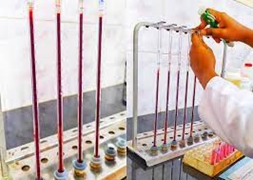
One other test that probably doesn’t make many “favorites” list is the Erythrocyte Sedimentation Rate (ESR), or sed rate. Remember old fashioned Westergren Sed Rates that took an hour to do, while the ER doctor kept calling looking for their “STAT” results? There are still labs that set up manual sed rates that take an hour, and modified manual methods that take “only” 15 or 30 minutes. Some semi-automated methods can give us results in a couple minutes, but still require techs to fill a capillary tube and load the instrument. Fortunately, real help may have arrived, in the form of fully automated ESR instruments! There are instruments now that actually make ESRs almost fun. I’ve never seen techs so excited about a new instrument as they were when we got iSeds. This thing is amazing! It’s like a little Ferris wheel for sed rates. You pop the whole tube in, they go for a little ride around the Ferris wheel, then drop out, in less than 30 seconds. And you can keep loading tubes even while it’s running. A truly Stat ESR. Now that’s amazing!

While these new instruments make ESR’s easier to run, with more reproducible results, and less hands-on time, they still don’t get much love, because, well…there are newer tests available for inflammation, and we know that the ESR is not a specific test for diagnosis. Across the years, some lab tests have become antiquated and obsolete…bleeding times come to mind, along with CK-MB. In 2010 an article was published that supported discontinuing laboratory tests that no longer have clinical utility in the lab. The ESR was on this list. Yet, many labs still perform ESRs. Should the ESR be phased out, or are there still valid reasons for ordering them?
Even though the test is considered non-specific, the ESR test is considered helpful in diagnosing two specific inflammatory diseases: temporal arteritis (TA) and polymyalgia rheumatica. A high ESR is one of the main test results used to confirm these diagnoses. It is also used to monitor disease activity and response to therapy in both these conditions. Almost all cases present with an elevated ESR, though a normal ESR should not be used to rule out these conditions.
Case 1: A 70 year old White female was admitted to the ER complaining of throbbing headache and blurry vision. She stated that the headache started 2 days ago, had been at her temples at first but in the past few hours was getting worse. She stated that she was prompted to come to the ER because now her whole scalp hurt, and her vision was blurry. A CBC, Basic panel, CRP and ESR were ordered. The CBC results were unremarkable, other than and increased platelet count of 480,000/µL. ESR was 110 mm/hr. Basic panel results were normal. CRP was 2.51 md/dL.
The patient was started on prednisone immediately, and a temporal artery biopsy was scheduled, with a suspicion of temporal arteritis (TA), also known as giant cell arteritis (GCA). TA is an autoimmune disease that causes inflammation of the temporal arteries. Under the microscope, the inflamed cells of these arteries look giant, which is how the disease got its name. The inflammation causes constriction of the arteries, can affect chewing and eating, and may cause blindness if not treated promptly. Treatment of choice are corticosteroids, often prescribed for at least a year. Symptoms are monitored frequently and lab results, including the ESR, can be used to monitor the condition and response to treatment.
If you are still wondering if the ESR should be discontinued as a useful test, we are now seeing patients with COVID infection and elevated ESRs. Over the past 2 years, several articles have been written about elevated ESRs in COVID-19 patients. One study aimed to evaluate the usefulness of ESR in distinguishing severe from non-severe COVID-19 cases. The study suggests that severe COVID-19 cases are associated with higher elevations of ESR, as compared to non-severe cases. A case report of a patient recovering from COVID described an increased ESR. The high ESR persisted for a long time even after the patient recovered from COVID-19, while no other inflammatory processes or other conditions known to raise ESRs were found.
Case 2: My second case is a case of a 58 year old woman who presented with an earache and a pulsing temporal headache. Ear infection was ruled out and the patient was referred to ophthalmology for possible TA. The patient’s CRP was elevated but her ESR and platelet counts were within normal reference range. The patient was COVID tested as part of a pre-op workup before temporal artery biopsy and the COVID-19 test came back positive. There have been cases in literature in the last year of this new set of symptoms in COVID-19 patients. The conclusion from these cases is that if a patient appears with symptoms consistent with TA with an elevated CRP but with a normal ESR and platelet counts, that the patients should be tested for COVID.
The ESR is one of the oldest laboratory tests still in use. The study of the sedimentation of blood was one of the principles on which ancient Greek medicine was based. In the 1700’s, physicians noticed that the rate of red blood cell sedimentation changed during illness. This theory was first introduced as a laboratory test over 100 years ago. Depending on the historic accounts and articles you read, it was first described by a Polish physician, Edmund Biernacki, in 1897, or by a Swedish physician, Robert Fahraeus, in 1915. Biernacki proved the connection between the rate of sedimentation and the amount of fibrinogen in the blood and suggested using the ESR in diagnostics. Alf Vilhelm Albertsson Westergren (a familiar name!) also presented a similar description of the ESR. In the early 1920’s. Dr Westergren went further to develop the blood drawing technique and defined standards for the ESR. To this day, the Westergren Erythrocyte Sedimentation Rate method is recognized as the gold standard reference method for ESR measurement.
The sed rate measures the rate at which erythrocytes sediment by gravity, in mm/hour. RBCs usually repel each other due to zeta potential and aggregation is inhibited. In conditions with increased fibrinogen or immunoglobulins, these proteins coat the RBCs, promoting aggregation. The RBCs form rouleaux which settle faster than individual RBCs. In conditions such as anemia, the ESR will be high because with a lower hematocrit, the velocity of the upward flow of plasma is altered and red blood cell aggregates fall faster. In polycythemia the increased blood viscosity can cause a lower ESR. In sickle cell anemia, and other conditions such as spherocytosis, the RBCs are abnormally shaped and will not form rouleaux easily, thus decreasing the ESR.
The ESR is an easy, inexpensive, non-specific test that has been used for many years to help diagnose conditions associated with acute and chronic inflammation. An elevated ESR is not associated with a specific diagnosis; therefore, it must be used in conjunction with other tests. Conversely, a normal ESR cannot be used to exclude the presence of significant disease. The ESR should also not be used as a screening test in asymptomatic patients. Since fibrinogen is an acute-phase reactant, the ESR is increased in many inflammatory and neoplastic conditions that increase fibrinogen, including diabetes, infection, pelvic inflammatory disease, lupus. rheumatoid arthritis, acute coronary syndrome, and neoplasms. However, noninflammatory factors such as older age, female gender, and pregnancy can also cause elevation of the ESR.
Historically, the ESR was used to indicate inflammatory conditions and monitor disease progression or response to treatment. More specific tests have been developed for many of these conditions, but the ESR still has its advantages. Interestingly enough, for a test that 12 years ago was on the ‘antiquated’ list, in the past 2 years there have been over 50 scientific journal articles written about the ESR. The ESR can eliminate unnecessary testing and help decrease medical costs. It has its advantages in small labs and in rural areas because it can provide quick results without expensive instrumentation. For labs that do not perform more sophisticated tests such as CRP and procalcitonin, the ESR can provide answers without waiting for results from reference laboratories. Even though an ESR may take 1 hour, it is much faster than send out testing. It can therefore expedite a diagnosis, or normal results can give the physician and patient timely reassurance.
What is your favorite or least favorite test in hematology? Let me know and I can highlight it in a future blog!
References
- Au, Benjamin Wai Yin MBBS, MMed (Ophth); Ku, Dominic J. BMed, MSurg; Sheth, Shivanand J. MBBS, MS (Ophthal) Thinking Beyond Giant Cell Arteritis in COVID-19 Times, Journal of Neuro-Ophthalmology: March 2022 – Volume 42 – Issue 1 – p e137-e139
- Brigden ML. Clinical utility of the erythrocyte sedimentation rate. Am Fam Physician. 1999 Oct 1;60(5):1443-50. PMID: 10524488.
- Hale AJ, Ricotta DN, Freed JA. Evaluating the Erythrocyte Sedimentation Rate. JAMA. 2019;321(14):1404–1405. doi:10.1001/jama.2019.1178
- Pu, Sheng-Lan et al. “Unexplained elevation of erythrocyte sedimentation rate in a patient recovering from COVID-19: A case report.” World journal of clinical cases vol. 9,6 (2021): 1394-1401. doi:10.12998/wjcc.v9.i6.1394
- Tishkowski K, Gupta V. Erythrocyte Sedimentation Rate. [Updated 2021 May 9]. In: StatPearls [Internet]. Treasure Island (FL): StatPearls Publishing; 2022 Jan-. Available from: https://www.ncbi.nlm.nih.gov/books/NBK557485/
- Alan H. B. Wu, PhD, Kent Lewandrowski, MD, et al. Antiquated Tests Within the Clinical Pathology Laboratory. The American Journal of Managed Care. September 2010, Volume 16, Issue 9
- https://emedicine.medscape.com/article/332483-workup

-Becky Socha, MS, MLS(ASCP)CMBBCM graduated from Merrimack College in N. Andover, Massachusetts with a BS in Medical Technology and completed her MS in Clinical Laboratory Sciences at the University of Massachusetts, Lowell. She has worked as a Medical Technologist for over 40 years and has taught as an adjunct faculty member at Merrimack College, UMass Lowell and Stevenson University for over 20 years. She has worked in all areas of the clinical laboratory, but has a special interest in Hematology and Blood Banking. She currently works at Mercy Medical Center in Baltimore, Md. When she’s not busy being a mad scientist, she can be found outside riding her bicycle.
