New at the Multiplex: The Syphilis Shuffle

Hello again everyone and welcome back!

If you caught my previous post it was a rather long twofer for my March rotation at the Mayo Clinic’s Department of Pathology and Laboratory Medicine as well as a great case study of a patient’s therapy-related AML in the setting of Li-Fraumeni Syndrome. Now, as I inch closer and closer to my last months of medical school, I’m doing another pathology clerkship at Danbury Hospital in southwestern Connecticut. It’s an excellent community-based pathology program with a great staff. As you’ve read in my posts before, community hospitals are no stranger to the leading edge of laboratory innovation. A fellow medical scientist in a recent ASCP membership video on social media said, “Laboratory medicine is at the precipice of change.” As a beacon of translational medicine, labs turn routine medical unknowns into answers. Often, they act as leaders in the lab medicine community because of certain population-specific testing and reporting that goes on at their institutions. You might recall my discussion of Bronx-Care Hospital leading the charge in New York City with the newest, 5th generation troponin testing or my experiences at Swedish Covenant Hospital in Chicago with lab automation, software innovation, and CQI.

But back to medical school: the coordinators of this rotation asked be about my interests in pathology and we discussed my past as a medical laboratory scientist. As such, they offered some special projects for me to be a part of in their lab! Specifically, and in addition to my subspecialty objectives and observership, I’ve been helping them with three small projects. First, I assisted in calibrating a freshly validated second Bio-Rad Bioplex2200 analyzer to correlate to a second instrument for some very interesting testing. Second, I’m helping gather ongoing inter-instrument data for a Sebia serum protein electrophoresis instrument. And finally, I’ve been assisting the histopathology section with cross-instrument validation of immunohistochemical stains as well as gathering data for the validation of a great new IHC that would replace PSA.  All of these have both used skills from my MLS foundational experience but taken that one step further into the scope of a pathologist by going over the clinical implications and testing outcomes provided by these analyses.

The Syphilis Shuffle

I already mentioned that Danbury’s lab uses the Bioplex2200, so let me tell you a bit about the analyzer and a bit more about an interesting way to do syphilis testing. It’s an interesting immunoassay instrument that uses something called “multiplex” technology. I won’t go into the details about footprint, throughput, and timing because, well, I’m not exactly expecting any Bio-Rad checks over here. But what I do want to talk to you about is the testing methodology. So, what exactly is “multiplex” testing. Basically, by using magnetic beads with fluorescent dyes associated with various tests you can create bead-set assays. Those beads are coated with detection proteins for each assay and exist in a single reagent pack. That allows numerous analytes to be detected at the same time from a single aspirated specimen. A laser in the instrument detects these immunologic events and reports the values. Now, in my experience with immunoassays for tests like syphilis, hepatitis, EBV, or HIV, there are usually honorable mentions of Abbott’s ARCHITECT and Siemens ADVIA Centaur alongside the Bioplex2200. I’m much more familiar with the former two, so getting a chance to work with the latter was great. But what did I learn?

Figure 1. Immunoassay guests-of-honor from left-to-right: Siemens ADVIA Centaur, Abbott ARCHITECT, and Bio-Rad BioPlex2200. I guess clinical immuno is all about the swivel workstation screen placement. But all three instruments are excellent at what they do; an aspect of laboratory medicine is analyzing your analyzers. Depending on the needs of the institution, your lab might have different demands of what limits of detection, turn-around, and sensitivity/specificity statistics are required for populations of patients.
Figure 2. Final reaction mixture with coated, tagged beads passing through the detector simultaneously. (Source: Bio-Rad)

First of all, other analyzers like the ADVIA Centaur use chemiluminescent immunoassay technology and the Abbott ARCHITECH use photometric, potentiometric, or turbidimetric detection for immuno and chemistry tests. There are published demonstrations of how the three instruments compare regarding various metrics and detection statistics. One of those papers from 2017 demonstrated the sensitivity and specificity for HIV testing across the analyzers. Overall, it said the ARCHITECT was the best performing instrument (spoilers: this study was funded by Abbott), though each had their strengths—read the paper here. The multiplex method, although similar in principle, is unique to the BioPlex. And one of the tests I find interesting is their syphilis assay. On the BioPlex, its tested as a total immunoassay with RPR that improves accuracy and precision in real-time. It’s a dual treponemal/non-treponemal test. Those bead packs contain two types of beads that qualitatively detect IgG and IgM antibodies to Treponema pallidum while also qualitatively detecting RPR antibodies in serum or plasma. It comes with its own internal control and verification beads for internal QC. Fast, simple, easy, unique and just as accurate as most syphilis testing—that’s what I’m talking about.

Figure 3. Multiplex dual treponemal/non-treponemal immunoassay acts as a one-step test with less steps and less requirments for confirmatory testing. Just set it and forget it! Unless your test comes back positive, then call the local health department and get that penicillin injection, STAT! (Source: Bio-Rad)

(Immuno) Fixing Everyone’s Problems

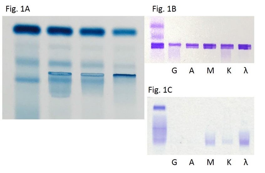
I remember learning a lot about gel electrophoresis, serum/urine protein electrophoresis, and immunofixation in MLS school but never really spent too much time with gels. I didn’t get to do much work with electrophoresis because I spent my time in labs either on a microscope doing diffs, a blood bank shaking tubes, or monitoring chemistry outputs and validating reports. Despite spending years with an ID badge that often said “chemistry” on it, gels were a less common test and not just for me. The primary care clinician usually only orders SPEP and subsequent gels in the investigation of multiple myeloma or other paraproteinemia from a vast array of disease process. As such, the results are often more challenging to interpret and require reporting and education from the pathology department. There are, however, a myriad of interpretable patterns and information within the gels of an SPEP. The principle is standard—proteins separate in media based on charge and mass—and particular patterns tell us more information than one might realize. For example, chronic inflammatory processes might increase production of acute phase reactants like alpha-1-antitrypsin or haptoglobin. Where do the peptide building blocks for those new proteins come from? Wherever the most protein is: Albumin, obviously for those playing at home. So your friendly normal neighborhood SPEP in a chronic inflammatory process might morph into something with a fainter albumin band and some extra attenuation in the alphas 1 and 2. If you see that, you might correlate with an ESR or CRP (or hs-CRP, you fancy laboratorian) …

Figure 4. We’ve all seen one of these before, but there are so many patterns to interpret which translate to real pathology and correlate well with concomitant serum values implicated in anything from myeloma, to infection, inflammation, or hepato-renal disorders. (Source: Univ. of Washington)

NKX3.1, the Lexus of Prostate IHCs

Now for something different. Let’s talk about prostates. Fun! More specifically, let’s talk about prostate-related immunohistochemical stains. The first one you’ll think off right away is probably …drumroll… PSA, and you’d be absolutely correct. That’s a standard IHC for detecting prostatic adenocarcinoma and is especially useful in finding metastases from a prostate primary. Though not brand new, there is another stain for prostatic IHC detection that, in some recent studies, has been shown to be more sensitive for prostate malignancy than PSA by about 5%. It’s called NKX3.1 and it has PSA’s sensitivity of 94.2% beat at around 98.6%–that could translate to plenty of earlier diagnoses and better outcomes for patients.

Image 1. Immunohistochemical staining of prostatic tissue. In IHC stains, brown is positive for expression. The immunologic technique of marking a specific antigen with a detectable antibody can be translated to a long list of tissue typing and can identify nuclear, cytoplasmic, or membranous patterns. For something like prostate tissue (seen here) sometimes PSA and/or NKX3.1 can identify prostatic malignancy or distant, suspicious metastases. (Source: BioCare medical)

What’s that got to do with my current pathology rotation? Well, I’ve gotten a lot of anatomical pathology exposure in my time here and I even helped correlate IHC stain quality across two instruments. With that done, I’m currently collecting specimens of saved tissue blocks that were both positive and negative for the lab’s current prostate IHC, PSA, and retesting them all with NKX3.1 in order to switch protocols to the new, more sensitive test. At the very least, the addition of a secondary validated prostatic stain would be useful. What’s important in gathering specimens for this kind of correlation is understanding when and where this new stain would be positive and negative and making sure it behaves in your patient population the way you would expect. NKX3.1 is supposed to be positive in nearly 99% of prostatic adenocarcinomas whether they’re primary or metastatic. It is a Chromosome 8 protein which is expressed in the prostate and testis and can even be found in the salivary glands, bronchial submucosal glands, and regions of the ureters. It can be positive in 27% of invasive lobular carcinoma, 25% of metastatic lobular carcinoma, 2-9% of invasive ductal carcinoma, and 5% of metastatic ductal carcinoma. (Source: Pathology Outlines) While I’m looking over specimens with historical orders for PSA IHCs, not all are positive, and not all are prostatic tissue. Conducting validation studies like these in pathology really require a good understanding of how to clinically correlate data with useful decision-making and tailor it to your patient population.

I wrote about a lot of topics this month, I know, but I think there’s a common theme. As a medical laboratory scientist, like many of you, I’ve worked out countless QC problems and instrument validations per protocol. Now that I’m making the transition to medicine in pathology, there’s a lot of forethought and planning that goes into validating or calibrating any test. In chemistry you need to get your limits of detection just right and match your throughput with the test volume your population needs. In hematology, you better know exactly how cells get detected by your analyzers and have a solid algorithm for working up and understanding aberrant flags. When it comes to anatomic pathology, speaking a common language of morphology and pattern-recognition is vital to reporting reliable and critically important data. Laboratory medicine always exists at the forefront of medical testing and methodology, and what that translates to on a day-to-day basis is being able to know how to find, make, or confirm a good, reliable test. As for me, medical school is full of unique experiences and rewarding opportunities to learn. This month, I couldn’t be happier to use my skills in the lab to connect my time at the bench to my work learning, calibrating, and validating for the next step.

And, after all, aren’t we all looking for a little validation now and then?

Thanks to Danbury Hospital’s Department of Pathology and Laboratory Medicine for having me this month and thank you all for reading.

See you next time!

–Constantine E. Kanakis MSc, MLS (ASCP)CM graduated from Loyola University Chicago with a BS in Molecular Biology and Bioethics and then Rush University with an MS in Medical Laboratory Science. He is currently a medical student actively involved in public health and laboratory medicine, conducting clinicals at Bronx-Care Hospital Center in New York City.

Insulin Resistance Caused by Insulin Antibodies

Insulin antibodies are seen in two conditions: 1, in insulin-naïve type-1 diabetic patients, insulin antibodies are developed together with some other autoantibodies against pancreatic islet cells; 2, in patients being treated with insulin, antibodies can be developed against exogenous insulins, in both type-1 and type-2 diabetes. These antibodies against exogenous insulins are found in >95% of patients treated with porcine and bovine insulins (1). Although the prevalence has decreased after the introduction of human insulin and insulin analogues, it is still not uncommon to detect these antibodies in insulin treated patients (2). However, these antibodies are rarely of clinical significance and laboratory test for insulin antibodies in insulin-treated patients has limited clinical value, except in rare cases where these antibodies are found to have immunologic role, causing insulin resistance. In some of these cases, postprandial hyperglycemia and nighttime hypoglycemia are both described due to reversible binding of insulin from antibodies (3), and patients were reported to respond to immunosuppressive therapies, and plasmapheresis in severe cases. 

We recently worked up a case for possible immunologic insulin resistance caused by insulin antibodies. In this case, patient is a 45 years old female with uncontrolled type-1 diabetes. She was found to have all four antibodies positive, including zinc transporter 8, islet antigens glutamate decarboxylase 65 (GADA), IA-2A, and insulin antibodies. Patient has been on multiple dose insulin injection (MDI) therapy, including insulin determir, aspart and lispro. She was reported to be compliant with medications and low carb diet. However, patient has poor glycemic control and presents with recurrent diabetic ketoacidosis. She was given high doses of insulins, but still presented with recurrent DKA and occasional hypoglycemia. Her HbA1c was consistently at >10% with daily glucose measured up to 500 mg/dL.

Immunologic insulin antibody and insulin receptor antibody were considered after ruling out more common causes of her uncontrolled diabetes. These two tests were then performed at a reference laboratory and patient was found to have positive insulin antibodies to analog insulin (determir and lispro) and negative insulin receptor antibodies. Significant insulin resistance by insulin antibodies was not found and the antibodies level did not suggest immunosuppressive therapy. Still, given her poor controlled diabetes, patient’s insulin was switched to human insulin and she was also recommend for pancreas transplant.

  1. Greenfield JR, Tuthill A, Soos MA, Semple RK, Halsall DJ, Chaudhry A, O’Rahilly S. Severe insulin resistance due to anti-insulin antibodies: response to plasma exchange and immunosuppressive therapy. Diabet Med. 2009 Jan;26(1):79-82. doi: 10.1111/j.1464-5491.2008.02621.x.
  2. Hall TR, Thomas JW, Padoa CJ, Torn C, Landin-Olsson M, Ortqvist E, Hampe CS. Longitudinal epitope analysis of insulin-binding antibodies in type 1 diabetes. Clin Exp Immunol. 2006 Oct;146(1):9-14.
  3. Hao JB, Imam S, Dar P, Alfonso-Jaume M, Elnagar N, Jaume JC. Extreme Insulin Resistance From Insulin Antibodies (Not Insulin Receptor Antibodies) Successfully Treated With Combination Immunosuppressive Therapy. Diabetes Care. 2017 Feb;40(2):e19-e20. doi: 10.2337/dc16-1975. Epub 2016 Dec 1.
Xin-small

-Xin Yi, PhD, DABCC, FACB, is a board-certified clinical chemist, currently serving as the Co-director of Clinical Chemistry at Houston Methodist Hospital in Houston, TX and an Assistant Professor of Clinical Pathology and Laboratory Medicine at Weill Cornell Medical College.

Chemistry Case Study: Protein Bands in All Lanes of the Immunofixation Electrophoresis

Waldenstrom Macroglobulinemia (WM) is defined as lymphoplasmacytic lymphoma (LPL) with IgM paraprotein and bone marrow involvement. The IgM paraprotein is an important serum marker for WM diagnosis, symptom prediction, disease burden assessment, treatment decision and drug response evaluation. Serum protein electrophoresis (SPEP) in conjunction with immunofixation electrophoresis (IFE) are the routine laboratory tests for IgM paraprotein detection, quantitation and characterization. A monoclonal protein typically presents as a sharp band on SPEP and selective lanes of IFE, allowing characterization of the immunoglobulin heavy chain isotypes and light chain classes. In rare situations, a monoclonal band is seen on all immunofixation lanes, suggesting cryoglobulin and/or soluble immune complex. We encountered a recent case of WM with a strong demarcated band on all immunofixation lanes.

The patient is a 76-year-old Chinese man diagnosed as WM/LPL by bone marrow biopsy. Peripheral blood showed pancytopenia with rouleaux formation. The serum IgM was up to 6900 mg/dL. Serum viscosity was increased up to 3.1 cP (normal range 1.5-1.9 cP). Serum rheumatoid factor was negative (<10 IU/ml). Serum protein electrophoresis (SPEP) on Sebia Hydrasys 2 showed a wide smearing pattern (Fig 1A). Serum protein immunofixation electrophoresis (IFE) showed a monoclonal band on all lanes with equal intensity, preventing isotype identification (Fig 1B). This pattern is generally believed to be due to cryoglobulin and/or polymerization of monoclonal proteins, similar to rheumatoid factor activity. Urine electrophoresis was consistent with an overflow pattern and urine immunofixation showed monoclonal free lambda light chain.

Previously it was demonstrated that cryogolublin dissolution was achieved by pre-treatment of serum samples with Fluidil. The IgM polymer can be disrupted by adding reducing agents such as beta-mercaptoethanol (bME) to disrupt the disulfide bonds (1-2). In our case, despite pretreatment with Fluidil and bME, no isotype resolution was achieved on serum IFE, prompting us to develop a novel method through the addition of sodium dodecyl sulfate (SDS) to the pretreatment process. Different combinations of reaction conditions were tested, including SDS concentration ranging from 0.01 to 1%, three different temperatures (37, 56 and 95 °C), three different concentrations of bME (1%, 2% and 4%) and three different serum volume (25 µL, 50 µL and 75 µL). Optimal isotype resolution was achieved using 0.1% SDS/0.25%bME/Fluidil incubated at 56°C for 30 mins (Fig 1C).

WMimg1


References

  1. Attaelmannan M, Levinson SS. Understanding and identifying monoclonal gammopathies. Clin Chem. 2000 Aug; 46(8 Pt 2):1230-8.
  2. Yusra Othman. Protein Bands in All Lanes of the Immunofixation Electrophoresis Pattern of Serum From a 50-Year-Old Saudi Woman. Lab Med (2006) 37 (3): 152-154.


HL

-Huifei Liu, MD, PhD. Former PGY4 resident in the Department of Pathology and Genomic Medicine, Houston Methodist Hospital. She currently serves as the associate medical director at Hematologics, Inc., Seattle, WA.  

Xin-small

-Xin Yi, PhD, DABCC, FACB, is a board-certified clinical chemist, currently serving as the Co-director of Clinical Chemistry at Houston Methodist Hospital in Houston, TX and an Assistant Professor of Clinical Pathology and Laboratory Medicine at Weill Cornell Medical College.

Microbiology Case Study: A 14 Year Old Female with Neck Swelling

Case History

A previously healthy 14-year-old female presents to the emergency department with three days of progressive facial and neck swelling. The swelling started on the left side. Two days ago she visited her primary care physician where she had negative monospot and mumps IgM testing.  She is fully vaccinated, but was exposed to a mumps outbreak at school.

Discussion

Our patient was diagnosed with mumps by positive RT-PCR from a buccal swab. The mumps virus is a member of the Paramyxoviridae family which includes notable human pathogens parainfluenza, Hendra, and Nipah viruses. Members of this family are enveloped, helical viruses with single-stranded, non-segmented RNA genomes with negative polarity. Mumps is an obligate human pathogen that replicates in the epithelial cells of the upper respiratory tract and subsequently moves to regional lymph nodes. It is spread from person to person via direct contact with respiratory secretions or contact with contaminated fomites. Mumps is a highly contagious disease with as high as 85% of naïve individuals becoming infected after contact with a mumps infected individual. It spreads most efficiently in areas where there is close contact among individuals for prolonged periods of time such as college campuses and close-knit religious communities.

Prior to vaccination for mumps in the 1960s, greater than 150,000 cases of mumps occurred each year in the US. The incubation period for infection is 16-18 days, with the majority of infected persons being asymptomatic or having mild respiratory symptoms. Orchitis causing sterility in post-pubescent males is the main concern of mumps infection but other rare but serious complications include mastitis and oophoritis in females, meningoencephalitis, pancreatitis, and deafness.

Due to sporadic outbreaks of measles since the introduction of the vaccine, the vaccine schedule has been revised from one dose of the MMR (measles, mumps, and rubella) vaccine at age 12-15 months to include another MMR booster at age 4-6 years. We are currently in the middle of yet another outbreak with nearly 6,000 cases of mumps reported to the CDC in 2016 and a high rate of infections reported thus far in 2017 (Figures 1 and 2).

mumps1
Figure 1. Number of cases identified by the CDC in 2017 by state. (Figure courtesy of the CDC Mumps website at https://www.cdc.gov/mumps/outbreaks.html. Content source: National Center for Immunization and Respiratory Diseases [NCIRD], Division of Viral Diseases)
mumps2
Figure 2. Number of cases of mumps per year identified by the CDC.
(Figure courtesy of the CDC Mumps website at https://www.cdc.gov/mumps/outbreaks.html. Content source: National Center for Immunization and Respiratory Diseases [NCIRD], Division of Viral Diseases)


Diagnostic Testing for Mumps

Serological testing for IgM and RT-PCR from a buccal swabs are the mainstay of mumps diagnosis. IgM becomes positive in the first 3-4 days after symptom onset and will remain positive for 8-12 weeks. IgG becomes positive 7-10 days following symptom onset and will remain at high levels for many years and detectable for life. In a vaccinated individuals, IgM testing has less utility as it may be non-reactive or weakly positive following a secondary immune response.

RT-PCR from a buccal swab specimen is the most sensitive test for diagnosis of mumps. It should be performed as soon as a patient is symptomatic, as testing by this method is the most sensitive in the first few days following symptom onset and becomes less sensitive as time goes on.

Urine specimens can be used to isolate mumps in viral culture. Urine is not positive for mumps until greater than 4 days post symptom onset and is less sensitive than PCR performed on the bucal swab. For these reasons, viral isolation from urine is no longer a commonly used test for diagnosis of mumps, although viral culture is still considered the gold standard for mumps conformation.

Resolution

The patient and her family were counseled on the infectious nature of mumps. She was instructed to remain in isolation at home for 6 days after resolution of swelling.


References

  1. Manual of Clinical Microbiology, 11th edition
  2. CDC Mumps Website (www.cdc.gov/mumps/index.html)


Erin McElvania TeKippe, PhD, D(ABMM), is the Director of Clinical Microbiology at Children’s Medical Center in Dallas Texas and an Assistant Professor of Pathology and Pediatrics at University of Texas Southwestern Medical Center.

Serum Protein Electrophoresis in Children

Although most of the testing performed and the methodologies utilized in a clinical laboratory which serves a pediatric institution are very similar to those found in adult laboratories, a few differences stand out. Differences include devising ways to deal with small test volumes and different test menus than those found in laboratories that serve adult patients. One such test menu differences is the lack of serum protein electrophoresis (SPEP) testing in pediatric labs.

SPEP’s are essentially performed for the main purpose of helping to diagnose and then monitor the treatment of multiple myeloma. SPEPs provide this help by detecting, identifying by reflex immunofixation electrophoresis (IFE), and quantifying monoclonal gammopathies. Children don’t get multiple myeloma. After 20 years of signing out SPEP results at the county hospital next door, the youngest person with a diagnosis of multiple myeloma that I’ve seen was 24 years old. Thus in general SPEP’s are not ordered on children, nor performed in pediatric labs.

Recently however, I learned that although children don’t get multiple myeloma, they do in fact get monoclonal gammopathies. An SPEP ordered on a 7 month old patient in my institution came back with a very clear biclonal gammopathy, identified by IFE as an IgG kappa and an IgA kappa. This child has no bone marrow indication of abnormality, although she does have a deficiency of B-cells along with plasma cell infiltrates in the liver and duodenum.

A little searching determined that apparently, monoclonal spikes on SPEP’s in children are not at all unusual. A study published in 2014 (1) looked at 695 children who had SPEPs performed, and 11% of those children had a monoclonal gammopathy, although none of them had multiple myeloma. The most common associated diagnosis was ataxia-telangiectasia (22%), with a wide range of other diagnoses being found in these children, including some immunodeficiencies, autoimmune diseases, various hematological disorders and a few solid organ malignancies.

Thus it appears that monoclonal gammopathies are present in children and have an entirely different meaning than they do in adults. In addition, currently monoclonal gammopathies in children have no clear diagnostic utility. Perhaps that is the real reason we don’t routinely perform them in the pediatric population.

  • Karafin MS, Humphrey RL, Detrick B. Evaluation of monoclonal and oligoclonal gammopathies in a pediatric population in a major urban center. AJCP 141:482-487. 2014

-Patti Jones PhD, DABCC, FACB, is the Clinical Director of the Chemistry and Metabolic Disease Laboratories at Children’s Medical Center in Dallas, TX and a Professor of Pathology at University of Texas Southwestern Medical Center in Dallas.