Up to Date or Up for Debate?

Hello again everyone! Welcome back.

This month I think it’s important to take a step back from clinical pearls, developing our interpersonal skills, and interdisciplinary dynamics and go back to what I started writing about here on Lablogatory: public health and shaping policy. (Sorry, no Transformers, Simpsons, or Star Trek this time.)

Now, you may or may not have heard in recent news that the United States Preventive Services Task Force (USPSTF) updated their long-standing guidelines for screening women for cervical cancer. Normally I wouldn’t file this away under “exciting must-read,” but I was piqued when I also read that ASCP along with the College of American Pathology (CAP), American Society of Cytopathology (ASC), American Society for Colposcopy and Cervical Pathology (ASCCP), the Society of Gynecologic Oncology, the American College of Physicians (ACP), the American Society for Cytotechnology (ASCT), the American Cancer Society (ACS) the Papanicolaou Society of Cytopathology, as well as the American College of Gynecologists (ACOG) and other professional institutions and individuals voiced concerns over the changes to the USPSTF standard.

This is a topic that can be discussed for days, but I’ll do my best to give you the readers’ digest and present the main contentions regarding this standard of patient care and laboratory methodology.

Woah. What’s going on?

Basically, because of some new research and recommendations, the USPSTF—a body which publishes the standard of care for nearly every conceivable aspect of preventive care in the US—rolled back on the algorithms for screening women for HPV and cervical cancer. It all comes down to the utilization of co-testing (doing both Pap smear cytology and HPV testing for certain age demographics) as a point of contention. Under a banner of addressing keywords like “cost” and “harm,” these new recommendations have left clinicians both in and out of the lab in stirrups—sorry, couldn’t resist that one. But don’t worry, I wouldn’t be able to track these changes or even understand them without some sort of visualization. When it comes to recommendations, standards, and guidelines I’m about as proficient as a broken manual diff counter…

cotesting1
Figure 1. These are the guidelines as they stand from each of the major professional organizations concerned with cervical cancer screenings. Dissenting/recommended opinions are highlighted. To the untrained eye this is very unexciting. The bottom line is that the USPSTF no longer recommends co-testing for screening. Source: adapted from UpToDate

Slow down. Explain co-testing and primary testing? What exactly do the old and new recommendations mean?

Okay. When women undergo routine cervical cancer screening they receive Pap smears (cytologic examinations) every three years. This testing has been the standard for a number of years and is adequately sensitive for women up to the age of 30. Often times, these younger women may have slight intraepithelial changes (LGIL) which are considered low grade and remiss on their own. After that age it has become standard practice to add the additional test (while collecting the Pap specimen) of HPV DNA testing. This adds an increased level of sensitivity/specificity and is called co-testing. The new recommendations depart from this co-testing model, citing that there are increased harms (in the form of false positives) which ultimately lead to waste and unnecessary testing for women after the age of 30. Primary testing would mean only screening with HPV DNA assays after 30. According to the National Cancer Institute, all available literature on the subject of HPV and cervical cancer testing adequately demonstrates that co-testing is the best option. A number of studies were compiled to address the harm vs. benefit of Pap and HPV testing. Together, however, these tests decrease the incidence of cervical cancer. New guidelines were made based off mathematical projections and cost-benefit analyses which try and minimize losses for screening. Dr. J. Kim, a public health researcher at Harvard, was integral in contributing models which projected the cost/benefit of changing HPV guidelines. Essentially, the study projected that, when considering “harm” (i.e. colposcopy/false negative) abandoning co-testing changed the mortality rate from 0.3-0.76 per 1000 women with co-testing, to 0.23-0.29 per 1000 women with primary HPV testing. An impressive and significant statistical advantage. However, the total number of unscreened women with mortality rates was between 1-2%. This study was a microsimulation done from historical data within rates of cytologic detection and retrospective testing data on women, projected for a future hypothetical 5 year interval. Fascinated by this study, I tried to reach out to Dr. Kim to discuss limitations in using models and simulations and public health evidence to change practices, but I’m sure she is busy and could not respond in time.

So, to co-test or not to co-test, that’s the question. Right?

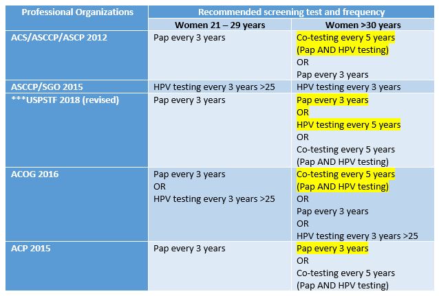
In its simplest sense, yes. The major medical professional societies also publish their most current recommendations for practice standards—and the issue is that the USPSTF took a departure from what most of the professional societies recommend regarding co-testing. Late last year, the CAP, ASC, ASCT, ASCP, and the PSC issued a statement under their independent collaboration called the Cytopathology Education and Technology Consortium (or CETC). In this response to the USPSTF guideline changes, they discussed their concerns. Specifically, their objections center around the fact that without co-testing for screening, cancer prevention might be impacted negatively. The CETC claimed that sensitivity is already maximized with previously recommended co-testing guidelines. They also cite that there is only one FDA approved HPV primary screening test available—and not all labs have it! More so, CETC discussed the need to keep morphological testing continuous for women who have histories of Pap smears, the potential to overwhelm colposcopy services for screening all positive HPV patients, and the honest reality that not all clinicians would be compliant with the way the USPSTF recommends testing. The bottom line from this consortium:

  1. Cytology and high-risk HPV co-testing should be kept as the standard screening for women aged 30-65
  2. Primary HPV screening should only be done with validated, FDA approved testing methodologies
  3. HPV screening methods should continue their current schedule until longitudinal data can offer new evidence for changes

So, what’s the current technological climate for how we test for HPV?

Currently, most clinicians do co-testing. The standard for Pap smears utilizes a physical tool to collect epithelial cells from the cervix at vaginal, ectocervical, and endocervical sites. The swabs are prepped on 1-2 slides, fixed with alcohol or other spray cell-preservatives and sent to labs for cytologic examination. The basic Papanicolaou staining procedure uses hematoxylin for nuclear staining, and two cytoplasmic counterstains.  This is essentially a modified H&E stain to clearly visualize morphology.  Staining is rarely done manually and some instruments offer stain/prep combination capability. I couldn’t find too much information on this, but I remember there not being too many official FDA approved prep machines for Pap specimens. Cytotechs and pathologists read the slides and issue sign outs on morphology according to the Bethesda system—very heavy read, don’t bother; essentially it has three main categories of normal, benign changes, and abnormal. According to ASC “for squamous lesions, TBS terminology includes atypical squamous cells of undetermined significance (ASCUS), low grade intraepithelial lesion (LGSIL or LSIL), high grade intraepithelial lesion (HGSIL or HSIL) and squamous cell carcinoma.  Some laboratories also incorporate other terminologies of dysplasia and/or cervical intraepithelial neoplasia (CIN) into their reports.  For glandular lesions, TBS terminology includes atypical glandular cells of undetermined significance (AGUS) and adenocarcinoma.”

As of now FDA approval for HPV primary testing for high-risk strains is limited to the Roche Cobas hrHPV test. I could link you to their website, but you’ll be sold right away. They tout the future of HPV screening is HPV primary testing and to do away with the Pap! Their graphs and figures are impressive (just like their price tag!) and there’s no doubt that sensitivity is something that real-time PCR provides more than cytologic examination. But, as always, more assays will be approved, and advancements will tweak the sensitivity and specificity higher and higher.

Got it. So, technology and lab tests are always advancing, why can’t we make this change?

It’s not so easy to change the method or assay we use to screen or diagnose patients in the lab. If you recall, I talked about how the hospital I’m currently rotating in is leading the region in advancing the new high-sensitivity troponin assays. It’s still a hard sell to many even though the data and projections seem to all point to a green light. But that’s a paradigm shift that involves side-stepping from one immunochemical assay to a more sensitive immunochemical assay. It’s the same stuff, just sharper and with more clinical data to interpret with regards to acute coronary symptoms and clinical risk stratification. Swapping an old car for a new car. This conversation is a bit more complex. The recommendations for cervical cancer screening suggest that we should move away from mostly morphologically-driven, human-based cytology interpretation and move toward PCR-based assays for detection. Literally apples to oranges. We might think we know which one is better right now, but longitudinal studies are the only way to really tease out if this change in practice to improve patient outcomes in the long run.

Where do we go from here?

Ultimately, I think a few things need to happen for this recommendation to become standard practice. First, professional societies in every discipline from gynecology to cytology need to come to an agreement. It remains to be seen whether certain agencies will adopt and recommend the USPSTF guidelines, and statements from groups like CETC reveal a vote of no confidence in this current climate. Ultimately, because of numerous objections (including the ones from ASCP and the CETC) the USPSTF does say that co-testing is still optional between patient and provider, so we’re not really in crisis mode. But what happens when the advancements and the recommendations catch up to our ability to abandon the cytologic contributions of a future useless Dr. Papanicolou? We could probably deal with that when it comes to fruition, but until then we have a real discussion about what “harm” really is. Is colposcopy flat out harm? Or are the false positives that reflex to further testing? Is the current practice a safety-net for populations across socio-economic tiers with varying access to screening in the United States? When compared to other countries, HPV prevention, vaccination, and screening is much more easily facilitated. Is this a contributing factor for our messy guidelines? Will there be more options for FDA approved methodology in the near future? There remain a number of good questions which require examining cross-sections of data and patient outcomes. And, I believe, we may see change soon—but not quite yet.

What are your thoughts? What have you experienced in your lab or clinic? Leave your comments below!

Thank you and see you next month!

References

  1. ASCP One Lab. 2018. ASCP Declares Victory for Patients with Revised USPSTF Cervical Cancer Recommendation. Aug 21, 2018. Accessed Sep 2018: http://labculture.ascp.org/community/news/2018/08/21/ascp-declares-victory-for-patients-with-revised-uspstf-cervical-cancer-recommendation
  2. 2018. Vaccines and Preventable Diseases. HPV Vaccine Recommendations. Centers for Disease Control and Prevention. Atlanta, GA. Accessed Sep 2018. https://www.cdc.gov/vaccines/vpd/hpv/hcp/recommendations.html
  3. 2017. Response to New USPSTF Draft Guidelines for Cervical Cancer Screening. Cytopathology Education and Technology Consortium. Accessed Sep 2018: https://s3.amazonaws.com/ascpcdn/static/ONELab/pdf/2017/CETC+-USPSTF+Letter+10-2-17.PDF
  4. Basu, S. 2013. Complexity in Mathematical Models of Public health: A Guide for Consumers of Models. PLOS, Medicine. Oct 29, 2013. https://doi.org/10.1371/journal.pmed.1001540
  5. Felscher, K. 2018. The science behind new screening guidelines for cervical cancer. An Interview with Dr. J. Kim. Harvard T.H. Chan, School of Public Health. Accessed Sep 2018: https://www.hsph.harvard.edu/news/features/science-behind-new-screening-guidelines-cervical-cancer/
  6. Kim, J. 2017. Screening for Cervical Cancer in Primary Care. Journal of the American Medical Association (JAMA). 2018;320(7):706-714. Doi:10.1001/jama.2017.19872
  7. Lerman, L. 2018. Screening for Cervical Cancer – New Tools and Opportunities. Journal of the American Medical Association (JAMA) – Opinion, Editorial. Vol. 320(7):647-649
  8. Nayar, R. 2017. Primary HPV Cervical Cancer Screening in the United States: Are We Ready? Journal of the American Society of Cytopathology (2017) 7, 50e55
  9. Nelson, R. 2018. USPSTF Updated Recommendations for Cervical Cancer Screening. Medscape. Accessed Sep 2018: https://www.medscape.com/viewarticle/900985#vp_3
  10. 2018. Cervical Cancer Screening (PDQ) Health Professional Version. National Cancer Institute. Accessed Sep 2018: https://www.cancer.gov/types/cervical/hp/cervical-screening-pdq#link/_133_toc
  11. Sawaya, G. 2018. Cervical Cancer Screening—Moving from the Value of Evidence to the Evidence of Value. Journal of the American Medical Association (JAMA), Internal Medicine. doi:10.1001/jamainternmed.2018.4282
  12. Up To Date. 2018. Cervical cancer screening recommendations from United States professional organizations. Accessed Sep 2018: https://www.uptodate.com/contents/image?topicKey=7575&search=&source=outline_link&imageKey=PC%2F82951
  13. 2018. Cervical Cancer: Screening. Recommendation Summary. August 2018. Accessed Sep 2018: https://www.uspreventiveservicestaskforce.org/Page/Document/UpdateSummaryFinal/cervical-cancer-screening2
  14. USPSTF. 2018. Screening for Cervical Cancer, US Preventive Task Force Recommendation Statement. US Preventive Task Force. Journal of the American Medical Association (JAMA) 2018;320(7):674-686. Doi:10.1001/jama/2018.10897


ckanakisheadshot_small

–Constantine E. Kanakis MSc, MLS (ASCP)CM graduated from Loyola University Chicago with a BS in Molecular Biology and Bioethics and then Rush University with an MS in Medical Laboratory Science. He is currently a medical student actively involved in public health and laboratory medicine, conducting clinicals at Bronx-Care Hospital Center in New York City.

History of Generations: GenZ

The newest generation, Generation Z, is born in the 21st century. The oldest are now 18, which means that some have started entering the work force in entry-level positions. This generation is even more comfortable with technology than Millennials, as they grew up with computers, laptops, cellphones, internet and social media all around them.

The older Gen Zers are aware of the financial crisis that occurred, which created a strong focus on saving money. This generation was brought up with a sense of “Stranger Danger” so they are concerned with their own and public safety. They have a strong family orientation and consider themselves global citizens. They are characterized by an entrepreneurial spirit, the idea that anyone can be famous, are open-minded, and care deeply about the environment.

Because of the rising cost of education, many are worried about the price of college and about saving money for their parents. It is a little too early to tell because this generation is still young, but they could have feelings of unsettlement and insecurity due to the state of the economy, environment, and world. They are very loyal, compassionate and independent and have friends around the world, even if they have never traveled abroad themselves.

lotte-small

-Lotte Mulder earned her Master’s of Education from the Harvard Graduate School of Education in 2013, where she focused on Leadership and Group Development. She’s currently working toward a PhD in Organizational Leadership. At ASCP, Lotte designs and facilitates the ASCP Leadership Institute, an online leadership certificate program. She has also built ASCP’s first patient ambassador program, called Patient Champions, which leverages patient stories as they relate to the value of the lab.


I think we’re embarking on an incredible generation. I interviewed someone from each of our generations about how they observed, interacted with, learned from the Generation Zs. Here are their thoughts.

The Traditionalist: Ned the Grandpa

As the grandpa of two Gen Z grandsons, Ned found them to have an expanded knowledge base of the entire world. They are sophisticated in their analysis and critical thinking because of their exposure to information that their phones and computers provide them.

Lastly, they value human diversity far more than his own generation.

The Baby-boomer: Donna the Grandma

Donna is a “Grandma Boomer” and finds the Gen Z grandchildren’s vocabulary amazing. She says they are obsessed with the mechanical stuff and are used to doing 2-3 things at the same time. They still love sports, however, it’s like a class that they study. They attend practices but still play with their friends on their computers or phones. However, they “only” text. They don’t talk on the phone.

The Gen Z’s are far more sophisticated than the Boomers, yet they can’t write or spell as well as other generations. They don’t know cursive, and the first question they ask when going somewhere is, “do they have WIFI?” Oh, and “do you have a charger?”

Another Boomer: Susan the Grandma

Susan’s greatest concern was that many high-schoolers were being treated for levels of anxiety. Why? There’s no “turn off switch” with the world. They are almost required to stay tuned to respond or react to friends 24/7. Life is all about them from Instagram to Twitter, and Snapchat and tracking the number of followers.

The GenXer, Kim the Aunt

Her nephews are definitely focused on technology. They do not like talking on the phone and prefer to only text. They have incredible access to information, but they still like to play family games because they value tradition. Her nephews are great travelers and most comfortable with airports, planes and trains, Vs. just cars or bicycles. This is attributed to their expanded world. So what’s their greatest fear? A dead battery!

Maddie the Millennial

Maddie was shocked when she noticed that her sister, who is a Gen Z, was communicating via texting with her friend who was in the same room!


Stakenas-small

-Catherine Stakenas, MA, is the Senior Director of Organizational Leadership and Development and Performance Management at ASCP. She is certified in the use and interpretation of 28 self-assessment instruments and has designed and taught masters and doctoral level students.  

Hematopathology Case Study: A 43 Year Old Man with History of Latent TB

Case History

43 year old Vietnamese speaking man with a history of treated latent TB who presented with one month of fevers, night sweats, weight loss, and acute left facial swelling with associated pain, nasal congestion and 2 nose bleeds. The patient was found to have a polypoid mass within the left interior nasal cavity.

Biopsy Left Nasal Mass

nkt-he10x
H&E 10x
nkt-he40x
H&E 40x
nkt-cd3
CD3
nkt-cd5
CD5
nkt-cd56
CD56
nkt-perforin
Perforin
nkt-67
KI-67
nkt-eber.jpg
EBER

Diagnosis

The biopsy shows nasal mucosa with a dense submucosal lymphoid infiltrate and large areas of necrosis. The lymphocytes are somewhat pleomorphic, medium to large in size with irregular nuclear contours, vesicular chromatin and inconspicuous nucleoli. There are scattered mitoses and apoptotic cells.

By immunohistochemistry, CD3 highlights the lymphoma cells, which comprise the majority of the lymphoid infiltrate. The lymphoma cells co-express CD56 and CD7 (dim) and are negative for CD2 and CD5. The lymphoma cells also express cytotoxic markers perforin and granzyme (major subset). CD20 highlights only rare small clusters of B-cells. The lymphoma cells are also positive for EBER (Epstein-Barr Virus encoded RNA) in situ hybridization.  The Ki67 (MIB1) proliferation index is 60% with focal areas exhibiting up to 80%.

Taken together, the morphologic and immunophenotypic findings are consistent with an extranodal NK/T cell lymphoma, nasal type.

Discussion

Extranodal NK/T cell lymphoma, nasal type is an aggressive lymphoma that is more prevalent in Asian and South American populations. It occurs most often in adults and is more common in men than women. It is generally located in the upper aerodigstive tract, with the nasal cavity being the prototypical site. Patients tend to present in a manner similar to the patient described in this case, with rhinorrhea, pain, nasal obstruction and epistaxis due to a mass lesion. 1 The term “lethal midline granuloma” was once used to describe this entity because patients can present with locally destructive mid-facial necrotizing lesions. The early non-specific symptoms can pose a diagnostic challenge, and often result in treatment delays, which makes the aggressive disease more lethal. 2

The entity is described as NK/T cell lymphoma because although most cases are of NK-cell origin, some cases are comprised of cytotoxic T-cells. Natural killer (NK) cells are non-T and non-B lymphocytes that are part of the innate immune system. They respond immediately to antigenic challenge and are able to directly kill virally infected cells without the help of antigen presenting cells. They also secrete cytokines to increase the innate immune response. NK cells are classically positive for cytoplasmic CD3 and CD56, as well as cytotoxic molecules granzyme and perforin. Of note, NK-cells lack recombination activating gene enzymes and therefore have no clonal molecular marker for gene rearrangement such as the T-cell receptor or Immunoglobulin heavy chain.  3

Microscopically, the involved sites generally have widespread mucosal destruction. There is an angioentric and angiodestructive growth pattern that results in extensive necrosis. Another important diagnostic consideration is the very strong association with EBV. EBV is present in a clonal episomal form. This means that the infection occurs prior to and likely plays a pathogenic role in the development of NK/T cell lymphomas. 3

Following diagnosis, staging and management of the disease involves quantification of circulating EBV DNA. This can be used as a laboratory marker for disease status and progression or remission of disease. PET/CT is performed for accurate staging and patients are most commonly treated with a combination of radiotherapy and the SMILE regimen, which includes dexamethasone, methotrexate, ifosfamide, L-asparaginase and etoposide. NK/T cell lymphomas are aggressive and patients tend to have a short survival and poor overall response to therapy. 3

A recent study by Kwong, et al. showed the potential use of PD1 (programmed death ligand 1) blockade drug pembrolizumab in the treatment of relapsed or refractory NK/T-cell lymphoma. As mentioned above, the lymphocytes in this entity are invariably infected with EBV. PD1 is known to be up regulated in cells infected with EBV. In the study, seven patients who had failed treatment with the SMILE regimen were treated with pembrolizumab. After a medium follow-up of 5 months, 5 patients remained in complete remission and all patients had objective responses to treatment. 4 This shows promise as a potential new treatment for patients with this uncommon, but deadly disease.


References

  1. Swerdlow SH, Campo E, Harris NL, et al. WHO Classification of Tumours of Haematopoetic and Lymphoid Tissues (Revised 4th edition). IARC: Lyon 2017.
  2. Mallya V, Singh A, Pahwa M. Lethal midline granuloma. Indian Dermatology Online Journal. 2013;4(1):37-39. doi:10.4103/2229-5178.105469.
  3. Tse, E, Kwong, Yok-Lam. The diagnosis and management of NK/T-cell lymphomas. Journal of Hematology and Oncology. 2017:10:85. Doi: 10.1186/s13045-017-0452-9.
  4. Yok-Lam, K, Thomas, S.Y. Chan, Daryl Tan, et. al. PD1 blockade with pembrolizumab is highly effective in relapsed or refractory NK/T-cell lymphoma failing L-asparaignase. Blood. 2017:129:2437-2422. Doi: 10/1182/blood-2016-12-758641.


Marcus, Chelsea_099-Edit

Chelsea Marcus, MD is a third year resident in anatomic and clinical pathology at Beth Israel Deaconess Medical Center in Boston, MA and will be starting her fellowship in Hematopathology at BIDMC in July. She has a particular interest in High-grade B-Cell lymphomas and the genetic alterations of these lymphomas.

Regulatory Inspections: Are You Ready?

Whether your laboratory is accredited by CAP, COLA, JCAHO, or simply adheres to local state and federal CLIA regulations, all laboratories are subject to regular inspections from their accrediting agencies. Normally the thought of an inspection places staff into panic or hiding mode (“Whew, glad I’ll be off that week!”), but if you prepare ahead of time, the inspection process can be an extremely valuable tool to access the overall quality of your laboratory program. Over the next 3 blog posts we’ll review tips on 1) how to prepare before your inspection, 2) what to expect during the inspection itself, and 3) how to address any deficiencies identified by the inspection team.

Part One: The Inspectors Are Coming!

Know the Regulations. As technology evolves and new laboratory methodology is introduced, the requirements for your respective regulatory agencies will be updated as well. Know what version of your checklists or standards you will be getting inspected on, and ensure all staff are familiar with any updates and changes. By engaging all staff in the inspection process from lab assistants up through management, everyone will be aware of what the requirements are and can actively participate to ensure the lab is meeting those requirements.

Focus on Previous Citations. Your inspectors will have access to your previous inspection results, and will be following up on any citations. Ensure that the corrective actions and preventive actions you said you were going to implement have actually gone into effect. For any procedural changes, have documentation (Read & Understand) ready to show that all staff were made aware of and have been trained on the changes. Make sure that those corrections have been sustained and are effective at addressing the noted citation. It’s great to add on new forms to document instrument maintenance – but not if your staff doesn’t have the time to complete them. Again, engage your staff to see what is working, and what needs to be reevaluated.

What is New? New regulatory requirements, new staffing, new instruments, new testing methodologies… These are all key areas that the inspectors will focus on. Have you kept current with your regulatory updates and implemented any necessary changes to address the new requirements? Do you have documented training and competency for each new staff member for each task they are performing? For new instruments, ensure they have been fully validated and correlated to similar instruments prior to being placed into use for patient testing. When adding on new tests, ensure you have a full validation summary with medical director approval and sign off, and that your testing activity menu has been updated as well.

Have a Plan. Depending upon the size and scope of your laboratory, there can be a lot to cover for your inspection preparations. If you wait until your inspection window opens to start getting ready, things will be overlooked or simple “quick fixes” will be implemented instead of finding a long term sustainable solution to any potential issues. Instead, schedule tasks throughout the year to continually review your quality assurance program. Ask management to review 3 – 5 SOPs each month for content (does SOP match the current manufacturer package insert, does SOP match current practice in use), rather than a mass annual sign off. Perform quarterly reviews of your maintenance documentation to ensure all logs have been filled out completely with corrective action documented when appropriate. Utilize calendar reminders to track proficiency testing sample results, and ensure proper follow-up for any non-satisfactory results.

Perform Meaningful Self-Audits. For most regulatory agencies, performing self-audits on your non-inspection years is a requirement. Make this task meaningful by using a fresh set of eyes to review your documents. Ask the hematology staff to inspect the chemistry department; chemistry to inspect urinalysis; urinalysis to inspect microbiology…. You don’t need to understand how to actually perform a specific weekly maintenance task on a particular instrument; you just need to ensure that all those weekly tasks have been documented every 7 days (or less). You don’t need to understand what reagent ‘XYZ’ is used for, you just need to ensure that the vial is properly labeled with an open and expiration date, and that it is not currently expired and still being used. Self-audits should not be punitive; they are meant to catch things that you may be taking for granted are compliant, when in fact they truly are not.

Stay tuned for part 2 coming out next month, where we’ll discuss the inspection process itself and what to expect from the inspection team.


Nevins-small

-Kyle Nevins, MS, MLS(ASCP)CM is one of ASCP’s 2018 Top 5 in the 40 Under Forty recognition program. She has worked in the medical laboratory profession for over 18 years. In her current position, she transitions between performing laboratory audits across the entire Northwell Health System on Long Island, NY, consulting for at-risk laboratories outside of Northwell Health, bringing laboratories up to regulatory standards, and acting as supervisor and mentor in labs with management gaps.

A Response to “Offline: Why has global health forgotten cancer?”

I read with great interest Richard Horton’s comment, “Offline: Why has global health forgotten cancer?” ASCP applauds his bringing light to this issue and his strong call to action for both the global health community and governments to take up the challenge of dealing with cancer. There is no doubt that the world needs a “Global Fund for Cancer” or the “President’s Emergency Plan for Cancer.” There is no question on what those funds could be spent—

prevention, screening, diagnosis, and treatment of cancer has been well worked out in high-income countries (HIC). There is definitely a question of how best to spend those funds, what is the most effective approach in a given population, and what special circumstances exist in a population that must be considered. We thank him for shouting about this and being so direct and for using the Lancet as a platform for this important message.

We would like to clarify, however, that Richard is certainly not the first person to shout this call (and hopefully he will not be the last!). Please review the 17 references below; one of the earliest was authored by pathologists and appeared in Lancet in 2012.  In addition, the three most recent were from a Lancet Series. When I was in Malawi working in a diagnostic laboratory in 2000, more than 75% of what I saw was cancer. Although, at the time, a lot of cases found their etiology in untreated HIV. My senior colleagues told me I was wasting my time because there was “no way to treat cancer in Africa.” As I continued to visit Malawi over the next 15 years, the percentage of cases that were cancers increased. The HIV-related cancers decreased. Lung cancer never crossed the scope because there were no resources to biopsy or resect patients; yet, lung cancer was a leading cause of death in cancer registries. Today, the limited oncologists in Blantyre are overwhelmed by breast cancer cases. A similar story is found in Butaro, Rwanda and Mirebalais, Haiti.

But in all three places, patients can access a diagnosis because pathology services have been installed, bolstered, or maintained through commitments of NGOs, academic institutions, and governments. More importantly, they have access to treatment because oncologists and oncology nurses have joined the fight against cancer in global health in these units. There are many organizations in the United States and around the world that focus on cancer in low- and middle-income (LMIC) countries including (but not limited to) ASCP, PIH, UICC, ACS, CHAI, BVGH, ICCP, ICCR, NIH, APECSA, ASLM, ASCO, and, yes, the WHO. Do all of these organizations need more resources to make their missions more effective? Absolutely! Do more organizations need to join the fight? Absolutely! But, even with limited resources, huge progress can be made for individuals and populations.

In his comment, Richard points out two arguments used to explain why global health has forgotten cancer. The first is that cancer is not a statistical priority in LMICs. This is actually untrue. Advances in treatment for communicable diseases, especially HIV, have “unmasked” cancer in every one of these nations with clear evidence that many are preventable, many are curable, and many require palliative care. Mortality in Africa from cancer reaches 80% compared with only 35% for all cancers in the US. We clearly have a goal to focus on in mortality reduction with measurable targets. The WHO has announced a cancer resolution at the World Health Assembly. National Cancer Control Plans have been written for most LMICs. The stage is set for any one or all LMICs to develop, build, and expand cancer centers of excellence with people in and out of those countries eager to help. What is missing is not desire or resolve. What is missing is funding. And in this challenge, we find an actual barrier for advancing cancer care. Many organizations are drunk with funding for infectious diseases. They have no experience with cancer and no capacity to tackle it. If funding were suddenly diverted from these communicable disease organizations (CDO) to NCD organizations that could deal with cancer, many CDOs would have to close their doors. And millions would suffer at the loss of infrastructure and capacity that these organizations have created. But THAT is the ultimate barrier—the assumption that we have to divert funding. We don’t need to move funding from one program to another. We must find creative ways to finance cancer for every patient everywhere around the world.

Richard second points out that global health people tout “building systems” rather than focusing on specific cancer types (e.g., breast or cervix) as an excuse to not start cancer care. However, this is not accurate because a) health systems ARE needed to treat cancer and b) it is impossible to treat a single entity cancer and maintain an ethical program. For example, focusing on breast cancer or cervical cancer “first” or “only” is highly unethical because all the tools for those cancers also allow one to partially move a non-breast/non-cervical cancer patient through the system (the main difference being the chemotherapy types used). I do not disagree with the concept of “you have to start somewhere” but, if we think back to HIV and malaria, there is a precedent for why this is a flawed approach. HIV was a single test that diagnosed a single disease but the pre-test probability was high (since very few things looked like HIV at the height of the epidemic). RDTs for malaria were a single test that diagnosed a single disease but the pre-test probability was medium (many things look like malaria that are not). But we focused on HIV diagnosis and treatment and we focused on malaria diagnosis and treatment. Now we have HIV patients who are doing great—and getting cancer. We have malaria patients that are doing great with RDTs and ACTs—but any child with a fever of another cause probably dies. If you ask anyone with an understanding of biology or epidemiology to look at the history of the HIV epidemic or malaria in the modern age, they would all predict these findings. It’s not an epiphany…it was deliberate ignorance. Building systems is hard but it IS the answer. So, I 100% disagree with Richard that treating a single cancer will have an impact beyond those few patients that benefit from that disease. Do those patients with a specific cancer deserve treatment? Of course! But so do patients with all cancers. So, the answer IS still systems.

In order to treat cancer, clinicians must have a pathological diagnosis. For example, if clinicians decided that they would by assumption treat all women with Stage 4 breast cancer in Peru (with positive lymph nodes on palpation), 20% of patients would actually have tuberculosis. But a % of the patients will also have metastasis from other tumor types (such as lymphoma, benign lesions, and soft tissue tumors). If we provide chemotherapy for invasive ductal carcinoma and a pathology service to biopsy the patients to prove the diagnosis, what do we do with those that don’t actually have invasive ductal cancer? How is that ethical? Once we expand our breast tumor regiment to cover all tumors that MAY occur in the breast, now we must treat patients that have those tumors in other locations, otherwise we are in an ethical nightmare.

At the heart of this issue is the pathological diagnosis. There is no treatment without a pathological diagnosis and, once you have the ability to make a pathological diagnosis, there is not justifiable excuse for not treating patients who present with any cancer. The curse of a tissue biopsy processed for histology is that it is one test with, literally, thousands of possible results. Remember HIV and Malaria? They are each one test with one actionable result. A histology slide can present thousands of actionable results! So, no, it is not possible within an ethical construct of healthcare or within a paradigm of equity to focus on one cancer. We can deploy thousands of oncologists and nurses across LMICs with truckloads of every chemotherapy known to humankind and there would be NO IMPACT—absolutely none—unless every patient was pathologically diagnosed before treatment was begun. Surgeons could enter a country and remove every breast with a lump in it—the number of women with inappropriate surgical treatment would result in criminal charges. Pathology is the central tool for diagnosing cancer and creating an appropriate treatment plan, but it is also a single tool that can diagnose EVERY cancer so we must be able to fulfill every appropriate treatment plan.

It is for this reason that PIH with assistance from Dana-Farber Cancer Institute and Brigham and Women’s Hospital began diagnosing and treating patients in Haiti, Lesotho, and Rwanda in 2005 with cancer. By 2011, the trickle of patients that would find their way to PIH clinics had become a flood. It was now necessary to not only build pathology laboratories in countries that could handle the volume and range of diagnoses but also import nurses and oncologists to formulate and run programs. Before the pathology laboratory was built in Butaro, Rwanda, patients may have waited for up to 6 months (if ever) to receive a result which may have been incomplete or inaccurate due to the limitation of staffing. In Butaro today, after the construction of a laboratory, training of staff, addition of immunohistochemistry, installation of telepathology, and residence of a permanent Rwandan pathologist, the turnaround time is < 72 hours. There are other success stories like this but these systems need to be replicated within country and in other countries at a rate of at least one cancer treatment center per 5 million people or less. And, as Richard rightly points out, these centers need to have resources to treat every patient.

ASCP has been in the global health arena working with PEPFAR since its inception. In 2015, ASCP launched Partners for Cancer Diagnosis and Treatment in Africa (including Haiti) which was built on the premise that telepathology would be a key tool to diagnose patients more rapidly and accurate in LMICs. Butaro, Rwanda was the first site to receive telepathology with ASCP but there were many examples of other labs with telepathology in place prior to that; however, the bulk of them were focused on single-entity or research-based programs. The ASCP program starts with the premise that the site where telepathology is placed plans to treat all cancers that are diagnosed. Thus, ASCP requires that a system for cancer care is at least planned or in process. So, the old adage, “you have to start somewhere” is great but, for cancer, that first start must be the provision of pathology services. The ethical framework that follows will require that all cancer move into the realm of treatment.

Again, ASCP thanks Richard Horton for bringing this issue up with the Lancet audience and ASCP hopes that we, all shouting together, can move the needle much further along towards funding for cancer across the systems spectrum.

References

  1. Horton S, Sullivan R, Flanigan J, Fleming KA, Kuti MA, Looi LM, Pai SA, Lawler M. Delivering modern, high-quality, affordable pathology and laboratory medicine to low-income and middle-income countries: a call to action. Lancet. 2018 May 12;391(10133):1953-1964. doi: 10.1016/S0140-6736(18)30460-4. Epub 2018 Mar 15. Review. PubMed PMID: 29550030.
  2. Sayed S, Cherniak W, Lawler M, Tan SY, El Sadr W, Wolf N, Silkensen S, Brand N, Looi LM, Pai SA, Wilson ML, Milner D, Flanigan J, Fleming KA. Improving pathology and laboratory medicine in low-income and middle-income countries: roadmap to solutions. Lancet. 2018 May 12;391(10133):1939-1952. doi: 10.1016/S0140-6736(18)30459-8. Epub 2018 Mar 15. Review. PubMed PMID: 29550027.
  3. Wilson ML, Fleming KA, Kuti MA, Looi LM, Lago N, Ru K. Access to pathology and laboratory medicine services: a crucial gap. Lancet. 2018 May 12;391(10133):1927-1938. doi: 10.1016/S0140-6736(18)30458-6. Epub 2018 Mar 15. Review. PubMed PMID: 29550029.
  4. Sayed S, Cherniak W, Lawler M, Tan SY, El Sadr W, Wolf N, Silkensen S, Brand N, Looi LM, Pai SA, Wilson ML, Milner D, Flanigan J, Fleming KA. Improving pathology and laboratory medicine in low-income and middle-income countries: roadmap to solutions. Lancet. 2018 May 12;391(10133):1939-1952. doi: 10.1016/S0140-6736(18)30459-8. Epub 2018 Mar 15. Review. PubMed PMID: 29550027.
  5. Milner DA Jr. Pathology: Central and Essential. Clin Lab Med. 2018 Mar;38(1):xv-xvi. doi: 10.1016/j.cll.2017.11.001. Epub 2017 Dec 12. PubMed PMID: 29412893.
  6. Milner DA Jr. Global Health and Pathology. Clin Lab Med. 2018 Mar;38(1):i. doi: 10.1016/S0272-2712(17)30139-7. Epub 2018 Feb 3. PubMed PMID: 29412888.
  7. Orozco JD, Greenberg LA, Desai IK, Anglade F, Ruhangaza D, Johnson M, Ivers LC, Milner DA Jr, Farmer PE. Building Laboratory Capacity to Strengthen Health Systems: The Partners In Health Experience. Clin Lab Med. 2018 Mar;38(1):101-117. doi: 10.1016/j.cll.2017.10.008. Epub 2017 Dec 28. Review. PubMed PMID: 29412874.
  8. Milner DA Jr, Holladay EB. Laboratories as the Core for Health Systems Building. Clin Lab Med. 2018 Mar;38(1):1-9. doi: 10.1016/j.cll.2017.10.001. Epub 2017 Dec 1. Review. PubMed PMID: 29412873.
  9. Dayton V, Nguyen CK, Van TT, Thanh NV, To TV, Hung NP, Dung NN, Milner DA Jr. Evaluation of Opportunities to Improve Hematopathology Diagnosis for Vietnam Pathologists. Am J Clin Pathol. 2017 Nov 20;148(6):529-537. doi: 10.1093/ajcp/aqx108. PubMed PMID: 29140404.
  10. Mpunga T, Hedt-Gauthier BL, Tapela N, Nshimiyimana I, Muvugabigwi G, Pritchett N, Greenberg L, Benewe O, Shulman DS, Pepoon JR, Shulman LN, Milner DA Jr. Implementation and Validation of Telepathology Triage at Cancer Referral Center in Rural Rwanda. J Glob Oncol. 2016 Jan 20;2(2):76-82. doi: 10.1200/JGO.2015.002162. eCollection 2016 Apr. PubMed PMID: 28717686; PubMed Central PMCID: PMC5495446.
  11. Sayed S, Lukande R, Fleming KA. Providing Pathology Support in Low-Income Countries. J Glob Oncol. 2015 Sep 23;1(1):3-6. doi: 10.1200/JGO.2015.000943. eCollection 2015 Oct. PubMed PMID: 28804765; PubMed Central PMCID: PMC5551652.
  12. Nelson AM, Milner DA, Rebbeck TR, Iliyasu Y. Oncologic Care and Pathology Resources in Africa: Survey and Recommendations. J Clin Oncol. 2016 Jan 1;34(1):20-6. doi: 10.1200/JCO.2015.61.9767. Epub 2015 Nov 17. Review. PubMed PMID: 26578619.
  13. Mpunga T, Tapela N, Hedt-Gauthier BL, Milner D, Nshimiyimana I, Muvugabigwi G, Moore M, Shulman DS, Pepoon JR, Shulman LN. Diagnosis of cancer in rural Rwanda: early outcomes of a phased approach to implement anatomic pathology services in resource-limited settings. Am J Clin Pathol. 2014 Oct;142(4):541-5. doi: 10.1309/AJCPYPDES6Z8ELEY. PubMed PMID: 25239422.
  14. Mtonga P, Masamba L, Milner D, Shulman LN, Nyirenda R, Mwafulirwa K. Biopsy case mix and diagnostic yield at a Malawian central hospital. Malawi Med J. 2013 Sep;25(3):62-4. PubMed PMID: 24358421; PubMed Central PMCID: PMC3859990.
  15. Berezowska S, Tomoka T, Kamiza S, Milner DA Jr, Langer R. Surgical pathology in sub-Saharan Africa–volunteering in Malawi. Virchows Arch. 2012 Apr;460(4):363-70. doi: 10.1007/s00428-012-1217-z. Epub 2012 Mar 10. PubMed PMID: 22407448.
  16. Roberts DJ, Wilson ML, Nelson AM, Adesina AM, Fleming KA, Milner D, Guarner J, Rebbeck TR, Castle P, Lucas S. The good news about cancer in developing countries–pathology answers the call. Lancet. 2012 Feb 25;379(9817):712. doi: 10.1016/S0140-6736(12)60306-7. PubMed PMID: 22364759.
  17. Carlson JW, Lyon E, Walton D, Foo WC, Sievers AC, Shulman LN, Farmer P, Nosé V, Milner DA Jr. Partners in pathology: a collaborative model to bring pathology to resource poor settings. Am J Surg Pathol. 2010 Jan;34(1):118-23. doi: 10.1097/PAS.0b013e3181c17fe6. PubMed PMID: 19898229.


milner-small

-Dan Milner, MD, MSc, spent 10 years at Harvard where he taught pathology, microbiology, and infectious disease. He began working in Africa in 1997 as a medical student and has built an international reputation as an expert in cerebral malaria. In his current role as Chief Medical officer of ASCP, he leads all PEPFAR activities as well as the Partners for Cancer Diagnosis and Treatment in Africa Initiative.

Hematopathology Case Study: A 48 Year Old Woman with Left Upper Quadrant Pain

Case History

A 48-year-old female presents with a one-month history of left upper quadrant pain. Laboratory investigation reveals pancytopenia. Radiology work-up demonstrates splenomegaly. CT scan confirms splenomegaly at 22 cm. There is no lymphadenopathy appreciated in the abdomen. A bone marrow biopsy is performed.

Figure HSTCL
Image 1. H&E and CD3 stains at varying magnification.

Bone Marrow Findings

The bone marrow core biopsy reveals a normocellular marrow space (approximately 50% cellular marrow) with progressive trilineage hematopoiesis. Clusters of small, slightly irregular, mature-appearing lymphocytes are seen within the sinusoids. The marrow aspirate smears reveal mild erythroid hyperplasia without morphologic evidence of dysplasia. There is no increase in blasts. Lymphocytes comprise 18% of a 500-cell differential count on the marrow aspirate smears.

The sinusoidally distributed lymphocytes demonstrate immunopositivity (flow and/or IHC) for CD2, CD3, CD7, CD16, CD56, and γδ. These neoplastic lymphocytes are negative for granzyme B, CD4, CD5, CD8, CD57, and αβ.  PCR for T-cell receptor clonality was positive. Cytogenetics revealed a normal female karyotype. FISH for 5p/5q and 7p11/7q31 was normal.

Diagnosis

Taken together, the patient’s clinical presentation along with the presence of an abnormal gamma-delta population of T cells in a sinusoidal distribution with PCR evidence of T-cell clonality is diagnostic of a T-cell lymphoma. The pattern of distribution, granzyme B negativity, lack of concurrent adenopathy favor a diagnosis of Hepatosplenic T-cell lymphoma.

Discussion

Hepatosplenic T-cell lymphoma (HSTCL) is an uncommon entity that represents <1% of all non-Hodgkin lymphomas and 1%-2% of all T/natural killer cell lymphomas. It most commonly affects young adult men, with a median age of 35 years. This high-grade malignancy is most often characterized by γδ T-cells. The most consistent symptoms among patients are fever, splenomegaly, hepatomegaly, bone marrow involvement, peripheral blood cytopenia, and less commonly, adenopathy. Hepatosplenic T-cell lymphoma has a poor prognosis with median survival rates varying from a few months to 16 months in different studies.

Immune suppression (such as solid- organ transplant, or immune dysregulation secondary to malignancy or infection) is thought to play a role in the lymphomagenesis in around 20% of cases. Inflammatory bowel disease and the use of immunosuppressive agents (e.g., antitumor necrosis- α agents) and antimetabolite therapy (e.g., 6TG, 6MP) had also been associated with development of HSTCL.

HSTCL initially infiltrates the cords and sinusoids of the splenic red pulp. The white pulp is often atrophic or absent. Eventually, the neoplastic T cells diffusely replace the spleen. The lymphoma cells often involve the liver and bone marrow sinusoids. At the time of diagnosis, the bone marrow is almost always involved and commonly hypercellular. The neoplastic cells are mostly intermediate in size, with pale agranular cytoplasm and round nuclei with condensed chromatin and inconspicuous nucleoli.

Cytogenetic studies in HSTCL most commonly show isochromosome 7q and trisomy 8. Molecular analysis of HSTCL characteristically shows expression of a γδ T-cell type and flow cytometric analysis typically reveals a CD2+, CD3+, CD7+/−, CD4−, CD5−, and CD8− phenotype with positivity for natural killer cell-associated markers CD11b, CD16, and CD56.

Activating mutations in PI3KCD and STAT signaling genes have also been described in HSTCL, providing potential molecular target therapies for this aggressive lymphoma.

The differential diagnosis of HSTCL includes other types of T-cell lymphoma and leukemia, and non-neoplastic such as immune thrombocytopenia or acute hepatitis. In most instances, the distinctive presentation of spleen, liver and bone marrow involvement, the immunophenotype and T-cell monoclonality distinguishes HSTCL from other entities.

The outcomes of the patients using standard chemotherapy regimens are dismal, and allogeneic SCT appears to be a reasonable approach to achieve the best possible patient outcome.

References

  1. Yabe M, Miranda RN, Medeiros LJ. Hepatosplenic T-cell Lymphoma: a review of clinicopathologic features, pathogenesis, and prognostic factors. Hum Pathol. 2018 Apr; 74:5-16.
  2. McThenia SS, Rawwas J, Oliveira JL, Khan SP, Rodriguez V. Hepatosplenic γδ T-cell lymphoma of two adolescents: Case report and retrospective literature review in children, adolescents, and young adults. Pediatr Transplant. 2018 Aug;22(5): e13213.


levent photo

-Levent Trabzonlu, MD is a postdoctoral researcher in the department of pathology at Johns Hopkins University in Baltimore, MD. Follow Dr. Trabzonlu on twitter @aflevent

Mirza-small

-Kamran M. Mirza, MD PhD is an Assistant Professor of Pathology and Medical Director of Molecular Pathology at Loyola University Medical Center. He was a top 5 honoree in ASCP’s Forty Under 40 2017. Follow Dr. Mirza on twitter @kmirza.

The Pyramid and the Power

In 1950 the National Safety Council began describing a safety system known as the “hierarchy of controls.”  This new model was created to show that that design, elimination and engineering controls are more effective in reducing risk to workers than ‘lower level controls’ such as warnings, training, procedures and personal protective equipment (PPE). The National Institute for Occupational Safety and Health (NIOSH) began to use the hierarchy of controls, and it has been an effective safety teaching tool for that organization and others over the years. The philosophy of the hierarchy- or the pyramid- is simple: “Controlling exposures to occupational hazards is the fundamental method of protecting workers.” It is simple, and although it may not be rocket science, it’s a powerful idea.

While this hierarchy is represented differently by multiple organizations, the basic protection levels of the pyramid remain the same; Elimination, Substitution, Engineering Controls, Administrative Controls, and PPE. The most effective part of the pyramid (Elimination) is at the sharp end, or the top, and the least effective (PPE) lies at the bottom.

Unfortunately, the top two most-effective layers of the safety pyramid do not work well in the laboratory setting. We can’t eliminate or substitute the biohazards we work with- that would mean not being able to perform our work. Laboratorians handle and analyze patient samples and chemicals, and they are a necessary hazardous part of the job. There is some substitution possible in the lab when considering chemicals (the use of a non-hazardous xylene substitute, for example), but for the most part, this level of the hierarchy of controls is not very helpful to the lab.

Engineering Controls involve the use of engineered machinery or equipment which reduces or eliminates exposure to a chemical or physical hazard. Engineering Controls are definitely favored over other levels on the pyramid for controlling existing worker exposures in the workplace because they are designed to remove the hazard at the source, before it comes in contact with the worker. Well-designed engineering controls can be very effective in protecting lab employees, and they are typically independent of worker interactions so they can provide that high level of protection. Sometimes the initial price of certain engineering controls can be high, but over the longer term, operating costs are frequently lower, and the controls can ultimately provide a cost savings. Good examples of engineering controls include Biological Safety Cabinets, Chemical Fume Hoods, centrifuges, and glove boxes.

The next level of the hierarchy is represented as Administrative Controls.  These controls seek to improve workplace safety by creating safer policies and procedures in the workplace. Administrative Controls can range from the placement of warning signs throughout a lab, the provision of safety training programs, and the implementation of proper ergonomics. The part of the pyramid may be the most difficult to manage. The onus of workplace safety here begins to shift from management over to staff, and sometimes the results can be… unpredictable.

An off-shoot of Administrative Controls that is discussed often in safety models is known as Work Practice Controls. These controls are not truly part of hierarchy, but they can be important safety practices in the lab setting. OSHA describes Work Practice Controls as “procedures for safe and proper work that are used to reduce the duration, frequency or intensity of exposure to a hazard.” These are the not the actual written procedures, but the actions that put those written policies into action. Following proper hand hygiene and preventing eating or drinking in the laboratory are good examples of those actions.

PPE is at the bottom of the hierarchy of controls- by definition that means that it is the least effective method to keep employees from hazard exposure. It is the last resort for safety in the lab. That’s a powerful point, and it should be discussed when providing lab safety training. All too often lab staff carelessly perform tasks without wearing PPE, and the danger is immediate and potentially disastrous. Even though this level of protection is considered the least effective, this last barrier between the employee and the hazardous material is crucial. Lab staff are required to have PPE education, and they should be able to provide a return demonstration for the proper donning and doffing of that PPE.

The Hierarchy of Controls is typically represented as a pyramid. It’s a simple symbol, but it’s really a powerful and complex model for safety. When you look at each separate level, you can see that there is a great deal of information that can provide a lab safety professional with helpful resources. As a lab leader, you can use the model to provide education, train staff, and help to enforce good safety behaviors which will improve the lab safety culture and keep employees from harm.


Scungio 1

Dan Scungio, MT(ASCP), SLS, CQA (ASQ) has over 25 years experience as a certified medical technologist. Today he is the Laboratory Safety Officer for Sentara Healthcare, a system of seven hospitals and over 20 laboratories and draw sites in the Tidewater area of Virginia. He is also known as Dan the Lab Safety Man, a lab safety consultant, educator, and trainer.

Compliments in Disguise, More than Meets the Eye

Hello again everyone!

As with most clinical situations, there is often more going on than you can see on the surface. The classic example being the lab values that might have derangements that aren’t apparent clinically; something we rely on heavily in medicine. While most of the situations in these cases apply to diagnostic methods in patient care, sometimes those nuances exist outside of patient care. For example, a simple comment or phrase can hint at an individual’s potential biases and/or carry with them a weight of opinion that means more than what it sounded like.

comp1
Image 1. Emerging from their laboratory, a pathologist, lab manager, and shift supervisor arrive ready to discuss clinical lab metrics with hospital administration. Many of us transform our roles within and outside of the lab, creating a complex team of clinicians all for the sake of our patients. (Source: Transformers: The Movie, obviously)
comp2
Image 2. Instruments get routine service visits from industry reps, while supervisors oversee, and bench techs commiserate all in matching lab coats. Laboratorians often enjoy the exclusivity of the laboratory, working mostly “with your own” can sometimes facilitate an easier experience. But beware of comfort zones: if you don’t spend your time learning about others’ scopes, they won’t learn about yours. (Source: The Simpsons)


Last January, I brought up the topic of stereotypes in pathology which seem to reflect common misconceptions about the field of laboratory medicine. This time I think I’d like to explore that topic a little more in-depth, as I’ve noticed a few things during my clinicals as a medical student. Those of us with careers or histories of lab work or pathology experience know that we’re mostly regarded as a “behind the scene” crowd. That can be true, and to a certain extent a necessary part of patient care, but what happens when these stereotypes catch up with you? What happens when they become a part of your training? Since I have the great luck to have been on both sides of this question, here are my thoughts on what it really means when lab folks are thought of as a mysterious secret hospital-basement society.

First of all, these stereotypes aren’t anything new. We’ve all been sharing and resharing the same story every couple of years from article to article. I shared a few last January: Dr. Lori Rasca’s “Lonely Life of a Clinical Pathologist,” Dr. Sarah Riley’s call to bring the lab to the forefront of medical practice, and survey after survey about things like burn-out and wages. Go ahead and google things about careers in pathology and you’ll get a mixed bag. Often times, you’ll see programs or departments tout the importance of a profession in clinical pathology. Yale University School of Medicine conducted a survey last March where they asked middle-school students “what does a pathologist do?” The responses varied—and were mostly wrong. So the department wrote a piece about the clinical roles of those in laboratory medicine addressing specialization, patient contact, and tech-innovation.  One line that stuck out to me: “[you’ll] sometimes hear a surgeon say, ‘I’m only as good as my pathologist.’” Fantastic, I wrote about that last June where I talked about how the relationships between surgery and pathology are critical. The fact of the matter is, pathology is always changing; and with it, the roles of pathologists do too. An article from April 2011 in the College of American Pathology’s CAP Today featured Dr. Sylvia L. Asa and she wrote at length about the future of pathology in response to current stereotypes:

“The 2020 pathologist should not be someone who hides in the basement of a hospital and looks at glass slides or even whole-slide images, but someone who’s able to take all the information from the clinical pathology lab, from radiology, from endoscopy, from slides and the molecular lab, and sit down with the patient to explain the disease he or she has. That is how we will stay relevant in the public eye and every patient will know who their pathologist is. And we should make sure that the patient’s pathologist is the person who, when the patient searches the Internet, is an expert in the field.”

Next, medical students experience a myriad of sifted and specialized knowledge which changes scope and tone from one month/service/attending to another. When you’re in internal medicine, ID specialists are lazy; when you’re in surgery, IM residents are flustered; when you’re in ED, the other attendings don’t have as many thrilling stories; and when you’re in clinic with family medicine staff, you know no one else can handle the “front lines” like you guys do…right? Basically, everyone has a point of view and we naturally find ourselves working with other professionals who have specialized in the same field as us. But when you get too comfortable with your homogenous staff, that’s when those (otherwise normal) opinions can get complicated. Most of the time, pathology is viewed as an outsiders’ specialty. People might think you’re socially inept, or don’t like patients, or even can’t “cut it” on the wards. (That was harder for me to type than for you to read, trust me.) But it does happen; and when it becomes a conversation piece, med students have two classic options: Smile and agree with everything your attending says because their word is gold and they ultimately sign your evaluations or take the chance to address misconceptions and stereotypes—which do you think is easier? Earlier this year, a medical student from Ireland named Robert Ta wrote about his path to pathology in an article published in the International Journal of Medical Students (yes, it’s a real thing—and it’s great!). In it he discussed his enlightening experiences observing laboratory medicine for the first time and falling for the interdisciplinary work and diagnostic algorithms pathology offers. He even cited all-too-familiar classics we’ve all heard such as ““you must really hate dealing with people,” “[you must] have no clinical skills,” “[you have] no social skills,” “[you are] only interested in research,” “[you] must love working with dead people,” and everyone’s favorite “but you’re great with patients … why you would want to go into pathology?”

comp3
Image 3. “I know you just finished—and honored—your surgery rotation but those scalpel-jockeys don’t really know how to take care of patients. Room 12B has gout and I am not about to cut his toe off!” Every time we switch rotations, medical students hear what everyone thinks about everyone else’s specialties. It can be exhausting keeping it all straight—I think by the time you graduate it just means you’ve lost track of who’s who… (Source: Medscape)

For the minority of students that figure out what specialty they like early on, those siren-songs can be a barrage to your patience. What ultimately happens is you could create a narrative of why you like pathology as an ad nauseum auto-pilot response, or you could try and engage people for their viewpoints and glean what insights you can—maybe you could even share some insight yourself. But something really interesting happens when you pursue these conversations a bit further: you learn a little more about the other person(s) and a little more about yourself in the process. I had heard the lattermost in the above list of “hits” a million times, and I used to think of it as a sort-of backhanded compliment. It wasn’t until I heard it from an attending I really respected, that my perception changed. I had done a full day’s worth of med student work in a particular clinic alongside my attending. It was full of difficult cases, challenging patients, biopsies, spot diagnoses, etc. On a few occasions I nailed a couple questions (a med student feather-in-cap moment) alongside interns and other students. At the end of the day, a conversation came up about interest in specialties, and I said pathology. Being greeted with a few comments/questions about it, along with a brief but great conversation, the attending finally said that they were impressed with me and to say that my skills would be wasted in the lab is a misnomer. Rather, my “clinical skills/work ethic” wherever I’d end up would be a valuable asset to patients anywhere in the hospital. (Um, that was a gold-star day. I think it was also a Friday, so just amazing overall.) So these stereotypic comments that used to make me feel frustrated, just got turned into one of my most memorable compliments—and I couldn’t be more grateful.

Turns out, medicine is full of moments like this. Where suddenly you learn or adjust a small piece of information and your point-of-view shifts to a new outlook. Dr. Justin Kreuter, a clinical pathologist, at the Mayo Clinic in Rochester, MN, recently wrote a perspectives piece for Mayo Medical Laboratories. It was all about taking the time to critically reflect. He linked to a few interesting articles and talked about how he takes time each day to reflect on moments and experiences he had. A mindfulness of “deliberate practice” (one of the various ways we can practice becoming better at something) shows us that being aware of opinions, cause-and-effect relationships, and our roles in certain situations can shape how we move forward from various experiences. Check his articles out and take his advice; who knows what you might learn about frustrating moments in your day, when instead you might change the entire conversation?

See you all next time!


ckanakisheadshot_small

–Constantine E. Kanakis MSc, MLS (ASCP)CM graduated from Loyola University Chicago with a BS in Molecular Biology and Bioethics and then Rush University with an MS in Medical Laboratory Science. He is currently a medical student actively involved in public health and laboratory medicine, conducting clinicals at Bronx-Care Hospital Center in New York City.

Hematopathology Case Study: An 85 Year Old Man with Pancytopenia

Case History

An 85 year old man presented with pancytopenia and weakness. His labs include WBC of 3.2, HgB of 9.9 and platelets of 137.

Bone Marrow Biopsy

hairycellbm10x
Bone Marrow Aspirate, 10x
hairycellbm40x
Bone Marrow Aspirate, 40x
hairycellcore10x
Core Biopsy, 10x
hairycellcore40x
Core Biopsy, 40x

Flow Cytometry

hairycellflow

hairycellplasmacell

hairycellplasmacellgate

Diagnosis

The bone marrow aspirate shows multiple cellular spicules with a prominent population of lymphoid cells with oval to reniform nuclei, dispersed chromatin and abundant pale cytoplasm. Scattered plasma cells are also present.

The core biopsy shows an infiltrating population of atypical lymphocytes with moderate amounts of pale eosinophilic cytoplasm and mature chromatin that stain positive for CD20. Frequent mononuclear cells consistent with plasma cells are also seen scattered throughout the bone marrow and stain positive for CD138.

Flow cytometry revealed that 80% of the lymphoid gate represented a kappa light chain restricted population that co-expressed B-cell markers CD19, CD20 and CD22 along with classic hairy cell leukemia specific markers CD11c, CD25 and CD103. A second population of kappa restricted cells fell in the plasma cell gate. The cells co-expressed CD138, CD56 and were largely negative for CD19 and CD20.

Overall, there is a hypercellular bone marrow with a prominent mononuclear lymphoid infiltrate consistent with hairy cell leukemia and a concurrent population of plasma cells consistent with plasma cell neoplasm.

Discussion

Hairy cell leukemia is a rare lymphoid neoplasm that accounts for only 2% of lymphoid leukemias. Patients tend to be in their 50s-60s with a 4:1 male predominance. The tumor is generally found in the bone marrow and spleen with rare circulating cells in the peripheral blood. Patients are generally cytopenic at presentation and symptoms include weakness and fatigue. Splenomegaly is common and hepatomegaly can also be seen.. 1

Hairy cell leukemia involves the clonal expansion of B-cells with a unique immunophenotypic profile. They are bright for CD19, CD20, CD22 and CD200, negative or dim for CD5, CD23 and CD10 and positive for CD11c, CD103, CD123 and CD25. Hairy cell leukemia must be distinguished from two provisional entities, hairy cell leukemia-variant and splenic diffuse red pulp lymphoma. These two entities do not have the classic morphology or staining profile of hairy cell leukemia.2

BRAF V600E mutations are detected in more than 80% of cases of classic hairy cell leukemia. The mutation is considered to be a driver mutation, but additional mutations are usually present that lead to disease progression. Hairy cell leukemia-variant is usually negative for BRAF mutations and has a more aggressive clinical course.3

Patients with hairy cell leukemia are given purine analogues as first line treatment and generally do well. However, patients who do not respond or who undergo relapse have few options. Increasingly, BRAF V600E inhibitors are being used for patients with hairy cell leukemia. Multiple studies have now confirmed the efficacy of vemurafenib and dabrafenib, however patients can be quick to relapse once off the drugs. Combination approaches should be considered for the most effective treatment. 4

References

  1. Swerdlow SH, Campo E, Harris NL, et al. WHO Classification of Tumours of Haematopoetic and Lymphoid Tissues (Revised 4th edition). IARC: Lyon 2017.
  2. Troussard X, Cornet E. Hairy cell leukemia 2018: Update on diagnosis, risk‐stratification, and treatment. American Journal of Hematology. 2017;92(12):1382-1390. doi:10.1002/ajh.24936.
  3. Maitre E, Bertrand P, Maingonnat C, et al. New generation sequencing of targeted genes in the classical and the variant form of hairy cell leukemia highlights mutations in epigenetic regulation genes. Oncotarget. 2018;9(48):28866-28876. doi:10.18632/oncotarget.25601.
  4. Roider T, Falini B, Dietrich S. Recent advances in understanding and managing hairy cell leukemia. F1000Research. 2018;7:F1000 Faculty Rev-509. doi:10.12688/f1000research.13265.1.


Marcus, Chelsea_099-Edit

Chelsea Marcus, MD is a third year resident in anatomic and clinical pathology at Beth Israel Deaconess Medical Center in Boston, MA and will be starting her fellowship in Hematopathology at BIDMC in July. She has a particular interest in High-grade B-Cell lymphomas and the genetic alterations of these lymphomas.

Leading in a VUCA World

Leading people can be a challenging task regardless of the industry or size of an organization. Adding volatile, uncertain, complex, and ambiguous (VUCA) environment into the mix and the leadership challenge increases. Today’s organizations are increasingly complex, ambiguous, uncertain, and volatile because change is accelerating and intensifying. How can leaders equip themselves to manage a VUCA workplace? The first step is understanding what each terms means.

Volatile Situations describe circumstances that change constantly and unexpectedly, and a certain level of instability of a task or challenge is present. However, the best leadership approach is to use available information, be proactive, and have multiple plans and strategies in place. An example of a volatile circumstance is a natural disaster. In such a circumstance not only is the natural disaster a volatile situation, but also the constantly changing nature of the aftermath; which emergency agencies are coming and when, where are people stuck, etc. There are a lot of changes occurring in a volatile situation.   Being proactive and prepared in volatile circumstances can be expensive, but that preparation is necessary to handle these situations.

Uncertain Situations are situations known for a lack of information, so on some level they are the opposite of volatile situations. In uncertain circumstances there is no reliable information about cause and effect and it is not known if change will happen, can happen, or have a positive effect if it does happen. The best approach in these circumstances is to find more information, more data, and more analytics. Once leaders have access to more data, they need to make sure the data is analyzed and implemented into new strategies and change processes. An example of an uncertain situation is when a competitor suddenly emerges that takes direct aim at your company by undercutting prices. In this case, it is important to collect as much data and information as possible to respond to the situation appropriately through new strategies.

Complex Situations have several interconnected and interdependent aspects which have a clear relationship. In these situations, there is partial information available but because everything is interlinked, it is a challenge to process the information in a way that reliably predicts the future. The approach is to reduce the number of linkages, or at least to make them clearer, so the complexity of the situation or task is easily understood and managed. An example of a complex situation is when implementing a process change affects all departments in an organization. In such a circumstance, everything is interconnected and it can be hard to predict how this change will impact everyone and to prepare for it. The key here is to make the change as simple as possible and to assess the impact it makes on every aspect of the organization before implementing the change.

Ambiguous Situations are situations which have relationships that are completely unknown and ambiguous; there appears to be no rhyme or reason. The phrase that comes to mind in these situations is “you don’t know what you don’t know.” In such ambiguity, leaders need to learn from mistakes, hypotheses, and test rounds so it is important to experiment in order to gain information. An example of an ambiguous situation is when you are launching a new product or starting a new business. There are a lot of unknowns in these circumstances so making hypotheses and learning from mistakes is essential for leaders’ success.

In order to lead in a VUCA world, leaders need to analyze these four situation types to confirm which one they are currently leading in. Next is to find the right approach in order to lead people, a department, or an organization through the volatile, uncertain, complex, or ambiguous situation. Knowing is half the answer, so the next time you find yourself in a VUCA situation, start by not only analyzing the situation and possible solutions, but also by analyzing your own reaction to each of the four situations. Being able to understand and control your own reaction will increase your leadership skills in all VUCA and non-VUCA worlds.


lotte-small

-Lotte Mulder earned her Master’s of Education from the Harvard Graduate School of Education in 2013, where she focused on Leadership and Group Development. She’s currently working toward a PhD in Organizational Leadership. At ASCP, Lotte designs and facilitates the ASCP Leadership Institute, an online leadership certificate program. She has also built ASCP’s first patient ambassador program, called Patient Champions, which leverages patient stories as they relate to the value of the lab.