Hematopathology Case Study: A 36 Year Old Woman with an Incidental Neck Mass

Case History

A 36 year old female underwent thyroidectomy for multinodular goitre that led to the fortuitous discovery of a neck mass. The neck mass specimen submitted comprised two lymph nodes measuring 2.2 cm and 1.3 cm in the greatest dimensions, with a fleshy tan cut surface.

Biopsy Findings

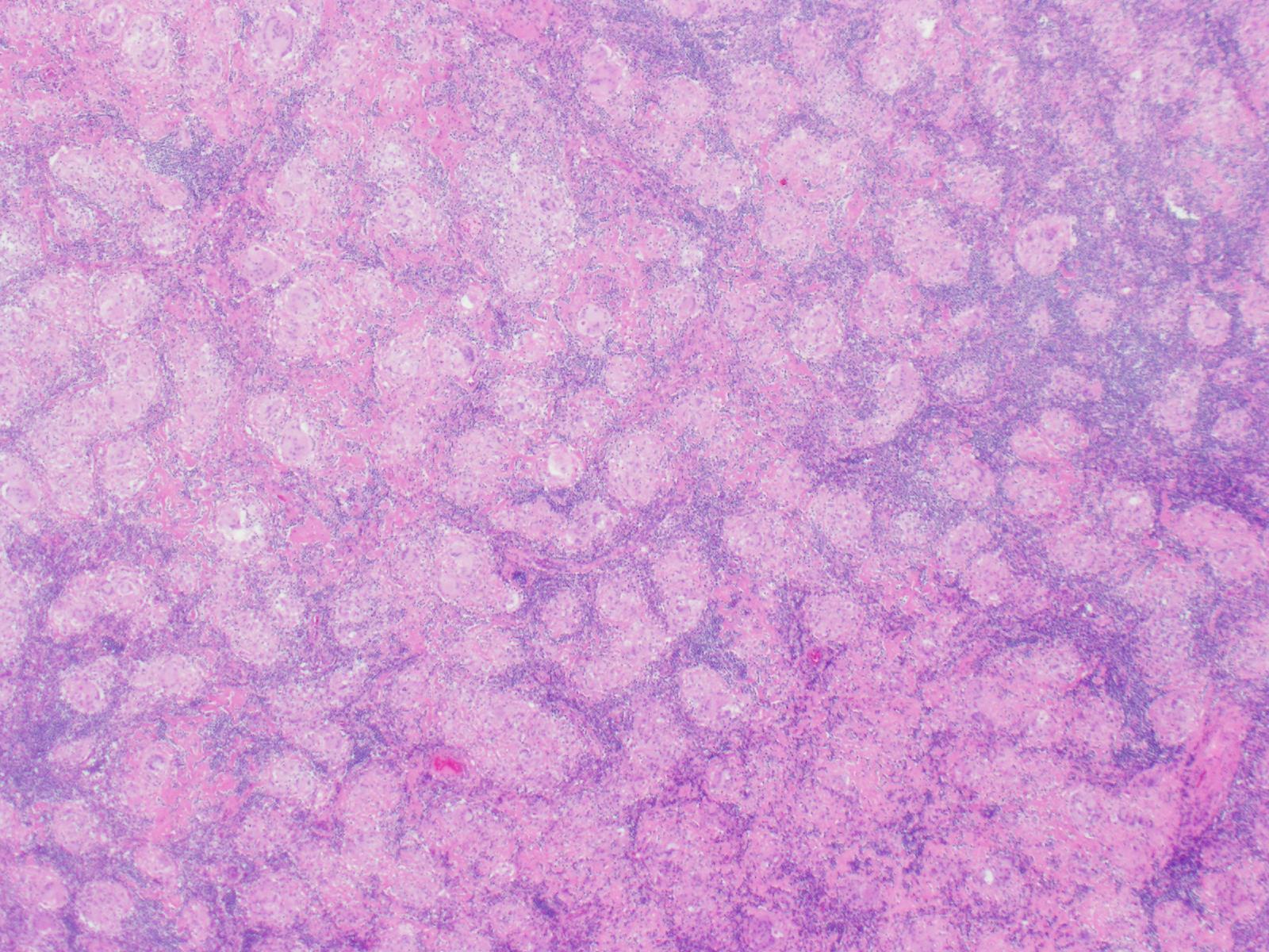
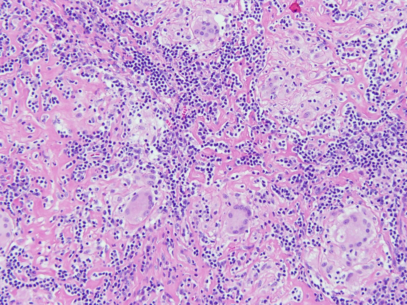
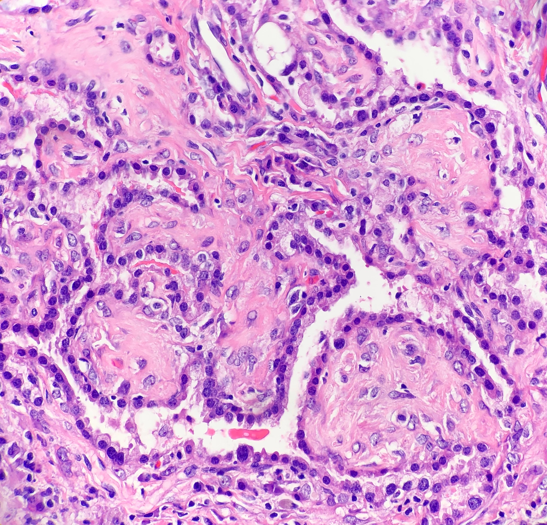
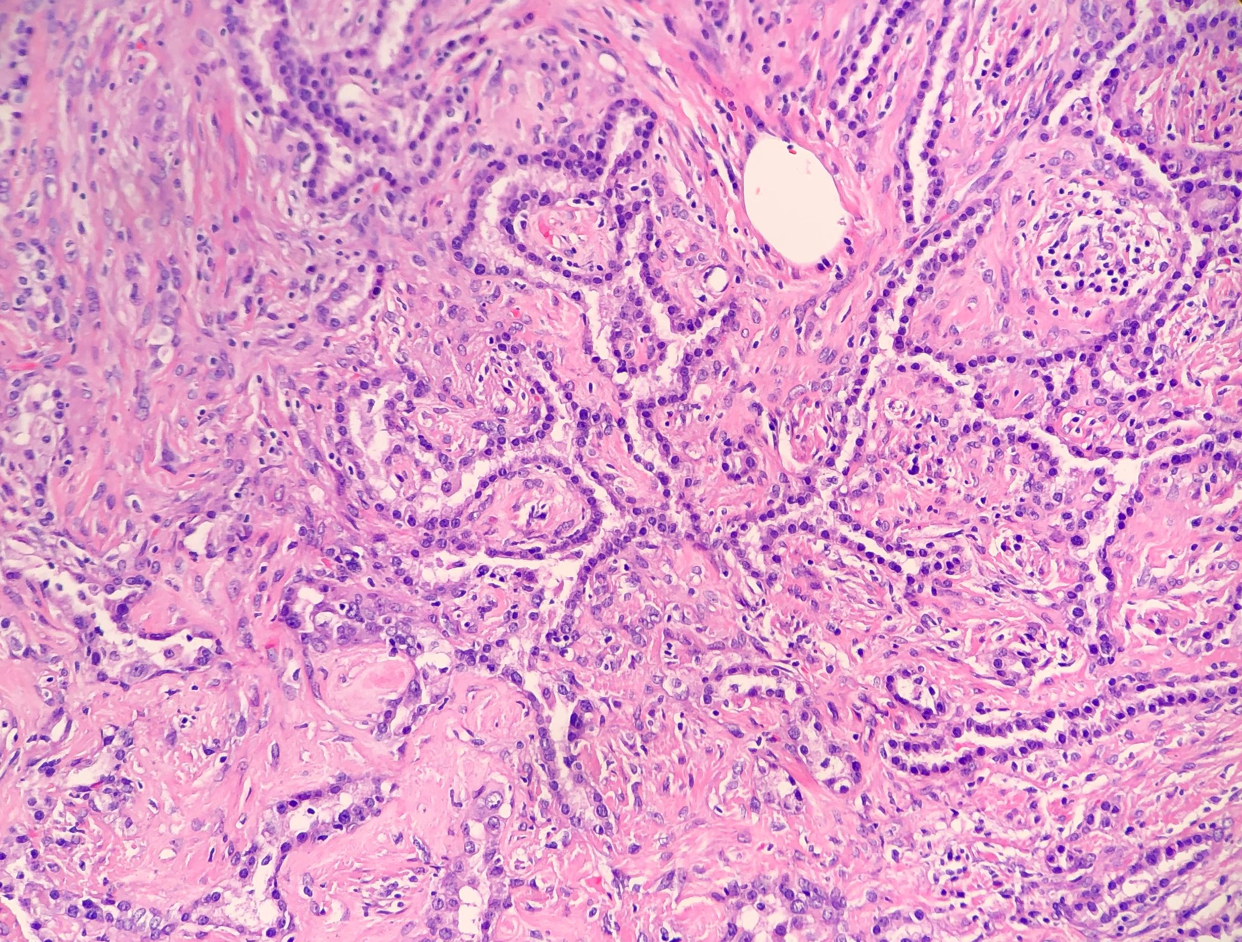
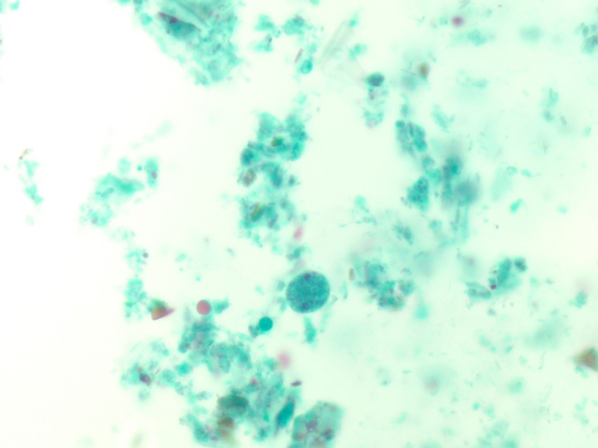
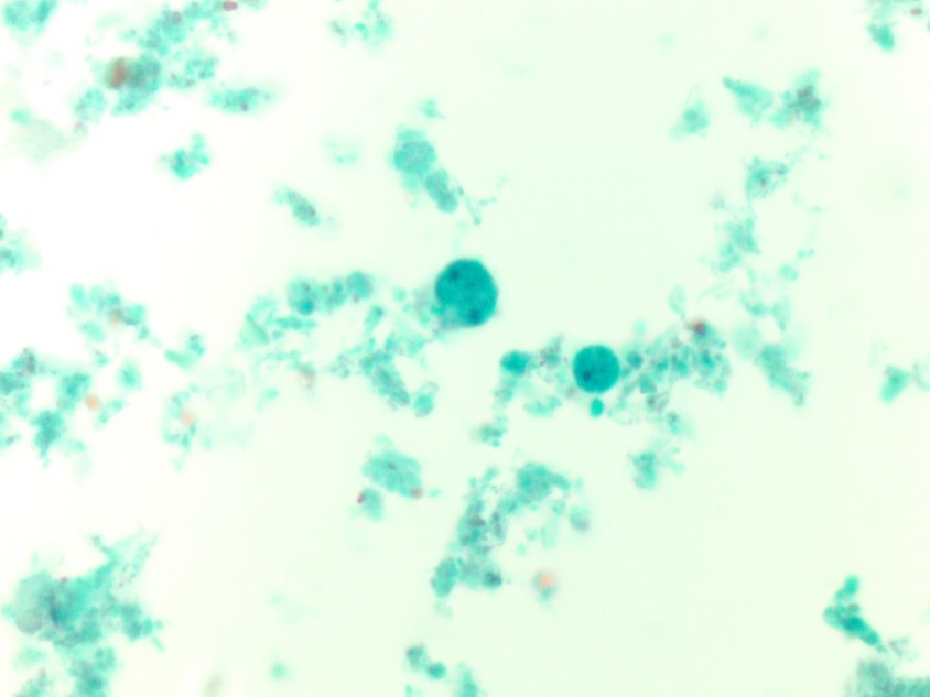
H&E stained sections revealed numerous non-necrotizing granulomas effacing and replacing normal lymph node architecture. These consisted of pale epithelioid histiocytes and Langhans type of giant cells. The granulomas lacked a peripheral rim of lymphocytes. AFB and GMS stains were negative for microorganisms

Diagnosis

A diagnosis of non-necrotizing granulomatous lymphadenitis was rendered noting that in the correct clinical context the findings could represent sarcoidosis.

Discussion

Granulomatous inflammation is a special type of chronic inflammatory response characterised by the formation of discrete collections of histiocytes called granulomas. Activated histiocytes appear as epithelioid cells with round to oval nuclei, often with irregular contours and abundant granular eosinophilic cytoplasm with indistinct cell borders. They may coalesce to form multinucleated giant cells. When found in the lymph node, the reaction pattern is called granulomatous lymphadenitis. It can be caused by a variety of different conditions, and therefore, requires thorough workup to come to a conclusive diagnosis.

On the basis of presence or absence of necrosis, granulomatous lymphadenitis can be classified as necrotizing or non-necrotizing. Additionally, the presence of an abscess, usually central, indicates a suppurative lymphadenitis.

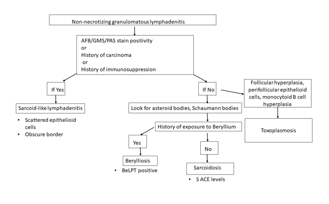
Non-necrotizing granulomatous lymphadenitis:

Sarcoidosis lymphadenitis is the prototype of non-necrotizing granulomatous lymphadenitis. It shows the presence of discrete granulomas without a peripheral rim of lymphocytes, called “naked granulomas”. The early phase shows follicular hyperplasia and sinus histiocytosis, followed by appearance of epithelioid cell nodules toward the end of this phase. The peak phase shows well-demarcated granulomas composed of epithelioid cells with scattered multinucleated giant cells observed throughout the lymph node. Granulomas may occasionally coalesce. In the late phase, increased collagen fibers result in fibrosis and hyalinization. There are no neutrophils and it is uncommon to find small foci of central necrosis. Numerous inclusions such as asteroid, Schaumann, or Hamazaki-Wesenberg bodies can be seen. In this case, we observed well-demarcated granulomas throughout the lymph node, typical of the peak phase without any caseous necrosis or suppuration.

Other causes of granulomatous lymphadenitis can be ruled out as follows.

Sarcoid-like lymphadenitis: It shows a similar pattern of non-necrotizing lymphadenitis like sarcoidosis. However, classically sarcoid like reaction shows scattered small epithelioid granulomas with sparsely arranged epithelioid cells. The border of the granulomas is usually obscure. The CD4:CD8 ratio ranges from 0.8 to 2.25 while in sarcoidosis, it is >3.5. These findings help distinguish sarcoid-like lymphadenitis from sarcoidosis.

Sarcoid-like adenitis may be seen in numerous conditions such as carcinoma, Toxoplasmosis, fungal infections, tuberculosis, immunocompromised states, pneumoconiosis etc. The fact that tuberculosis and fungal infections can present with a non-necrotizing granulomatous lymphadenitis highlights the importance of performing fungal (PAS & GMS) and AFB (Ziehl Neelson) stains in non-necrotizing lymphadenitis as well. In this case, the granulomas had distinct borders, numerous epithelioid cells, no organisms were identified on special stains, nor was there any history of immune compromise; ruling out a sarcoid-like reaction.

Berylliosis: The lymph node picture in Berylliosis is identical to that of sarcoidosis. We may even see asteroid bodies or Schaumann bodies. A diagnosis can be established by eliciting a history of chronic exposure to Beryllium. Beryllium lymphocyte proliferation test (BeLPT) is a test that measures Beryllium sensitization and is very specific for Beryllium exposure. There was no known history of exposure to Beryllium in this case.

Toxoplasmosis: A classic triad of follicular hyperplasia, small granulomas composed of epithelioid cells within and around hyperplastic follicles and, monocytoid B cell hyperplasia, is observed in toxoplasmosis lymphadenitis. This case did not show follicular hyperplasia, ruling out toxoplasmosis.

Necrotizing granulomatous lymphadenitis

Even though we did not find any necrosis in this case, yet, it is worthwhile to review briefly the various causes of necrotizing lymphadenitis.

  • Non-suppurative

Tuberculosis: Histology of a tuberculous lymph node is characterised by central caseous necrosis surrounded by an epithelioid cell layer. The outermost layer is comprised of lymphocytes and fibrosis. Plasma cells are not observed. Diagnosis can be established by performing an AFB stain that demonstrates acid fast rod shaped bacteria in the areas of necrosis. Organisms can also be detected by PCR.

BCG lymphadenitis: About 0.7 to 2.3% of BCG vaccinated children may develop BCG lymphadenitis that is smaller than tuberculous lymphadenitis. Early phase shows follicular hyperplasia and sinus histiocytosis. Later, there is development of micronodules of epithelioid granulomas without necrosis and epithelioid cell granulomas with central caseous necrosis. Langhans giant cells are rare.

Fungal infections: Fungal infections by Histoplasma, Cryptococcus, coccidiodomycosis, pneumocystis may also cause a necrotizing granulomatous inflammation. There are numerous neutrophils, and fungal structures can be seen. GMS and PAS can be used in cases where it is difficult to the find the fungal elements on H&E.

  • Suppurative

Tularemia: There are three forms of histological changes, Abscess form, showing abscess with central necrosis and mononuclear cells, Abscess-granulomatous form with granulomas with central necrosis, which form large lesions with central abscesses, and granulomatous form with caseating necrosis at the centre of the granulomas.

Cat Scratch disease: Similar to tularemia, there are three phases of histologic presentation, an early phase of follicular hyperplasia, intermediate phase of microabscess, and a late phase of granulomatous inflammation. Monocytoid B cell clusters are observed close to the abscess.

Conclusion

Sarcoidosis is usually diagnosed by excluding other causes of granulomatous inflammation, as we did in this case. Characteristic non-necrotizing, discrete granulomas were seen throughout the lymph node. The age of the patient and female gender epidemiologically support the diagnosis. This case reflects an example work up of a granulomatous lymphadenitis that is a morphologic presentation of myriad diseases.

-Swati Bhardwaj, MD has a special interest in surgical pathology and hematopathology. Follow her on Twitter at @Bhardwaj_swat.

Kamran M. Mirza, MD, PhD, MLS(ASCP)CM is an Assistant Professor of Pathology and Medical Education at Loyola University Health System. A past top 5 honoree in ASCP’s Forty Under 40, Dr. Mirza was named to The Pathologist’s Power List of 2018. Follow him on twitter @kmirza.

Global Health Narratives Interview Series: Meet Dr. Kumarasen Cooper.

Kumarasen Cooper, MD, PhD completed his medical training from his home country in South Africa and his PhD at Oxford. He now works as a surgical pathologist at the University of Pennsylvania and is responsible for leading the initiative to engage the pathology department in the Botswana-UPenn partnership through the Perelman School of Medicine Center for Global Health. He has over 260 publications and has lectured in 5 continents. Despite this busy schedule, Dr. Cooper devotes two separate months of the year to work in Botswana’s only academic pathology department, where he pours his energy into helping the department advance.

I met Dr. Cooper through email when I heard about the work he was doing in Africa. He generously agreed to come visit my department to give an excellent Grand Rounds lecture on his experiences working in Global Pathology, and he led a much-appreciated resident slide session of unusual and difficult cases from his work in Botswana. Humility and grace envelop Dr. Cooper despite his brilliant accomplishments. He also proved to be incredibly generous with a refusal of his speaker honorarium, in exchange for an agreement that we would collect pathology textbooks to send to the under-supplied residency program in Botswana. I’m excited to share the inspiring work that he does through the Botswana-UPenn partnership with all of you today, as I think this program could be used as a model for all institutes to involve their pathology departments in global health opportunities.

Q: What began your interest in global health?

A: I was born, raised, and completed my medical training in South Africa. I spent 15 years working as a Pathologist and served as the Chair of Pathology in Johannesburg until I was recruited to the US to work as Vice-Chair at the University of Vermont. I knew when I left Africa that I would always come back, and that I could use what I learned abroad to give back in some way. I wasn’t sure in what form that would take at the time, but I knew there was work that still needed to be done. This was also influenced by my visits to the pathology departments in many different countries over the years…I was able to gain a sense of the ‘haves and have-nots’, and so developed a strong feeling that I needed to give back.

Q: How did you hear about the Botswana-University of Pennsylvania (BUP) partnership and was pathology an active part in that already?

A: When I first discovered the partnership, I thought that this may be an avenue for me to participate in global pathology. At the time, the pathology department was not involved in any of the ongoing BUP projects, though other clinical departments at UPenn were. After my initial assessment of the Botswana pathology department and its resources in April of 2016, I was able to identify ways that I could help. Together with the Director of BUP, I approached the Chairman of my department with the proposal, and we started the pathology partnership program in October of that year. Since then, I travel to Botswana twice a year for one month at a time, and each time I take 1-2 residents from UPenn along with me.

Q: Can you describe the pathology department in Botswana?

A: To serve a population of just over 2 million people, Botswana has only one academic pathology department, a College of the University of Botswana (UB) School of Medicine, which consists of six pathologists who are all from other countries. There are currently no Botswana pathologists working in the department. There are about six technicians working in the laboratory, all of whom were trained internationally. The laboratory receives around 7,000 surgical specimens yearly, plus cytology, and autopsy. They work with an extremely limited panel of immunostains that are not routinely used but are spared for the rare case that cannot be diagnosed with morphology alone.

The residency program is still very new. There are six residents in the program at the present time, and the program is designed so that they will spend the first two years in Botswana and then they will continue their final years of training in South Africa. I look forward with anticipation to the first Botswana trained pathologists in the country.

Q: What is your role when visiting Botswana?

A: We try to help with everything we can. I sign out cases with the residents during the time I am there, and I teach the residents using these cases every day. The UPenn residents that I bring with me are eager to teach as well, so they deliver didactics regularly also. We all participate in tumor boards and the FNA clinic. We each take on projects that we can partner with them to tackle…things like improving turnaround time, quality improvement, and SOP preparations.  We also work on developing academic programs, grossing templates and manuals (A UPenn pathology PA spent two weeks working in Botswana on this project), synoptic reports, cancer guidelines…anything they need I try to help them with.

Q: How are the UPenn pathology residents given credit in their home program to join you?

A: As of this year, the BUP pathology program is now offered as one of the official electives that residents are allowed to choose from. They are able to use elective time and their travel expenses are paid for by a resident travel grant.

Q: In your role as supervisor of the UPenn residents, what do you see the residents gaining from the experience?

A: The residents that have come with me to Botswana are very compassionate and are eager to contribute in any way they can. Experiencing pathology in Botswana, where people are trying to achieve so much with so little resources, it makes the UPenn residents even more grateful for all of the resources they have available to them. They also have the opportunity to not only learn from the unusual cases that present in Botswana, but also the opportunity to contribute their own unique set of skills – some have focused on teaching autopsy technique, others give enthusiastic  and detailed lectures, and one gave a talk about successful study techniques. [For more information about the resident experience, one can read more about it in the UPenn blog here: https://pathology.med.upenn.edu/department/blogs/residency-matters/penns-pathology-residency-program-reaches-botswana]

Q: How do you see the BUP pathology partnership affecting the trainees in Botswana? What changes have you seen since you started working with them?

A: The residents in Botswana really appreciate the partnership that we have formed.  I have seen the residents develop so much since working with them. At first, they were reserved and now they actually request lectures on topics they feel they could improve on. They are still very humble and respectful, but I have encouraged them to be advocates for themselves. They have really embraced their program and I’m very proud of them. We have a deep appreciation for each other and are proud of what we have achieved together.

We’ve also started hosting Botswana residents at UPenn for a one month rotation so they have the opportunity to supplement their training even further. We fly them to the US, house them, and include them in our residency training program for the month. They have the opportunity to sit in on sign-outs, shadow grossing and autopsy, attend conferences, and be exposed to the advanced testing that we routinely perform in the US.

Q: How do you see the pathology partnership growing in years to come?A: I’m currently helping them find placements in South Africa or possibly partnering with private laboratories to help expose the residents to a greater diversity and volume of cases. As the program continues to grow, we look forward to seeing the fruits of the partnership for many years to come.


-Dana Razzano, MD is a Chief Resident in her third year in anatomic and clinical pathology at New York Medical College at Westchester Medical Center and will be starting her fellowship in Cytopathology at Yale University in 2020. She was a top 5 honoree in ASCP’s Forty Under 40 2018 and was named to The Pathologist’s Power List of 2018. Follow Dr. Razzano on twitter @Dr_DR_Cells.

Surgical Pathology Case Study: A 43 Year Old Female with a Lung Nodule Noted on Imaging Following Chest Congestion

Case History

The patient is a 43 year old woman who experienced chest congestion and presented to her local physicians office. A chest X-ray was ordered and demonstrated a lung abnormality. A follow-up CT scan confirmed a 1.9 cm smoothly marginated nodule in the upper lobe with no adenopathy and a normal liver and adrenal glands. The nodule was mildly hypermetabolic on PET scan. A bronchoscopy was performed, which was non-diagnostic. Two subsequent CT scans demonstrated no change in the size of the nodule. Overall, the patient feels well and denies cough, hemoptysis, dyspnea on exertion, and weight loss. Due to the suspicion of cancer, the patient has decided to undergo a lung lobectomy.

Diagnosis

Received in the Surgical Pathology lab for intraoperative consultation is a 30.0 x 7.2 x 2.2 cm lung lobectomy specimen. There is an attached 6.2 cm staple line, which is removed and the subjacent resection margin is inked blue. The entire pleural surface is inked black. The specimen is sectioned revealing a 2.1 x 1.7 x 1.0 cm white-tan, firm, round nodule that is 0.5 cm from the blue inked resection margin and 0.2 cm from the black inked pleural surface. The remainder of the specimen is composed of red-tan, spongy, grossly unremarkable lung parenchyma without nodules or other lesions. Photographs of the specimen are taken (Figure 1). A representative section of the nodule is submitted for frozen section and read out as “diagnosis deferred”. Representative sections of the specimen are submitted as follows:

A1FS:   Frozen section remnant

A2-A7:   Nodule, entirely submitted

A8-A10:   Grossly unremarkable lung parenchyma

Immunohistochemical stains show the epithelial cells in the lesion to be positive for CK7, TTF-1, and surfactant proteins A and B which supports these cells to be type 2 pneumocytes (all controls are appropriate). Based on the immunohistochemical stains and routine H&E slides, the case was signed out as a sclerosing pneumocytoma

Image 1. Gross presentation of the well-defined, round sclerosing pneumocytoma.

Discussion

Sclerosing pneumocytoma (SP) is a rare, benign pulmonary tumor that was first described in 1956 as a vascular tumor, but has since been found to be of primitive respiratory epithelium origin. In the past, SP has also been referred to as sclerosing hemangioma, pneumocytoma, and papillary pneumocytoma, but the 2015 World Health Organization classification of lung tumors states that the agreed upon term for this tumor should be a sclerosing pneumocytoma. SP is commonly seen in middle aged adults, with a female to male ratio of 5:1. There is no racial bias. Patients are usually asymptomatic, with the tumor incidentally found on screening chest radiographs. If the patient was to present with any symptoms, they would usually include a cough, hemoptysis and chest pain. Radiographically, SP appears as a solitary, well-defined, homogenous nodule along the periphery of the lung.

Grossly, most SPs appear as a solitary, firm, well-circumscribed, yellow-tan mass generally arising along the periphery of the lung. The majority of these tumors appear within the lung parenchyma, but there have been cases reported of endobronchial and pleural based SP tumors. Multifocal unilateral tumors and bilateral tumors are uncommon.

Histologically, SP consists of two epithelial cell types: surface cells and round cells. Surface cells are cuboidal, resembling type II pneumocytes, with finely stippled nuclear chromatin, indistinct nuclei, occasional nuclear grooves, and inclusions. The stromal round cells will have bland oval nuclei with coarse chromatin and eosinophilic cytoplasm (Figure 2). Both the surface cells and round cells will have a low mitotic rate, but can have moderate to marked nuclear atypia. Ciliated bronchial epithelium is often identified in the tumor. There are four architectural patterns identified within SP: papillary, sclerotic, solid and hemorrhagic, with over 90% of SPs displaying three of the patterns, and all of the tumors containing at least two of the patterns.

  • Papillary pattern: Complex papillae composed of surface cells covering a stroma of round cells
  • Sclerotic pattern: Papillae containing hyalinized collagen, either in solid areas or along the periphery of hemorrhagic areas (Figure 3)
  • Solid pattern: Sheets of round cells bordered by surface cells
  • Hemorrhagic pattern: Large blood filled spaces
Image 2. Photomicrograph demonstrating the cuboidal surface cells and round stromal cells.
Image 3. Photomicrograph of the papillary and sclerotic architectural patterns.

Immunohistochemical stains can be helpful in the diagnosis of SP, with both the surface cells and round cells exhibiting expression of thyroid transcription factor 1 (TTF-1) and epithelial membrane antigen (EMA). It should be noted that TTF-1 is also used for the diagnosis of pulmonary adenocarcinoma, increasing the risk of misdiagnosing SP. The surface cells will also express both pancytokeratin (AE1/AE3) and Napsin A, with the round cells being negative for AE1/AE3, but having a variable expression of cytokeratin 7 and the low molecular weight cytokeratin (CAM 5.2). Molecular pathology has demonstrated a frequent loss of heterozygosity at 5q, 10q and 9p, and an allelic loses at p16 in the surface and rounds cells. Although the immunohistochemical stains and molecular pathology results can be very helpful, diagnosis of a SP is still largely based on routine H&E slides showing the two epithelial cell types and four architectural patterns.

Electron microscopy will show abundant lamellar bodies similar to those in type II pneumocytes in the surface cells. Round cells will lack the lamellar bodies and instead will contain variably-sized electron-dense bodies that have been thought to represent the different stages of lamellar body maturation.

The differential diagnosis for SP includes a variety of benign and malignant neoplasms, which can be difficult to distinguish on cytology, small biopsies and intraoperative consultations. The cytologic features include moderate to high cellularity with a bloody background and foamy macrophages, occasional nuclear pleomorphism in the round cells, absent mitotic figures, and occasional necrosis with cholesterol clefts and calcifications. In the case of small biopsies, making a diagnosis of SP can be difficult if the papillary pattern is highly prevalent without one of the other three patterns present. With intraoperative consultations, the frozen section artifact can make it difficult to appreciate the two epithelial cell types or the four architectural patterns. The gross examination, as well as the radiographic findings of a well-circumscribed tumor can help point the Pathologist to favoring a benign neoplasm over a malignant one. The benign neoplasms that should be considered in the differential diagnosis include:

  • Clear cell tumor, which will have clear cells with scant stroma, thin-walled vessels and a strong expression of HMB-45
  • Pulmonary hamartoma, which will have a combination of cartilage, myxoid stroma, adipose tissue and trapped respiratory epithelium
  • Hemangiomas, which are rare in the lung, and will lack epithelial cells and contain either a cavernous or capillary morphology

The malignant neoplasms that should be considered in the differential diagnosis include:

  • Bronchioalveolar carcinoma, which can have a papillary pattern, but will not contain the two epithelial cell types and combination of the four architectural patterns
  • Metastatic papillary thyroid carcinoma, which is distinguished from SP by the presence of the characteristic Orphan Annie nuclei
  • Metastatic renal cell carcinoma, which will contain nuclear atypia and striking vascularity
  • Carcinoid, which will contain organoid and ribbon-like growth patterns

Currently, with the benign nature of SP, surgical excision is the preferred treatment choice to cure the patient. There have been cases reported of lymph node metastasis and recurrence, but neither of these appear to effect the prognosis. This just helps to highlight the need for a multidisciplinary approach to this benign tumor.

References

  1. Hisson E, Rao R. Pneumocytoma (sclerosing hemangioma), a Potential Pitfall. Diagn Cytopathol. 2017;45(8):744-749
  2. Keylock JB, Galvin JR, Franks TJ. Sclerosing Hemangioma of the Lung. Arch Pathol Lab Med. 2009;133(5):820-825.
  3. Travis WD, Brambilla E, Nicholson AG, et al. The 2015 World Health Organization Classification of Lung Tumors: Impact of Genetic, Clinical and Radiologic Advances Since the 2004 Classification. J Thorac Oncol. 2015;10(9):1243-1260.
  4. Wu R. Sclerosing Pneumocytoma (Sclerosing Hemangioma). Pathology Outlines. http://www.pathologyoutlines.com/topic/lungtumorsclerosingheman.html. Revised February 19, 2019. Accessed June 6, 2019.

-Cory Nash is a board certified Pathologists’ Assistant, specializing in surgical and gross pathology. He currently works as a Pathologists’ Assistant at the University of Chicago Medical Center. His job involves the macroscopic examination, dissection and tissue submission of surgical specimens, ranging from biopsies to multi-organ resections. Cory has a special interest in head and neck pathology, as well as bone and soft tissue pathology. Cory can be followed on twitter at @iplaywithorgans.

Microbiology Case Study: A 58 Year Old Female with Abdominal Pain

Clinical History

A 58 year old female with no significant past medical history presented her primary care physician with chief complaint of abdominal pain. She reported continued vague abdominal symptoms for the past two months, with intermittent diarrhea and increased flatulence. No recent travel history or significant exposures were identified. An ultrasound of the right upper quadrant was unremarkable and no gallstones were present. The patient was scheduled for a screening colonoscopy. A stool specimen was submitted to the microbiology laboratory for stool culture and ova & parasite exam.  

Laboratory Identification

Image 1. Trichrome stained fecal smear illustrating a binucleated trophozoite with fragmented karyosomal material from a stool ova & parasite exam.
Image 2. Additional trichrome fecal smear image highlighting both uninucleate and binucleate trophozoites that range in size from 5 to 15 um.

The findings from the ova and parasite exam were consistent with Dientamoeba fragilis, an intestinal flagellate. The stool culture was negative for Salmonella, Shigella, and Escherichia coli 0157:H7. Stool enzyme immunoassays were negative for Campylobacter spp.and Shiga toxin 1 and 2.

Discussion

Dientamoeba fragilis is an intestinal flagellate with worldwide distribution and causes asymptomatic and symptomatic infections, predominantly in small children. Symptoms of infection may include intermittent diarrhea, abdominal pain, anorexia, weight loss, and flatulence.  While the pathogenesis is not completely understood, transmission is thought to occur via the fecal oral route and it is hypothesized that the trophozoites are transmitted via the eggs of nematodes, Enterobius vermicularis and Ascaris lumbricoides, due to a higher incidence of co-infections between these organisms than expected.

In the laboratory, the diagnosis of D. fragilis is made by ova and parasite exam. The trophozoite resembles amebae and is typically 9-12 µm. Most trophozoites are binucleate with finely granular cytoplasm and the within the nuclei there are 4-8 fragments of karyosomal granules (Figure 1). Due to the fact that 30-40% of D. fragilis trophozoites are uninucleate (Figure 2) and they lack external flagella, they must be differentiated from Endolimax nana and Entamoeba hartmanni, which are both non-pathogenic amebae. Historically, no cyst phase was known for D. fragilis; however, recent studies have identified precyst forms or putative cysts. Permanently trichrome stained slides are essential to diagnosing D. fragilis infection, as the organism is hard to detect in concentrated smears.

Since our patient was symptomatic, she was treated with iodoquinol, the drug of choice for D. fragilis infections. Her symptoms resolved and colonoscopy did not reveal additional pathology.  

-Debbie Walley, MD, is a 4th year Anatomic and Clinical Pathology chief resident at the University of Mississippi Medical Center. 

-Lisa Stempak, MD, is an Assistant Professor of Pathology at the University of Mississippi Medical Center in Jackson, MS. She is certified by the American Board of Pathology in Anatomic and Clinical Pathology as well as Medical Microbiology. She is the Director of Clinical Pathology as well as the Microbiology and Serology Laboratories. Her interests include infectious disease histology, process and quality improvement, and resident education.

Hey, What’s the Buzz on Zika?

Hello everyone and welcome back!

Last month, it was as fun to write about hematology peripheral smear differentials as it was to address the importance of interdisciplinary collaboration. I found myself in a unique position both as a medical student as well as a former medical laboratory scientist in what was a great clinical training rotation in hematology/oncology. Now, with just one rotation left until the end of my medical school journey, I want to take you on a look back at some of the very first posts I made here on Lablogatory and update you on the intersectional, collaborative topic that I shared with you almost two years ago: Zika!

Image 1. ASCP’s official professional society partner, The Pathologist. I’ve been getting them in my mailbox since the official partnership was announced. It’s an excellent platform for laboratory professionals across scopes to discuss relevant topics in pathology. I was particularly excited to see Zika make an appearance last month! (Source: The Pathologist [online] https://thepathologist.com/diagnostics/our-powers-combined)

In a recent digital article on ASCP’s partner, The Pathologist, author and staff editor Michael Schubert wrote about the connectivity between public health, epidemiologic research, laboratory medicine, and clinical patient outcomes. He examined the effectiveness and accuracy of Zika testing availability in commercially available assays and spoke with a leading virologist in the field from Berlin. You may recall one of those “ancient” posts I made about Zika, where I was part of a research team that used the same methodology! Combined immunoglobulin-specific assays, arbovirus detection in the heat of a public health epidemic’s epicenter, and lab medicine that complimented my concurrent immunology class in med school—what more could you ask for?

And, since the last tagged Lablogatory Zika update I can see was by Dr. Sarah Riley in February of 2017, here’s my update! Dr. Riley’s post was a fantastic summary of the Zika epidemic, its troublesome diagnostic assessments, and the recommendations and plans of organizations like the World Health Organization (WHO). She was, and still is, right—the “struggle is still real’ when it comes to Zika testing. Curious about what it was like during the 2016 epidemic? Who was doing testing, what kind of testing, and what was the lab data climate? Well…it feels like it’s time for a…

*** FLASHBACK ***

An Arbovirus Abroad

Hey! My inaugural post! It was fun to go back and see the data from the work then (Spoilers: epidemiologic updates are on your horizon). We were just getting started to take an assessment of the situation and address it as a public health concern. My then Caribbean location was a great place to study Zika trends coming from Brazil, Puerto Rico, and Florida. As a snapshot, at that time (Dec 2016) there were a purported almost 2,000 cases, however less than a fifth of those cases were serologically confirmed by lab testing. Before the recommendations to move toward RT-PCR, most labs in the region were requesting commercially available screening tests for IgG/IgM assays.

Image 2a. These were the (then) suspected Zika viral infection cases per epidemiological week, Pan-American Health Organization (PAHO) and World Health Organization (WHO) 2016. My wife and I are included in these statistics—that mosquito virus rash is awful!
Image 2b. Remember that spoiler I promised above? Well here’s the updated WHO epidemiologic data for confirmed Zika cases in the region we worked in. Seems like the mosquitoes…buzzed off. (Source: WHO)

Healthy Me

How do you reach people when you’ve got compelling public health lab data that translates to possible prevention of infection and spread of disease? Easy: go to where the people are and engage them when and where they’re comfortable. One of the overarching themes in public health is mitigating barriers to change by way of utilizing social humility. This a certainly a type of interdisciplinary collaboration because if we’re the experts on IgM and IgG trends in testing confirmations, the public are the experts in social determinants of health within their communities.

Image 3. Want to make sure a message gets home to every family? Bug their kids about Zika bugs in fun, educational ways. That’s me delivering one of my “Healthy Me” presentations to children, October 2016.

Laboratory Data and Global Health Security

As my team and I were busy preparing SOPs, conducting a new project aimed at improving local health literacy and source reduction, securing IRB approval, and collecting data about the residents of Sint Maarten to correlate with local Health Ministry projections, one of the officials—who now serves as a regional director for PAHO—took our work to the Global Health Security Agenda Summit. Talk about motivation! In and out of the lab, I worked with teams who were getting some fantastic work done on the ground with respect to mosquito-borne virus research.

Image 4. IgM and IgG seroprevalence of Zika virus (along with other Arboviruses i.e. West Nile, Chikungunya, Dengue, and Yellow Fever etc.) within the community around my medical school. We used commercially available IgG and IgM assays from Germany with great success. Internal controls and known cases were fantastic ways to include internal validation.

IRBs and Public Heath Pathology

For those of us who work in laboratory medicine, it’s easy to talk about the best way to test, detect, and treat an epidemiologic threat—it’s even exciting when it’s a current threat. But to really be successful, you’ve got to collaborate with those outside of the lab, and often this means thinking “outside the box.” Public health is different from lab medicine in that while lab-work is based around results, testing, and organized data-driven decisions, success in public health is highly determined by community buy-in in the form of partnerships!

Figure 1. There’s a method to the community “buy-in” concept. With a foundation in evidence-based practices, any project aimed at improving public health outcomes must include some critical components like clear objectives, attainable goals, sustainability, and effective (and constant) re-evaluation.

*** FLASH … FORWARD? ***

So, after my time in Sint Maarten, I came to New York City to rotate through my clinical clerkships. And, if you’ve seen some more recent post-Zika posts on this website, you know they’ve been going great! Within a few months of being here, my wife brought back some swag from a training session she attended. (Side note: she’s a graduate-level nurse, working in the public health non-profit sector with vulnerable populations in the inner city—she’s too busy to blog.) After months of both of us working and learning about Zika and public health initiatives in the Caribbean, we were greeted by this fantastic toolkit from the New York State Department of Public Health!

Image 5. Empowering a large number of patients with highly variable demographics is challenging. The NYS DOH distributed “Prevention Kits” for Zika Virus which included: Zika Virus educational materials in 8 languages, pamphlets on reducing mosquito activity, travel related information for pregnant women, 2 larvicide pellets with instructions for using larvicide, picaridin insect repellent, and condoms.
Image 6. That’s us! My wife Kathryn and I presenting on the importance of Disaster Planning and Implementation of Preparedness Programs at the 2019 Caribbean Conference of Disaster Medicine. Disasters are bad on their own, but think about what happens months after flooding, hurricanes, or destruction—transmittable diseases. And that includes standing-water-borne mosquito viruses!

The take home message: collaboration is key, both inside and out of the lab. Schubert’s piece in The Pathologist created a fantastic dialogue in addressing the clinical needs for interdisciplinary collaboration. The best testing means finding out exactly where the needs are and using data-driven decisions to implement change or action. In the lab, that means constantly working for higher quality and better patient outcomes in every test, result, report, and (mosquito) byte of data. In the field, it means the same thing, but instead of metrics like sensitivity, specificity, and TAT it’s about cultural humility, attainable goals, and dynamic timing.

Thanks for reading! Hope most of our national heat wave spared you, but if it didn’t remember: don’t keep standing water around, wear light loose clothing, and use appropriate insect repellent!

See you next time!

–Constantine E. Kanakis MSc, MLS (ASCP)CM graduated from Loyola University Chicago with a BS in Molecular Biology and Bioethics and then Rush University with an MS in Medical Laboratory Science. He is currently a medical student actively involved in public health and laboratory medicine, conducting clinicals at Bronx-Care Hospital Center in New York City.

An Introduction to Laboratory Regulations: Part 1

Everyone who works in a laboratory knows that there are certain rules and regulations to be followed to ensure accuracy in testing, and the safety of both the patient and testing personnel. With all the acronyms floating around (CLIA, FDA, CAP, CMS, TJC) it can get confusing to keep track of who controls what, and which rules apply to your specific lab. In the first installment of this 3-part series on regulations, we’ll review the different federal agencies responsible for oversight and moderation of the laboratory. In part 2 we’ll go further in-depth to demystify testing complexity (waived, non-waived, PPM) and why it’s important to know the correct classification for the tests you perform. Lastly, we’ll review the optional accreditations available to labs, and how accreditation differs from certification.

CLIA

CLIA refers to the Clinical Laboratory Improvement Amendments of 1988. These amendments were drafted to the Public Health Services Act, in which the federal program was revised to include certification and oversight of clinical laboratory testing. Although there have been two additional amendments made after 1988 (1997, 2012), the law still continues to be cited as CLIA ’88 as it is named within legislation.

These CLIA regulations helped to establish quality standards for all U.S. laboratory testing performed on human specimens (except for research) for the purpose of assessment of health, or the diagnosis, prevention, or treatment of disease. The regulations cover all aspects of testing including general laboratory requirements, quality monitors, pre-analytics, analytic performance, post-analytics, and personnel requirements.

In addition to setting the basic ground rules for performing quality laboratory testing, the CLIA regulations also require clinical laboratories to be certified by their state as well as the Center for Medicare & Medicaid Services (CMS) before accepting human samples for diagnostic testing. Laboratories can obtain multiple types of CLIA certificates, based on the kinds of diagnostic tests they perform. In order for laboratories to receive payments from Medicare or Medicaid, laboratories must be properly certified for the testing they are performing and billing for.

There are 3 federal agencies responsible for enforcing the CLIA regulations: The Food & Drug Administration (FDA), Center for Medicaid Services (CMS) and the Center for Disease Control and Prevention (CDC). Each agency has a unique role in assuring quality laboratory testing.

CMS

The Centers for Medicare & Medicaid Services (CMS) is the federal agency responsible for ensuring that the CLIA standards are upheld and enforced. Their responsibilities include the following:

  • Issuing laboratory certificates
  • Collecting user fees
  • Conducting inspections and enforcing regulatory compliance
  • Approving private accreditation organizations (such as CAP) for performing inspections, and approves state exemptions
  • Monitoring laboratory performance on Proficiency Testing (PT) and approving PT programs
  • Publishing CLIA rules and regulations

FDA

The Food & Drug Administration (FDA) is primarily responsible for reviewing and approving new tests, instruments, and equipment used in diagnostic laboratories. They also perform the following tasks:

  • Categorize tests based on complexity
  • Review requests for Waiver by Application from manufacturers
  • Develop rules/guidance for CLIA complexity categorization

CDC

The Center for Disease Control and Prevention (CDC) responsibilities include the following tasks:

  • Provide analysis, research, and technical assistance
  • Develop technical standards and laboratory practice guidelines, including standards and guidelines for cytology
  • Conduct laboratory quality improvement studies
  • Monitor proficiency testing practices
  • Develop and distribute professional information and educational resources
  • Manage the Clinical Laboratory Improvement Advisory Committee (CLIAC)

To summarize, CLIA establishes the rules and guidelines that laboratories must follow to ensure they are providing accurate laboratory results. Federal agencies then work together to support the CLIA amendments and enforce compliance. All certified laboratories will be subject to inspection by regulatory agencies to ensure compliance with the rules. In some cases, your local state Department of Health (DOH) or accrediting agency may be more stringent or have additional requirements to be followed – always go with the stricter requirement to ensure compliance with all agencies.

Coming up next we’ll review how the FDA decides the complexity of each test, and how this designation will affect the CLIA rules to be followed.

References

  1. Electronic Code of Federal Regulations: https://www.ecfr.gov/cgi-bin/text-idx?SID=1248e3189da5e5f936e55315402bc38b&node=pt42.5.493&rgn=div5
  2. Interpretive Guidelines for Laboratories: https://www.cms.gov/regulations-and-guidance/legislation/clia/interpretive_guidelines_for_laboratories.html

†


-Kyle Nevins, MS, MLS(ASCP)CM is one of ASCP’s 2018 Top 5 in the 40 Under Forty recognition program. She has worked in the medical laboratory profession for over 18 years. In her current position, she transitions between performing laboratory audits across the entire Northwell Health System on Long Island, NY, consulting for at-risk laboratories outside of Northwell Health, bringing laboratories up to regulatory standards, and acting as supervisor and mentor in labs with management gaps.

Blood Bank Case Study: “What’s Your Type?”

The general public doesn’t always know a lot about laboratory testing in general, but most people know a little about blood types, even if it’s what they have learned from TV! Blood types do seem to come up in casual conversation. We might hear a conversation about blood type after someone has donated blood, or between family members comparing notes, who ask “What’s your type?” Yet, even with medical technologists, there can still be some confusion about blood types and blood typing, particularly if one has not worked in Blood Bank in many years. I recently received an email from a colleague who had a few questions about blood types, as she has not worked in Blood Bank for over 40 years. I always tell my students that no question is a bad question, and indeed, she asked some very good questions, which I will address with this case study.

  • What blood type is listed on a patient’s chart if they type “O Du”?
  • What blood type is recorded on a donated unit of blood typed “O Du”?
  • What type of blood does an “O Du” patient receive?
  • Can an “O Du” patient have a transfusion reaction if they are transfused with O positive blood? Would she need to receive O negative blood in a transfusion?
  • Does an “O Du” patient need to receive RhoGAM if she pregnant and her husband is Rh positive?

If you have ever wondered or can’t remember details about any of these questions, you’re in the right place. So, what’s new, if anything, with blood types?

Landsteiner discovered the ABO blood group system in 1901, and identified A, B and O blood types, using experiments performed on blood from coworkers in his laboratory. The discovery of the codominant AB blood type soon followed, but it was not until around 1940 that the Rh blood group was first described. In 1946, Coombs and coworkers described the use of the antihuman globulin (AHG) to identify weak forms of Rh antibodies in serum. For us old blood bankers, the original name for this test was the Coombs’ test. (You will still find physicians ordering a Coombs’ test!) The current and proper name for this is the direct antibody test (DAT), which is used to detect in vivo sensitization of RBCs. AHG can also be used to detect in- vitro sensitization of RBCs using the 2 stage indirect antibody test (IAT).

Since Landsteiner’s work, we have not discovered any new blood groups that are part of the routine blood type. The ABO and Rh blood groups are still the most significant in transfusion medicine, and are the only groups consistently reported. However, we currently recognize 346 RBC antigens in 36 systems.1 Serological tests determine RBC phenotypes. Yet, today we can also determine genotype with family studies or molecular testing. This case study and 2 part blog reviews some terminology in phenotyping, some difficulties and differences encountered, and explores the possibility of RHD genotyping to assess a patient’s true D status.

Our case study involves a 31 year old woman who is newly married. She is not currently pregnant, has never been pregnant, is not scheduled for surgery but has had a prior surgery 15 years ago, and has never received any blood products. She and her husband recently donated blood and, as first time blood donors, just got their American Red Cross (ARC) blood donor cards in the mail. The husband noted that his card says that he is type O pos. The woman opens her card, and, with a puzzled look on her face, says “My card says I’m an O Pos, too. There must be a mistake.” She knows she has been typed before and checks her MyChart online. Sure enough, her blood type performed at a local hospital is listed in her online MyChart as O negative. She further checks older printed records and discovers that 15 years ago, before surgery, she was typed at a different hospital as “O Du”. She is very upset, wondering how she can have 3 different blood types. She is additionally concerned because they are planning to have children and recalls being told that because she is Rh negative, that she would need Rhogam. Is she Rh negative or positive, and what does Du mean? Will she need Rhogam when pregnant? She has many questions and calls the ARC donor center for an explanation.

What blood type is listed on a patient’s chart if they type “O Du”?

What is happening here, what is this woman’s actual blood type, and what testing can be done to ensure accuracy in Rh typing? From the patient reports, it appears that this woman has what today we call a “weak D.” Du is an older terminology that should no longer be used, and that has been replaced by the term “weak D.” But, why does she have records that show her to be an O neg, a type O, Du (today, this would be written O weak D), and now, a card from ARC stating she is O pos?

RhD negative phenotypes are ones that lack detectable D antigen. The most common Rh negative phenotype results from the complete deletion of the RHD gene. Serologic testing with anti-D is usually expected to produce a strong 3+ to 4+ reaction. A patient with a negative anti-D at IS and at IAT would be Rh negative. If the patient has less than 2+ strong reaction at immediate spin (IS), but reacts at IAT, they would be said to have a serologically weak D.1 Historically, weak D red blood cells (RBCs) are defined as having decreased D antigen levels which require the IAT for detection. Today’s reagents can detect many weak D types that may have been missed in the past, without the need for IAT. However, sometimes IAT is still necessary to detect a weak D. When this is necessary is dependent on lab SOPs and whether this is donor testing or patient testing. The reported blood type of this patient also depends on the SOPs of the laboratory that does the testing. And, the terminology used for reporting is also lab dependent. It is not required by AABB to test patient samples for weak D (except for babies of a mother who is D negative). There is also no general consensus as to the terminology to be used in reporting a weak D. Some labs would result this patient as O negative, weak D pos. Some labs may result O pos, weak D pos. Others may show the individual reactions but the resulted type would be O pos. Labs who do not perform weak D testing would report this patient as O, Rh negative. The following chart explains why this patient appears to have 3 types on record.

Figure 1. Tube typing results of same patient from different labs with different SOPs.

What blood type is recorded on a donated unit of blood typed “O Du?”

AABB Standards for Blood Banks and Transfusion Services requires all donor blood to be tested using a method that is designed to detect weak D. This can be met through IAT testing or another method that detects weak D. If the test is positive, the unit must be labeled Rh positive. This is an important step to prevent alloimmunization in a recipient because weak D RBCs can cause the production of anti-D in the recipient. This also explains why the ARC donor card this patient received lists her type as O pos.

What type of blood does an “O Du” patient receive?

Historically, weak D red blood cells (RBCs) were defined as having decreased D antigen levels which require the IAT for detection. A patient who is serologic weak D has the D antigen, just in fewer numbers. This type of weak D expression primarily results from single-point mutation in the RHD gene that encodes for a single amino acid change. The amino acid change causes a reduced number of D antigen sites on the RBCs. Today we know more about D antigen expression because we have the availability to genotype these weak D RBCs. More than 84 weak D types have been identified, but types 1, 2, and 3 represent more than 90% of all weak D types in people of European ethnicity.2 An Rh negative patient has no D antigen and should, under normal circumstances, only receive Rh negative blood. Yet, there has been a long history of transfusing weak D patients with Rh positive RBCs. 90% of weak D patients genotype as Type 1, 2 or 3 and may receive Rh positive transfusions because they rarely make anti-D. 2

It is now known that weak D can actually arise from several mechanisms including quantitative, as described above, position effect, and partial D antigen. Molecular testing would be needed to differentiate the types, but, with the position effect, the D antigen is complete and therefore the patient may receive Rh positive blood with no adverse effects. On the other hand, a partial D patient may type serologically as Rh negative or Rh positive and can be classified with molecular testing. It is important to note that these partial D patients are usually only discovered because they are producing anti-D. If anti-D is found, the patient should receive Rh negative blood for any future transfusions.

Thus, 3 scenarios can come from typing the same patient. With a negative antibody screen, and because 90% of weak D patients have been found to be Type 1, 2 or 3 when genotyped, many labs do not routinely genotype patients and will report the blood type as Rh pos and transfuse Rh pos products. However, depending on the lab medical director and the lab’s SOPs, these same patients may be labeled Rh neg, weak D and receive Rh negative products. There is no general consensus on the handling and testing of weak D samples. The 3rd scenario is that many labs do not test for weak D in patients at all, and a negative D typing at IS would result in reporting the patient as Rh neg, with no further testing. In this case, the patient would be transfused with Rh negative products.

Can an “O Du” patient have a transfusion reaction if they are transfused with O positive blood? Would she need to receive O negative blood in a transfusion?

This question was covered somewhat in the above discussion. Policies regarding the selection of blood for transfusion are lab dependent, dictated by the lab medical director, and are based on the patient population, risk of developing anti-D, and the availability or lack of availability of Rh negative blood products. Anti-D is very immunogenic. Less than 1 ml of Rh pos blood transfused to an Rh negative person can stimulate the production of anti-D. However, not all patients transfused with Rh positive blood will make and anti-D. As discussed above, 90% of weak D patients are types 1, 2 or 3, would be unlikely to become alloimmunized to anti-D. If a weak D patient with a negative antibody screen receives a unit of D pos RBCs, there is a very small possibility that they are a genotype who could become alloimmunized to the D antigen and produce anti-D. However, as stated above, the majority of weak D patients can be transfused with D positive RBCs. Thus, with few exceptions, from a historical perspective, one can safely classify the weak D as D positive.

This question gets a little trickier when dealing with females of childbearing age. We particularly want to avoid giving Rh positive blood to females to avoid anti-D and the complications of Hemolytic Disease of the Fetus and Newborn. Therefore, when dealing with these patients, lab policies and physicians tend to be more conservative in their approach to transfusion. The consequences, however, in males and older females are less serious and these patients could be given Rh positive blood if there exists a shortage of Rh negative units. Any patient who becomes alloimmunized to the D antigen, would thereafter be transfused with Rh negative products.

Does an “O Du” patient need to receive RhoGAM if she pregnant and her husband is Rh positive?

This, again, would be up to the medical director, the lab’s SOPs or the patient’s physician. Depending on lab practice, the lab may or may not perform weak D testing. If the lab does not perform weak D and results this patient as Rh neg, the patient would get Rhogam. If the lab does do weak D testing and finds a weak D phenotype, the decision whether or not to give Rhogam would be up to lab practices and the practitioners involved. The lab’s policy on terminology used in resulting the type may also reflect the decision whether or not to give Rhogam. This brings up a lot of questions in the lab because we know that a patient who would not make anti-D would not need Rhogam. So, what is the best course of action? Read my next blog to learn more about troubleshooting and resolving D typing discrepancies!

From the discrepancies in reported type in this individual, and putting all the pieces of the puzzle together, we can conclude that this patient is a weak D phenotype. However, the type reported and the terminology used varies from lab to lab. Molecular testing is available, yet most labs are still using serological testing for blood types for both donors and patients. This is based on several factors within the lab setting. Stay tuned for my next Blood Bank blog exploring D typing discrepancies and the financial aspects of performing genotype on pregnant patients to clarify Rh type.

-Becky Socha, MS, MLS(ASCP)CM BB CM graduated from Merrimack College in N. Andover, Massachusetts with a BS in Medical Technology and completed her MS in Clinical Laboratory Sciences at the University of Massachusetts, Lowell. She has worked as a Medical Technologist for over 30 years. She’s worked in all areas of the clinical laboratory, but has a special interest in Hematology and Blood Banking. When she’s not busy being a mad scientist, she can be found outside riding her bicycle.

Microbiology Case Study: A 15 Year Old Male with Endocarditis

Case History

A 15 year old male with a past medical history significant for Tetralogy of Fallot (congenital heart defect), multiple valve replacements, chronic kidney disease, and prior Bartonella endocarditis. He presented with a “flu-like” illness including muscle aches, fevers, fatigue, and night sweats. His symptoms slowly dissipated after about three days. However, he had labs drawn including multiple blood culture sets which were all positive for growth.

Laboratory Findings

Gram stain showed gram positive bacilli and culture plates grew two morphologies of slow growing gray, granular and opaque colonies.This organism was identified by MALDI-TOF as Corynebacterium pseudodiphtheriticum.

Image 1. Gram stain with gram positive bacilli .
Image 2. Culture with small, grayish colonies with granular appearance and opaque centers (growth at day 2).

Discussion

The genus Corynebacterium comprises a collection of irregular-formed, rod-shaped or coccoid bacteria that are non-motile, catalase-positive, and non-spore-forming.

Corynebacterium pseudodiphtheriticum (previously designated as Corynebacterium hofmannii) is a nonlipophilic, nonfermentive, urease- and nitrate-positive Corynebacterium species.1 C. pseudodiphtheriticum is part of the usual oropharyngeal bacterial flora, including the nares and throat. It appears to play a role in preventing colonization of oropharyngeal epithelia by pathogenic bacteria.

Most commonly, C. pseduodiptheriticum is a pathogen of the respiratory tract with cases of nosocomial and community-acquired pneumonia, bronchitis, tracheitis, pharyngitis, and rhinosinusitis. Endocarditis is the second most common infection site, although very rare. Cases of urinary tract and wound infections have also been reported.

Treatment is usually with penicillin alone or in combination with aminoglycosides. Antibiotic susceptibility profiling of C. pseudodiphtheriticum isolates showed that resistance to oxacillin, erythromycin, clindamycin, and macrolides are common.1

References

  1. Burkovski A. Corynebacterium pseudodiphtheriticum: Putative probiotic, opportunistic infector, emerging pathogen. Virulence. 2015;6(7):673–674. doi:10.1080/21505594.2015.1067747

-Nicole Mendelson, MD is a 1st year Anatomic and Clinical Pathology resident at the University of Vermont Medical Center.

-Christi Wojewoda, MD, is the Director of Clinical Microbiology at the University of Vermont Medical Center and an Associate Professor at the University of Vermont.

Think S.P.I.L.L.E.D.

Large biological and chemical spills are not a common occurrence in the laboratory. That’s a good thing, but when they do occur, they can create a very dangerous situation. It is vital that lab staff know how to handle such events even though they may not be commonplace.

Some laboratories differentiate between large and small spills. They may have an emergency number to call for a hazardous spill response team. Other smaller facilities simply don’t have that in place. Either way, it’s important for laboratory professionals to know they are the experts about the biological and chemical materials they use, and they need to be in charge as the experts when a spill situation needs to be managed.

Most laboratory spills can be managed using a standardized step-wise process known as the S.P.I.L.L.E.D. procedure. I don’t usually ask lab staff to memorize the acronym, but having the information contained on a poster with the lab spill kits can make a clean-up procedure go smoothly.

S = Secure the Site – Make sure no one walks through the area where a spill has occurred. It could be a dangerous situation if a hazardous chemical is spilled, and you would never want someone slipping in the area or tracking the spilled material to another area.

P = Protect Yourself – Arm yourself with the appropriate Personal Protective Equipment (PPE). In a lab spill event, this would mean using a lab coat, gloves, and face protection to prevent accidental splashes.

I = Inspect the Spill – Look to see what was spilled. If it is a hazardous chemical, is there a concern about fumes? Obtain a Safety Data Sheet to see if section 6 will give any special information about handling the accidental release or spill of that chemical. Consider other spill concerns such as broken glass or possible ignition sources if flammable material is involved.

L = Lay Down a Barrier – If the spill is large and spreading, lay down spill pillows or booms designed to contain a flow of liquids. Surround the spill area with these materials. Sometimes, the use of an emergency shower can create the need for a barrier to be made.

L = Lay Down Absorbents – No matter the size of the spill, the next step is to place any absorbent powders, granules or clean-up pads to soak up the spilled material. If the absorbent is also a neutralizer, make sure you allow the necessary time for neutralization to occur.

E = Extract the Mess – Use implements to pick up the materials used for stopping and absorbing the spill.

D = Dispose of the Waste – Properly dispose of all materials involved with the spill clean-up. If there was glass involved, be sure to use a sharps container.  Biohazard material should go into an appropriate container, and chemical waste materials may need to be disposed of separately for pick-up by a chemical waste vendor.

Lab staff should be able to access spill control materials quickly, and the necessary items should be stored in a location designated by signage. Perform an inventory of spill supplies and make sure there are adequate materials that could handle spills of the biohazards and chemicals stored and used in the department. Be sure items in the spill kit are not expired, and if there is no expiration date for absorbent powders, check them at least annually for effectiveness.

All laboratory staff need to have complete spill clean-up training. Give information about the types and locations of spill kits and how to handle various types of spills that can occur. Once that training is done, it will become important to perform spill drills in the department. Drills can be performed a number of different ways, but a common method involves having a “victim” spill water onto the floor and claim the material splashed into their eyes. Watch from a distance to see how the staff reacts. Do they provide appropriate first aid? Do they inspect the container label? Do they access the correct clean-up supplies and facilitate cleaning efficiently? Make notes of how the drill went, discuss them with the staff, and repeat the drills until all staff are comfortable with a spill situation. Biological and chemical spills should not be a common occurrence in the lab. When they do occur, however, the situation can become serious quickly, and a fast and effective clean-up needs to occur. Because these events are rare, it becomes important to provide regular spill training and drills so staff can remain ever-ready to handle them.

Dan Scungio, MT(ASCP), SLS, CQA (ASQ) has over 25 years experience as a certified medical technologist. Today he is the Laboratory Safety Officer for Sentara Healthcare, a system of seven hospitals and over 20 laboratories and draw sites in the Tidewater area of Virginia. He is also known as Dan the Lab Safety Man, a lab safety consultant, educator, and trainer.

The Paperwork of Transgender Care

I don’t think anyone enjoys filling out the paperwork at a doctor’s office. For transgender individuals, this can be an experience that ranges from irksome to offensive. Most intake forms don’t allow for expression of their gender identity. Furthermore, confusion on gender and sex can create real confusion and healthcare failures in several places that laboratory medicine encounters a transgender individual.

Arguably the first place the lab encounters a transgender patient is via the phlebotomist. These professional collectors of blood must confirm two patient identifiers, which are often name and date of birth. The “name” used is the legal name. Using a transgender person’s “dead name” (name given at birth) represents a gender they do not want to be associated with and can be a very offensive experience. “Isn’t it obvious that name is not what I look like?”

While names can be legally changed, this happens with varying difficulty and legal cost in different states. A solution is to improve training of phlebotomists to explain the necessity of confirming a legal name so lab results are properly matched to the patient. Additionally, front-desk intake workers should be similarly trained to interact with transgender patients when recording demographic information. This can be aided by electronic health records (EHR) becoming more flexible and inclusive of the gender diversity.

Traditionally, EHR would only include one field for SEX: M or F.

Several in the laboratory community have asked how many different gender options should be included? Facebook included up to 71 options in 2017. That’s a big step up from the 2 traditional EHRs are built around.

The World Professional Association for Transgender Health (WPATH) executive committee in 2011 outlined the recommended fields to include in EHR: preferred name, sex assigned at birth, gender, and pronoun preference. EHRs are evolving and can be flexible depending on the user requirements. At my program, we use EPIC at 3 different different sites (children’s, county and university hospitals) and each has a different version.

From what I’ve seen preferred name is an easy addition and would not interfere with functions of the EHR or Laboratory Information Systems (LIS), which is the Lab’s version of EHR.

If the field for sex assigned at birth is different from gender, then it would clear up any confusion about whether the person is transgender and then they should be addressed by the pronouns matching the gender. While there is a spectrum of genders, only transgender males and transgender females are of a high enough prevalence to have medically relevant recommendations. Plus, if a system at least starts here, they could expand further as necessitated by their population.

EHR could include preferred pronouns, but I haven’t seen this implemented in an EHR yet. Ideally, you would just use the pronouns that match the intended appearance of the individual (ma’am to someone wearing a dress, etc.).

Lastly, I think Legal sex should be added to the EHR as well. One of our hospitals has this and it makes several processes easier such as processing hormone medication.

Legal (or administrative) sex, sex assigned at birth, and gender data fields provide the clearest and simplest picture of a patient and should be a minimum for labs making recommendations for changes to HER.

Next month I will describe in greater detail the issues that can arise in the lab when gender or sex are entered incorrectly in the system for transgender patients and how this can negatively affect care delivery.

References

  1. Deutsch MB, Green J, Keatley J, Mayer G, Hastings J, Hall AM, World Professional Association for Transgender Health EMR Working Group. Electronic medical records and the transgender patient: recommendations from the World Professional Association for Transgender Health EMR Working Group. J Am Med Inform Assoc. 2013 Jul-Aug; 20(4):700-3.
  2. Gupta S, Imborek KL, Krasowski MD. Challenges in Transgender Healthcare: The Pathology Perspective. Lab Med. 2016 Aug; 47(3):180-188.

-Jeff SoRelle, MD is a Chief Resident of Pathology at the University of Texas Southwestern Medical Center in Dallas, TX. His clinical research interests include understanding how the lab intersects with transgender healthcare and improving genetic variant interpretation.