A 72 year old man was admitted to the hospital for an aneurysm repair. The physician ordered a type and crossmatch for 6 units of blood in preparation for surgery. The patient history included surgery in 2016 during which he was transfused with 4 units of RBCs.
patient’s blood type: A positive
antibody screen: negative
history: anti Jkb (2016)
6 Jkb negative units were found and full crossmatches were performed. One of the 6 donor units was incompatible. What is the most probable explanation for these findings?
If the patient has a negative antibody screen, and no history of an antibody, most facilities would do an electronic crossmatch or an immediate spin crossmatch. The immediate spin (abbreviated) crossmatch will simply verify ABO compatibility. However, if the patient has a positive antibody screen, we must identify the antibody, phenotype the patient, and do a full AHG crossmatch with donor units that are antigen negative for the corresponding antibody. In this case, the patient had a history of an antibody, so the antibody must be honored, and antigen negative units must be chosen for transfusion.
Kidd antibodies demonstrate dosage, are often weak, and can be found in combination with other antibodies. Because if this, they can be notoriously difficult to detect. They are usually IgG and are made in response to transfusion or pregnancy. Jkb has an antigen frequency of about 73% in the white population and about 43% in the black population. To find antigen negative blood, we consider that about 27% of units would be antigen negative. The tech working on the sample screened 21 units and found 6 that were Jkb negative.
AHG crossmatch results:
unit 1: compatible
unit 2: compatible
unit 3: compatible
unit 4: 3+ at AHG
unit 5: compatible
unit 6: compatible
There are 2 possible scenarios for the above results. A crossmatch is a test between donor’s red blood cells and patient’s plasma. Antigens, we know, are on red blood cells and antibodies are detected in the plasma. So, with a negative antibody screen, crossmatch incompatibility is due either to a patient antibody to a low incidence antigen on the donor red blood cells, or a donor cells with a positive direct antiglobulin test. We can easily rule in or out a positive donor DAT by performing a DAT on the segment. If the donor unit has a positive DAT, the unit should be quarantined and the positive DAT reported to the collecting facility. If the donor unit has a negative DAT, the patient likely has an antibody to a low incidence antigen.
Low frequency antigens are uncommon, but antibodies that recognize them are less rare. Fortunately, for patients with these antibodies to low frequency antigens, finding antigen negative compatible blood is easy. As we can see, 5 of the 6 chosen units were negative for the unknown low frequency antigen and were antiglobulin crossmatch compatible. The low prevalence of the antigen makes compatible blood readily available. If transfusion is necessary, it should not be delayed while waiting for identification of the antibody.
In this case, the antibody screen was repeated and the negative result was verified. In many cases, it may not be possible for a lab to identify the antibody because the lab may not have the necessary panel cells or typing reagents. Yet, these antibodies to low incidence antigens that react at AHG can be clinically significant and cause severe hemolytic transfusion reactions. To identify the antibody, you may need to send the sample to a reference lab for testing against a panel of reagent red cells that express low incidence antigens. Alternately, the donor red cells that were incompatible can be tested against known antibodies to low prevalence antigens to help identify the antibody.
In this patient, anti-Wra was identified. The incompatible donor unit was verified to be Wra positive. Wra is part of the Diego system, usually IgG, and has ben implicated in hemolytic transfusion reactions.
One of the reasons I have written up this case is questions my Transfusion Medicine students often ask about exam and exam prep questions concerning incompatibility. Below are 2 questions to give examples of the confusion.
“At the indirect antiglobulin phase of testing, there is no agglutination between patient serum and screening cells. One of 3 donor units was incompatible.. The most probable explanation for these findings is that the:
patient has an antibody directed against a high incidence antigen
patient has an antibody directed against a low incidence antigen
donor has an antibody directed against donor cells
donor has a positive antibody screen”5
answer: b
“Which of the following would most likely be responsible for an incompatible antiglobulin crossmatch?
recipient’s red cells possess a low incidence antigen
anti-K antibody in donor serum
recipient’s red cells are polyagglutinable
donor red cells have a positive direct antiglobulin test”4
answer: d
I am asked why is one answer “low prevalence antigen” and one answer “positive DAT”? I typically ask questions of my students to let them reason out the answer. Take a careful look at the words antigen and antibody. Remember that a DAT is a test of red cells, the IAT tests for antibodies in plasma. A crossmatch uses donor red cells against patient plasma. Therefore, even though these are both reasons for the incompatibility of one out of multiple units, each question only has one answer of a common reason for such incompatibility. Be sure to read questions and use your theory and knowledge of testing when encountering discrepancies and problems in Blood Bank. To all of my students: Happy Studying for your ASCP exam!
References
Fung, Mark K., Technical Manual 18th ed, Bethesda: AABB, 2014.
Harmening, Denise M. Modern Blood Banking and Transfusion Practices, 7th edition, 2019.
Schonewille, Henk, et al. “The importance of antibodies against low‐incidence RBC antigens in complete and abbreviated cross‐matching”. The Journal of AABB. 20 June 2003.
-Becky Socha, MS, MLS(ASCP)CM BB CM graduated from Merrimack College in N. Andover, Massachusetts with a BS in Medical Technology and completed her MS in Clinical Laboratory Sciences at the University of Massachusetts, Lowell. She has worked as a Medical Technologist for over 30 years. She’s worked in all areas of the clinical laboratory, but has a special interest in Hematology and Blood Banking. When she’s not busy being a mad scientist, she can be found outside riding her bicycle.
Good morning!
We’re entering the holiday season, and it’s an exciting time for all. I love
seeing the ethnic and cultural diversity as we all celebrate our favorite
holidays with family and friends. I myself look forward to the holiday season.
It’s a festive time and a season of giving and sharing. It’s a favorite time of
year to share traditions and create new ones. However, at a time when stores
have Christmas candy on the shelves, holiday lights up and holiday music playing
on the day after Halloween, I feel a bit rushed and want to slow down and find
better ways to celebrate and enjoy the season. Over the past few years I have
been making a special effort to become more environmentally conscious;
remembering my reusable bags at stores, purchasing more reusable products, and
reusing, recycling, and upcycling whenever I can. I belong to a community ‘buy
nothing’ group and am warmed by the generosity of strangers to others in the
community. It’s wonderful to give from our abundance and to receive wish list
items from neighbors without having to exchange money. And it’s great for the
environment, too. Used items are being put to use by others, and not into
landfills. People in the community have asked for or gifted furniture,
clothing, tools, toys and many other goods and services. I have gifted no
longer needed clothing, household items, excess fabric from my fabric stash,
and donated my time to participate in a career fair at a local high school. I
have been given a car set for my grandchildren when they visit, toys, and
someone even loaned me a bike trailer so we could take my granddaughter out for
a bike ride. The generosity makes it feel like the holiday season all year
round.
So, you may be
asking, “where is this blog going?” I saw a memo from Red Cross this week that
there is a critical need for blood and platelets and thought that giving to our
community with the gift of blood would be a wonderful way to make this holiday
season even better! It’s one of the most generous gifts we can give, and costs
nothing. Every 2 seconds in the US, someone needs a blood product. That’s about
36,000 units of red blood cells, 7,000 units of platelets and 10,000 units of
plasma needed every day. 21 million blood products are transfused every year.1
That’s a lot of blood. And, these blood
products cannot be manufactured, so must come from volunteer donors.
In
the US, we need to collect about 13,000 units a day to meet demand. Approximately
14 million units of whole blood are collected each year from roughly
7 million donors.1 The blood is processed into components and
used in the treatment of surgical, obstetric, oncology, and other patients. One
unit of whole blood can be made into up to 3 components and used to help up to
3 patients. Yet, even with all these donations we still cannot keep up with
demand. Weather, holidays, illness and travel can all affect blood donations. Shortages
are not just apparent during the winter holiday season. This past summer, the
Red Cross announced a critical blood shortage around the July 4th
holiday. Compared to other weeks, there were 17,000 fewer blood donations
during the week of July 4th. As of July 9, the Red Cross had less than a
three-day supply of most blood types and less than a two-day supply of Type O
blood. 2 During the summer, and particularly during the holiday
week, people are busy with other activities or traveling. In the winter, busy
schedules, holiday travel, winter weather and seasonal illnesses contribute to
fewer blood and platelet donations. Severe weather can also cause the
cancellation of blood drives which greatly impact the blood supply.
Some people donate blood because they see this critical need
and hear the calls for blood. Others donate because a classmate or friend asked
them to. Some people feel it’s their civic duty. For some, it just makes them
feel good to help another person. And, others donate for the cookies and tee
shirt. Yet, for all donors, it is a form of volunteerism and giving to the
community. But, did you know that, other than the benefits from helping others,
there are benefits to the donor, as well? Helping others can improve our
emotional and physical health. It can help reduce stress, improve emotional well-being
and help people feel a sense of belonging. A study conducted in Sweden
concluded that regular blood donors enjoy better than average health.Blood
donors had an overall mortality 30% lower and a cancer incidence 4% lower than
the control population.3 Donating blood may help reduce high iron
stores, a risk factor for heart attack. In addition, there have been several
studies over the past few years, exploring the hypothesis that regular blood
donations may help in the management of hypertension and high cholesterol.
Another
interesting benefit of blood donation is being able to contribute to science
and research. For example, there is currently a study being conducted on donor
blood to test an investigational nucleic acid test for Babesia microti. Babesia
microti is responsible for most transfusion-transmitted babesiosis cases in the
United States, but there is no licensed test for screening for B. microti in
donated blood. Participation in this study can help obtain FDA approval for a
screening test. By giving
your consent to use your blood sample, there is no additional blood taken and
no further time commitment, but you can help protect the public health by
supporting the development of a new blood safety test.
How
can we, as individuals, help? About 38% of the population is eligible to donate
blood, but less than 10% of the population actually donates. To be eligible to
donate, you should be in good general health and feeling well. You
must be at least 17 years old in most states
(16 years old with parental
consent in some states) but there is no age limit to donation. Adult doors must
weigh 110 lbs, but there are additional height and weight requirements for
donors 18 years old and
younger. There have also been some recent changes to blood donor requirements. I
will not be able list all of them here, but some of them don’t change a
deferral, only the reasoning behind the deferral. One of the most prominent
changes is, as of 2016, the indefinite deferral for men who have had sex with
men, has been changed to a 12 month deferral since the last sexual contact with
another man . Also changed is the minimum hemoglobin for male donors. This has been
raised from 12.5g/dl to 13.0 g/dl. Until this time, the cutoff was the same for
both males and females. Males with a Hgb below 13.0 g/dl are considered anemic
and are no longer eligible to donate blood. On the other hand, the criteria for
females to be mildly anemic is a Hgb below 12.0 g/dl, so females between 12.0
g/dl and 12.5 g/dl, though not considered anemic, are still not eligible to
donate. The minimum hemoglobin for females has not changed and remains 12.5
g/dl. To review other eligibility requirements, visit https://www.redcrossblood.org/donate-blood/how-to-donate/common-concerns/first-time-donors.html
So, in this busy season, we often find ourselves with little time to get our own “to do” lists done, yet alone volunteer our time for others. But most of us would welcome an hour to reduce stress and improve our emotional well-being. Please consider a gift of self this season. It takes about an hour of your time, you get to sit and relax with your feet up, to feel good about yourself, and you’ll even get a snack!
Edgre, G et al. Improving health profile of blood donors as a
consequence of transfusion safety efforts. Transfusion. 2007 Nov;47(11):2017-24.
Kamhieh-Milz
S, et al.Regular blood donation may help in the
management of hypertension: an observational study on 292 blood donors. Transfusion. 2016 Mar;56(3):637-44. doi: 10.1111/trf.13428.
Epub 2015 Dec 8.
-Becky Socha, MS, MLS(ASCP)CM BB CM graduated
from Merrimack College in N. Andover, Massachusetts with a BS in
Medical Technology and completed her MS in Clinical Laboratory Sciences
at the University of Massachusetts, Lowell. She has worked as a Medical
Technologist for over 30 years. She’s worked in all areas of the
clinical laboratory, but has a special interest in Hematology and Blood
Banking. When she’s not busy being a mad scientist, she can be found
outside riding her bicycle.
The general public doesn’t always know a lot about laboratory
testing in general, but most people know a little about blood types, even if
it’s what they have learned from TV! Blood types do seem to come up in casual
conversation. We might hear a conversation about blood type after someone has
donated blood, or between family members comparing notes, who ask “What’s your
type?” Yet, even with medical technologists, there can still be some confusion
about blood types and blood typing, particularly if one has not worked in Blood
Bank in many years. I recently received an email from a colleague who had a few
questions about blood types, as she has not worked in Blood Bank for over 40
years. I always tell my students that no question is a bad question, and indeed,
she asked some very good questions, which I will address with this case study.
What blood type is listed on a patient’s chart if they type “O
Du”?
What blood type is recorded on a donated unit of blood typed “O
Du”?
What type of blood does an “O Du” patient receive?
Can an “O Du” patient have a transfusion reaction if they are
transfused with O positive blood? Would she need to receive O negative blood in
a transfusion?
Does an “O Du” patient need to receive RhoGAM if she pregnant and
her husband is Rh positive?
If you have ever wondered or can’t remember details about any of
these questions, you’re in the right place. So, what’s new, if anything, with
blood types?
Landsteiner discovered the ABO blood group system in 1901, and
identified A, B and O blood types, using experiments performed on blood from
coworkers in his laboratory. The discovery of the codominant AB blood type soon
followed, but it was not until around 1940 that the Rh blood group was first described.
In 1946, Coombs and coworkers described the use of the antihuman globulin (AHG)
to identify weak forms of Rh antibodies in serum. For us old blood bankers, the
original name for this test was the Coombs’ test. (You will still find
physicians ordering a Coombs’ test!) The current and proper name for this is
the direct antibody test (DAT), which is used to detect in vivo sensitization
of RBCs. AHG can also be used to detect in- vitro sensitization of RBCs using
the 2 stage indirect antibody test (IAT).
Since Landsteiner’s work, we have not discovered any new blood groups
that are part of the routine blood type. The ABO and Rh blood groups are still
the most significant in transfusion medicine, and are the only groups consistently
reported. However, we currently recognize 346 RBC antigens in 36 systems.1 Serological tests determine RBC
phenotypes. Yet, today we can also determine genotype with family studies or
molecular testing. This case study and 2 part blog reviews some terminology in
phenotyping, some difficulties and differences encountered, and explores the
possibility of RHD genotyping to assess a patient’s true D status.
Our case study involves a 31 year old woman who is newly married.
She is not currently pregnant, has never been pregnant, is not scheduled for
surgery but has had a prior surgery 15 years ago, and has never received any
blood products. She and her husband recently donated blood and, as first time
blood donors, just got their American Red Cross (ARC) blood donor cards in the
mail. The husband noted that his card says that he is type O pos. The woman
opens her card, and, with a puzzled look on her face, says “My card says I’m an
O Pos, too. There must be a mistake.” She knows she has been typed before and
checks her MyChart online. Sure enough, her blood type performed at a local
hospital is listed in her online MyChart as O negative. She further checks
older printed records and discovers that 15 years ago, before surgery, she was
typed at a different hospital as “O Du”. She is very upset, wondering how she
can have 3 different blood types. She is additionally concerned because they
are planning to have children and recalls being told that because she is Rh
negative, that she would need Rhogam. Is she Rh negative or positive, and what
does Du mean? Will she need Rhogam when pregnant? She has many questions and calls
the ARC donor center for an explanation.
What blood type is listed on a patient’s chart
if they type “O Du”?
What is happening here, what is this woman’s actual blood type, and what testing can be done to ensure accuracy in Rh typing? From the patient reports, it appears that this woman has what today we call a “weak D.” Du is an older terminology that should no longer be used, and that has been replaced by the term “weak D.” But, why does she have records that show her to be an O neg, a type O, Du (today, this would be written O weak D), and now, a card from ARC stating she is O pos?
RhD negative phenotypes are ones that
lack detectable D antigen. The most common Rh negative phenotype results from
the complete deletion of the RHD gene. Serologic testing with anti-D is usually
expected to produce a strong 3+ to 4+ reaction. A patient with a negative
anti-D at IS and at IAT would be Rh negative. If the patient has less than 2+ strong
reaction at immediate spin (IS), but reacts at IAT, they would be said to have
a serologically weak D.1 Historically, weak D red blood cells (RBCs)
are defined as having decreased D antigen levels which require the IAT for
detection. Today’s reagents can detect many weak D
types that may have been missed in the past, without the need for IAT. However,
sometimes IAT is still necessary to detect a weak D. When this is necessary is
dependent on lab SOPs and whether this is donor testing or patient testing. The
reported blood type of this patient also depends on the SOPs of the laboratory
that does the testing. And, the terminology used for reporting is also lab
dependent. It is not required by AABB to test patient samples for weak D
(except for babies of a mother who is D negative). There is also no general
consensus as to the terminology to be used in reporting a weak D. Some labs would
result this patient as O negative, weak D pos. Some labs may result O pos, weak
D pos. Others may show the individual reactions but the resulted type would be
O pos. Labs who do not perform weak D testing would report this patient as O, Rh
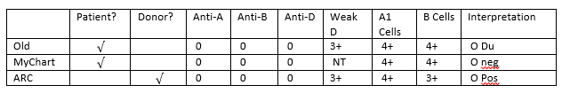
negative. The following chart explains why this patient appears to have 3 types
on record.
Figure 1. Tube typing results of same patient from different labs with different SOPs.
What blood type is recorded on a donated unit
of blood typed “O Du?”
AABB Standards for Blood Banks and
Transfusion Services requires all donor blood to be tested using a method that
is designed to detect weak D. This can be met through IAT testing or another
method that detects weak D. If the test is positive, the unit must be labeled
Rh positive. This is an important step to prevent alloimmunization in a
recipient because weak D RBCs can cause the production of anti-D in the
recipient. This also explains why the ARC donor card this patient received
lists her type as O pos.
What type
of blood does an “O Du” patient receive?
Historically, weak D red blood cells
(RBCs) were defined as having decreased D antigen levels which require the IAT
for detection. A patient who is serologic weak D has the D antigen, just in
fewer numbers. This type of weak D expression primarily results from
single-point mutation in the RHD gene that encodes for a single amino acid
change. The amino acid change causes a reduced number of D antigen sites on the
RBCs. Today we know more about D antigen expression because we have the
availability to genotype these weak D RBCs. More than 84 weak D types have been
identified, but types 1, 2, and 3 represent more than 90% of all weak D types
in people of European ethnicity.2 An Rh negative patient has no D
antigen and should, under normal circumstances, only receive Rh negative blood.
Yet, there has been a long history of transfusing weak D patients with Rh
positive RBCs. 90% of weak D patients genotype as Type 1, 2 or 3 and may
receive Rh positive transfusions because they rarely make anti-D. 2
It is now known that weak D can actually
arise from several mechanisms including quantitative, as described above, position
effect, and partial D antigen. Molecular testing would be needed to
differentiate the types, but, with the position effect, the D antigen is
complete and therefore the patient may receive Rh positive blood with no
adverse effects. On the other hand, a partial D patient may type serologically
as Rh negative or Rh positive and can be classified with molecular testing. It
is important to note that these partial D patients are usually only discovered
because they are producing anti-D. If anti-D is found, the patient should
receive Rh negative blood for any future transfusions.
Thus, 3 scenarios can come from typing
the same patient. With a negative antibody screen, and because 90% of weak D
patients have been found to be Type 1, 2 or 3 when genotyped, many labs do not routinely
genotype patients and will report the blood type as Rh pos and transfuse Rh pos
products. However, depending on the lab medical director and the lab’s SOPs,
these same patients may be labeled Rh neg, weak D and receive Rh negative
products. There is no general consensus on the handling and testing of weak D
samples. The 3rd scenario is that many labs do not test for weak D
in patients at all, and a negative D typing at IS would result in reporting the
patient as Rh neg, with no further testing. In this case, the patient would be
transfused with Rh negative products.
Can an “O
Du” patient have a transfusion reaction if they are transfused with O positive
blood? Would she need to receive O negative blood in a transfusion?
This question was covered
somewhat in the above discussion. Policies regarding the selection of blood for
transfusion are lab dependent, dictated by the lab medical director, and are
based on the patient population, risk of developing anti-D, and the
availability or lack of availability of Rh negative blood products. Anti-D is
very immunogenic. Less than 1 ml of Rh pos blood transfused to an Rh negative
person can stimulate the production of anti-D. However, not all patients
transfused with Rh positive blood will make and anti-D. As discussed above, 90%
of weak D patients are types 1, 2 or 3, would be unlikely to become
alloimmunized to anti-D. If a weak D patient with a negative antibody screen
receives a unit of D pos RBCs, there is a very small possibility that they are
a genotype who could become alloimmunized to the D antigen and produce anti-D. However,
as stated above, the majority of weak D patients can be
transfused with D positive RBCs. Thus, with few exceptions, from a historical
perspective, one can safely classify the weak D as D positive.
This question gets a little trickier
when dealing with females of childbearing age. We particularly want to avoid
giving Rh positive blood to females to avoid anti-D and the complications of
Hemolytic Disease of the Fetus and Newborn. Therefore, when dealing with these
patients, lab policies and physicians tend to be more conservative in their
approach to transfusion. The consequences, however, in males and older females
are less serious and these patients could be given Rh positive blood if there
exists a shortage of Rh negative units. Any patient who becomes alloimmunized
to the D antigen, would thereafter be transfused with Rh negative products.
Does an “O
Du” patient need to receive RhoGAM if she pregnant and her husband is Rh
positive?
This, again, would be up to the medical
director, the lab’s SOPs or the patient’s physician. Depending on lab practice,
the lab may or may not perform weak D testing. If the lab does not perform weak
D and results this patient as Rh neg, the patient would get Rhogam. If the lab
does do weak D testing and finds a weak D phenotype, the decision whether or
not to give Rhogam would be up to lab practices and the practitioners involved.
The lab’s policy on terminology used in resulting the type may also reflect the
decision whether or not to give Rhogam. This brings up a lot of questions in
the lab because we know that a patient who would not make anti-D would not need
Rhogam. So, what is the best course of action? Read my next blog to learn more
about troubleshooting and resolving D typing discrepancies!
From the discrepancies in reported type in this individual, and putting all the pieces of the puzzle together, we can conclude that this patient is a weak D phenotype. However, the type reported and the terminology used varies from lab to lab. Molecular testing is available, yet most labs are still using serological testing for blood types for both donors and patients. This is based on several factors within the lab setting. Stay tuned for my next Blood Bank blog exploring D typing discrepancies and the financial aspects of performing genotype on pregnant patients to clarify Rh type.
-Becky Socha, MS, MLS(ASCP)CM BB CM graduated
from Merrimack College in N. Andover, Massachusetts with a BS in
Medical Technology and completed her MS in Clinical Laboratory Sciences
at the University of Massachusetts, Lowell. She has worked as a Medical
Technologist for over 30 years. She’s worked in all areas of the
clinical laboratory, but has a special interest in Hematology and Blood
Banking. When she’s not busy being a mad scientist, she can be found
outside riding her bicycle.
A 36 year old woman presented to the delivery room at a local county hospital at 39 weeks’ gestation. The doctor ordered a type and screen on the patient, the blood was drawn and sent to the Blood Bank lab. The Blood Bank tech looked up the patient’s Blood Bank history and noted that an antibody screen done at 28 weeks was positive, with an anti-Lea identified. The Blood Bank’s policy is to have 2 units of blood available for any patient with an antibody. As the Blood Bank tech was working on the sample, the physician sent a STAT order for 2 units RBCs for intrapartum hemorrhage.
Are Lewis antibodies clinically significant? AABB defines a clinically significant antibody as one that causes decreased red blood cell survival of transfused cells, one that causes hemolytic transfusion reaction or one that causes Hemolytic Disease of the Fetus and Newborn (HDFN).3 In the Blood Bank, we would always be cognizant of all three criteria, but in this case, we are particularly concerned with HDFN.
The Lewis system is of great interest in immunohematology because of its unique characteristics. The Lewis blood group system is the only one where the antigens are not produced by the red blood cell itself. We learn in immunohematology that red cell antigens are structures that are usually formed on red blood cell membranes, but Lewis stands alone in that the antigens are glycolipids that are formed in the plasma and then passively absorbed onto the red blood cell membrane. This forms a loose attachment and these antibodies can shed or elute off the RBCs in certain circumstances.
Because Lewis antigens are not formed on RBCs, Lewis antigens are not present at birth and therefore not found on cord blood cells. Cord blood and RBCs from newborns will phenotype as Le(a-b-). The saliva of these newborns will have Lea and/or Leb antigens depending on the genes inherited, but the RBCs will test negative for these antigens at birth. By about 10 days of age, the Lewis antigens can be detected in plasma, and they will shortly thereafter begin to be absorbed onto the RBCs. Yet, children do not exhibit their true Lewis phenotype until about age 6.
The development of Lewis antigens is also unique. Lewis antigens are not antithetical, as they result from the interaction of two fucosyltransferases encoded by the Le and Se genes. The Le gene is needed for the production of Lea antigen and the Se gene is needed to form Leb antigen. The three common Lewis phenotypes, Le(a+b-), Le(a-b+) and Le(a-b-) indicate the presence or absence of the Le and Se transferase enzymes.
In pregnancy a mother’s plasma volume increases, and because Lewis antigens are not an integral part of the RBC membrane, they can elute off her RBCs. This causes a decrease in Lewis antigen and some pregnant women, regardless of their true Lewis antigen type, will temporarily type as Le(a-b-). At the same time, because they are now typing Le(a-b-), pregnant women often acquire Lewis antibodies.
Anti-Lea is the most frequently found Lewis antibody, is IgM, and is usually detected at room temperature. In most cases, it is acceptable to give patients with Lewis antibodies RBC units that are crossmatch compatible at 37C without giving antigen negative units. One reason for this is that, as we saw above, Lewis antigens are merely absorbed onto RBCs and can be eluted from transfused red cells within days of transfusion. In addition, when Lewis antigen positive blood is given to Lewis-negative recipients, the Lewis substance in plasma neutralizes antibodies in the recipient. This is why it is extremely rare for anti-Leato cause hemolysis of transfused RBCs. Regardless of Lewis phenotype, RBCs would be expected to have normal in vivo survival.
For an antibody to cause HDFN it must be able to cross the placenta. The antibody must also react with antigens on the red blood cells. Because Lewis antibodies are IgM and do not cross the placenta, and because Lewis antigens are not present on fetal and neonatal erythrocytes, Lewis antibodies have not been implicated in HDFN and this baby is not at risk.
What does this all means in practice? Though the presence of anti-Lewis antibodies in pregnant women is fairly common, both anti-Leaand anti-Leb are naturally occurring IgM antibodies that are not generally considered to be clinically significant. They have low immunogenicity, they do not cause HDFN, they rarely cause hemolysis and do not cause decreased survival of transfused RBCs. This baby is not at risk for HDFN. The mother can safely be transfused with crossmatch compatible RBCs. Her Lea antibodies may be neutralized with a transfusion or will naturally disappear, and her true Lewis phenotype should return within about 6 weeks after delivery.
References
Harmening DM: The Lewis System. In Harmening DM, (6th ed): Modern Blood Banking and Transfusion Practices. FA Davis, Philadelphia 2012, pp. 177-180
Fung, Mark K, ed.: The Lewis System. 18th ed: AABB Technical manual, Bethesda, Md. 2014, pp 304-306
D. Radonjic et al, The Presence of antibodies in anti-Lewis system in our pregnant women. Giorn.It.Ost.Gin. Vol. XXXII-n.4.Luglio-Agosto 2010.
-Becky Socha, MS, MLS(ASCP)CM BB CM graduated from Merrimack College in N. Andover, Massachusetts with a BS in Medical Technology and completed her MS in Clinical Laboratory Sciences at the University of Massachusetts, Lowell. She has worked as a Medical Technologist for over 30 years. She’s worked in all areas of the clinical laboratory, but has a special interest in Hematology and Blood Banking. When she’s not busy being a mad scientist, she can be found outside riding her bicycle.
A 26 year old African American female with sickle cell anemia presented to a New York emergency room with cough, chest pain, fever and shortness of breath. Laboratory results showed an increased white blood cell count, slightly decreased platelet count and a hemoglobin of 6.2 g/dl. Her reticulocyte count was 7%, considerably below her baseline of 13%. Consulting the patient’s medical records revealed history of stroke as a child and subsequent treatment with chronic blood transfusions. She was admitted to the hospital for acute chest syndrome and aplastic crisis and care was transferred to her hematologist. Two units of RBCs were ordered for transfusion.
The blood bank technologists checked the patient’s blood bank history and noted her blood type was A, Rh(D) positive, with a history of a warm autoantibody and anti-E. The current blood bank sample confirmed the patient was blood type A, RH(D) positive with a negative DAT but the antibody screen was positive. Anti-E was identified. Per request of the hematologist, phenotypically similar units were found and the patient was transfused with 2 units of A RH(negative), C/E/K negative, HgS negative, irradiated blood. The patient’s hemoglobin rose to 8g/dl and she was discharged from the hospital 3 days after transfusion.
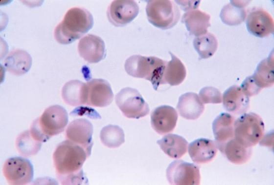
Ten days after discharge the patient returned to the emergency room with symptoms including aching muscles, fever and chills. A delayed transfusion reaction was suspected. A type and screen was immediately sent to the blood bank. The post transfusion type and screen remained positive for anti-E, DAT was negative. No additional antibodies were identified. However, a CBC sent to the lab at the same time revealed malarial parasites on the peripheral smear. The patient was consulted for a more complete medical history and reported that she had never traveled outside of the country. A pathology review was ordered and the patient was started on treatment for Plasmodium falciparum.
Discussion
Red Blood cell transfusions can be life saving for patients with sickle cells anemia. These patients are frequently transfused by either simple transfusion of red cell units or by exchange transfusion. Because of this, alloimmunization is reported to occur in 20% to 40% of sickle cell patients.1 Blood bank technologists are very diligent in adhering to strict procedures and follow a standard of practice aimed to prevent transfusion reactions. While preventing immune transfusion reactions may be the most forefront in our minds when transfusing the alloimmunized patient, it is important to consider transfusion transmitted diseases as a potential complication of blood transfusions.
Malaria is caused by a red blood cell parasite of any of the Plasmodium species. Mosquito transmitted infection is transmitted to humans through the bite of an infected mosquito. Transfusion-transmitted malaria is an accidental Plasmodium infection caused by a blood transfusion from a malaria infected donor to a recipient.
Donors, especially those from malarial endemic countries who may have partial immunity, may have very low subclinical levels of Plasmodium in their blood for years. Even these very low levels of parasites are sufficient to transmit malaria to a recipient of a blood donation. Though very rare, transfusion-transmitted malaria remains a serious concern for transfusion recipients. These transfusion-transmitted malaria cases can cause high percent parisitemia because the transfused blood releases malarial parasites directly into the recipient’s blood stream.
Blood is considered a medication in the United States, and, as such, is closely regulated by the FDA. Blood banks test a sample of blood from each donation to identify any potential infectious agents. Blood donations in the US are carefully screened for 8 infectious diseases, but malaria remains one infectious disease for which there is no FDA-approved screening test available. For this reason, screening is accomplished solely by donor questioning.2 A donor is deferred from donating if they have had possible exposure to malaria or have had a malarial infection. Deferral is 12 months after travel to an endemic region, and 3 years after living in an endemic region. In addition, a donor is deferred from donating for 3 years after recovering from malaria. It is important, therefore, for careful screening to take place by questionnaire and in person, to make sure that the potential donor understands and responds appropriately to questions concerning travel and past infection.
Malaria was eliminated from the United States in the early 1950’s. Currently, about 1700 cases of malaria are reported in the US each year, almost all of them in recent travelers to endemic areas. From 1963-2015, there have been 97 cases of accidental transfusion-transmitted malaria reported in the United States. The estimated incidence of transfusion-transmitted malaria is less than 1 case in 1 million units.4 Approximately two thirds of these cases could have been prevented if the implicated donors had been deferred according to the above established guidelines.3 While the risk of catching a virus or any other blood-borne infection from a blood transfusion is very low, a blood supply with zero risk of transmitting infectious disease may be unattainable. With that being said, the blood supply in the United Sates today is the safest it has ever been and continues to become safer as screening tests are added and improved. Careful screening of donors according to the recommended exclusion guidelines remains the best way to prevent transfusion-transmitted malaria.
References
LabQ, Clinical laboratory 2014 No.8, Transfusion Medicine. Jeanne E. Hendrickson, MD, Christopher Tormey, MD, Department of Laboratory Medicine, Yale University School of Medicine
Technical Manual, editor Mark K. Fung-18th edition, AABB. 2014. P 201-202
The New England Journal of Medicine. Transfusion-Transmitted Malaria in the United States from 1963 through 1999. Mary Mungai, MD, Gary Tegtmeier, Ph.D., Mary Chamberland, M.D., M.P.H., June 28, 2001. Accessed April 2018
Malaria Journal. A systematic review of transfusion-transmitted malaria in non-endemic areas. 2018; 17: 36. Published online 2018 Jan 16. doi: 1186/s12936-018-2181-0. Accessed April 2018
-Becky Socha, MS, MLS(ASCP)CM BB CM graduated from Merrimack College in N. Andover, Massachusetts with a BS in Medical Technology and completed her MS in Clinical Laboratory Sciences at the University of Massachusetts, Lowell. She has worked as a Medical Technologist for over 30 years. She’s worked in all areas of the clinical laboratory, but has a special interest in Hematology and Blood Banking. When she’s not busy being a mad scientist, she can be found outside riding her bicycle.