Hematopathology Case Study: Two Cases with Surprising Hematopoetic Elements

Case 1 History

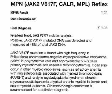
Sixty-one year old man with new diagnosis of Bud-Chiari syndrome and extensive peripheral, splenic and hepatic venous thrombosis with increasing fatigue, abdominal discomfort and abnormal liver function tests. A liver biopsy was performed and a hypercoagulability work-up, including JAK2 mutation analysis was initiated.

2-cases-liver2x
Liver core biopsy 2X
2-cases-liver10x
Liver core biopsy 10X

Diagnosis

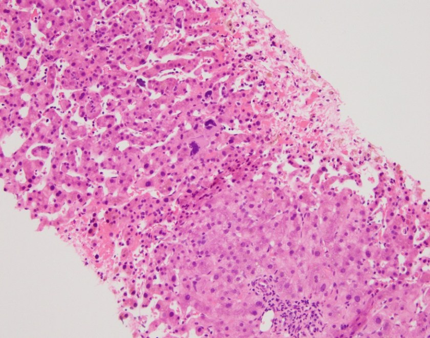
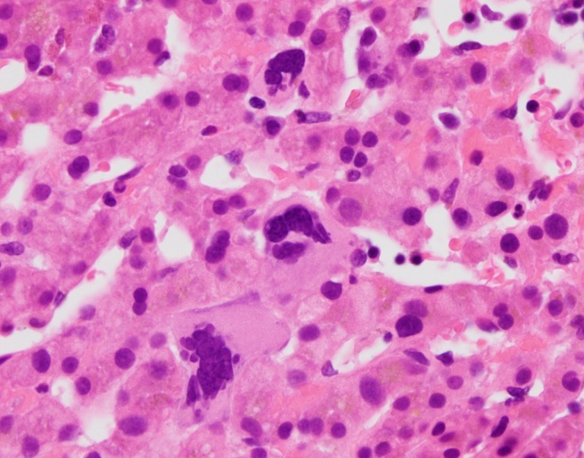
The liver biopsy showed extensive hemorrhage, hepatocellular necrosis and collapse with mild portal and lobular mixed inflammation. Occasional megakaryocytes and nucleated red blood cell precursors were noted. The case was sent to hematopathology for further review.

2-cases-liver40x
Liver core biopsy 40X
2-cases-liver40x2
Liver core biopsy 40X
2-cases-jak2analysis
JAK2 Mutations Analysis

Hematopathology Diagnosis

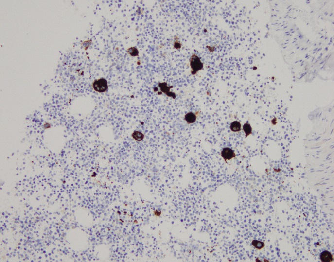
Sections show liver parenchyma with changes of the patient’s known history of venous outflow obstruction, as well as extramedullary hematopoiesis, including scattered megakaryocytes (arrows) and erythroid precursors (circle). In the setting of a positive JAK2 V617F mutation, this constellation of findings is consistent with a myeloproliferative neoplasm.

Case 2 History

Fifty-nine year old man with a history of hypertension and alcohol abuse with posterior mediastinal lymphadenopathy. Recent bone marrow biopsy showed mildly hypercellular bone marrow with megakaryocytic and myeloid hyperplasia, and increased stromal reticulin with concern for primary myelofibrosis. A lymph node biopsy was performed.

2-cases-lymph10x.jpg
Lymph node biopsy 10X
2-cases-lymph40x
Lymph node biopsy 40X
2-cases-myelo.jpg
Myeloperoxidase
2-cases-CD71
CD71
2-cases-CD61
CD61
2-cases-CD34
CD34
2-cases-CD3
CD3
2-cases-CD20
CD20

Diagnosis

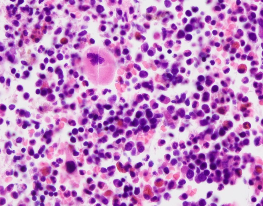
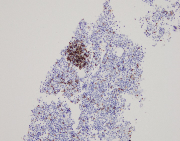
The lymph node biopsy shows fragments composed of adipocytes and maturing trilineage hematopoiesis. Multiple small to medium sized lymphoid aggregates are also seen, composed of small and mature appearing lymphocytes. The lymphocytes are a mixture of CD3 positive T cells and CD20 positive B cells with focal B cell predominance. Myeloperoxidase highlights myeloid precursors, which comprise 70-80% of the cellularity. CD71 highlights erythroid precursors, which comprise 20-30% of the cellularity. CD61 highlights megakaryocytes. CD34 highlights vessels and only rare CD34-positive cells are seen. Taken together, the findings are consistent with extramedullary hematopoiesis.

Discussion

Extramedullary hematopoiesis (EMH) is defined as hematopoiesis that occurs outside of the bone marrow. It can occur in both normal and pathologic states and has been seen in several hematologic disorders including chronic myeloproliferative neoplasms. Myeloproliferative neoplasms (MPN) are a group of clonal hematopoetic stem cell disorders that include polycythemia vera (PV), essential thrombocythemia (ET) and primary myelofibrosis (PMF).1 JAK2V617F mutation is the most frequent mutation associated with MPNs, found in roughly 96% of patients with PV and 65% of patients with ET and PMF. This mutation leads to constitutive activation of the JAK/STAT pathway and is a driver of myeloproliferation.2  The patient in case 1 was found to have a JAK2 mutation during the work-up for hypercoagulability. This suggests that he may have an underlying MPN, however JAK2 mutations have been found in patients with venous thrombosis, but without overt evidence of MPNs.3 The patient in case 2 had a bone marrow biopsy with features concerning for primary myelofibrosis. In PMF, there is generally a proliferation of myeloid cells in addition to marrow fibrosis. Increasing fibrosis can eventually result in pancytopenia as the fibrosis takes over the marrow space in addition to altering the bone marrow environment so that it is unable to support normal hematopoiesis. Ultimately, this can lead to extramedullary hematopoesis. EMH most commonly occurs in the spleen and liver, but has been described in many other sites including the mediastinum and lymph nodes. In addition to being a driver of proliferation, it is thought that JAK2 mutations make hematopoetic stem and progenitor cells more sensitive to growth factors and can cause the cells to mobilize to the liver and spleen.4  Patients with EMH can have symptoms related to the site of involvement. Depending on the extent of involvement and location, EMH may require treatment with low dose radiation. While EMH is a rare finding, it should prompt an investigation for an underlying MPN.

References

  1. Imai K, Aoi T, Kitai H,et al. A case of perirenal extramedullary hematopoiesis in a patient with primary myelofibrosis. CEN Case Reports. 2017;6(2):194-199. doi:10.1007/s13730-017-0274-1.
  2. Kim CH. Homeostatic and pathogenic extramedullary hematopoiesis. Journal of blood medicine. 2010;1:13-19.3https://www.ncbi.nlm.nih.gov/pubmed/17263783.
  3. De Stefano, V, Fiorini, A, Rossi, E, et al. Incidence of JAK2 V617F mutation among patients with splanchnic or cerebral venous thrombosis and without overt chronic myeloproliferative disorders. Journal of Thrombosis and Haemostasis. 2007;5(4):708-14. https://www.ncbi.nlm.nih.gov/pubmed/17263783.
  4. Passamonti F, Maffioli M, Caramazza D, et al. Myeloproliferative neoplasms: From JAK2 mutations discovery to JAK2 inhibitor therapies. Oncotarget. 2011;2(6):485-490.

Marcus, Chelsea_099-Edit

Chelsea Marcus, MD is a third year resident in anatomic and clinical pathology at Beth Israel Deaconess Medical Center in Boston, MA and will be starting her fellowship in Hematopathology at BIDMC in July. She has a particular interest in High-grade B-Cell lymphomas and the genetic alterations of these lymphomas.

Cut it Out … No, Really, I Need Margins

Hello everyone! Back again with another post about that interesting space between my experiences working in laboratory medicine as an MLS and my current path through medical school toward a career in pathology. Last month, I discussed how the new 5th generation cardiac enzyme assays are evolving and reaffirming the relationships between lab data and clinical decision making. This month, as I adjust to a very different circadian rhythm, I’d like to talk about some topics in my surgery rotation as they relate to surgical pathology and the lab.

Just to summarize, besides epidemiological research and public health initiatives I’ve written about here on this blog, I had several years of lab work before medical school. In my experience, I have seen the gamut of required steps for pathology specimens peri/post-operatively. Everything from placenta, bone, blood, marrow, skin, brain, lung, GI, to any other organ system’s tissue is processed, blocked, stained and examined on glass by pathologists who write reports for their clinical colleagues.  Often, we in the lab receive phone calls from providers inquiring about turn-around times and results as they  follow-up on their patients and cases. In Chicago, I was able to see and train in a great trauma center at Northwestern, community hospitals like Swedish Covenant and Weiss Memorial, and an academic hospital centers like Rush and UIC. What I learned there is just how much really depends on those pathology reports. Cytology, diagnostic immunohistochemistry, morphology, margins, and gross analysis all contribute to a final diagnosis. After an extended observership at UAB Medical Center, I was fortunate to see first-hand the critical process involved in signing out dermatology consults, examining gross pathology, and even frozen neuropathology specimens. Sitting with attendings in the OR and frozen rooms deciding between glioblastoma multiforme, lymphoma, or something benign (read: defer to permanent slide diagnosis later) was fascinating. Meanwhile, I’m now a month into formal surgical rotations at Bronx-Care Hospital in NY and I get to see the other side of the pathology report.

The Relationships Between Surgeons and Pathologists are Critical

Many surgical interventions and procedures require resection of known or suspected pathologic tissue. Whether it’s malignancy, benign growth, obstruction, adhesion, or otherwise mechanically compromising tissue, many patients require a surgeon to remove the entity in question. And, while the difficulty of these excisions and resections may vary depending on location, cases rely heavily on the pathologist-surgeon collaboration. Virtually all neoplasms are diagnosed through anatomic pathology assessment under a microscope. Fine needle aspirates, pap smears, bone marrow biopsies, and countless other tissues must go through pathology before being finalized. This interdisciplinary collaboration between the surgical team and the pathology team is, of course, by nature acutely critical. In proper circumstances, open cases in the operating room are consulted to a pathologist STAT. The effective communication between the pathologist and surgeon awaiting the intraoperative consultation is key to effectively treating their shared patient. Sometimes operating rooms will have live microscopic image-casting, sometimes there is an intercom system, sometimes its solely based on electronic forms in the EHR, and sometimes pathologists need to go into the surgical field to examine the resection intraoperatively in person. However it happens, this is a very important relationship that patients might not be aware of.

The Point of View Between Surgical Pathology and Clinical Surgeons Are Different

So this sounds like a perfect match, right? Surgeons and pathologists living in harmony? Unfortunately, harmony isn’t part of regular onboarding at many institutions so, as with any staff, there are different scopes and sometimes this can be a challenge. Getting a frozen notification as a pathologist is a serious task. They are emergent and must be addressed immediately and diagnoses are made with serious gravity, often consulting with other pathologists. This is also, however, a singular teaching moment as every frozen section is different and pathologists use these learning opportunities to teach their residents and medical students. In the interests of accurate diagnoses, educational value, and appropriate response to the OR, pathologists take measures to ensure success. For example, frozen specimens will be received, a history and presentation of the patient is discussed, the specimen is partitioned for frozen section (STAT), permanent section, and further studies (routine). So, for the pathologist it’s all about accuracy, reliability, and what they can confidently report. The surgeon has a different point of view: they are operating with a specific physical goal in mind by either resecting a tumor, or isolating good margins from a known malignancy, or ensuring the tissue being removed is correct/adequate for its therapeutic purpose. Fun fact: surgical pathology was a field originally developed by surgeons! There are things a pathologist only knows, and there are things a surgeon only knows—but when working together, the overlap of medical knowledge increases the coverage of care for their shared patients’ outcomes.

surgpath1
Image 1. A pathologist processes a frozen specimen on a cryostat machine. A summary of frozen sections from JAMA, 2005;294(24):3200. doi:10.1001/jama.294.24.3200

The Cold Truth About Frozen Sections

Frozen specimens aren’t perfect. In these specimens, tissue gets stiffened by freezing instead of routine paraffin embedding, and because of that a frozen section could be distorted by folds, tears, and other artifacts that might appear because of mechanical manipulation during processing. Frozen samples also leave artifacts where water would crystallize and freeze, but one of the caveats regarding artifacts in frozen sections is that FAT DOES NOT FREEZE. Instead, specimens that have large fat content (i.e. brain tissue) have to be examined carefully to not confuse findings with inflammation or other pathologic processes. Ultimately, it takes numerous cases to properly hone the skills required to confidently diagnose from frozen section. While they might not be perfect, it is a critical tool used between the surgical and pathology teams. Challenges in this handoff process relate to proper use of this surgical tool. For instance, if a frozen is called for and the surgery is closed by the time a pathology report is filed, then (assuming there were no serious delays) this may have been an inappropriate specimen decision. Furthermore, specimens must be discussed prior to receipt for appropriateness and clinical relevance. Fatty lipomas aren’t going to go to frozen section, they shouldn’t be ordered. A thyroid lobectomy? That’s a better utilization of resources and tools.

surgpath2
Image 2. A demonstration of water-related crystal formation causing distortion and artifact (LEFT) on frozen section of muscle tissue, compared to normal (RIGHT). From Northwestern, source: http://www.feinberg.northwestern.edu/research/docs/cores/mhpl/tissuefreezing.pdf

Ultimately, with proper training and experience a pathologist can effectively use the frozen section as a useful clinical tool to improve patient outcomes. Surgeons operating in the best interests of their patients, should strive to create a functional and successful communication between both services. My experiences in NY with surgeons of various kinds reveals a common truth among them: pathology is a critical player in surgical interventions, and without margins, diagnostic stains, and other work-ups, those interventions would be much more difficult and risky.

Thanks again! See you next time!

Bonus: for more content specifically detailing some of the cellular morphologies and cytology I discussed above, please check out I Heart Pathology, a compendium website my friend and colleague at UAB, Dr. Tiffany Graham, manages. It’s meant for other pathology residents to review and refresh on material and it’s updated as often as possible. Check out the link here: https://www.iheartpathology.net/


ckanakisheadshot_small

–Constantine E. Kanakis MSc, MLS (ASCP)CM graduated from Loyola University Chicago with a BS in Molecular Biology and Bioethics and then Rush University with an MS in Medical Laboratory Science. He is currently a medical student actively involved in public health and laboratory medicine, conducting clinicals at Bronx-Care Hospital Center in New York City.

Microbiology Case Study: A 10 Year Old with Fever and Chills

Case History

A 10 year old female presented to the pediatric emergency department (ED) with a chief complaint of persistent fever and chills for the past 10 days. Her mother reported the fevers reached up to 103°F and temporarily would respond to ibuprofen.  She also noted a decrease in the patient’s appetite, tiredness and a bumpy rash on her truck and extremities. In the ED, she was clinically stable but her temperature reached a max of 104.7°F. On physical examination, shotty cervical lymphadenopathy was noted and there was no appreciable enlargement of the liver or spleen. Initial laboratory testing showed a white blood cell count of 10.6 TH/cm2 (normal range: 4.3-11.4 TH/cm2) and elevated acute phase proteins (ESR 45 mm/HR and CRP 2.6 mg/dL). Blood cultures were collected and the patient was started on ceftriaxone. Pediatric infectious disease was consulted and a thorough infectious work up was completed.

Laboratory Identification 

  • Rapid influenza antigen test: Negative
  • Rapid Group A Strep antigen test: Negative
  • Rapid Monospot: Negative
  • HIV antigen/antibody (4th generation) test: Negative
  • Legionella urinary antigen: Negative
  • Histoplasma urinary antigen: Negative
  • Antinuclear antibody: Negative
  • Rheumatoid factor: Negative
  • Urine culture: Negative
  • Blood cultures: Negative
  • Bartonella henselae IgM: ≥1:20 (normal <1:20)
  • Bartonella henselae IgG: ≥1:1024 (normal <1:128)

Infectious disease and rheumatologic work ups, as listed above, were negative with the exception of a positive IgM and IgG serologic testing for Bartonella henselae, with the results suggesting a recent infection based on the elevated titers. Upon further questioning, the family did have many outdoor cats and dogs; however, the child denied any recent bites or scratches.

Discussion 

Bartonella henselae is a facultative, Gram negative coccobacillary rod that is the causative agent of cat scratch disease and bacillary angiomatosis. The main reservoir for B. henselae is cats and the disease is spread from cat to cat via the cat flea. Feral cats, outdoor cats and young kittens, especially those living in hot, humid environments where fleas are plentiful, are more likely to be infected and spread the disease to humans via infective flea feces during a scratch or bite from the cat.

The incubation period for B. henselae ranges from 1-3 weeks and the majority of patients present with systemic symptoms including fever, chills, malaise, anorexia and headache. In addition, painful lymphadenopathy, on the side of the body where the scratch occurred (most common upper extremity), can be present in the epitrochlear, axillary and cervical regions. Less frequently, B. henselae causing bacillary angiomatosis can result in the proliferation of vessels in organs (liver and spleen). Though rare, encephalopathy and endocarditis due to B. henselae are the most severe manifestations of disease.

In the microbiology laboratory, the diagnosis of B. henselae is challenging due to the fact it is slow growing, highly hemin dependent and requires high humidity conditions for growth. The organism will grow on chocolate and heart infusion agars containing 5% fresh rabbit blood. Plates should be incubated at 35°C with 5% CO2 with high humidity for at least 4 weeks. Colonies are irregular and off-white in color and B. henselae is negative for both catalase & oxidase and asaccharolytic.

Due to the identification difficulties with culture, serologic testing is the main methodology for the diagnosis of B. henselae. Enzyme linked immunosorbent assays (ELISA) are relatively easy to perform and provides good results, although the provider should be aware of the sensitivity of the particular platform, the fact that cross reactivity with other Bartonella spp. can occur and seronegative infections can sometimes occur. Warthin-Starry silver stain on fixed tissue sections from lymph nodes and other organs can be helpful as well; however, it is relatively insensitive and not highly specific.   

 With regards to treatment, there are no agreed upon breakpoints for B. henselae published by CLSI or EUCAST. Microdilution or Etests can be used for testing and isolates have been susceptible to many antibiotics. In general, for cat scratch disease, it does not respond to antibiotic therapy and there is only a minimal benefit of antimicrobial agents. In the case of our patient, she was switched from ceftriaxone to a five day course of azithromycin with a gradual improvement of her fever curve. She was scheduled to follow up with pediatric infectious disease in 2-3 weeks.


Stempak

-Lisa Stempak, MD, is an Assistant Professor of Pathology at the University of Mississippi Medical Center in Jackson, MS. She is certified by the American Board of Pathology in Anatomic and Clinical Pathology as well as Medical Microbiology. She is the Director of Clinical Pathology as well as the Microbiology and Serology Laboratories.  Her interests include infectious disease histology, process and quality improvement and resident education.

Tissue is the Issue: Microorganism Identification (part 3)

In parts 1 and 2, we discussed pre-analytical and analytical issues that can be faced when culturing tissue specimens. Part 3, the final part of the Tissue is the issue series, will review analytical and post-analytical issues of tissue culture requests. 

The Issue

Let’s consider a case of “culture-negative” endocarditis (1), in which organism was detected during direct observation of the specimen (2); but as you would expect for suspected “culture-negative” endocarditis, the culture does not yield an organism. This can happen for a variety of reasons. Perhaps the culture request was for a routine aerobic culture, but the organism was a strict anaerobe and therefore could not grow. Maybe the patient was on antibiotics, so the organism observed was not viable. Or it is possible that the organism in question is fastidious and requires special media or growth conditions which were not met. Another frequent occurrence is that tissue is sent for pathology, but not for culture. This is more common when malignancy is expected, but the pathological findings suggest an infectious process. These scenarios may seem hopeless, but don’t despair; there is a non-culture alternative that can aid in identifying the causative agent. 

The Solution

Microbial (bacterial, fungal, and viral) DNA can be detected from fresh, frozen, or fixed tissue. Simply put, DNA is first extracted from the specimen, of which the microbial DNA of interest (bacterial, fungal, viral) is then amplified via PCR. Broad-range or pathogen-specific PCRs are commonly available from a variety of reference laboratories. If broad-range PCR is performed, then sequencing of the amplicon is required to determine the organism identification. Sequences are queried against a library of known microbial genomes to obtain a match.

Depending on the DNA of interest, different primer sets are utilized. For broad-range bacterial PCR, the 16S ribosomal RNA gene is typically used. Although mycobacteria are bacteria, they require additional gene targets for optimal detection and identification (16S rRNA, rpoB, and hsp65). For fungi, the 28S rRNA and the ITS (internal transcribed spacer; ITS1 and ITS2) genes are used.

The organism burden and specimen type can affect the probability of detecting an organism and obtaining its identification. The likelihood of a positive outcome is proportional to the organism burden. For example, if organism observed in the direct exam (i.e., Gram or acridine stain), then the organism can usually be detected and identified. However, if no organism is observed, then the chances of a positive result is unlikely. Therefore, our protocol is to only send specimens for microbial DNA sequencing on specimens in which organisms were observed in the direct exam. This is true for all specimen types (fresh, frozen, fixed).

It is important to note that fixed specimens may not yield as good results as a fresh or frozen specimen. This is because the process of fixation can degrade the microbial DNA (3). Additionally, because detection of microbial DNA is the basis for pathogen identification, susceptibility results are not going to be available. Treatment options will need to be based on known empiric therapies.

The Conclusion

Microbial DNA sequencing is a viable option for the identification of etiological agents of infection from a variety of sources, such as culture-negative infections. Other uses include slow-growing organisms and organisms that are unidentifiable by traditional methods (4). In my experience, this is a valuable tool that should be considered when culture does not yield a result and a result is necessary to drive clinical decisions. 

References

  1. Tissue is the Issue, Part 1
  2. Tissue is the Issue, Part 2
  3. Martinez, R.M. Genes in your tissue: probe identification and sequencing microbial targets from formalin-fixed, paraffin-embedded tissue. Clin. Microbiol. Newslett. 36: 139-147.


Martinez Headshot-small 2017

-Raquel Martinez, PhD, D(ABMM), was named an ASCP 40 Under Forty TOP FIVE honoree for 2017. She is one of two System Directors of Clinical and Molecular Microbiology at Geisinger Health System in Danville, Pennsylvania. Her research interests focus on infectious disease diagnostics, specifically rapid molecular technologies for the detection of bloodstream and respiratory virus infections, and antimicrobial resistance, with the overall goal to improve patient outcomes.

Learning Organizations and Systems Thinking

For organizations to grow and sustain themselves, it is essential that they take a learning stance. What is a “learning stance,” you ask? Well, learning organizations: encourage new ways of thinking and doing business, focus on employee learning, and build the organizational capacity. These companies focus on learning about the organizational systems within a business, such as interconnected actions and patterns of behavior. However, only understanding the systems themselves are not enough. Systems thinking requires the creation of a shared vision within and between teams, because teams are the core learning units in organizations. Leaders cannot lead and learn without a deep understanding of these systems and the interconnectedness of them.

Therefore, it’s important to understand the concept of systems, as people are influenced by their environment. Open systems have a continuous outflow and inflow and maintain a steady state (not to be confused with a state of equilibrium) as long as the system is alive. Closed systems only interact with themselves; there is no outside influence and all information is only shared within the system. An example of a closed system in an organization is intranet; this system is only accessible to employees and the information is not shared outside of the intranet system. An example of an open system is an HR department, which is constantly influenced by governmental policies, organizational changes, personal issues, and internal ideas and suggestions. Another example of an open system is the medical laboratory, where samples are moved between multiple people and specialties within a system. A chemistry analyzer that tests cholesterol levels might be a closed system in and of itself, but in order for it to be effective (namely, diagnosing a patient) it needs to be open because a phlebotomist collects the specimen, a laboratory professional inspects the specimen and releases the results to the clinician, who then communicates the results to the patient, who then makes adjustments to their diet (which creates a whole additional open system). It is clear from this example how intricate open systems are and how they are all connected to other aspects and possibly other systems.

In order to create an effective organizational culture, leaders need to see people and events as systems. There are twelve key systems, namely: role description, selection to role, task review, performance planning and review, performance evaluation, salary admin, career assessment, career development, succession planning, discipline, and fair treatment. When implementing a new process, structure, or project it is important to consider the impact on all these systems to check if you need to take them into account. A change in one of these key systems can have a tremendous impact. For example, having the wrong job title can not only be demotivating it can also be detrimental to productivity and outside communication.

It is also important to note that small changes in systems can become catastrophic, especially over a longer period of time. Errors and conflicts that seem inconsequential can indeed be the reason why companies fail. Such critical points often become clear in hindsight, because the impact of these points was overlooked. However, using a systems thinking approach can bring these critical points to the surface before the results are catastrophic. Systems thinking allows organizations to locate these seemingly random events, because it focuses on the underlying structures and actions that create the conditions for certain events. These events have impacts in the long-term and it allows leaders to understand and prepare for them before their negative impact occurs.


lotte-small

-Lotte Mulder earned her Master’s of Education from the Harvard Graduate School of Education in 2013, where she focused on Leadership and Group Development. She’s currently working toward a PhD in Organizational Leadership. At ASCP, Lotte designs and facilitates the ASCP Leadership Institute, an online leadership certificate program. She has also built ASCP’s first patient ambassador program, called Patient Champions, which leverages patient stories as they relate to the value of the lab.

Hematology Case Study: A 51 Year Old Woman with Fever and Chills

A 51 year old patient presented to the emergency room with abdominal pain and fever. Fever was associated with diaphoresis, chills and headaches. Patient was in Tanzania for 3 months. She was admitted to the hospital while she was there for some unknown infection, details of which are not available.

CBC done revealed normocytic normochromic anemia with a hemoglobin of 9.2 g/dl and thrombocytopenia. Platelet count was 100 K/uL. On review of peripheral blood revealed presence of malarial parasite (ring forms).

mal1.PNG

mal2

Malaria is an infectious disease caused by Plasmodium parasites. These parasites are primarily spread by the bite of infected female Anopheles mosquitos. There are four main types of Plasmodium (P) species that infect humans:

  • Plasmodium vivax and Plasmodium ovale, which cause a relapsing form of the disease, and
  • Plasmodium malariae and Plasmodium falciparum, which do not cause relapses.

mal3

Malaria must be recognized promptly in order to treat the patient in time.

Microscopy (morphologic analysis) continues to be the “gold standard” for malaria diagnosis. Parasites may be visualized on both thick and thin blood smears stained with Giemsa, Wright, or Wright-Giemsa stains. Giemsa is the preferred stain, as it allows for detection of certain morphologic features (e.g. Schüffner’s dots, Maurer’s clefts, etc.) that may not be seen with the other two. Ideally, the thick smears are used to detect the presence of parasites while the thin smears are used for species-level identification. Quantification may be done on both thick and thin smears.

Various antigen kits are available to detect antigens derived from malarial parasites. These rapid diagnostic tests (RDT) offer a useful alternative to microscopy in situations where reliable microscopic diagnosis is not available.

Vajpayee,Neerja2014_small

-Neerja Vajpayee, MD, is the director of Clinical Pathology at Oneida Health Center in Oneida, New York and is actively involved in signing out surgical pathology and cytology cases in a community setting. Previously, she was on the faculty at SUNY Upstate for several years ( 2002-2016) where she was involved in diagnostic work and medical student/resident teaching.

The Occurrence of Lewis Antibodies in Pregnancy

A 36 year old woman presented to the delivery room at a local county hospital at 39 weeks’ gestation. The doctor ordered a type and screen on the patient, the blood was drawn and sent to the Blood Bank lab. The Blood Bank tech looked up the patient’s Blood Bank history and noted that an antibody screen done at 28 weeks was positive, with an anti-Lea identified. The Blood Bank’s policy is to have 2 units of blood available for any patient with an antibody. As the Blood Bank tech was working on the sample, the physician sent a STAT order for 2 units RBCs for intrapartum hemorrhage.

Are Lewis antibodies clinically significant? AABB defines a clinically significant antibody as one that causes decreased red blood cell survival of transfused cells, one that causes hemolytic transfusion reaction or one that causes Hemolytic Disease of the Fetus and Newborn (HDFN).3 In the Blood Bank, we would always be cognizant of all three criteria, but in this case, we are particularly concerned with HDFN.

The Lewis system is of great interest in immunohematology because of its unique characteristics. The Lewis blood group system is the only one where the antigens are not produced by the red blood cell itself. We learn in immunohematology that red cell antigens are structures that are usually formed on red blood cell membranes, but Lewis stands alone in that the antigens are glycolipids that are formed in the plasma and then passively absorbed onto the red blood cell membrane. This forms a loose attachment and these antibodies can shed or elute off the RBCs in certain circumstances.

Because Lewis antigens are not formed on RBCs, Lewis antigens are not present at birth and therefore not found on cord blood cells. Cord blood and RBCs from newborns will phenotype as Le(a-b-). The saliva of these newborns will have Lea and/or Leb antigens depending on the genes inherited, but the RBCs will test negative for these antigens at birth. By about 10 days of age, the Lewis antigens can be detected in plasma, and they will shortly thereafter begin to be absorbed onto the RBCs. Yet, children do not exhibit their true Lewis phenotype until about age 6.

The development of Lewis antigens is also unique. Lewis antigens are not antithetical, as they result from the interaction of two fucosyltransferases encoded by the Le and Se genes. The Le gene is needed for the production of Lea antigen and the Se gene is needed to form Leb antigen. The three common Lewis phenotypes, Le(a+b-), Le(a-b+) and Le(a-b-) indicate the presence or absence of the Le and Se transferase enzymes.

In pregnancy a mother’s plasma volume increases, and because Lewis antigens are not an integral part of the RBC membrane, they can elute off her RBCs. This causes a decrease in Lewis antigen and some pregnant women, regardless of their true Lewis antigen type, will temporarily type as Le(a-b-). At the same time, because they are now typing Le(a-b-), pregnant women often acquire Lewis antibodies.

Anti-Lea is the most frequently found Lewis antibody, is IgM, and is usually detected at room temperature. In most cases, it is acceptable to give patients with Lewis antibodies RBC units that are crossmatch compatible at 37C without giving antigen negative units. One reason for this is that, as we saw above, Lewis antigens are merely absorbed onto RBCs and can be eluted from transfused red cells within days of transfusion. In addition, when Lewis antigen positive blood is given to Lewis-negative recipients, the Lewis substance in plasma neutralizes antibodies in the recipient. This is why it is extremely rare for anti-Leato cause hemolysis of transfused RBCs. Regardless of Lewis phenotype, RBCs would be expected to have normal in vivo survival.

For an antibody to cause HDFN it must be able to cross the placenta. The antibody must also react with antigens on the red blood cells. Because Lewis antibodies are IgM and do not cross the placenta, and because Lewis antigens are not present on fetal and neonatal erythrocytes, Lewis antibodies have not been implicated in HDFN and this baby is not at risk.

What does this all means in practice? Though the presence of anti-Lewis antibodies in pregnant women is fairly common, both anti-Leaand anti-Leb are naturally occurring IgM antibodies that are not generally considered to be clinically significant. They have low immunogenicity, they do not cause HDFN, they rarely cause hemolysis and do not cause decreased survival of transfused RBCs. This baby is not at risk for HDFN. The mother can safely be transfused with crossmatch compatible RBCs. Her Lea antibodies may be neutralized with a transfusion or will naturally disappear, and her true Lewis phenotype should return within about 6 weeks after delivery.

References

  1. Harmening DM: The Lewis System. In Harmening DM, (6th ed): Modern Blood Banking and Transfusion Practices. FA Davis, Philadelphia 2012, pp. 177-180
  2. Fung, Mark K, ed.: The Lewis System. 18th ed: AABB Technical manual, Bethesda, Md. 2014, pp 304-306
  3. Fung, Mark K, ed.: PreTransfusion testing. 18th ed: AABB Technical manual, Bethesda, Md. 2014, pp 376
  4. D. Radonjic et al, The Presence of antibodies in anti-Lewis system in our pregnant women. Giorn.It.Ost.Gin. Vol. XXXII-n.4.Luglio-Agosto 2010.


Socha-small

-Becky Socha, MS, MLS(ASCP)CM BB CM graduated from Merrimack College in N. Andover, Massachusetts with a BS in Medical Technology and completed her MS in Clinical Laboratory Sciences at the University of Massachusetts, Lowell. She has worked as a Medical Technologist for over 30 years. She’s worked in all areas of the clinical laboratory, but has a special interest in Hematology and Blood Banking. When she’s not busy being a mad scientist, she can be found outside riding her bicycle.

Microbiology Case Study: An 88 Year Old Male with Headache

Case History

An 88 year old male presents with fever, nausea, and headache. The patient reported a diffuse headache accompanied by malaise, fatigue, and nausea without vomiting. He denied confusion, irritability, or a personal and family history of headaches. According to the patient, he frequently attends cookout parties and enjoys fruits, salads, wine, and cheese. Temperature is 38.2 degrees Celsius, blood pressure is 96/65 mmHg, pulse is 102 beats/minute, and respiratory rate is 20 breaths per minute. Physical exam is negative for nuchal rigidity and Kernig sign. Funduscopic exam is negative for papilledema. CBC shows leukocyte count of 16,000/mm3. The patient’s blood culture is positive.

Laboratory Identification

listmono1
Image 1. Short gram positive bacilli identified on Gram stain of blood culture (100x oil immersion).
listmono2
Image 2. Aerobic growth of round and translucent colonies with a narrow zone of beta hemolysis subcultured from positive blood culture bottle to sheep blood agar plate.

The blood culture was positive for short, gram positive bacilli. Sheep blood agar plate grew round and translucent colonies which have a narrow zone of beta hemolysis as shown on our plate. The organism was catalase positive and motile at 25 degrees Celsius. It showed end over end tumbling motility in a wet prep and an umbrella pattern in semi-solid motility medium. It was identified by MALDI-ToF as Listeria monocytogenes.

Discussion

Listeria monocytogenes is a gram positive bacillus that is isolated from the environment and a variety of animals. It is associated with foodborne outbreaks from dairy and meat products. The most common foods associated with listeriosis outbreaks include unpasteurized raw milk, cold deli meat, hot dogs, raw sprouts, smoked seafood, and soft cheese.1

Listeria commonly infects pregnant women, immunocompromised individuals, and elderly 65 years or older.1 Among pregnant women, Listeria can lead to miscarriages, stillbirths, and newborn meningitis resulting in death.1 In 1985, an outbreak of Listeria due to soft cheese resulted in 142 individuals sick, 10 newborn deaths, 18 adult deaths, and 20 miscarriages.1 Among the immunocompromised and elderly, Listeria can cause septicemia and meningitis. In 2011, a cantaloupe outbreak due to Listeria resulted in 147 people sick in 28 states and 33 deaths.1 The infected population was mostly over the age of 65 years.1 In addition, Listeria can cause acute febrile gastroenteritis in healthy individuals.2 Patients typically present with fever, watery diarrhea, nausea, headache, and pain in joints and muscles.2 Symptoms start 24 hours after the ingestion of bacteria and resolve by themselves in 2 days.2

Treatment of Listeria depends on the severity of symptoms. Although pregnant women with Listeria infection typically present with a self-limited flu-like illness, they are treated with IV ampicillin to prevent infection of the fetus.1 For patients other than pregnant women, the treatment of Listeria infection depends on the severity of symptoms.

References

  1. Information for Health Professionals and Laboratories. (2017, June 29). Retrieved on March 1st, 2018 from https://www.cdc.gov/listeria/technical.html
  2. Say Tat Ooi, Bennett Lorber; Gastroenteritis Due to Listeria monocytogenesClinical Infectious Diseases, Volume 40, Issue 9, 1 May 2005, Pages 1327 1332, https://doi.org/10.1086/429324


-Ting Chen, MD is a 1st year anatomic and clinical pathology resident at the University of Vermont Medical Center.

Wojewoda-small

-Christi Wojewoda, MD, is the Director of Clinical Microbiology at the University of Vermont Medical Center and an Associate Professor at the University of Vermont.

A Snap of the Fingers

In the latest Avengers movie (if you haven’t seen it, beware, there are spoilers ahead), the villain Thanos goes through much trouble to gather all six infinity stones from the far reaches of the galaxy. Once he has them, he snaps his fingers, and half of the people in the universe disappear. While that is not a very nice thing to do, the ability to get something done with a snap of the fingers is very intriguing- especially if that accomplishment could lead to something that improves your lab safety culture. Is that possible? Are there things that can be easily and quickly done that a safety professional can do to help reduce injuries or exposures and improve safety compliance? Of course there are!

One of the easiest safety snaps is a walk-through of the department. If you have developed your “Safety Eyes” enough to see lab safety issues in the department, then the immediate snap fix is taking action to rectify the issue. Many safety issues in the lab are clearly visible, but seeing them is useless if there is no follow-up. If it seems overwhelming, try to pay attention to one thing at a time. On day one, look for PPE issues. Are people wearing the correct shoes? Are their lab coats unbuttoned, or are the sleeves rolled up? What about face protection? Is it used with open specimens and chemicals? Once these issues are seen, make the corrections. On day two, focus on fire safety issues. On day three, look at the physical environment to make sure there are no trip hazards. If you focus on one safety subject each day, you can make quite an impact on safety in just one week. It can be quite powerful.

Another quick snap that can improve a safety culture involves safety drills. Not all drills have to include every staff member and take a long time to complete. Conduct mini drills by asking pointed questions and providing education. Ask one staff member where the spill clean-up kit is located and how to use it. Tell another her computer terminal just caught fire and ask how she would respond.  Tell a co-worker you splashed a chemical in your eyes and need to know the correct first aid response. Ask an employee how to respond if a tornado warning were sounded. If staff is unable to answer these quick quizzes or drills, provide them with the information on the spot. That will lead to a better staff knowledge of safety procedures.

A third quick snap is the five minute review. Many lab safety professionals struggle keeping up with the latest safety regulations and incorporating them to maintain up-to-date procedures. Set aside a quick five minutes every day, whether it is in the morning or at the end of the day. Use that time to peruse safety articles or news stories and updates. Use internet alerts or sign up for safety newsletters to get this information and stay in the know about the latest regulatory changes and updates. Take another five minutes and look at one safety policy each day. Updating all of them can be daunting, and it can be accomplished one fast piece at a time. Use the information you learn about updates and apply it each day to maintain a current set of lab safety procedures.

Lastly, use time with staff as a quick snap to raise safety awareness. Make sure you talk about safety at every staff huddle, at meetings, and even at on-on-one interactions. It doesn’t take long to bring up a safety topic or to tell a safety story at each meeting. You can even staff about their perception of the safety culture in conversations, in passing or during an annual evaluation. These quick injections of safety into these staff interactions are a powerful tool to raise safety awareness and to let the staff know where safety stands with departmental priorities.

While it would be fantastic if one snap of the fingers using magical stones could fix all lab safety problems, it’s not very realistic. However, even though the safety culture challenges in some labs seem daunting, if tackled one at a time, bit by bit every day, significant progress can be made. Choose one of the quick snaps above this week, and you will be surprised at the difference that can be made by the end of the week. Gather a team of “Safety Avengers,” and the process will go even faster!


Scungio 1

Dan Scungio, MT(ASCP), SLS, CQA (ASQ) has over 25 years experience as a certified medical technologist. Today he is the Laboratory Safety Officer for Sentara Healthcare, a system of seven hospitals and over 20 laboratories and draw sites in the Tidewater area of Virginia. He is also known as Dan the Lab Safety Man, a lab safety consultant, educator, and trainer.

Microbiology Case Study: A 45 Year Old Woman with Breast Abscess

Case History

Our patient is a 45-year-old female who presents to the Emergency department with breast pain. She was diagnosed with granulomatous mastitis 3 months prior. She was treated with 3 weeks of steroids, but they were stopped when the mass was unchanged and the patient was experiencing increasing breast tenderness. Since then she and has undergone several procedures to drain her right breast abscess, the most recent being five days prior. The woman has been treated with sequential courses of sulfamethoxazole–trimethoprim and metronidazole without improvement. On this visit, the abscess was again drained and sent to the microbiology laboratory for culture. The Gram stain showed no bacteria and 3+ polymorphonuclear cells. After 48 hours incubation there was scant growth on the blood agar plate and no growth on the chocolate, MacConkey or CNA plates. The colonies growing on the blood plate were tiny, white, and lipophilic (Image 1).

corynekrop1
Image 1. Small, white, lipophilic colonies growing on blood agar plate at 48 h incubation.  There was no growth of this organism on chocolate or MacConkey agars this time point.

Discussion

The organism was identified as Corynebacterium kroppenstedtii.

Colonies on the blood agar plate were identified as Corynebacterium kroppenstedtii using MALDI-TOF mass spectrometry for identification. C. kroppenstedtii is catalase positive, non-motile and a facultative anaerobe. It grows better on 5% sheep blood agar than chocolate agar, as is the case for many Corynebacterium spp. Corynebacterium come in two varieties, lipophilic such as Corynebacterium jeikeium, and luxuriantly growing, such as Corynebacterium straitum. C. kroppenstedtii is part of the former lipophilic group, forming small colonies after extended incubation.  Lipids such as Tween-80 can added to the media to improve growth of lipophilic Corynebacterium such as C. kroppenstedtii, but clinically this is not routinely performed. When viewed on a gram stain, the bacteria are rod-shaped gram positive diptheroids with typical coryneform morphology. Both MALDI-TOF and 16S rRNA sequencing can accurately identify C. kroppenstedtii to the species level.

C. kroppenstedtii is a relatively newly recognized species within the Corynebacterium genus. It was first described in a case series of young Polynesian women with histological evidence of lobar mastitis, from which C. kroppenstedtii was identified from >40% of the patients’ abscesses. Since that time, isolation of C. kroppenstedtii has been clinically associated with breast abscesses and granulomatis mastitis. C. kroppenstedtii is a highly lipophilic bacterium. Its cell wall lacks many mycolic acids, which may explain its propensity to grow in lipid-rich sites such as mammary glands. C. kroppenstedtii typically affects women of reproductive age and can be difficult to diagnose due to the slow growing nature of the lipophilic organism and the relatively few organisms present in abscess specimens.

Prior to identification by MALDI-TOF MS and 16s rRNA sequencing this patient’s culture would have been reported as rare or 1+ “dipthroid,” “coryneform,” or “Corynebacterium spp.” Without knowing the clinical significance of this organism, the culture results could easily be dismissed as contaminating skin flora.

It is very difficult to treat C. kroppenstedtii in abscesses, with the most effective treatment requiring both surgical drainage of the abscess and long term antibiotic use. It is fairly difficult to get antibiotics to the site of infection, so antibiotics that test as susceptible in the laboratory may not eradicate the pathogen. Our patient’s isolate of C. kroppenstedtii was susceptible to ciprofloxacin, clindamycin, doxycycline, and intermediate to penicillin. She remains on ciprofloxacin therapy, but has ongoing right breast tenderness. She had another surgical drainage of her breast abscess a week after this case, and the culture also grew 1+ C. kroppenstedtii with 3+ PMN seen on Gram stain, so her infection has not yet been resolved.
References

  1. Tauch, Andreas, et al. “A Microbiological and Clinical Review on Corynebacterium Kroppenstedtii.” International Journal of Infectious Diseases, vol. 48, 2016, pp. 33–39., doi:10.1016/j.ijid.2016.04.023. ScienceDirect.
  2. Johnson, Matthew G., et al. “The brief case: recurrent granulomatous mastitis due to Corynebacterium kroppenstedtii.” Journal of clinical microbiology 54.8 (2016): 1938-1941.
  3. Paviour, Sue, et al. “Corynebacterium species isolated from patients with mastitis.” Clinical Infectious Diseases 35.11 (2002): 1434-1440.


CW

-Carolyn Wiest, MT(ASCP) graduated from Michigan State University with a BS in molecular genetics and is a medical technologist at NorthShore University HealthSystem.  Her interests are in microbiology and molecular diagnostics. 

-Erin McElvania, PhD, D(ABMM), is the Director of Clinical Microbiology NorthShore University Health System in Evanston, Illinois.