Gram Stain Examination – Beyond Infectious Organisms

Case History

A 72 year old female with past medical history of stage IV ovarian adenocarcinoma treated with chemotherapy and interval debulking surgery, presented to emergency room with a one week history of confusion and worsening balance.

CT scan of the head showed new communicating hydrocephalus.  A magnetic resonance imaging couldn’t be performed initially because of patient’s uncontrolled agitation.  Lumbar puncture (LP) was performed.  Following this procedure the patient’s mental status showed some improvement and therefore neurosurgery team decided to insert ventriculoperitoneal (VP) shunt to treat her hydrocephalus and prevent recurrence of seizures.

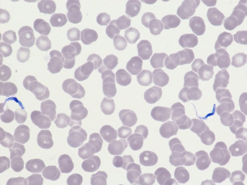
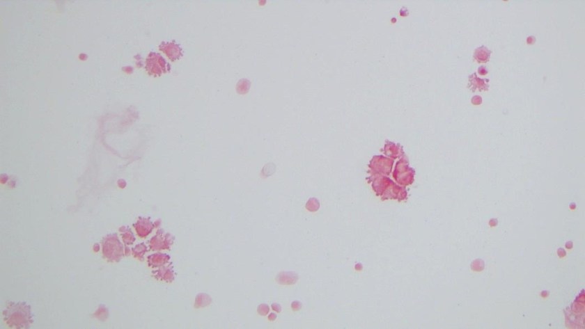
It was Friday afternoon when a microbiology technologist brought the patient’s cerebrospinal fluid (CSF) gram stain to be reviewed.  It was confirmed that no inflammatory cells and organisms were present.  However, cells in the background looked very atypical (Image 1a, b).

CSF1
Image 1:  Gram stain of CSF showing atypical epithelial cells at (a) 40x and (b) 100x with oil.
CSF2
Image 1b.

Discussion

The gram stain is used to provide preliminary information about the microorganism present in the specimen.  Gram stain differentiates bacteria into two fundamental varieties of cells.  Bacteria that retain the initial crystal violet stain (purple) are said to be “Gram-positive,” whereas those that are decolorized and stain red with carbol fuchsin (or safranin) are said to be “Gram-negative” (1).  An adequate examination of a gram-stained smear includes observing numerous representative fields and the fields containing neutrophils yield the most information (2).  Gram stain provides information about number of bacteria present, gram reaction and shape of the bacteria.  In background we can also see epithelial cells and inflammatory cells.  However, it’s a good practice to also appreciate and investigate any odd looking findings.

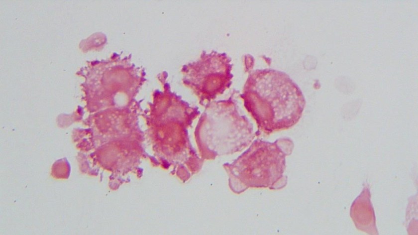
To investigate further, we visited the hematology laboratory to view their CSF slide to determine if these cells were a processing artifact.  After it was confirmed that hematopathology saw the same atypical cells, a cytopathologist was requested to review the gram stain since patient’s CSF cytology specimen was to be processed after the weekend.  Cytopathologist favored our suspicion and decided to process the cytology specimen late in the day on Friday and it was confirmed that those atypical cells were consistent with the metastatic adenocarcinoma.

Neurosurgery team was immediately contacted to reconsider insertion of the VP shunt as the shunt would drain fluid from the CSF into the peritoneal cavity and thus there was concern for transferring of malignant cells from central nervous system into abdomen/pelvis. However, after consulting oncology team it was later decided to proceed with the procedure since patient’s primary tumor originated in abdomen/pelvis and current intraabdominal tumor burden was not significant as compared to the symptoms driven by CNS involvement. Proceeding with this procedure was considered to be palliative and the best course of action to improve the patient’s quality of life.

References

  1. Beveridge TJ. Use of the gram stain in microbiology. Biotech Histochem.2001 May;76(3):111-8.
  2. Barenfanger J, Drake C. Interpretation of gram stains for the nonmicrobiologist. 2001 July;32(7):368–375.

KM

-Kiran Manjee, MD, is a 1st year anatomic and clinical pathology resident at University of Chicago (NorthShore).

-Erin McElvania, PhD, D(ABMM), is the Director of Clinical Microbiology NorthShore University Health System in Evanston, Illinois. Follow Dr. McElvania on twitter @E-McElvania. 


Hematology Case Study: A 51 Year Old Woman with Fever and Chills

A 51 year old patient presented to the emergency room with abdominal pain and fever. Fever was associated with diaphoresis, chills and headaches. Patient was in Tanzania for 3 months. She was admitted to the hospital while she was there for some unknown infection, details of which are not available.

CBC done revealed normocytic normochromic anemia with a hemoglobin of 9.2 g/dl and thrombocytopenia. Platelet count was 100 K/uL. On review of peripheral blood revealed presence of malarial parasite (ring forms).

mal1.PNG

mal2

Malaria is an infectious disease caused by Plasmodium parasites. These parasites are primarily spread by the bite of infected female Anopheles mosquitos. There are four main types of Plasmodium (P) species that infect humans:

  • Plasmodium vivax and Plasmodium ovale, which cause a relapsing form of the disease, and
  • Plasmodium malariae and Plasmodium falciparum, which do not cause relapses.

mal3

Malaria must be recognized promptly in order to treat the patient in time.

Microscopy (morphologic analysis) continues to be the “gold standard” for malaria diagnosis. Parasites may be visualized on both thick and thin blood smears stained with Giemsa, Wright, or Wright-Giemsa stains. Giemsa is the preferred stain, as it allows for detection of certain morphologic features (e.g. Schüffner’s dots, Maurer’s clefts, etc.) that may not be seen with the other two. Ideally, the thick smears are used to detect the presence of parasites while the thin smears are used for species-level identification. Quantification may be done on both thick and thin smears.

Various antigen kits are available to detect antigens derived from malarial parasites. These rapid diagnostic tests (RDT) offer a useful alternative to microscopy in situations where reliable microscopic diagnosis is not available.

Vajpayee,Neerja2014_small

-Neerja Vajpayee, MD, is the director of Clinical Pathology at Oneida Health Center in Oneida, New York and is actively involved in signing out surgical pathology and cytology cases in a community setting. Previously, she was on the faculty at SUNY Upstate for several years ( 2002-2016) where she was involved in diagnostic work and medical student/resident teaching.

Hematology Case Study: The Race to Save a 48 Year Old Man from a Rare Disease

A 48-year-old Caucasian male presented to a Baltimore Emergency Room complaining of fever, chills, and aches. He stated he had not been feeling well for the past week. His symptoms had progressed rapidly over the last 3 days to include night sweats, nausea and excessive somnolence. History taken in the ER revealed the patient had returned 10 days prior from a Safari in Botswana and Zambia. The patient was admitted to the ICU, in shock, with a BP of 75/50. Even though the patient had taken anti-malarial medication, the doctors suspected malaria. Blood was sent to the lab for a blood parasite exam and treatment for malaria was started while the doctors waited for the confirmation.

In the Hematology laboratory, technologists perform microscopy of thick and thin blood smears to look for malarial parasites. The thin smear is a typical Wright Giemsa stained wedge smear, and the thick smears are prepared and stained so that the red blood cells are lysed, and the sample is concentrated, making examination easier. Thorough, careful examination of the thick smear is aimed to identify whether a particular parasite is present, but they require a long drying period and take several hours to prepare and read. Thin smears can detect the parasites but also permit identification of particular species of malaria. While the thick smears were drying the technologist examined the thin smear.

The technologist who examined this patient’s thin smears saw parasites (image 1) under her microscope. She consulted with a supervisor and pathologist to confirm, and the patient’s doctor was notified that the patient did not have malaria, but instead, had Trypanosoma! This was an exciting find in the laboratory, as there have been only 40 cases seen in the US in the past 50 years.

tryp1
Image 1. This slide shows the parasite, in dark blue. The parasite causes
African trypanosomiasis, also known as sleeping sickness
(Courtesy of Greater Baltimore Medical Center).

The race for diagnosis and treatment did not stop there, as there are 2 types of African trypanosomiasis, or African sleeping sickness, and effective and appropriate treatment must be started in a timely fashion. Both types look identical on a blood smear and both are considered universally fatal, if not treated. West African trypanosomiasis and East African trypanosomiasis are caused by the tsetse fly, which only lives in rural Africa. The patient stated he did remember being bitten by tsetse flies, and because there had been such a short span of time between being bitten and the onset of symptoms, doctors concluded that the patient had the rarer and fast-acting East African trypanosomiasis, which can kill within months.

Epidemiologists at CDC were contacted, who then consulted other infectious disease specialists at CDC. There are 2 treatments depending the stage of the disease. Surinam is the first line of defense, but melarsoprol, which is arsenic-like and very toxic, must be used if the parasites have reached the central nervous system. Because of the urgent need to start treatment, emergency shipments of both drugs were flown to Baltimore. The patient was started on Surinam to reduce the number of parasites in his blood to a level low enough to allow a spinal tap to be performed. After confirming that the CSF showed no signs of the parasite, treatment with surinam was continued and the patient was discharged a week later and has made a full recovery.

Because of the excellent work done by the medical technologists who made the first discovery, the speed with which the critical calls were made, the actions of the doctors involved, and the cooperation of the CDC, this patient received his first dose of Surinam a little over 24 hours after his blood was sent to the lab. This case shows the importance of a thorough medical and travel history in differential diagnosis. It also illustrates the importance of the competency evaluations and surveys in which all laboratory professionals are required to participate. None of the technologists, doctors or scientists involved had ever actually seen a case of African Trypanosomiasis, they had only read about it in books and seen it on competency assessments.

This case is based on an actual case from 2016. My coworker, Gail Wilson, was the technologist who first saw the Trypanosoma on the slides. Many thanks to Gail for her keen eye and attention to detail!

tryp2
Image 2: L&R: Trypanosoma brucei in thin blood smears stained with Giemsa. Center: A close up of a tsetse fly. Credit: DPDx


References 

  1. Jon E. Rosenblatt Barth Reller Melvin P. Weinstein.pages 1103-1108, Laboratory Diagnosis of Infections Due to Blood and Tissue Parasites Clinical Infectious Diseases, Volume 49, Issue 7, 1 October 2009; retrieved March 2018 from https://academic.oup.com/cid/article/49/7/1103/316703
  1. Ivo Elliott, Trupti PatelJagrit Shah, and Pradhib Venkatesan. West-African trypanosomiasis in a returned traveller from Ghana: an unusual cause of progressive neurological decline BMJ Case Rep. 2014; 2014: bcr2014204451. Published online 2014 Aug 14.doi: 1136/bcr-2014-204451; retrieved March 2018 from https://www.ncbi.nlm.nih.gov/pmc/articles/PMC4139564/
  1. Lena H. Sun. Medical Detectives raced to save a man from a rare, ‘universally lethal’ disease; retrieved March 2018 from https://www.washingtonpost.com/news/to-your-health/wp/2016/12/22/medical-detectives-raced-to-save-a-man-from-a-rare-universally-lethal-disease/?utm_term=.16d7b136bc47
  1. Parasites – African Trypanosomiasis (also known as Sleeping Sickness). Retrieved March 2018 from https://www.cdc.gov/parasites/sleepingsickness/
  1. DPDx- Laboratory Identification of parasites of Public Health Concern; retrieved March 2018 from https://www.cdc.gov/dpdx/


Socha-small

-Becky Socha, MS, MLS(ASCP)CM BB CM graduated from Merrimack College in N. Andover, Massachusetts with a BS in Medical Technology and completed her MS in Clinical Laboratory Sciences at the University of Massachusetts, Lowell. She has worked as a Medical Technologist for over 30 years. She’s worked in all areas of the clinical laboratory, but has a special interest in Hematology and Blood Banking. When she’s not busy being a mad scientist, she can be found outside riding her bicycle.


Telemicroscopy: Applying Technology to Solve an Old Problem

The Gram Stain

Everyone knows that the Gram stain is an essential microbiological method which aids in the differentiation of bacteria. When a specimen is sent to the clinical microbiology laboratory for culture, the Gram stain result is frequently the first information provided to the clinician. It is used to first determine a) if infection is present and b) what type of infection (i.e., gram positive vs. gram negative? monomicrobial vs. polymicrobial?). Furthermore, if organisms are observed in a normally sterile fluid/tissue (i.e., blood, cerebral spinal, fluid, cardiac tissue, etc.) the Gram stain result can be a critical result. More importantly, the Gram stain result often drives patient care. 

The Issue

Although the Gram stain is an essential clinical tool, many laboratories struggle to maintain competent technologists, especially on off-shifts or in laboratories that lack microbiology expertise (generalists). The need for second review is common when performing Gram stains as they are often subject to variability due to inconsistent staining techniques, antibiotic pressure, as well as artifacts. Even under best case scenarios, Gram stain interpretation can be challenging and may require multiple reviewers. 

The Solution

Telemicroscopy offers an easy to use and relatively inexpensive solution to provide formal and informal second opinions to various sections of the laboratory (microbiology, hematology, pathology). With the proper tools, telemicroscopy allows Gram stain interpretation from anywhere there is internet access.  Every hospital laboratory has a microscope and a computer with internet, so the only item that may need to be purchased is a microscope camera (≥$5,000). There are also various microscope adapters available for phone cameras that provide equal results for less capital (≥$90). The microscope adapter encases the smart phone and then fits into the eyepiece of most microscopes.

Telemicroscopy utilizes technology to improve diagnostic accuracy, by providing expert consultation for technologists who are uncertain of their results. Telemicroscopy allows laboratories to “present” still or live images to a reference laboratory via a web-based software application such as Skype (or FaceTime if using an iPhone).

About Geisinger Medical Laboratories Telemicroscopy Program 

Geisinger Medical Laboratories is an eight hospital integrated health service organization, serving >2.6 million residents throughout 46 counties in Pennsylvania. Geisinger Medical Center serves as the reference laboratory for 4 minimal laboratories (Gram stain reading, no culture work-up) and 2 partial laboratories (Gram stain reading, limited culture work-up). The Telemicroscopy program consists of presenting still or live images [Olympus BX40, BX41 microscope, Nikon cellSense software (version 1.7.1)] to the reference laboratory via Skype [Logitech 920 camera (version 2013)]. The telemicroscopy result, which is a consensus finding, is manually recorded and followed up with culture review to determine patient impact. 

The Outcome

We evaluated the effect of implementing a telemicroscopy program on patient care.  A retrospective look back at our telemicroscopy data showed that nearly 40% of consults resulted in a change to the original interpretation. The consensus Gram stain result correlated with culture 85% of the time. Overall, 49% of the cases assessed by telemicroscopy were impacted by the consult. Of which, patient care was positively and negatively impacted in 72% and 28% of cases, respectively.

The Conclusion

Gram stain consultations via telemicroscopy from remote hospital sites can improve patient care. Telemicroscopy offers a simple, inexpensive, and innovative approach to providing expert consultation services to off-shift or inexperienced staff. This is also a great way to promote interdepartmental consultation and collaboration (i.e., between microbiology and hematology or pathology).

telemicro1
Image 1. Telemicroscopy via traditional microscope camera. Microscope with camera attached and computer screen showing Gram stain.
telemicro2
Image 2. Telemicroscopy via s mart phone. Close up of microscope adapter attached to microscope. Image of Gram stain displayed on phone screen.

References:

  1. Microbiology Strong: Enhancing Microbiology Services and Technical Support in an Integrated Laboratory System. ASCP.  Las Vegas, Nevada. September 2016. Oral presentation.
  2. Martinez, R.M., Shoemaker, B.C., Riley, J.A., and Wolk, D.M. 2016. The TeleGram of the 21st Century: the Digital Gram Stain. American Society for Microbiology (ASM) General Meeting. Boston, MA. Poster presentation.


Martinez Headshot-small 2017

-Raquel Martinez, PhD, D(ABMM), was named an ASCP 40 Under Forty TOP FIVE honoree for 2017. She is one of two System Directors of Clinical and Molecular Microbiology at Geisinger Health System in Danville, Pennsylvania. Her research interests focus on infectious disease diagnostics, specifically rapid molecular technologies for the detection of bloodstream and respiratory virus infections, and antimicrobial resistance, with the overall goal to improve patient outcomes.


The Importance of Manual Urine Microscopy

Research presented today at the National Kidney Foundation spring clinical meeting indicates that manual microscopy surpasses automated analyzers when assessing kidney injury. The abstract is titled “Manual Microscopy: Not a Lost Art” and says, in part: “In this study, we examined if a significant difference exists between the reported ranges of granular and muddy brown casts using manual microscopy as compared to an automated urine analyzer in an acute kidney injury cohort.”

According to one of the abstract’s authors, Dr. Sharda, “What our research has been able to show so far is that the automated system under reported the value of granular casts in our patient cohort of acute kidney injury. The automated system still has utility as a screening test, but manual microscopy should be done in all cases of abnormal kidney function, as accurate quantification of casts could have some prognostic benefit to patients.”

The poster is available online. The authors are currently writing a paper on their research; their contact information is here.