Surgical Pathology Case Study: A 43 Year Old Female with a Lung Nodule Noted on Imaging Following Chest Congestion

Case History

The patient is a 43 year old woman who experienced chest congestion and presented to her local physicians office. A chest X-ray was ordered and demonstrated a lung abnormality. A follow-up CT scan confirmed a 1.9 cm smoothly marginated nodule in the upper lobe with no adenopathy and a normal liver and adrenal glands. The nodule was mildly hypermetabolic on PET scan. A bronchoscopy was performed, which was non-diagnostic. Two subsequent CT scans demonstrated no change in the size of the nodule. Overall, the patient feels well and denies cough, hemoptysis, dyspnea on exertion, and weight loss. Due to the suspicion of cancer, the patient has decided to undergo a lung lobectomy.

Diagnosis

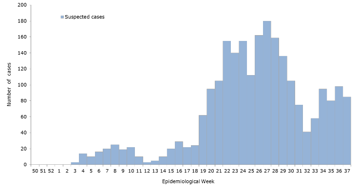
Received in the Surgical Pathology lab for intraoperative consultation is a 30.0 x 7.2 x 2.2 cm lung lobectomy specimen. There is an attached 6.2 cm staple line, which is removed and the subjacent resection margin is inked blue. The entire pleural surface is inked black. The specimen is sectioned revealing a 2.1 x 1.7 x 1.0 cm white-tan, firm, round nodule that is 0.5 cm from the blue inked resection margin and 0.2 cm from the black inked pleural surface. The remainder of the specimen is composed of red-tan, spongy, grossly unremarkable lung parenchyma without nodules or other lesions. Photographs of the specimen are taken (Figure 1). A representative section of the nodule is submitted for frozen section and read out as “diagnosis deferred”. Representative sections of the specimen are submitted as follows:

A1FS:   Frozen section remnant

A2-A7:   Nodule, entirely submitted

A8-A10:   Grossly unremarkable lung parenchyma

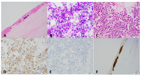
Immunohistochemical stains show the epithelial cells in the lesion to be positive for CK7, TTF-1, and surfactant proteins A and B which supports these cells to be type 2 pneumocytes (all controls are appropriate). Based on the immunohistochemical stains and routine H&E slides, the case was signed out as a sclerosing pneumocytoma

Image 1. Gross presentation of the well-defined, round sclerosing pneumocytoma.

Discussion

Sclerosing pneumocytoma (SP) is a rare, benign pulmonary tumor that was first described in 1956 as a vascular tumor, but has since been found to be of primitive respiratory epithelium origin. In the past, SP has also been referred to as sclerosing hemangioma, pneumocytoma, and papillary pneumocytoma, but the 2015 World Health Organization classification of lung tumors states that the agreed upon term for this tumor should be a sclerosing pneumocytoma. SP is commonly seen in middle aged adults, with a female to male ratio of 5:1. There is no racial bias. Patients are usually asymptomatic, with the tumor incidentally found on screening chest radiographs. If the patient was to present with any symptoms, they would usually include a cough, hemoptysis and chest pain. Radiographically, SP appears as a solitary, well-defined, homogenous nodule along the periphery of the lung.

Grossly, most SPs appear as a solitary, firm, well-circumscribed, yellow-tan mass generally arising along the periphery of the lung. The majority of these tumors appear within the lung parenchyma, but there have been cases reported of endobronchial and pleural based SP tumors. Multifocal unilateral tumors and bilateral tumors are uncommon.

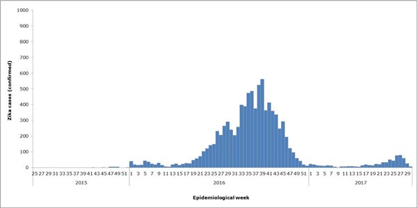
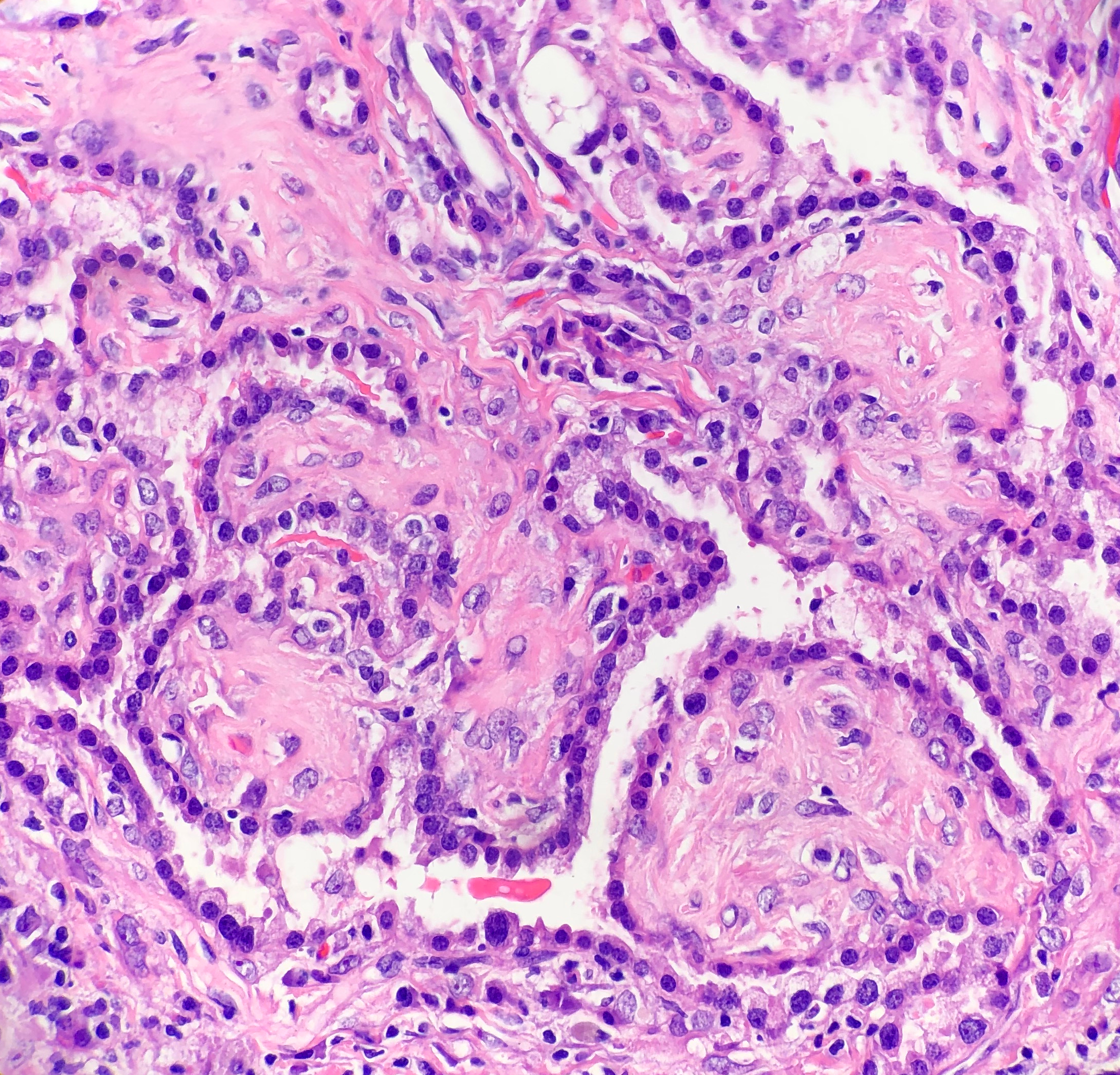
Histologically, SP consists of two epithelial cell types: surface cells and round cells. Surface cells are cuboidal, resembling type II pneumocytes, with finely stippled nuclear chromatin, indistinct nuclei, occasional nuclear grooves, and inclusions. The stromal round cells will have bland oval nuclei with coarse chromatin and eosinophilic cytoplasm (Figure 2). Both the surface cells and round cells will have a low mitotic rate, but can have moderate to marked nuclear atypia. Ciliated bronchial epithelium is often identified in the tumor. There are four architectural patterns identified within SP: papillary, sclerotic, solid and hemorrhagic, with over 90% of SPs displaying three of the patterns, and all of the tumors containing at least two of the patterns.

  • Papillary pattern: Complex papillae composed of surface cells covering a stroma of round cells
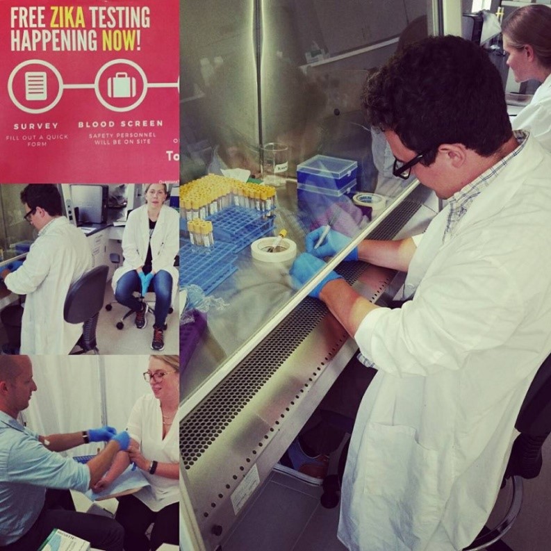
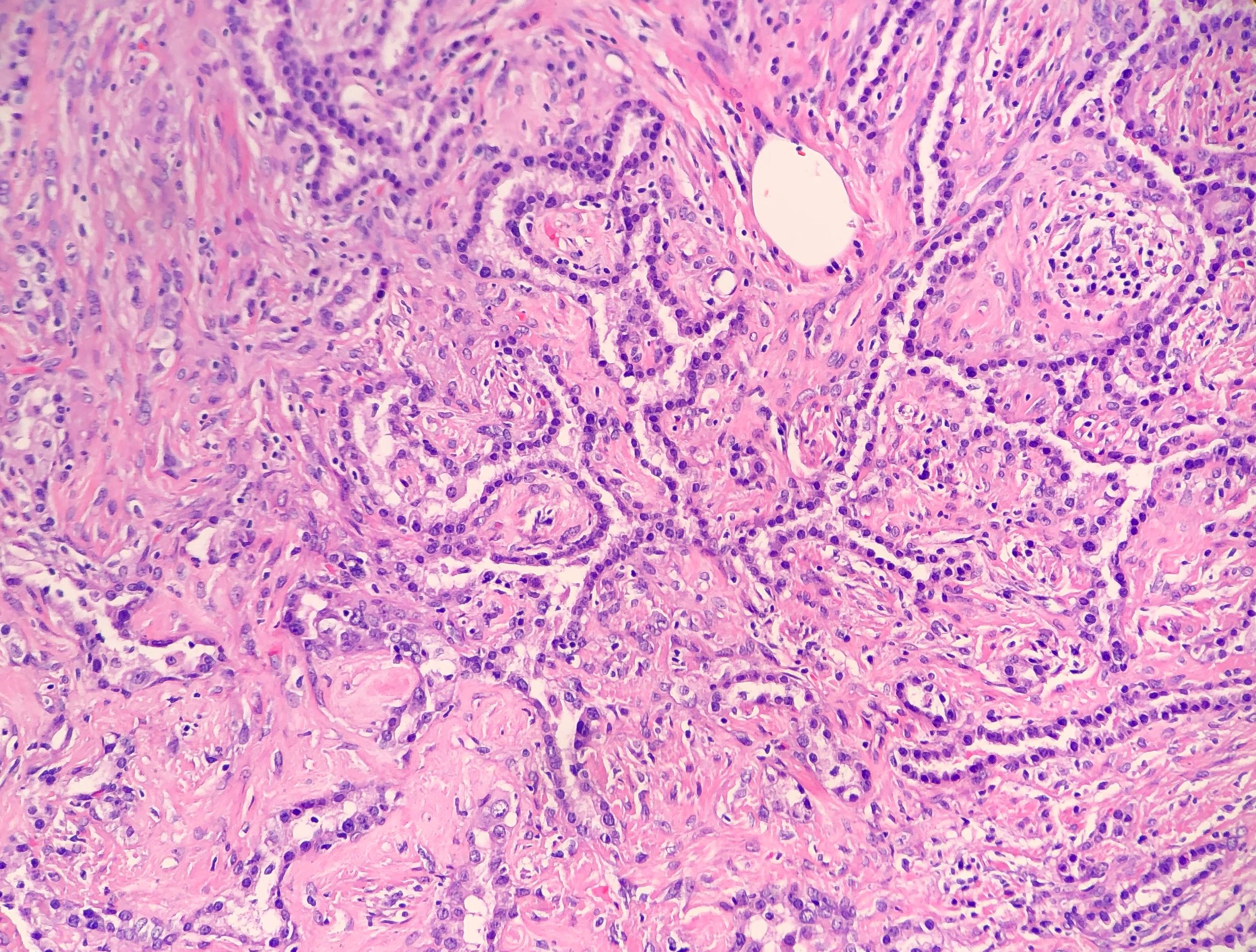
  • Sclerotic pattern: Papillae containing hyalinized collagen, either in solid areas or along the periphery of hemorrhagic areas (Figure 3)
  • Solid pattern: Sheets of round cells bordered by surface cells
  • Hemorrhagic pattern: Large blood filled spaces
Image 2. Photomicrograph demonstrating the cuboidal surface cells and round stromal cells.
Image 3. Photomicrograph of the papillary and sclerotic architectural patterns.

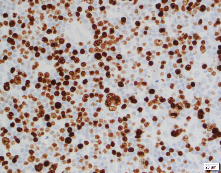
Immunohistochemical stains can be helpful in the diagnosis of SP, with both the surface cells and round cells exhibiting expression of thyroid transcription factor 1 (TTF-1) and epithelial membrane antigen (EMA). It should be noted that TTF-1 is also used for the diagnosis of pulmonary adenocarcinoma, increasing the risk of misdiagnosing SP. The surface cells will also express both pancytokeratin (AE1/AE3) and Napsin A, with the round cells being negative for AE1/AE3, but having a variable expression of cytokeratin 7 and the low molecular weight cytokeratin (CAM 5.2). Molecular pathology has demonstrated a frequent loss of heterozygosity at 5q, 10q and 9p, and an allelic loses at p16 in the surface and rounds cells. Although the immunohistochemical stains and molecular pathology results can be very helpful, diagnosis of a SP is still largely based on routine H&E slides showing the two epithelial cell types and four architectural patterns.

Electron microscopy will show abundant lamellar bodies similar to those in type II pneumocytes in the surface cells. Round cells will lack the lamellar bodies and instead will contain variably-sized electron-dense bodies that have been thought to represent the different stages of lamellar body maturation.

The differential diagnosis for SP includes a variety of benign and malignant neoplasms, which can be difficult to distinguish on cytology, small biopsies and intraoperative consultations. The cytologic features include moderate to high cellularity with a bloody background and foamy macrophages, occasional nuclear pleomorphism in the round cells, absent mitotic figures, and occasional necrosis with cholesterol clefts and calcifications. In the case of small biopsies, making a diagnosis of SP can be difficult if the papillary pattern is highly prevalent without one of the other three patterns present. With intraoperative consultations, the frozen section artifact can make it difficult to appreciate the two epithelial cell types or the four architectural patterns. The gross examination, as well as the radiographic findings of a well-circumscribed tumor can help point the Pathologist to favoring a benign neoplasm over a malignant one. The benign neoplasms that should be considered in the differential diagnosis include:

  • Clear cell tumor, which will have clear cells with scant stroma, thin-walled vessels and a strong expression of HMB-45
  • Pulmonary hamartoma, which will have a combination of cartilage, myxoid stroma, adipose tissue and trapped respiratory epithelium
  • Hemangiomas, which are rare in the lung, and will lack epithelial cells and contain either a cavernous or capillary morphology

The malignant neoplasms that should be considered in the differential diagnosis include:

  • Bronchioalveolar carcinoma, which can have a papillary pattern, but will not contain the two epithelial cell types and combination of the four architectural patterns
  • Metastatic papillary thyroid carcinoma, which is distinguished from SP by the presence of the characteristic Orphan Annie nuclei
  • Metastatic renal cell carcinoma, which will contain nuclear atypia and striking vascularity
  • Carcinoid, which will contain organoid and ribbon-like growth patterns

Currently, with the benign nature of SP, surgical excision is the preferred treatment choice to cure the patient. There have been cases reported of lymph node metastasis and recurrence, but neither of these appear to effect the prognosis. This just helps to highlight the need for a multidisciplinary approach to this benign tumor.

References

  1. Hisson E, Rao R. Pneumocytoma (sclerosing hemangioma), a Potential Pitfall. Diagn Cytopathol. 2017;45(8):744-749
  2. Keylock JB, Galvin JR, Franks TJ. Sclerosing Hemangioma of the Lung. Arch Pathol Lab Med. 2009;133(5):820-825.
  3. Travis WD, Brambilla E, Nicholson AG, et al. The 2015 World Health Organization Classification of Lung Tumors: Impact of Genetic, Clinical and Radiologic Advances Since the 2004 Classification. J Thorac Oncol. 2015;10(9):1243-1260.
  4. Wu R. Sclerosing Pneumocytoma (Sclerosing Hemangioma). Pathology Outlines. http://www.pathologyoutlines.com/topic/lungtumorsclerosingheman.html. Revised February 19, 2019. Accessed June 6, 2019.

-Cory Nash is a board certified Pathologists’ Assistant, specializing in surgical and gross pathology. He currently works as a Pathologists’ Assistant at the University of Chicago Medical Center. His job involves the macroscopic examination, dissection and tissue submission of surgical specimens, ranging from biopsies to multi-organ resections. Cory has a special interest in head and neck pathology, as well as bone and soft tissue pathology. Cory can be followed on twitter at @iplaywithorgans.

Hey, What’s the Buzz on Zika?

Hello everyone and welcome back!

Last month, it was as fun to write about hematology peripheral smear differentials as it was to address the importance of interdisciplinary collaboration. I found myself in a unique position both as a medical student as well as a former medical laboratory scientist in what was a great clinical training rotation in hematology/oncology. Now, with just one rotation left until the end of my medical school journey, I want to take you on a look back at some of the very first posts I made here on Lablogatory and update you on the intersectional, collaborative topic that I shared with you almost two years ago: Zika!

Image 1. ASCP’s official professional society partner, The Pathologist. I’ve been getting them in my mailbox since the official partnership was announced. It’s an excellent platform for laboratory professionals across scopes to discuss relevant topics in pathology. I was particularly excited to see Zika make an appearance last month! (Source: The Pathologist [online] https://thepathologist.com/diagnostics/our-powers-combined)

In a recent digital article on ASCP’s partner, The Pathologist, author and staff editor Michael Schubert wrote about the connectivity between public health, epidemiologic research, laboratory medicine, and clinical patient outcomes. He examined the effectiveness and accuracy of Zika testing availability in commercially available assays and spoke with a leading virologist in the field from Berlin. You may recall one of those “ancient” posts I made about Zika, where I was part of a research team that used the same methodology! Combined immunoglobulin-specific assays, arbovirus detection in the heat of a public health epidemic’s epicenter, and lab medicine that complimented my concurrent immunology class in med school—what more could you ask for?

And, since the last tagged Lablogatory Zika update I can see was by Dr. Sarah Riley in February of 2017, here’s my update! Dr. Riley’s post was a fantastic summary of the Zika epidemic, its troublesome diagnostic assessments, and the recommendations and plans of organizations like the World Health Organization (WHO). She was, and still is, right—the “struggle is still real’ when it comes to Zika testing. Curious about what it was like during the 2016 epidemic? Who was doing testing, what kind of testing, and what was the lab data climate? Well…it feels like it’s time for a…

*** FLASHBACK ***

An Arbovirus Abroad

Hey! My inaugural post! It was fun to go back and see the data from the work then (Spoilers: epidemiologic updates are on your horizon). We were just getting started to take an assessment of the situation and address it as a public health concern. My then Caribbean location was a great place to study Zika trends coming from Brazil, Puerto Rico, and Florida. As a snapshot, at that time (Dec 2016) there were a purported almost 2,000 cases, however less than a fifth of those cases were serologically confirmed by lab testing. Before the recommendations to move toward RT-PCR, most labs in the region were requesting commercially available screening tests for IgG/IgM assays.

Image 2a. These were the (then) suspected Zika viral infection cases per epidemiological week, Pan-American Health Organization (PAHO) and World Health Organization (WHO) 2016. My wife and I are included in these statistics—that mosquito virus rash is awful!
Image 2b. Remember that spoiler I promised above? Well here’s the updated WHO epidemiologic data for confirmed Zika cases in the region we worked in. Seems like the mosquitoes…buzzed off. (Source: WHO)

Healthy Me

How do you reach people when you’ve got compelling public health lab data that translates to possible prevention of infection and spread of disease? Easy: go to where the people are and engage them when and where they’re comfortable. One of the overarching themes in public health is mitigating barriers to change by way of utilizing social humility. This a certainly a type of interdisciplinary collaboration because if we’re the experts on IgM and IgG trends in testing confirmations, the public are the experts in social determinants of health within their communities.

Image 3. Want to make sure a message gets home to every family? Bug their kids about Zika bugs in fun, educational ways. That’s me delivering one of my “Healthy Me” presentations to children, October 2016.

Laboratory Data and Global Health Security

As my team and I were busy preparing SOPs, conducting a new project aimed at improving local health literacy and source reduction, securing IRB approval, and collecting data about the residents of Sint Maarten to correlate with local Health Ministry projections, one of the officials—who now serves as a regional director for PAHO—took our work to the Global Health Security Agenda Summit. Talk about motivation! In and out of the lab, I worked with teams who were getting some fantastic work done on the ground with respect to mosquito-borne virus research.

Image 4. IgM and IgG seroprevalence of Zika virus (along with other Arboviruses i.e. West Nile, Chikungunya, Dengue, and Yellow Fever etc.) within the community around my medical school. We used commercially available IgG and IgM assays from Germany with great success. Internal controls and known cases were fantastic ways to include internal validation.

IRBs and Public Heath Pathology

For those of us who work in laboratory medicine, it’s easy to talk about the best way to test, detect, and treat an epidemiologic threat—it’s even exciting when it’s a current threat. But to really be successful, you’ve got to collaborate with those outside of the lab, and often this means thinking “outside the box.” Public health is different from lab medicine in that while lab-work is based around results, testing, and organized data-driven decisions, success in public health is highly determined by community buy-in in the form of partnerships!

Figure 1. There’s a method to the community “buy-in” concept. With a foundation in evidence-based practices, any project aimed at improving public health outcomes must include some critical components like clear objectives, attainable goals, sustainability, and effective (and constant) re-evaluation.

*** FLASH … FORWARD? ***

So, after my time in Sint Maarten, I came to New York City to rotate through my clinical clerkships. And, if you’ve seen some more recent post-Zika posts on this website, you know they’ve been going great! Within a few months of being here, my wife brought back some swag from a training session she attended. (Side note: she’s a graduate-level nurse, working in the public health non-profit sector with vulnerable populations in the inner city—she’s too busy to blog.) After months of both of us working and learning about Zika and public health initiatives in the Caribbean, we were greeted by this fantastic toolkit from the New York State Department of Public Health!

Image 5. Empowering a large number of patients with highly variable demographics is challenging. The NYS DOH distributed “Prevention Kits” for Zika Virus which included: Zika Virus educational materials in 8 languages, pamphlets on reducing mosquito activity, travel related information for pregnant women, 2 larvicide pellets with instructions for using larvicide, picaridin insect repellent, and condoms.
Image 6. That’s us! My wife Kathryn and I presenting on the importance of Disaster Planning and Implementation of Preparedness Programs at the 2019 Caribbean Conference of Disaster Medicine. Disasters are bad on their own, but think about what happens months after flooding, hurricanes, or destruction—transmittable diseases. And that includes standing-water-borne mosquito viruses!

The take home message: collaboration is key, both inside and out of the lab. Schubert’s piece in The Pathologist created a fantastic dialogue in addressing the clinical needs for interdisciplinary collaboration. The best testing means finding out exactly where the needs are and using data-driven decisions to implement change or action. In the lab, that means constantly working for higher quality and better patient outcomes in every test, result, report, and (mosquito) byte of data. In the field, it means the same thing, but instead of metrics like sensitivity, specificity, and TAT it’s about cultural humility, attainable goals, and dynamic timing.

Thanks for reading! Hope most of our national heat wave spared you, but if it didn’t remember: don’t keep standing water around, wear light loose clothing, and use appropriate insect repellent!

See you next time!

–Constantine E. Kanakis MSc, MLS (ASCP)CM graduated from Loyola University Chicago with a BS in Molecular Biology and Bioethics and then Rush University with an MS in Medical Laboratory Science. He is currently a medical student actively involved in public health and laboratory medicine, conducting clinicals at Bronx-Care Hospital Center in New York City.

An Introduction to Laboratory Regulations: Part 1

Everyone who works in a laboratory knows that there are certain rules and regulations to be followed to ensure accuracy in testing, and the safety of both the patient and testing personnel. With all the acronyms floating around (CLIA, FDA, CAP, CMS, TJC) it can get confusing to keep track of who controls what, and which rules apply to your specific lab. In the first installment of this 3-part series on regulations, we’ll review the different federal agencies responsible for oversight and moderation of the laboratory. In part 2 we’ll go further in-depth to demystify testing complexity (waived, non-waived, PPM) and why it’s important to know the correct classification for the tests you perform. Lastly, we’ll review the optional accreditations available to labs, and how accreditation differs from certification.

CLIA

CLIA refers to the Clinical Laboratory Improvement Amendments of 1988. These amendments were drafted to the Public Health Services Act, in which the federal program was revised to include certification and oversight of clinical laboratory testing. Although there have been two additional amendments made after 1988 (1997, 2012), the law still continues to be cited as CLIA ’88 as it is named within legislation.

These CLIA regulations helped to establish quality standards for all U.S. laboratory testing performed on human specimens (except for research) for the purpose of assessment of health, or the diagnosis, prevention, or treatment of disease. The regulations cover all aspects of testing including general laboratory requirements, quality monitors, pre-analytics, analytic performance, post-analytics, and personnel requirements.

In addition to setting the basic ground rules for performing quality laboratory testing, the CLIA regulations also require clinical laboratories to be certified by their state as well as the Center for Medicare & Medicaid Services (CMS) before accepting human samples for diagnostic testing. Laboratories can obtain multiple types of CLIA certificates, based on the kinds of diagnostic tests they perform. In order for laboratories to receive payments from Medicare or Medicaid, laboratories must be properly certified for the testing they are performing and billing for.

There are 3 federal agencies responsible for enforcing the CLIA regulations: The Food & Drug Administration (FDA), Center for Medicaid Services (CMS) and the Center for Disease Control and Prevention (CDC). Each agency has a unique role in assuring quality laboratory testing.

CMS

The Centers for Medicare & Medicaid Services (CMS) is the federal agency responsible for ensuring that the CLIA standards are upheld and enforced. Their responsibilities include the following:

  • Issuing laboratory certificates
  • Collecting user fees
  • Conducting inspections and enforcing regulatory compliance
  • Approving private accreditation organizations (such as CAP) for performing inspections, and approves state exemptions
  • Monitoring laboratory performance on Proficiency Testing (PT) and approving PT programs
  • Publishing CLIA rules and regulations

FDA

The Food & Drug Administration (FDA) is primarily responsible for reviewing and approving new tests, instruments, and equipment used in diagnostic laboratories. They also perform the following tasks:

  • Categorize tests based on complexity
  • Review requests for Waiver by Application from manufacturers
  • Develop rules/guidance for CLIA complexity categorization

CDC

The Center for Disease Control and Prevention (CDC) responsibilities include the following tasks:

  • Provide analysis, research, and technical assistance
  • Develop technical standards and laboratory practice guidelines, including standards and guidelines for cytology
  • Conduct laboratory quality improvement studies
  • Monitor proficiency testing practices
  • Develop and distribute professional information and educational resources
  • Manage the Clinical Laboratory Improvement Advisory Committee (CLIAC)

To summarize, CLIA establishes the rules and guidelines that laboratories must follow to ensure they are providing accurate laboratory results. Federal agencies then work together to support the CLIA amendments and enforce compliance. All certified laboratories will be subject to inspection by regulatory agencies to ensure compliance with the rules. In some cases, your local state Department of Health (DOH) or accrediting agency may be more stringent or have additional requirements to be followed – always go with the stricter requirement to ensure compliance with all agencies.

Coming up next we’ll review how the FDA decides the complexity of each test, and how this designation will affect the CLIA rules to be followed.

References

  1. Electronic Code of Federal Regulations: https://www.ecfr.gov/cgi-bin/text-idx?SID=1248e3189da5e5f936e55315402bc38b&node=pt42.5.493&rgn=div5
  2. Interpretive Guidelines for Laboratories: https://www.cms.gov/regulations-and-guidance/legislation/clia/interpretive_guidelines_for_laboratories.html

†


-Kyle Nevins, MS, MLS(ASCP)CM is one of ASCP’s 2018 Top 5 in the 40 Under Forty recognition program. She has worked in the medical laboratory profession for over 18 years. In her current position, she transitions between performing laboratory audits across the entire Northwell Health System on Long Island, NY, consulting for at-risk laboratories outside of Northwell Health, bringing laboratories up to regulatory standards, and acting as supervisor and mentor in labs with management gaps.

Think S.P.I.L.L.E.D.

Large biological and chemical spills are not a common occurrence in the laboratory. That’s a good thing, but when they do occur, they can create a very dangerous situation. It is vital that lab staff know how to handle such events even though they may not be commonplace.

Some laboratories differentiate between large and small spills. They may have an emergency number to call for a hazardous spill response team. Other smaller facilities simply don’t have that in place. Either way, it’s important for laboratory professionals to know they are the experts about the biological and chemical materials they use, and they need to be in charge as the experts when a spill situation needs to be managed.

Most laboratory spills can be managed using a standardized step-wise process known as the S.P.I.L.L.E.D. procedure. I don’t usually ask lab staff to memorize the acronym, but having the information contained on a poster with the lab spill kits can make a clean-up procedure go smoothly.

S = Secure the Site – Make sure no one walks through the area where a spill has occurred. It could be a dangerous situation if a hazardous chemical is spilled, and you would never want someone slipping in the area or tracking the spilled material to another area.

P = Protect Yourself – Arm yourself with the appropriate Personal Protective Equipment (PPE). In a lab spill event, this would mean using a lab coat, gloves, and face protection to prevent accidental splashes.

I = Inspect the Spill – Look to see what was spilled. If it is a hazardous chemical, is there a concern about fumes? Obtain a Safety Data Sheet to see if section 6 will give any special information about handling the accidental release or spill of that chemical. Consider other spill concerns such as broken glass or possible ignition sources if flammable material is involved.

L = Lay Down a Barrier – If the spill is large and spreading, lay down spill pillows or booms designed to contain a flow of liquids. Surround the spill area with these materials. Sometimes, the use of an emergency shower can create the need for a barrier to be made.

L = Lay Down Absorbents – No matter the size of the spill, the next step is to place any absorbent powders, granules or clean-up pads to soak up the spilled material. If the absorbent is also a neutralizer, make sure you allow the necessary time for neutralization to occur.

E = Extract the Mess – Use implements to pick up the materials used for stopping and absorbing the spill.

D = Dispose of the Waste – Properly dispose of all materials involved with the spill clean-up. If there was glass involved, be sure to use a sharps container.  Biohazard material should go into an appropriate container, and chemical waste materials may need to be disposed of separately for pick-up by a chemical waste vendor.

Lab staff should be able to access spill control materials quickly, and the necessary items should be stored in a location designated by signage. Perform an inventory of spill supplies and make sure there are adequate materials that could handle spills of the biohazards and chemicals stored and used in the department. Be sure items in the spill kit are not expired, and if there is no expiration date for absorbent powders, check them at least annually for effectiveness.

All laboratory staff need to have complete spill clean-up training. Give information about the types and locations of spill kits and how to handle various types of spills that can occur. Once that training is done, it will become important to perform spill drills in the department. Drills can be performed a number of different ways, but a common method involves having a “victim” spill water onto the floor and claim the material splashed into their eyes. Watch from a distance to see how the staff reacts. Do they provide appropriate first aid? Do they inspect the container label? Do they access the correct clean-up supplies and facilitate cleaning efficiently? Make notes of how the drill went, discuss them with the staff, and repeat the drills until all staff are comfortable with a spill situation. Biological and chemical spills should not be a common occurrence in the lab. When they do occur, however, the situation can become serious quickly, and a fast and effective clean-up needs to occur. Because these events are rare, it becomes important to provide regular spill training and drills so staff can remain ever-ready to handle them.

Dan Scungio, MT(ASCP), SLS, CQA (ASQ) has over 25 years experience as a certified medical technologist. Today he is the Laboratory Safety Officer for Sentara Healthcare, a system of seven hospitals and over 20 laboratories and draw sites in the Tidewater area of Virginia. He is also known as Dan the Lab Safety Man, a lab safety consultant, educator, and trainer.

The Paperwork of Transgender Care

I don’t think anyone enjoys filling out the paperwork at a doctor’s office. For transgender individuals, this can be an experience that ranges from irksome to offensive. Most intake forms don’t allow for expression of their gender identity. Furthermore, confusion on gender and sex can create real confusion and healthcare failures in several places that laboratory medicine encounters a transgender individual.

Arguably the first place the lab encounters a transgender patient is via the phlebotomist. These professional collectors of blood must confirm two patient identifiers, which are often name and date of birth. The “name” used is the legal name. Using a transgender person’s “dead name” (name given at birth) represents a gender they do not want to be associated with and can be a very offensive experience. “Isn’t it obvious that name is not what I look like?”

While names can be legally changed, this happens with varying difficulty and legal cost in different states. A solution is to improve training of phlebotomists to explain the necessity of confirming a legal name so lab results are properly matched to the patient. Additionally, front-desk intake workers should be similarly trained to interact with transgender patients when recording demographic information. This can be aided by electronic health records (EHR) becoming more flexible and inclusive of the gender diversity.

Traditionally, EHR would only include one field for SEX: M or F.

Several in the laboratory community have asked how many different gender options should be included? Facebook included up to 71 options in 2017. That’s a big step up from the 2 traditional EHRs are built around.

The World Professional Association for Transgender Health (WPATH) executive committee in 2011 outlined the recommended fields to include in EHR: preferred name, sex assigned at birth, gender, and pronoun preference. EHRs are evolving and can be flexible depending on the user requirements. At my program, we use EPIC at 3 different different sites (children’s, county and university hospitals) and each has a different version.

From what I’ve seen preferred name is an easy addition and would not interfere with functions of the EHR or Laboratory Information Systems (LIS), which is the Lab’s version of EHR.

If the field for sex assigned at birth is different from gender, then it would clear up any confusion about whether the person is transgender and then they should be addressed by the pronouns matching the gender. While there is a spectrum of genders, only transgender males and transgender females are of a high enough prevalence to have medically relevant recommendations. Plus, if a system at least starts here, they could expand further as necessitated by their population.

EHR could include preferred pronouns, but I haven’t seen this implemented in an EHR yet. Ideally, you would just use the pronouns that match the intended appearance of the individual (ma’am to someone wearing a dress, etc.).

Lastly, I think Legal sex should be added to the EHR as well. One of our hospitals has this and it makes several processes easier such as processing hormone medication.

Legal (or administrative) sex, sex assigned at birth, and gender data fields provide the clearest and simplest picture of a patient and should be a minimum for labs making recommendations for changes to HER.

Next month I will describe in greater detail the issues that can arise in the lab when gender or sex are entered incorrectly in the system for transgender patients and how this can negatively affect care delivery.

References

  1. Deutsch MB, Green J, Keatley J, Mayer G, Hastings J, Hall AM, World Professional Association for Transgender Health EMR Working Group. Electronic medical records and the transgender patient: recommendations from the World Professional Association for Transgender Health EMR Working Group. J Am Med Inform Assoc. 2013 Jul-Aug; 20(4):700-3.
  2. Gupta S, Imborek KL, Krasowski MD. Challenges in Transgender Healthcare: The Pathology Perspective. Lab Med. 2016 Aug; 47(3):180-188.

-Jeff SoRelle, MD is a Chief Resident of Pathology at the University of Texas Southwestern Medical Center in Dallas, TX. His clinical research interests include understanding how the lab intersects with transgender healthcare and improving genetic variant interpretation.

Not Starbucks but the DMV

I merrily wait in line at Starbucks for my iced cappuccino with soy milk, pay $5+ for $0.25 worth of goods poured into my $14.00 souvenir mug, and walk out the door with my head held high, joyous with the privileges of conspicuous consumption. My server was super-cheery and the brief exchange we had was so pleasant—they really love me!  I need that high because I am off to the Department of Motor Vehicles (DMV) for a driving-related task and know–just know–that there will be an incredibly long line at the end of which sits a disgruntled government employee who doesn’t care if I show up or not. Their motivation to help us is non-existent. “Why would anyone ever work here?” I ask, sipping my delicious beverage.

Today, a doctor called someone in the United States (US) and told them the biopsy taken from their leg earlier this week has come back as invasive cancer. A bit distraught and nervous, the patient called up a nationally recognized cancer center, from which they only live a few miles, and on the end of the line is a caring, pleasant voice who informs them they can be seen today! The valet parking is gorgeous, the building is gleaming with glass and steel, and every face they see as they journey from check-in to clinic is smiling, compassionate, and sincere. Their nurse and then doctor are both genuine people with their patient’s best interest in mind, and they carefully and completely explain what has been found, what needs to be done, and how they are going to get through all of this together. As they depart, the receptionist grabs them for a brief moment to return their private insurance card and waves at them as they depart, adding, “We will see you soon!”

Today, someone in Africa went back to the hospital—an 8-hour journey from their home—where their biopsy was performed a month ago, hoping to get the result. After several people searched multiple offices and inquired with several people, the result is found and brought to them, a single piece of paper. Payment is required before they can receive the biopsy results. They have brought money with them, which they gathered from three neighbors, their brother, and by selling some chickens, and pays for the report. They read the report and, at the bottom, notices that it says additional testing is needed. Confused, they ask for help and a pathologist comes to find them. Respectfully, the pathologist explains that additional testing is needed, which is not available in the hospital despite the pathologist’s strong desire to have it, but they can send the biopsy to a lab elsewhere to do the testing which will cost about 3 times what they just paid for the primary report. They happen to have enough and pay the amount requested. The report will be back in about a month. Two months later, they have returned to the hospital for the 4th time and the report is now available. The testing that was done simply confirms that the primary diagnosis is accurate. They go to the oncology clinic on the same campus and sit in the waiting area with 3 dozen other people. They sleep at the clinic overnight outside with about a dozen people. The following afternoon, they are finally seen and the oncologist reviews the report. He notes that if the patient had come to the clinic as soon as they had the biopsy result three months ago, a simple surgery would have cured them of this lesion. But now, because they waited so long, there is only chemotherapy available which is expensive and, the oncologist reports, doesn’t actually work very well for this tumor.

Before you shed a tear for this terrible situation (while I sip my cappuccino and a nurse begins someone’s chemotherapy in a shiny, brightly lit, and expansively windowed infusion unit not far away), we have to ask ourselves what is really going on? First and foremost, this is an allegory to make a few points but the situation is repeated over and over again every day in the US and Africa. However, as a simple, superficial explanation, the person with cancer in the US is receiving their cancer therapy from Starbucks and the person in Africa had to go to the DMV.

Cancer care in the United States is almost entirely in the private sector, dispersed among the 1500 cancer treatment facilities, of which 70 are comprehensive cancer centers.[i] Based on the US population, the expected cancer rate, 100% detection, and 240 working days for a given cancer center, there are on average only 5 new patients per day per cancer center. Is that why one can often get that appointment right away in a major cancer center or is it really a concierge customer service effort? A standard private insurance plan for which I pay, for example, $250 per month and my employer pays $1300 per month is accepted by cancer centers and results in small co-pays for multiple appointments, which can be covered with a Flexible Spending Account (FSA) or Health Savings Account (HSA). On insurance statements after appointments, some of the services received cost thousands of dollars but the patient portion was only, say, a hundred dollars, again, which may be paid with FSA/HSA. It’s so great that we have insurance because the insurance company is bearing the brunt of costs. But are they?

In the United States, 79% of facilities providing health care are private, a mix of non-profit and for-profit.[ii] But 64% of all healthcare in the United States is paid for by the US government through Medicare, Medicaid, the Veterans Administration (VA) system, and Children’s Health Insurance Program (CHIP).[iii],[iv] Since almost every cancer care facility is private (or, stated another way, “not free”), that means that for every one of us at the cancer center getting treatment, for which we and our employer are paying through insurance, there are two people getting the same treatment at the same high-level quality of care for which the government is paying. Those other deductions from our paychecks for Medicare and Medicaid (which everyone pays, regardless of how old, as long as they are employed and regardless of their own health insurance plan) are going towards the 64% coverage. The point is not that the US healthcare system is expensive. The point is that there is a lot of revenue and resource being put into the healthcare system and, thus, there is a high-quality product or experience that is available.

If we look at any low GINI index country and compare their GDP with the US GPD and compare their spending on healthcare as a % of GDP, we don’t even need to do the math to see that there is very little money per person available in the system for any type of healthcare. The challenge in low-resourced settings (by which it is meant low-resourced patients in low-resources locations) is both a lack of funding available to provide healthcare services along with a lack of “stuff” to provide those services. We can invoke the law of supply and demand to try and argue that the people can rise up and demand more healthcare facilities and “someone” will meet that supply. In the US, this results in the Starbucks model. In a low-resourced setting who has the incentive to meet that supply? Where does the government get the money from to create such a system? What private corporation is going to start a healthcare program that provides universal coverage regardless of what you can pay?

The answer is really quite simple. This model of healthcare is insufficient for cancer and isn’t going to work for all patients. Moreover, the Starbucks model is not really applicable, sustainable, nor equitable. When we go to Starbucks for their coffee, to some degree, our choice of Starbucks is because of the a) flavor of the coffee, b) cost of the coffee, c) perception of the coffee, and/or d) convenience of the coffee. We could always choose Dunkin’, Peet’s, Tim Horton’s (maybe let’s not go there for this analogy), or Green Mountain coffee at a different location. There is variation in pricing and convenience. There is variation in the condiments we can use to doctor our coffee. An economy and series of markets exist which allow Starbucks to gather resources from dozens of other companies to provide your coffee. But, ultimately, we are all buying coffee which has caffeine which has a desired effect. We can go to a free AA meeting or to a soup kitchen and get some pretty basic coffee if we don’t have the money to pay. The point is we have choices and we can pay a high price, a low price, or no price and we get coffee.

The Starbucks model does work for a certain sector of the population but not everyone. Since vast majority of cancer care in the US is private, the Starbucks model falls down because we don’t actually have any free options as a society and “low-cost healthcare” is not typically appealing to most Americans with cancer because they have their mortality at stake (no one wants cancer nor does anyone want to die from cancer). In fact, desperation in the face of cancer is what makes the US one of the only places in the developed world where people go bankrupt trying to be treated for cancer. The ultimate inequity is that cancer care is “pay to play” in the US and there essentially aren’t safety nets for any populations that can’t pay (homeless) or are living below a certain income threshold (i.e., the ~10% of Americans without healthcare plus a large percentage with insufficient insurance).[v]

Please remember, these are human beings and they didn’t choose to get cancer (there is no demand for cancer… there is only demand for cancer care!). Since they didn’t have a choice in the disease they have to be burdened with, why is there an expectation that they should pay for the treatment? Moreover, if a patient has a stage I cancer, easily surgically removed and cured vs. a Stage III cancer requiring months of various therapies at a very high cost, how do we ethically explain an increased cost for a worst state of disease? It’s really an inverse quality spectrum and we make patients pay more for getting a lot less. We pay for insurance in case we ever do get cancer (or other major disease). It’s a risk reduction or risk aversion pre-payment. Like we do with our car or our house or our boat. Those last three things we choose to have (and are luxuries). We don’t get to choose to have health. It’s just an inherent part of being human so holding someone accountable for it because they didn’t have the resources to “prepare for the worst” is really the wrong attitude. Our healthcare system isn’t perfect but there are gaps that could be easily filled if resources are allocated efficiently to meet the whole populations needs—that’s the benefit of having a large resource supply into the system. We just have to find the operational efficiency to make the costs work.

However, when we remove the luxuries of insurance, Medicare, and Medicaid and other payments systems from the health sector or, worse, simply assume the government’s role is to provide healthcare 100% free to all citizens in a resource-limited or resource-constrained setting, we suddenly have an untenable situation. The economy and tax-base are not there to create the resources. We find overworked, underpaid, and undersupplied medical staff working in crowded conditions. For single entity care (e.g., HIV, tuberculosis, malaria), vertical programs have made great strides in combatting these diseases even in some of the poorest countries in the world. But cancer is anything but simple with the complexity of cross-discipline collaboration, spectrum of disease, range of treatments, and inherent costs creating huge gaps in the delivery of cancer care. Economic and physical infrastructure for the provision of care is what is needed to meet this challenge. Our current Starbucks model in the US would be extremely difficult to replicate in a low-resourced setting due to the lack of infrastructure. However, when this infrastructure is assessed, planned for, and implemented, cancer care can be delivered in these settings at a significantly lower cost per patient. Adding infrastructure implementation high-quality private facilities and public-private partnerships creates a way forward to pump resources into the system and insure that no patient is left behind. To round out this allegory, AAA locations (a commercial car-servicing company) in various parts of the US allow one to renew your driver’s license with them, rather than the DMV. I did this once, it was VERY fast, friendly, and efficient. This type of public-private partnership worked for me and I believe it will work for cancer if we are willing to try.

References

[i] NCI-designated Cancer Center. https://en.wikipedia.org/wiki/NCI-designated_Cancer_Center  Retrieved May 21, 2019.

[ii]  “Fast Facts on US Hospitals”. Aha.org. Retrieved December 1, 2016.

[iii] Himmelstein DU, Woolhandler S (March 2016). “The Current and Projected Taxpayer Shares of US Health Costs”. American Journal of Public Health. 106 (3): 449–52. doi:10.2105/AJPH.2015.302997. PMC 4880216. PMID 26794173. Government’s share of overall health spending was 64% of national health expenditures in 2013

[iv] ^ Leonard K (January 22, 2016). “Could Universal Health Care Save U.S. Taxpayers Money?”. U.S. News & World Report. Retrieved July 12, 2016.

[v] https://www.kff.org/uninsured/fact-sheet/key-facts-about-the-uninsured-population/

milner-small

-Dan Milner, MD, MSc, spent 10 years at Harvard where he taught pathology, microbiology, and infectious disease. He began working in Africa in 1997 as a medical student and has built an international reputation as an expert in cerebral malaria. In his current role as Chief Medical officer of ASCP, he leads all PEPFAR activities as well as the Partners for Cancer Diagnosis and Treatment in Africa Initiative.

Hematology Case Study: A 69 Year Old Female with Breast Implants

Case History

A sixty nine year old female who underwent right breast reconstruction about 13 years ago due to breast cancer presents to the doctor office with right breast pain and right breast enlargement over the last two months. She has lost some weight and does not recall any trauma to this area. She had a textured saline implant. Examination reveals no definite palpable masses. MRI of right breast showed intact saline implant with moderate amount of fluid surrounding the implant within the intact external capsule. No adenopathy was noted. Right breast implant was removed and complete capsulectomy was performed.

Image 1. A. Section of breast capsule with rare atypical hyperchromatic cells (arrow). B. Cytospin preparation of the fluid surrounding the implant with numerous atypical lymphocytes. C. Cell block of the fluid with large atypical lymphocytes. D, E. Lymphocytes are positive for CD30 (image D) and negative for ALK-1 (image E). F. CD30 positive cells in the section of the implant.

Diagnosis

Breast implant-associated anaplastic large cell lymphoma.

Discussion

Breast implant associated anaplastic large cell lymphoma is a provisional entity that is morphologically and immunophenotypically similar to ALK-negative anaplastic large cell lymphoma. It arises primarily in association with a breast implant. It is a very rare entity with an incidence of 1 in 500,000 to 3 million women with implants. Tumor cells may be localized to the seroma cavity or may involve pericapsular fibrous tissue. Sometimes it can form a mass lesion. Locoregional lymph node may be involved. The mean patient age is 50 years. Most patient presents with stage 1 disease, usually with peri-implant effusion. The mean interval from implant placement to lymphoma diagnosis is 10.9 years. There is no association with the type of implant. Histologic examination shows two different types of proliferations. In patients with seroma, the proliferation is confined to the fibrous capsule (“in situ” iALCL). However, the distribution of neoplastic lymphocytes could be heterogeneous with some cellular areas with numerous large pleomorphic cells of varying size and some fibrotic areas with rare atypical lymphocytes. It is beneficial to look at the seroma fluid in addition to capsule sections, because sometimes the neoplastic lymphocytes are predominantly present in fluid (as in our case). Patients presenting with tumor mass show more heterogeneous proliferations infiltrating surrounding tissues (“infiltrative” iALCL). They consists of either sheets are clusters of large neoplastic cells accompanied by a large number of eosinophils. By immunohistochemistry, the tumor cells are strongly positive for CD30. CD2 and CD3 are more often positive than CD5. CD43 is almost always expressed. Most cases are CD4 positive. The prognosis is very good in patients with disease confined to the capsule. The median overall survival is 12 years. However, patients with a tumor mass could have a more aggressive clinical outcome.

References

1. Swerdlow SH, Campo E, Harris NL, et al. WHO Classification of Tumours of Haematopoetic and Lymphoid Tissues (Revised 4th edition). IARC: Lyon 2017.

2. Jaffe, E , Arber, D, et al. Hematopathology (second edition) 2017.

-Junaid Baqai, MD, was born in Chicago, IL but spent most of his life in Karachi, Pakistan. He graduated from DOW Medical College in Pakistan and did his residency in anatomic and clinical pathology at Danbury Hospital, CT followed by hematopathology fellowship from William Beaumont Hospital, Michigan and oncologic-surgical pathology fellowship from Roswell Park Cancer Institute, New York. He currently serves as Medical Director of hematology, coagulation and flow cytometry at Memorial Medical Center and Medical Director of Laboratory at Taylorville Memorial Hospital.

Laboratory Test of Anti-Neutrophil Cytoplasmic Antibody in Sinonasal Inflammatory Disease

Case History

A 44 year old male with history of cocaine use presented with 1 year history of headache and progressive frontal lobe syndrome, including symptoms like apathy, personality changes, lack of ability to plan, poor working memory for verbal information or spatial information, Broca aphasia, disinhibition, emotional lability, etc. CT scan found extensive destruction of osteocartilaginous structures of the nasal cavity and MRI showed extensive edema of the frontal lobe. Biopsy showed chronic inflammation but negative for granulomatous inflammation. Patient’s CSF laboratory analysis was normal but ANCA was tested positive, in a P-ANCA pattern without MPO detectable. Patient was diagnosed as CIMDL. After stopping cocaine use, patient was doing better but still has mild frontal lobe syndrome.

Discussion

Anti-neutrophil cytoplasmic antibody (ANCA) are a group of autoantibodies that directed toward antigens expressed mainly in neutrophil granulocytes, such as proteinase 3 (RP3) and myeloperoxidase (MPO). The presence of ANCA is mainly associated with a distinct form of small vessel vasculitis, known as ANCA-associated vasculitis, but is also detected in other disease, like autoimmune hepatitis, primary sclerosing cholangitis, ulcerative colitis, and other chronic inflammatory disease. The gold standard laboratory method to screen ANCA is indirect immunofluorescence assay (IFA or IIF), which qualitatively capture antibodies in serum/or plasma bound to fixed human neutrophil granulocytes.

Two form of ANCA-associated vasculitis, granulomatous with polyangiitis (GPA) and eosinophilic granulomatous with polyangiitis (EGPA), are systemic diseases that commonly associated with necrotizing granulomatous vasculitis. GPA has a primary involvement of the upper and lower respiratory tract and kidney. Autoantibodies to PR3 are found in 90% of active GPA cases, which generates a cytoplasmic-ANCA (C-ANCA) pattern on ANCA IFA test. EGPA is a rare form of systemic necrotizing vasculitis characterized by asthma and eosinophilia. A perinuclear-ANCA (P-ANCA) IFA pattern directing towards MPO antibody are often seen in EGPA cases.

Both GPA and EGPA may also present with sinonasal involvement, causing non-infectious inflammatory lesions of the sinonasal tract. Sinonasal inflammatory disease can also result from bacterial and fungal infections, or other non-infectious process, such as sarcoidosis, polychondritis, or obstruction. ANCA is detected in the majority of GPA and EGPA case, therefore it provides useful information in differential diagnosis of sinonasal inflammatory disease. Both GPA and EGPA are autoimmune diseases, corticosteroids and immunosuppressive agents are effective treatment.

Sinonasal inflammation can also been seen in a subset of patients with cocaine abuse, who normally present with midline destructive lesions, known as cocaine-induced midline destruction lesions (CIMDL). Long-term cocaine use has been associated with ischemia of mucosal tissue, cartilage and bone, and cocaine abuser using intranasal inhalation route can have midline deformity and septal perforation. Interestingly, ANCA are also found in a large portion of CIMDL, and in contrast to GPA or EGPA, ANCA in CIMDL are primarily directed against neutrophil elastase, generate a P-ANCA or atypical P-ANCA pattern, without detection of MPO. Therefore, ANCA serology testing could help the differentiation between CIMDL and GPA although these two can overlap clinically and histopathologically. Also, CIMDL does not respond well to immunosuppressive therapy and only consistent removal of stimuli (cocaine) can halt the disease process.

References

  1. Montone KT. Differential Diagnosis of Necrotizing Sinonasal Lesions. Arch Pathol Lab Med. 2015 Dec;139(12):1508-14. doi: 10.5858/arpa.2015-0165-RA.
  2. Trimarchi M, Bussi M, Sinico RA, Meroni P, Specks U. Cocaine-induced midline destructive lesions – an autoimmune disease? Autoimmun Rev. 2013 Feb;12(4):496-500. doi: 10.1016/j.autrev.2012.08.009. Epub 2012 Aug 24.
  3. Madani G, Beale TJ. Sinonasal inflammatory disease. Semin Ultrasound CT MR. 2009 Feb;30(1):17-24.
  4. Timothy R. Helliwell Non-infectious Inflammatory Lesions of the Sinonasal Tract. Head Neck Pathol. 2016 Mar; 10(1): 32–39.
Xin-small

-Xin Yi, PhD, DABCC, FACB, is a board-certified clinical chemist, currently serving as the Co-director of Clinical Chemistry at Houston Methodist Hospital in Houston, TX and an Assistant Professor of Clinical Pathology and Laboratory Medicine at Weill Cornell Medical College.

Hematopathology Case Study: A 76 Year Old Man with Lymphadenopathy

Case History

76 year old man with a history of chronic lymphocytic leukemia/small lymphocytic lymphoma (CLL/SLL) with new anterior mediastinal mass and increasing lymphadenopathy.

Lymph Node Biopsy

H&E

Diagnosis

Tissue sections show a diffuse atypical lymphoid infiltrate that completely effaces the normal nodal architecture. The infiltrate is composed of numerous small lymphocytes with round to mildly irregular nuclei, clumped chromatin, inconspicuous nucleoli and scant cytoplasm. There are also expanded pale areas that contain intermediate sized cells with more open chromatin and distinct single to multiple nucleoli. These cells are most consistent with prolymphocytes/paraimmunoblasts and form the proliferation centers characteristic of CLL/SLL. Occasional centroblastic-type B-cells are noted within these proliferation centers. In addition, there are scattered single to multinucleated cells that have irregular nuclear membranes with pale, vesicular chromatin and prominent inclusion-like, eosinophilic nucleoli. These cells morphologically resemble Hodgkin cells, Reed-Sternberg cells, mummified forms and other variants. These large cells are more evident in areas with a histiocyte rich background and around foci of necrosis. Occasionally, apoptotic bodies and mitotic figures are seen.

 Immunohistochemical studies show that the vast majority of the small-intermediate lymphocytes express B-cell markers CD20 (dim) and PAX5 and co-express CD5 and CD23 (subset). This is consistent with a background of CLL/SLL. The large atypical cells are positive for CD30, PAX5 and CD20 (variable). CD3 highlights numerous scattered background small T-cells, which are increased in the areas with the large cells. In situ hybridization for Epstein Barr viral RNA (EBER ISH) is mainly staining the large atypical cells. By Ki-67, the proliferation fraction is overall increased (40%) with increased uptake by the large atypical cells.

The morphologic and immunophenotypic findings are consistent with involvement by the patient’s known small lymphocytic lymphoma/chronic lymphocytic leukemia (SLL/CLL) with aggressive morphological features. The aggressive features include expanded proliferation centers and an elevated Ki-67 proliferative index (40%). Additionally there are histiocyte/T-cell rich areas composed of multiple EBV positive large atypical cells with morphologic and immunophenotypic features compatible with Hodgkin/ Reed-Sternberg cells. These areas are most in keeping with evolving classic Hodgkin lymphoma. Sheets of large cells indicative of large cell transformation are not seen, although increased scattered large centroblastic-type B cells are present.

Discussion

Lymph node involvement by CLL/SLL will typically show a diffuse proliferation of small lymphocytes with effacement of the normal nodal architecture.  The small lymphocytes have round nuclei, clumped chromatin and scant cytoplasm. Scattered paler areas known as proliferation centers are characteristic of this entity. The proliferation centers are composed of a mixture of cell types including small lymphocytes, prolymphocytes and paraimmunoblasts. Prolymphocytes are small to medium in size with relatively clumped chromatin, whereas paraimmunoblasts are larger cells with round to oval nuclei, dispersed chromatin, eosinophilic nucleoli and slightly basophilic cytoplasm. Some cases show increased and enlarged proliferation centers with a higher proliferation rate. This must be distinguished from large cell transformation.1

Aggressive features of CLL/SLL include proliferation centers that are broader than a 20x field or becoming confluent. An increased Ki-67 proliferation >40% or >2.4 mitoses in the proliferation centers can also portend a more aggressive course. These cases tend to have worse outcomes than typical CLL/SLL and better outcomes than cases that have undergone Richter transformation to diffuse large B-cell lymphoma (DLBCL). Transformation to DLBCL occurs in 2-8% of patients with CLL/SLL. Less than 1% of patients with CLL/SLL develop classic Hodgkin lymphoma (CHL). In order to diagnose CHL in the setting of CLL/SLL, classic Reed-Sternberg cells need to be found in a background appropriate for CHL, which includes a mixed inflammatory background. The majority of these CHL cases will be positive for EBV.1

Richter’s transformation is defined as an aggressive evolution of CLL. While the most common type of transformation is to a high-grade B-cell Non-Hodgkin lymphoma, other histological transformations have been described. This includes CHL, lymphoblastic lymphoma, hairy cell leukemia and high-grade T-cell lymphomas. The prognosis for patients who present with transformation to CHL is poor compared to de novo CHL.2 A large study from the M.D. Anderson Cancer Center described 4121 patients with CLL/SLL and found that only 18 patients or 0.4% developed CHL. The median time from CLL to CHL diagnosis was 4.6 years. Fourteen of the patients received chemotherapy. The overall response rate was 44% with a complete response rate of 19%. The median overall survival was 0.8 years and all patients eventually died from disease recurrence or progressive disease.3 This dismal prognosis is similar to patients with Richter transformation to DLBCL and much worse than patients with de novo CHL, which is curable in >85% of cases.1

References

  1. Swerdlow SH, Campo E, Harris NL, et al. WHO Classification of Tumours of Haematopoetic and Lymphoid Tissues (Revised 4th edition). IARC: Lyon 2017.
  2. Janjetovic S, Bernd HW, Bokemeyer C, Fiedler W. Hodgkin’s lymphoma as a rare variant of Richter’s transformation in chronic lymphocytic leukemia: A case report and review of the literature. Mol Clin Oncol. 2016;4(3):390–392.doi:10.3892/mco.2016.727.
  3. Tsimberidou, AM, O’Brien, S and Kantarjian, HM, et. al. Hodgkin transformation of chronic lymphocytic leukemia. Cancer. 2006;107(6).doi.org/10.1002/cncr.22121.

Chelsea Marcus, MD is a Hematopathology Fellow at Beth Israel Deaconess Medical Center in Boston, MA. She has a particular interest in High-grade B-Cell lymphomas and the genetic alterations of these lymphomas.

Fecal Transplants in the News

An article posted today at The Atlantic discusses fecal transplants and FDA regulation. Dr. Colleen Kraft (co-author of a paper on fecal transplant protocols that appeared in Lab Medicine) is quoted in the article, and it’s worth a read.