Microbiology Case Study: A 55 Year Old Woman with Cough and Headache

Clinical History

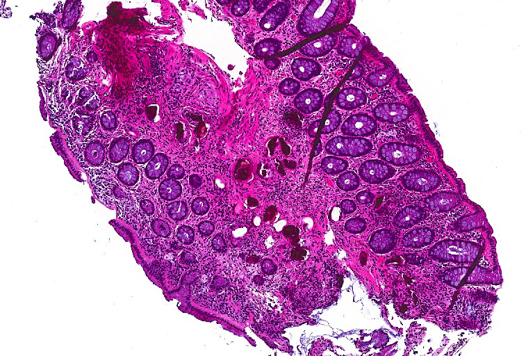
A 55 year old female presented to the gastroenterology clinic with a chief complaint of cough and headache. She reported no recent fevers, abdominal pain, or diarrhea. On further questioning she revealed she was originally from the Philippines and had a past history of a parasitic infection that was treated twice with praziquantel. She did not remember the name of the parasite but was concerned for a recurrent infection. A stool specimen for ova & parasite exam and basic laboratory work, including an IgE level, were collected and the patient was scheduled for a screening colonoscopy. Findings from the colonoscopy revealed no gross evidence of neoplastic or infectious disease; however, random rectal biopsies were obtained. 

Laboratory Identification

Image 1. Rectal biopsy showing unremarkable colonic mucosa with many calcified structures deep in the epithelium (H&E, 40x).
Image 2. Rectal granuloma surrounding a calcified structure (H&E, 400x).
Image 3. Intact parasitic egg with a small, inconspicuous spine measuring approximately 90 um in greatest dimension (H&E, 1000x oil immersion).

Discussion

Schistosoma japonicum is a trematode that can infect humans through direct penetration of the skin by the cercariae when wading or swimming in infected waters in the Far East, such as China, the Philippines, Indonesia, and Thailand. Infection can initially present as swimmer’s itch and then develop into Katyama syndrome, which includes fever, eosinophilia, muscle aches, lymphadenopathy, abdominal pain, and diarrhea.

S. japonicum migrates through tissues and the adult male & female forms take up residence in the mesenteric veins that drain the small intestine. The female lays eggs which travel to the lumen of the intestines and can be shed in the stool. The host immune response to the eggs is the major cause of clinical disease which presents as inflammation & ulceration in the intestines, portal fibrosis in the liver & splenomegaly, and more rarely, lesions in the central nervous system. As with all trematodes, snails serve as the intermediate host. 

In the microbiology laboratory, diagnosis is usually made by identification of the eggs in stool specimens. The eggs of S. japonicum are ovoid in shape with a transparent shell and a small, inconspicuous spine. The eggs typically measure between 70-100 um in greatest dimension. These eggs can commonly be visualized in rectal biopsies as well. It is important to get an accurate measurement of the size of the egg and multiple sections to be able to detect the location and morphology of the spine. The eggs of S. japonicum must be distinguished from those of S. mekongi which is similar in appearance; however, the latter is found along the Mekong River in Southeast Asia and is smaller in size (50-70 um in greatest dimension). Serology is also a viable diagnostic test in those that have traveled to endemic regions, but sensitivity and specificity of the assays vary depending on how the antigen is prepared and the Schistosoma species of interest.

Treatment of choices for those infected with S. japonicum is praziquantel divided into three doses over the course of one day and it should be administered at least 6 to 8 weeks after the last exposure to contaminated freshwater. Since our patient admitted to a recent visit to the Philippines with potential exposure to infected waters, she received another course of praziquantel therapy.

-Anas Berneih, MD, is a fourth year Anatomic and Clinical Pathology chief resident at the University of Mississippi Medical Center. 

-Lisa Stempak, MD, is an Assistant Professor of Pathology at the University of Mississippi Medical Center in Jackson, MS. She is certified by the American Board of Pathology in Anatomic and Clinical Pathology as well as Medical Microbiology. She is the Director of Clinical Pathology as well as the Microbiology and Serology Laboratories. Her interests include infectious disease histology, process and quality improvement, and resident education.

Proficiency Testing (PT)Part 1: Are You Doing it Right?

Every laboratory knows that they must participate in proficiency testing (PT) for all of the regulated analytes they report. But did you know that there is more to it than simply checking your overall score in each survey you participate in? Whether you utilize samples from the CAP, API, or have developed your own in-house blind sample testing algorithm, there is a lot of data available to help you assess the quality of your laboratory program. In the first of this 3-part series, we’ll review why PT testing is important and the rules that must be followed. In part 2 we’ll discuss how to properly perform an investigation when scores are <100%. Lastly, in part 3 we’ll look at how to review your results so that you get the most out of them for a successful quality laboratory.

Why participate? Well frankly, because you have to. It is a CLIA/CMS requirement, and if your lab has additional accreditations, those agencies will have their own rules and requirements as well (we’ll get to the rules in a little bit). But outside of the regulations stating you must participate; all labs should want to participate. It’s an opportunity to check your accuracy against peers who are using the same instrumentation as you. Similar to utilizing an affiliated QC report, this is a way to see what the “real” value is supposed to be (despite what a manufacturer may claim it to be), and how close/far off your lab is to that true value. It can help you identify potential problems before they become huge problems with patient values being affected, and it’s also a great way to satisfy competency requirements for your staff.

The rules:

  • Participation: For every regulated analyte being tested under your laboratory permit1, you must participate in a CMS-approved PT program2.

Key things to note: This only applies to testing performed using non-waived methodologies. Waived testing is exempt from PT requirements; although it is still recommended that participation occur if an evaluation program is available. Additionally, this only applies to your primary instrumentation. For example, if you have an automated urinalysis reader and your backup methodology is to read dipsticks manually, you are only required to participate in PT for the primary methodology. (Your backup method would then be evaluated for accuracy through semi-annual correlation studies.)

  • Routine Analysis: Unless otherwise instructed by the provider of your PT samples, PT samples are to be treated the same as patient samples. Meaning they are handled, prepared, processed, examined, tested and reported the same way you would perform patient testing; AND by the same staff who would handle patient testing.

Key things to note: If nursing staff perform a particular test within their unit (for example, ACT testing in the cardiac cath lab), it is those nursing staff members who must run the PT samples. You cannot have the laboratory perform PT testing unless the laboratory also performs the patient testing. Additionally, PT samples should be rotated among all staff members who perform patient testing. Meaning all shifts, and all days of the week that the test is performed – don’t let the day shift get all the fun.

  • Repeated Analysis: Similar to rule #2, unless you routinely perform duplicate testing on your patient samples, you cannot perform duplicate or repeat testing on your PT samples. You cannot run a PT sample in duplicate “just to make sure.” Patient samples are just as important to be accurate as a PT sample, which is why we participate in a PT program in the first place.

Key things to note: After the date that laboratories are required to report results back to the PT provider, you are then allowed to use the samples for repeat testing. This can be used to check for uniformity in grading of reactions among staff members, and to assess annual competency. But only after the submission date has passed.

  • Interlaboratory Communication: You cannot discuss the results or samples from a PT survey with any other laboratory (or Facebook user group) until after the results submission deadline has passed. Doing so before that time would be considered cheating. The point of PT testing is not to see how good your networking skills are, but to ensure accuracy of your own results. Plus, the other lab may not be as good as you think they are.

Key things to note: If your laboratory is part of a larger integrated health system, be careful that you have separate designated staff assigned to enter results from each location. Entering results for more than one permit number by the same person would be considered a violation of the interlaboratory communication rule as they could compare results from Lab A to Lab B prior to submitting. Also, be mindful of what you put on social media. User groups are a great networking resource and learning tool, but you still need to follow the rules. Violating them in a public arena such as Facebook for all the world to see would put yourself and your organization in great jeopardy if you were caught. 

  • Referral of Samples: You are not permitted to forward or share your PT samples with any other laboratory until after the result submission deadline has passed. Similarly, if your laboratory has received PT samples from another lab, state regulations may require you to notify your local Department of Health to inform them of the violation.

Key things to note: The intended purpose of performing PT testing is to verify the accuracy of your own laboratory testing. If you would routinely send a positive sample to a reference lab for additional confirmation testing, you would not do so in this case. Simply report out the values for the tests that your laboratory performs only. The reference laboratory will have their own PT samples to check accuracy for the confirmation testing they perform for you. Ensure your testing menu is up to date and accurate so that your PT provider is not expecting values for a confirmatory test if you do not physically perform it in-house.

  • Records Retention: Ensure that all records and documents related to the testing of PT samples are saved for the amount of time required by your regulatory agencies (typically 2-5 years). This includes instrument print outs, LIS chart copies of the filed results, QC records for the day of testing, and any associated worksheets used to document your results.

Key things to note: Retaining a copy of the instrument maintenance logs and QC records along with the actual PT results will help you investigate any scores that are less than 100%.

  • Attestation: Both the laboratory director and all personnel performing testing must sign the included attestation statement. This is not just a way to track who performed the test, but is a legal binding document assuring that testing was carried out appropriately as per the rules defined above.

The penalties for labs that are caught violating the rules (whether intentionally or not) can be quite severe. These penalties can include the revocation of your CLIA permit; a ban for the laboratory owner and laboratory director; as well as possible financial penalties and fines.

Coming up in the next blog we’ll review the rest of the rules related to evaluation of your scored PT results, and how to perform a thorough investigation into any unsuccessful survey events.

1: https://www.cms.gov/regulations-and-guidance/legislation/clia/downloads/cliabrochure8.pdf

2: https://www.cms.gov/Regulations-and-Guidance/Legislation/CLIA/Downloads/ptlist.pdf

-Kyle Nevins, MS, MLS(ASCP)CM is one of ASCP’s 2018 Top 5 in the 40 Under Forty recognition program. She has worked in the medical laboratory profession for over 18 years. In her current position, she transitions between performing laboratory audits across the entire Northwell Health System on Long Island, NY, consulting for at-risk laboratories outside of Northwell Health, bringing laboratories up to regulatory standards, and acting as supervisor and mentor in labs with management gaps.

Blood Bank Case Study: A 54 Year Old Woman with Lethargy

The patient is a 54 year old woman, presenting to the Emergency Room with complaints of abdominal cramps and feeling lethargic for the past few days. She also reports her stools have been black and sticky.  Her chart reveals a history of ulcers and GI bleeding.  She was transfused with 2 units packed RBCs 2 months ago for the same symptoms. CBC results are shown below.

The patient was admitted to the hospital and four units of blood were ordered. The patient is type A pos with a negative antibody screen. One unit of packed red blood cells would be expected to raise the Hgb by 1g/dl. Because the patient was actively bleeding, 4 units were crossmatched and transfused.

Two days later, the patient was discharged, with orders to follow up with her GI doctor for further testing and treatment. Three days after discharge she still felt weak and returned to the ER. On examination, it was noted that the patient’s eyes and skin appeared jaundiced. The patient had a fever of 100F. Repeat lab results are shown below.

The Physician ordered a type and crossmatch for 2 units of packed red blood cells. The patient’s antibody screen was now positive. A transfusion reaction workup was initiated

Transfusion workup

Clerical Check- No clerical errors found.

Segments from all 4 transfused units were phenotyped for Jka antigen. Three of the four units transfused typed as Jka positive.

A transfusion reaction is defined as any transfusion-related adverse event that occurs during or after transfusion of whole blood, or blood components. Transfusion reactions can be classified by time interval between the transfusion and reaction, as immune or non-immune, by presentation with fever or without fever, or as infectious or non-infectious.

A delayed transfusion reaction is defined as one whose signs or symptoms typically present days to several weeks after a transfusion. In Transfusion Medicine, we do not want to give the patient an antigen that is not present on their red blood cells. However, we do not routinely phenotype patients, so, in the patient with a negative antibody screen and history, it is always possible that the patient receives units with foreign antigens. The more immunogenic the antigen, and the greater number units received that expose the patient to this antigen, the greater likelihood that the patient will develop an antibody to the foreign antigen. Therefore, this type of reaction would also be categorized as immune.

In a delayed hemolytic transfusion reaction (DHTR) investigation, the units transfused would have appeared compatible at initial testing. This type of adverse event is fairly common in patients who have been immunized to a foreign antigen from previous transfusion or pregnancy. The antibody formed may fall to a very low level and therefore not be detected during pretransfusion screening. If the patient is subsequently transfused with another red cell unit that expresses the same antigen, an anamnestic response may occur.  The antibody level rises quickly and leads to the DHTR. In the transfusion reaction workup, this antibody can often be detected when testing is repeated. However, in some cases, particularly with Kidd antibodies, the levels again drop off so quickly they may not be detected!  The diagnosis of DHTR is often difficult because antibodies against the transfused RBCs are often undetectable and symptoms are inconclusive.

This case is a classical example of a DHTR.  Kidd antigens are notorious for causing DHT because their levels can drop off quickly and disappear, making them difficult to detect in screening. In this case, the transfusion two months earlier exposed the patient to the Jka antigen and the patient produced the corresponding antibody. The levels then dropped quickly, as elusive Kidds are known to do! When the patient returned to the ER in crisis, the antibody levels had dropped below detectable levels and the antibody screen was negative. The patient was given 4 units and returned to the ER five days after transfusion. This patient did exhibit mild jaundice and a low-grade fever. However, often, the only symptom of a DHTR is the unexpected drop in Hgb and Hct, making them even more difficult to diagnose.

The new antibody screen, sent to the Blood Bank on day 5, detected anti-Jka. The DAT was positive mixed field due to the transfused cells. Elution was performed and anti-Jka was recovered in the eluate. In the DHTR, only the transfused cells are destroyed. Phenotyping segments from the transfused units can estimate amount of transfused RBCs that may have shortened survival. Management of this case patient would be to provide antigen negative units for all future transfusions.

Kidd  (Anti-Jka and Anti-Jkb), Rh, Fy, and K have all been associated with DHTR and occur in patients previously immunized to foreign antigens through pregnancy and transfusion. These types of reactions are generally self-limiting but can be life threatening, especially in multiply transfused patients, such as those with sickle cell anemia. Antigen negative blood must always be given, even if the current sample is not demonstrating the antibody in question. For that reason, it is vitally important to always do a thorough Blood Bank history check on all samples!

-Becky Socha, MS, MLS(ASCP)CM BB CM graduated from Merrimack College in N. Andover, Massachusetts with a BS in Medical Technology and completed her MS in Clinical Laboratory Sciences at the University of Massachusetts, Lowell. She has worked as a Medical Technologist for over 30 years. She’s worked in all areas of the clinical laboratory, but has a special interest in Hematology and Blood Banking. When she’s not busy being a mad scientist, she can be found outside riding her bicycle.

Surgical Pathology Case Study: A 42 Year Old Woman with an Enlarging Mass of the Forearm

Case History

A 42 year old female with a history of neurofibromatosis, hypertension and Hashimoto’s thyroiditis had noted a mass on her forearm approximately 15 years ago. According to the patient, the mass did not change in size and did not cause her any discomfort during that time. Approximately 6 months prior to presenting to her primary physician, the mass began to increase in size and caused discomfort and pain. Upon examination with the Orthopedic Surgery department, a 20 x 20 cm firm, smooth mass on her forearm with mild pain on palpation was noted (Image 1). On MRI, the mass appeared to partially surround the radius and ulna, and encased the median, radial and ulnar nerves. A needle core biopsy was subsequently performed on the mass revealing a high grade malignant peripheral nerve sheath tumor (MPNST). A CT scan of the chest showed no evidence of metastatic disease. During her clinical visit, the use of neoadjuvant chemotherapy and chemoradiotherapy were discussed, but based on the large size of the mass, tumor response would have to be significant in order to allow for limb conserving surgery. At the time that the patient was seen, MPNSTs were not known to be chemosensitive and the chances of significant tumor response was very low (clinical drug trials have since shown some improvements in this area). In light of the poor response to systemic therapy of these tumors and the potentially toxic side effects of chemotherapy, the decision was made to proceed with amputation of the arm through the humerus.

Diagnosis

Frozen sections were sent from all the major peripheral nerves, including the ulnar, radial and median nerves. There was no evidence of any tumor consistent with a high-grade MPNST, although there was evidence of neurofibromas. There were atypical cells with hyperchromasia in the ulnar nerve margin, however, this was not considered to be consistent with a high grade MPNST. Received in the surgical pathology lab was an above elbow amputation consisting of a 30.0 cm long distal arm, an attached hand measuring 17.0 cm in maximum length., and a 4.5 cm long exposed humerus. The specimen is covered by grossly unremarkable skin, with a palpable mass in the mid-portion of the forearm. Sectioning reveals an 18.0 x 12.0 x 11.0 cm well-circumscribed mass composed of bulging, myxoid, white-tan tissue with central areas of hemorrhagic degeneration and yellow-tan friable tissue (Image 2). The bulging white-tan tissue is mainly found peripherally and encompasses approximately two-thirds of the mass. The mass is confined to a thin translucent lining and does not grossly invade neighboring soft tissue or overlying skin. The radial, median and ulnar nerves are adjacent to but not invaded by the mass, although the distal aspect of the mass shares a translucent, myxoid-like tissue with the peripheral nerve sheath of the ulnar and median nerves.

In addition to the standard bone and soft tissue margins that are taken, representative sections of the mass with the closest approach to the overlying skin are submitted. Sections demonstrating the relationship of the distal mass to the radial, median and ulnar nerves are submitted in separate cassettes. Lastly, representative sections sampled from various areas of the mass are submitted in an additional 15 blocks.

Histologically, the tumor consisted of spindle cells arranged in a fascicular pattern with intermittent whorled areas. The cells contained pleomorphic, hyperchromatic nuclei and intervening myxoid hypocellular areas. Mitotic figures were observed with sparse areas of necrosis and hemorrhage. S-100 was ordered on the prior biopsy of the mass, which was weakly positive. Based on these findings, the specimen was signed out as a malignant peripheral nerve sheath tumor.

Image 1. Above elbow amputation with a large forearm mass.
Image 2. Longitudinal cross section of arm demonstrating a bulging, white-tan mass with areas of hemorrhage and necrosis.

Discussion

Malignant peripheral nerve sheath tumors (MPNST) are locally invasive tumors that are associated with medium to large nerves (as opposed to cranial or distal small verves) and commonly recur with eventual metastatic spread. Common sites for metastatic spread include lung, liver, brain, bones and adrenals. They are usually found in adults between the second and fifth decades of life, and account for only 5% of malignant soft tissue tumors. Approximately half of MPNSTs will occur sporadically, with the other half generally arising in the setting of neurofibromatosis type 1 (such as in this case). There is a high clinical suspicion for MPNST if the patient has a history of neurofibromatosis type 1 or if the tumor arises within a major nerve component.

Grossly, MPNST will present as a large, poorly defined, fleshy tumor that runs along a nerve and involves adjacent soft tissue. Often, these tumors will have areas of hemorrhage or necrosis and can track along the length of a nerve. Histologically, the tumors are composed of monomorphic spindle cells arranged in fascicles, palisades and whorls, with compact comma-shaped, wavy or buckled hyperchromatic nuclei with alternating hypocellular foci. (Image 3 and 4). Mitotic figures and necrosis are common, and although S-100 is considered the best marker for MPNST, there is a lack of specificity and sensitivity for immunohistochemical markers. Due to the lack of immunohistochemical markers and molecular findings, as well as the variability associated with the cells, it has traditionally been difficult to diagnose MPNST. The differential diagnosis includes fibrosarcoma, monophasic synovial sarcoma, desmoplastic melanoma, and pleomorphic liposarcoma. Goldblum et al put forth the idea that a diagnosis of MPNST can be made if the tumor falls into any one of the following three categories:

  1. The tumor arises along a peripheral nerve
  2. The tumor arises from a pre-existing benign nerve sheath tumor, such as a neurofibroma
  3. The histologic features are consistent with a malignant Schwann cell tumor

Unfortunately, due to the aggressiveness of the tumor and high recurrence rate, MPNST has a poor prognosis with a 2 year overall survival rate of around 57% and a 5 year survival rate around 39%.

Image 3. Low power photomicrograph showing a spindle cell neoplasm arranged in a fascicular pattern.
Image 4. High power photomicrograph demonstrating spindle cells with hypercellular nuclei in a whorled arrangement and adjacent myxoid hypocellular areas.

References

  1. Case of the week #443. Pathology Outlines. http://www.pathologyoutlines.com/caseofweek/case443.htm. Published November 15, 2017. Accessed March 10, 2019.
  2. Frosch MP, Anthony DC, De Girolami U. Malignant Peripheral Nerve Sheath Tumor. In: Kumar V, Abbas AK, Fausto N, Aster JC. Robbins and Cotran Pathologic Basis of Disease, 8th edition. Philadelphia, PA: Elsevier, Inc. 2010: 1341-1342
  3. Guo A, Liu A, Wei L, Song X. Malignant Peripheral Nerve Sheath Tumors: Differentiation Patterns and Immunohistochemical Features – A Mini-Review and Our New Findings. J Cancer. 2012; 3:303-309. http://www.jcancer.org/v03p0303.html. Accessed March 9, 2019.
  4. Hirbe AC, Cosper PF, Dahiya S, Van Tine BA. Neoadjuvant Ifosfamide and Epirubicin in the Treatment of Malignant Peripheral Nerve Sheath Tumors. Sarcoma. https://www.hindawi.com/journals/sarcoma/2017/3761292/cta/. Accessed March 10, 2019.
  5. Ramnani, DM. Malignant Peripheral Nerve Sheath Tumor. WebPathology. https://www.webpathology.com/case.asp?case=499. Accessed March 9, 2019.
  6. Shankar V. Malignant peripheral nerve sheath tumor (MPNST). Pathology Outlines. http://www.pathologyoutlines.com/topic/softtissuempnst.html. Revised September 12, 2018. Accessed March 9, 2019.

-Cory Nash is a board certified Pathologists’ Assistant, specializing in surgical and gross pathology. He currently works as a Pathologists’ Assistant at the University of Chicago Medical Center. His job involves the macroscopic examination, dissection and tissue submission of surgical specimens, ranging from biopsies to multi-organ resections. Cory has a special interest in head and neck pathology, as well as bone and soft tissue pathology. Cory can be followed on twitter at @iplaywithorgans.

Microbiology Case Study: A 65 Year Old Female with Altered Mental Status

Case History

A 65 year old female with a past medical history significant for hypertension, type 2 diabetes, and asplenia, presented to a regional hospital with shortness of breath and altered mental status after the patient’s spouse found them obtunded. Emergency room workup was significant for hypoxia, acidosis, leukocytosis, and acute kidney injury with CT chest imaging showing multifocal right lung infiltrates concerning for aspiration pneumonia. The patient was started on broad spectrum antibiotics but subsequently developed increasing respiratory distress requiring intubation. The patient was transferred to our institution for further management.

Laboratory Findings

Additional testing revealed negative RSV, influenza PCR, and S. pneumoniae and Legionella urinary antigen tests. A sputum culture grew E. coli susceptible to several antibiotics and the antibiotic therapy was narrowed to doxycycline. The patient improved clinically and was transferred back to the regional hospital. Two days after transfer, one blood culture bottle which was collected upon admission to our institution (about 5 days prior), flagged positive for microbial growth. Gram stain revealed budding yeast forms, which were subsequent identified as Candida glabrata.

Image 1. Gram stain from blood culture showing budding yeast forms.
Image 2. Wet mount from a growth plate showing yeast without pseudohyphae
Image 3. Potato flake agar showing smooth creamy white colonies.

Discussion

Candida glabrata is a yeast which can cause infections of the bloodstream as well as urogenital tract and infections of the mucosal surfaces such as the oral cavity. Patients at increased risk for infection include immunocompromised patients, especially patients on immunosuppressant drugs, chemotherapeutic agents which cause neutropenia, and antibiotics. There has been an increasing incidence of systemic and mucosal yeast infection, thought to be due to the increasing use of immunosuppressing therapies as well as broad spectrum antibiotics. Candida species have been reported in some centers as the fourth most common cause of bloodstream infections and after Candida albicans, Candida glabrata has been reported as the second most frequent Candida species to cause nosocomial yeast infections. Candida glabrata is a significant pathogen as it can be resistant to azole antifungals, amphotericin, and echinocandins. There are several mechanisms of resistance to azoles which Candida glabrata utilizes including upregulation of genes CgCDR1 and CgCDR2 which encode drug efflux pumps. In addition, Candida glabrata also can have mutations in gene CgERG11, the usual enzymatic target of the azole antifungal drugs.

Candida glabrata appears as a budding yeast on gram stain with smooth white to cream colonies on agar plates. Microscopically, Candida glabrata appears as an oval yeast with single terminal budding. Distinguishing morphologic features of Candida glabrata include not forming pseudohypae at 37 degrees and not producing germ tubes in in vitro assays. Candida glabrata will also utilize the carbohydrate trehalose.

In some clinical cases, it is difficult to determine whether a Candida species recovered from culture represent an actual pathogen or a normal colonizer which was carried into the culture during collection. Candida species are normal commensal organisms of mucosal surfaces as well as skin, making it important to correlate clinical findings which microbial culture results. In this case, the patient had been stabilized and demonstrated great clinical improvement a few days prior to the positive blood culture result. Thus, this positive blood culture for Candida glabrata most likely represents a case where normal skin flora was inadvertently cultured.

-Liam Donnelly, MD is a 1st year anatomic and clinical pathology resident at the University of Vermont Medical Center.

-Christi Wojewoda, MD, is the Director of Clinical Microbiology at the University of Vermont Medical Center and an Associate Professor at the University of Vermont.

The More You Know: Brucella and Laboratory Safety

Did you know that despite zoonotic transmission of Brucella spp. within United States being rare, Brucella is one of the most common laboratory transmissible infections?

This is why is important to be aware that on February of 2019 the Center of Disease Control and Prevention (CDC) emitted a food safety alert regarding potential exposures to Brucella RB51 in 19 states (https://www.cdc.gov/brucellosis/exposure/drug-resistant-brucellosis-linked-raw-milk.html).

The diagnosis of brucellosis can be a challenge if it is not suspected due to many reasons. Brucella spp. can be easily aerosolized and the infective dose to cause disease is very low. Clinically, the symptoms can present from 5 days to 6 months after exposure. The acute symptoms are flu-like and non-specific. Although brucellosis is rarely a deadly disease, it can cause chronic infections with multiple complications including arthritis, epididymitis/orchitis, hepatosplenomegaly, endocarditis, and CNS disease.  Despite completing the recommended treatment course of antibiotics, 5-10% of patients with brucellosis have relapse of disease and need additional antibiotic therapy.

In the laboratory, identification of Brucella is also a challenge: they appear as gram negative coccobacilli on Gram stain.  On solid agar they form small (1-2 mm in diameter) smooth and glistening colonies after 24 to 72 h of aerobic incubation. Biochemically they are oxidase, catalase, and urease positive. But there are some caveats: Brucella can stain gram variable or even gram positive (1).  Also, Brucella is one of the few gram negative bacteria that grows well on Colistin-Nalidixic Acid (CNA) agar, so it can be easily mistaken for a gram positive organism. Because of its slow-growing nature, performing biochemical tests to rule Brucella too early can yield false-negative results due to the paucity of organism tested.

The most common reasons for accidental Brucella exposure in the laboratory are lack of experience working with the organism and work performed on an unsuspected Brucella isolate on an open bench.  Not only is the person working with the isolate is exposed, but all those whom are within 5 feet due to aerosolized particles.  The whole laboratory section can be potentially infected depending of the aerosolizing event (https://www.cdc.gov/brucellosis/laboratories/risk-level.html).

If you work in a sentinel microbiology laboratory and think an isolate may be Brucella, you are advised to perform rule out biochemical tests (oxidase, catalase, and urea) in a biosafety hood using proper PPE and proper disposal techniques for items contaminated with the organism (glass slides, loops, etc).  Keep manipulation of the organism to a bare minimum and do not set up testing such as MALDI-TOF MS, API, or any additional biochemicals until Brucella can be ruled out.  Brucellosis is a mandatory reportable disease, and sentinel laboratories are required to send isolates that are suspicious for Brucella to their local public health departments instead of trying to pursue identification themselves.

What about MALDI-TOF, you may be thinking? Glad you asked! There are methods to inactivate highly infectious pathogens such as Brucella before utilization of MALDI-TOF, but this is not recommended for sentinel laboratories.  Definitive identification is best left to public health and other specialty laboratories with enhanced facilities for working with highly infectious pathogens. In addition, some MALDI-TOF MS databases do not contain spectra for Brucella spp., making it likely that testing will give you no identification or an organism misidentification. 

There are several tests to diagnose brucellosis and serology is a common method.  The CDC requirements for definitive diagnosis are (1) identification from culture or (2) Brucella antibody titers demonstrating a four-fold or greater rise in two weeks or more.  Presumptive diagnosis is defined as (1) Brucella detection by PCR, (2) Brucella total antibody titers of ≥ 1:160 by standard tube agglutination test (SAT), or (3) Brucella microagglutination test (BMAT) in one or more serum specimens.

Now to the present food safety alert: the causative agent is Brucella strain RB51 (RB51), a live-attenuated vaccine for cattle. RB51 can be shed in the milk and people can contract brucellosis after ingestion of raw or unpasteurized dairy products. The presence of RB51 infection is not detected by serological tests and the RB51 stain is resistant to rifampin. This is problematic in two ways: (1) for people exposed to RB51, serology cannot be relied upon for diagnosis and (2) doxycycline plus rifampin for six weeks is one of the most common drug regimens for treatment of brucellosis, but due to rifampin resistance this regimen is not optimal therapy against the RB51 strain of Brucella. 

References

  1. West, Tsubasa, Rhonda A. Warren, and Siu-Kei Chow. “Photo Quiz: A 66-Year-Old Man with Bloodstream Infection and Back Pain.” (2019): e00381-18.
  2. National Center for Emerging, Zoonotic and Infectious Disease. Centers for Disease Control and Prevention. Brucellosis Reference Guide: Exposures, Testing, and Prevention. February 2017. https://www.cdc.gov/brucellosis/pdf/brucellosi-reference-guide.pdf
  3. Food safety alert: Exposures to Drug-Resistant Brucellosis Linked to Raw Milk, February 8, 2019.  https://www.cdc.gov/brucellosis/exposure/drug-resistant-brucellosis-linked-raw-milk.html
  4. Third Case of Rifampin/Penicillin-Resistant Strain of RB51 Brucella from Consuming Raw Milk https://emergency.cdc.gov/han/HAN00417.asp
  5. Solera J. Update on brucellosis: therapeutic challenges. Int J Antimicrob Agents. 2010;36:18–20.

-Dennise E. Otero Espinal, MD is a Medical Microbiology Fellow at the University of Chicago (NorthShore).

-Erin McElvania, PhD, D(ABMM), is the Director of Clinical Microbiology NorthShore University Health System in Evanston, Illinois. Follow Dr. McElvania on twitter @E-McElvania. 

2019 Call for Abstracts

The Abstract Submission site is open for ASCP 2019. Last year, ASCP had a record number of submissions and we aim to bring in even more this year. ASCP’s selection process is known as highly competitive, and as a result, presenters receive close attention from big-name faculty and industry contacts. Some have even gained immediate funding opportunities to expand their research.

Abstracts can be submitted until 11:59pm CST on March 20th. Submit your abstract HERE.

Call for Abstracts 2019

The Educational Audit

The Lab Safety Officer (LSO) had years of experience, and he was proud about how far he had advanced the lab safety culture. He had focused on fire safety for a long time because when he started, very few staff members knew how to respond to fire drills or alarms. He studied fire regulations and educated staff about them. He performed safety audits, looked for and corrected potential fire safety issues, and overall felt fairly certain that he had learned all there was to know about fire safety.

When the hospital accreditation inspector walked through the laboratory, the safety officer accompanied her. The inspector opened a freezer containing patient specimens in one of the specialty labs. The safety officer had opened that freezer many times during audits, but this time the inspector asked a staff member if anything other than serum was stored in the specimen tubes. The staff member stated that there was methanol and other reagents added to the tubes. The inspector turned to the lab safety officer and stated she would need to cite the lab for inappropriate storage of flammable materials. According to NFPA-45, a national fire code for labs using flammable materials, these specimens need to be stored in a freezer that is designated as explosion-proof. In all his years, the LSO had never seen that regulation. Upon further investigation, he also learned that every laboratory refrigerator needs to be labeled as to whether or not it is capable of storing flammable materials.  

Later during the accreditation walk-through, the inspector noticed that the flammable cabinets in the laboratory did not have self-closing doors. The LSO asked if that was a requirement, and if so, where was it stated. The inspector said that self-closing doors was a requirement of the International Fire Code (IFC), and it was required if the state adopted the code. Again, upon further study, the LSO learned that 48 U.S. states had adopted IFC, and he now needed to consider replacing his flammable storage cabinets with self-closing units.

When the auditor reviewed the lab’s Exposure Control Plan, she asked how education about Bloodborne Pathogens was given to the staff. The LSO was happy to show the inspector staff education records which showed that every employee viewed a mandatory computer-based training program which covered all aspects of bio-hazard education. When the inspector asked how employees could inter-actively ask questions about bloodborne pathogens as required by the standard, the LSO could not answer. When he researched the OSHA standard, he found the requirement, and he told the inspector he would work with the hospital to figure out how to make the changes to their annual education.

As you might imagine, the safety officer wasn’t feeling quite as proud of his lab safety program after this inspection. In fact, he felt more than a little surprised that after so many years in the field that there was so much he still had to learn about lab safety regulations. He was disheartened, but he was able to turn that feeling around into a resolve to make the necessary corrections, to learn more about the regulations, and to continue to make improvements to the lab safety program.

One of the benefits of having an outside auditor come through your lab is having that new set of eyes in an area that you may see every day. Maybe the inspector has a very different background- perhaps they were a fire inspector previously – and they can enlighten you about specific regulations you hadn’t considered before. Be sure to look at audits as an educational opportunity, even if (or especially if) you receive several citations you were not expecting. The world of safety is always changing, and there will be changing regulations and other regulatory agencies you just didn’t know about. Take that as an opportunity to learn, to grow, and to always be working to improve your lab’s safety culture.

Dan Scungio, MT(ASCP), SLS, CQA (ASQ) has over 25 years experience as a certified medical technologist. Today he is the Laboratory Safety Officer for Sentara Healthcare, a system of seven hospitals and over 20 laboratories and draw sites in the Tidewater area of Virginia. He is also known as Dan the Lab Safety Man, a lab safety consultant, educator, and trainer.

Genetic Results: Set in Stone or Written in Sand?

This month, I’m switching gears to another interest of mine: Molecular Pathology. I am currently in fellowship for Molecular Genetic Pathology which exposes me to unique, thought-provoking cases.  

Advances in genomic sequencing has allowed multiple genes to be analyzed in a single laboratory test. These so-called gene panels have increased diagnostic yield when compared to serial gene sequencing in syndromic and non-syndromic diseases with multiple genetic etiologies. However, interpretation of genetic information is complicated and evolving. This has led to wide variation in how results are reported. A genetic test result can either be positive (pathogenic or likely pathogenic), negative (benign or likely benign) or uncertain (variant of uncertain significance- VUS). A VUS may just be part of what makes each individual unique and doesn’t have enough evidence present to say that it is pathogenic or benign. Many results come back like this and can be frustrating for patients to hear and for genetic counselors and clinicians to explain.

Initial approaches to exclude benign variants through sequencing 100 “normal people” to determine the frequency of common variants in the population was fraught with bias. The “normal population” initially was constructed mostly of individuals with white European descent. Not surprisingly, lack of genetic diversity in control populations lead to errors in interpretation.

Fortunately, there are now several publicly available databases that exist to help determine whether gene variants are damaging. The first important piece comes from population sequencing efforts. These projects performed whole exome sequencing of hundreds or thousands of individuals to find variants that might be rarely expressed in a more genetically diverse population. If a variant occurs in a normal health population at a frequency >1%, then it likely doesn’t cause a severe congenital disease that would in turn prevent that genetic variant from being passed on.

The Exome Association Consortium (ExAC)1, which has been rolled into the larger gnomAD (genome aggregation database) database now contains sequencing information on 120,000 individuals (Figure 1). The smaller ESP (Exome Sequencing Project) was a project by the NHLBI division of NIH and sequenced several patients with different cardiovascular and pulmonary diseases.

Figure 1. Number and percent of various ethnicities present in 4 major population sequencing projects.

While there is ethnic diversity present in this database, the 1000 genomes project2 furthered efforts by searching all over the world to get genetic information from around 100 ethnically and geographically distinct sub-populations (Figure 2).

Figure 2. Geographic map of populations sequenced by the 1000 Genomes Project.

With use of these databases, we can effectively rule out rare polymorphisms as benign when they are expressed in several healthy individuals and especially when expressed in the homozygous state in a healthy individual. Before, it was common for a person of an ethnic minority to have different variants compared to predominantly European cohorts. In many cases, this led to uncertain test results.

One way to deal with these VUSs is for a lab to periodically review their test results in light of new knowledge. Although the CAP has a checklist3 item that requires a lab to have a policy about reassessing variants and actions taken. However, this item doesn’t require a lab to communicate the results with a physician and doesn’t specify how often to reanalyze variants. Before last year, there weren’t even any studies that indicated how often variant reanalysis should occur. Variant reanalysis had only been studied in a limited context of whole exome sequencing for rare diseases to improve the diagnostic yield4. However, this did not address the issue of frequent VUSs to determine how often they were downgraded to benign or upgraded to pathogenic.

One example of how reclassification can occur is illustrated in the case of a young African American boy who had epilepsy and received a genomic test that covered a panel of genes known to be involved in epilepsy in 2014. Two heterozygous VUS were reported back for EFHC1 (EFHC1 c.229C>A p. P77T and EFHC1 c.662G>A p. R221H), which causes an autosomal dominant epilepsy syndrome when one allele is damaged. However, this variant could later be reclassified as benign by looking at population databases. The ExAC database showed an allele frequency of 2.5% in African Americans and the 1000 Genomes database showed an 8.8% frequency in the GWD subpopulation (Gambian Western Divisions).

This case demonstrates the importance of reanalyzing genetic test results as medical knowledge continues to evolve. Recently studies looking at reclassification rates of epilepsy5 and inherited cancer syndromes6 have been published in JAMA journals and demonstrate that reclassification of variants is common. It is thus important for laboratories to periodically review previously reported variants to provide optimal quality results and patient care. I will elaborate on this further in the next blog post.

References:

  1. Lek M, Karczewski KJ, Minikel EV, et al. Analysis of protein-coding genetic variation in 60,706 humans. Nature. 2016;536:285-291.
  2. The 1000 Genomes Project Consortium, Auton A, Brooks LD, et al. A global reference for human genetic variation. Nature. 2015;526:68-74.
  3. Sequence Variants – Interpretation and Reporting, MOL.36155. 2015 College of American Pathologists (CAP) Laboratory Accreditation Program Checklist.
  4. Costain G, Jobling R, Walker S. Periodic reanalysis of whole-genome sequencing data enhances the diagnostic advantage over standard clinical genetic testing. Eur J Hu Gen. 2018.
  5. SoRelle JA, Thodeson DM, Arnold S, Gotway G, Park JY. Clinical Utility of Reinterpreting Previously Reported Genomic Epilepsy Test Results for Pediatric Patients. JAMA Pediatr. 2018 Nov 5:e182302.  
  6. Mersch J, Brown N, Pirzadeh-Miller, Mundt E, Cox HC, Brown K, Aston M, Esterling L, Manley S, Ross T. Prevalence of Variant Reclassification Following Hereditary Cancer Genetic Testing. JAMA. 2018 Sep 25;320(12):1266-1274.

-Jeff SoRelle, MD is a Molecular Genetic Pathology fellow at the University of Texas Southwestern Medical Center in Dallas, TX. His clinical research interests include understanding how the lab intersects with transgender healthcare and advancing quality in molecular diagnostics.

This work was produced with the guidance and support of:

Dr. Jason Park, MD, PhD, Associate Professor of Pathology, UT Southwestern Medical Center

Dr. Drew Thodeson, MD, Child Neurologist and Pediatric Epileptologist

Hematopathology Case Study: A 39 Year Old Woman Presenting with Persistent Cough and Pericardial Effusion

Case history

The patient is a 39 year old woman presenting with a persistent cough. Upon work up, a pericardial effusion is noted. Pericardiocentesis is performed and a smear made from the pericardial fluid reveals atypical lymphoid cells.

Cytology of the Pericardial Fluid

Image 1. Pericardial fluid cytology showing reactive mesothelial cells surrounded by benign small lymphocytes and atypical large lymphocytes.

Additional imaging reveals an anterior mediastinal mass measuring 12.6 cm. Excision of the mediastinal mass is performed. Sections of mediastinal mass show a variable population of lymphoid cells ranging from small to medium lymphocytes and some atypical large lymphocytes. These atypical large lymphocytes have irregular nuclear contours with abundant cytoplasm, vesicular chromatin and prominent nucleoli. These atypical large lymphoid cells are consistent with Hodgkin Reed-Sternberg cells. Abundant eosinophilic and scattered neutrophilic infiltration are noted within the nodules. These nodules are surrounded by dense collagen bands.

Image 2. H&E sections showing small to medium sized lymphoid cells with scattered large Hodgkin Reed-Sternberg cells infiltrating through fibrosis (frozen section A) and inflammatory cells predominantly eosinophilic infiltration (B) Fascin (C) and CD30 (D) are positive for atypical lymphoid cells.

Immunohistochemistry studies are performed, atypical large lymphoid cells are positive for CD30, Fascin and PAX5, while rare small to medium sized lymphocytes are positive for CD20, however, large atypical lymphoma cells are negative for CD20. Tumor cells are negative for CD3, CD5, CD15, LCA, ALK and EBER ISH. CD3 and CD5 highlight the reactive T cells in the background.

Image 3. PAX5 is positive in some tumor cells.

Overall, the case is consistent with nodular sclerosis classic Hodgkin lymphoma.  The presence of sheets of large lymphoma cells is suggestive of the syncytial variant.

Discussion

Nodular sclerosis classic Hodgkin’s lymphoma (NSCHL) subtype has a distinct epidemiology, clinical presentation and histology. NSCHL is more common in females with peak aged between 15 and 34 years. The risk is higher in high socioeconomic status. The patients are presenting with particularly mediastinal mass and 40% B symptoms.

NSCHL can be distinguished from the other subtypes of Hodgkin’s lymphoma (HL) with characteristic histologic features. There is a nodular growth pattern and the nodules are surrounded by collagen bands representing nodular sclerosis.  The lymphoma is composed of variable number of Hodgkin Reed-Sternberg (HRS) cells, small to medium sized lymphoid cells and non-neoplastic inflammatory cells, predominantly eosinophils, neutrophils and histiocytes. HRS cells have multinucleated or binucleated with irregular nuclear contours and prominent nucleoli. HRS cells induce fibroblastic activity by expressing IL-13 and the fibrosis begins in the lymph node by invaginating into the lymph node along vascular septa.

Immunophenotypically, the lymphoma cells are mostly positive for CD30 and 75-85% positive for CD15. Association with EBV can be demonstrated with EBER in-situ hybridization.  The malignant lymphocytes in NSCHL are variably expressing CD20, PAX5 and CD79a, however, T cell antigen markers, particularly CD4 and CD2 are aberrantly expressed in NSCHL.

NSCHL is classified mostly as grade 2 and the prognosis is better than the other subtypes of HL.  Doxorubicin, bleomycin, vinblastine and dacarbazine (ABVD) is the most frequent induction regimen for NSCHL patients with over 70% response rate.

Patients with Syncytial Variant Nodular Sclerosis Classic Hodgkin Lymphoma experience a lower than expected rate of complete therapeutic response with shorter progression-free than non-SV NSCHL treated with standard therapy. Syncytial Variant NSCHL should therefore be recognized as a high-risk subgroup within the otherwise traditionally docile NSCHL classification. This case fits the classic presentation for syncytial variant with presentation as bulky (mediastinal) disease.

References

  1. Eberle FC, Mani H, Jaffe ES. Histopathology of Hodgkin’s Lymphoma. Cancer J. 2009 Mar-Apr;15(2):129-37.
  2. Swerdlow SH, Campo E, Harris NL et al. WHO Classification of Tumors of Haematopoietic and Lymphoid Tissues (Revised 4th Edition). IARC: Lyon 2017.
  3. Sethi T, Nguyen V, Li S, Morgan D, Greer J. Differences in outcome of patients with syncytial variant Hodgkin Lymphoma compared with typical nodular sclerosis Hodgkin Lymphoma. Ther Adv Hematol 2017, Vol. 8(1):13-20.

Ayse Irem Kilic is a 2nd year AP/CP pathology resident at Loyola University Medical Center. Follow Dr. Kilic on twitter @iremessa.

Kamran M. Mirza, MD, PhD, MLS(ASCP)CM is an Assistant Professor of Pathology and Medical Education at Loyola University Health System. A past top 5 honoree in ASCP’s Forty Under 40, Dr. Mirza was named to The Pathologist’s Power List of 2018. Follow him on twitter @kmirza