Microbiology Case Study: 35 Year Old Male with Chest Pain

Case History

A 35 year old man presented to the Emergency Department (ED) with intermittent chest pain for 3-4 days, abdominal pain, fatigue, and lightheadedness over the same time period. Additionally, his family reported symptoms of progressive malaise for about a month, worse over the last week. In the ED, he was found to have ST elevations in the inferior leads of the electrocardiogram, which can be indicative of a heart attack. He was given 325 mg of aspirin and was emergently taken to the catheterization lab. He was found to have multiple complete occlusions in the distal left anterior descending artery (LAD), posterior descending artery (PDA), and posterior left ventricular artery (PLV). He underwent aspiration thrombectomy and the resulting clots were thought to be emboli; segments were sent to pathology for histopathologic evaluation and to microbiology for culture. There was no evidence of underlying plaque. He was admitted for management of ST-elevation myocardial infarction (STEMI). While in the ED, he was found to have white blood cell count of 23,000 and tachycardia to 110 beats per minute. A transthoracic echocardiogram demonstrated thickened aortic valve leaflets with evidence of leaflet destruction, severe aortic insufficiency, and right coronary cusp perforation which are consistent with endocarditis. Blood cultures were obtained and he was started on broad spectrum antibiotics (Vancomycin and Cefepime).

He has a past medical history significant for previous shoulder abscess with Methicillin-resistant Staphylococcus aureus (MRSA) and intravenous drug use (IVDU) (heroin, last use ~6 days prior to admission).

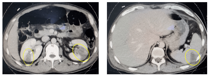
Computed tomography (CT) of his abdomen and pelvis revealed multiple renal infarctions and a splenic infarction (Image 1). In addition, the CT of the brain showed: “Multifocal scattered supratentorial and infratentorial subarachnoid hemorrhages and findings suggestive of evolving ischemic infarct involving the right inferior frontal gyrus, without evidence of hemorrhagic transformation currently. No midline shift or other complication identified.”

Image 1. Computed tomography of the abdomen demonstrating multiple renal infarctions (left, circled) and a splenic infarction (right, circled).

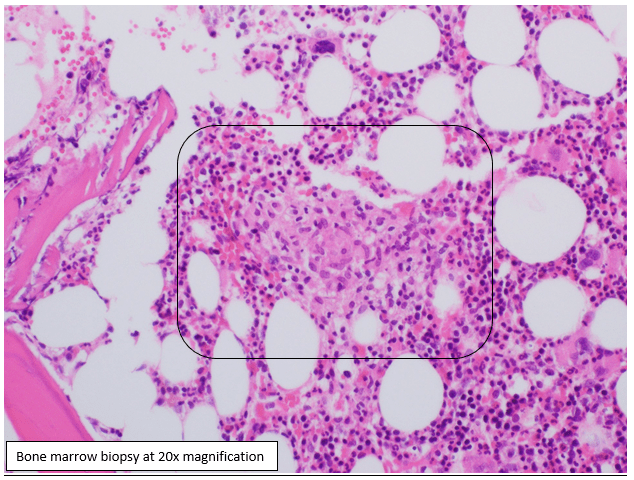
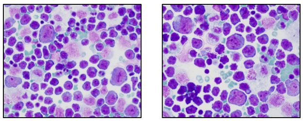
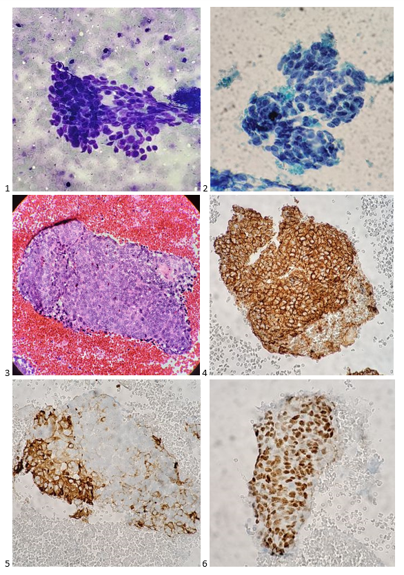
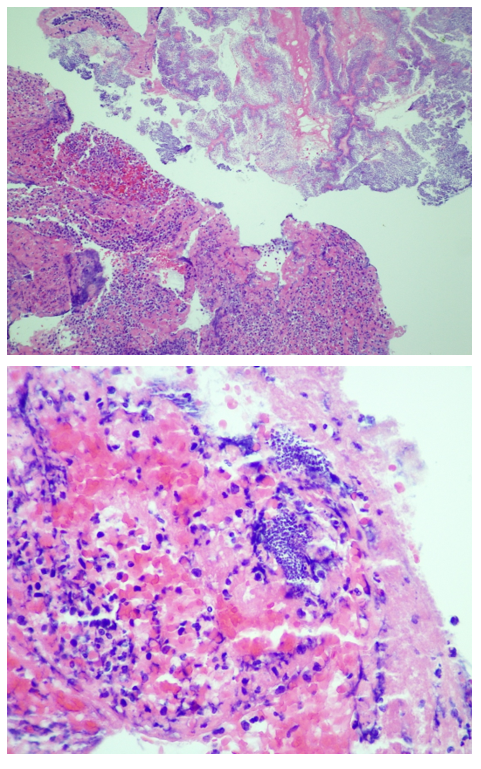
On hospital day 1 (HD1), both sets of initial blood cultures turned positive with gram positive cocci (GPC) in clusters and thrombectomy cultures were also growing GPC in clusters (Image 2). On HD2, the GPC in the thrombectomy culture was identified as Rothia mucilaginosa. GPC growing in the blood cultures were also Rothia mucilaginosa (Image 2). The patient was continued on Vancomycin. Repeat blood cultures were obtained after catheterization on HD0, and HD2, which were negative. On HD2, the pathology of the initial clots showed “fibrinopurulent debris and fibrin plaques with innumerable cocci in clusters” (Image 3).

Image 2. Microscopic and culture morphology of Rothia mucilaginosa. Left: Gram stain from a blood culture demonstrating groups of Gram-positive cocci in small clusters (1000x magfication, oil immersion). Right: Blood agar plate with mucoid light pink-gray colonies.
Image 3. Hematoxylin and eosin stained slide of formalin fixed paraffin embedded tissue of the thrombus removed during the initial emergent catheterization procedure. Sections demonstrate fibrinous material with entrapped white cells and innumerable cocci. Top: 100x magnification; Bottom: 400x magnification.

On HD3, the patient developed 10/10 chest pain with troponin elevation and T-wave inversion. He was taken back to the catheterization lab for another procedure where he was found to have recurrent, complete occlusion of the PDA with unsuccessful recanalization due to the dense thrombus. On HD6, he developed tamponade physiology due to a large pericardial effusion that was drained. Cultures of the pericardial fluid were negative. Given the recurrent embolization events, the patient was transferred to another hospital to undergo aortic valve replacement surgery and coronary artery bypass graft surgery. Cultures taken at the time of the valve replacement surgery were negative and the valve tissue was not sent for pathologic evaluation.  

Discussion

We present an uncommon case of extensive Rothia mucilaginosa sepsis with septic emboli and endocarditis. Rothia mucilaginosa has experienced the scientific name-change game over the last several decades. It was first identified as Micrococcus mucilaginosus, then became Stomatococcus mucilaginosus, was also known as Staphylococcus salivarius before finally arriving to today’s name of Rothia mucilaginosa.1,2 R. mucilaginosa is a normal inhabitant of the oropharynx and is often associated with dental caries.3 R. mucilaginosa can cause invasive infections, typically in patients with compromised immune systems, disrupted mucosal barriers or injection drug use.4

R. mucilaginosa is a facultatively anaerobic, gram positive, non-fastidious coccus that is coagulase negative but with variable catalase positivity. Colony morphology is usually white to gray nonhemolytic colonies with a mucoid appearance. Although the variable catalase reaction may point toward a Streptococcus spp., the Gram stain morphology of clusters helps to rule it out. Although not all strains are mucoid, the classic colony morphology is wet and is due to polysaccharide capsule.

The organism is generally susceptible to antibiotics designed to target gram positive bacteria including, penicillin, ampicillin, cefotaxime, rifampin and vancomycin.4 It is important to note that R. mucilaginosa is not predictably susceptible to clindamycin, trimethoprim-sulfamethoxazole or ciprofloxacin.5 The patient presented in this case received intravenous vancomycin in part due to the extensive disease on presentation, but also because he was at risk for methicillin-resistant Staphylococcus aureus (MRSA) sepsis and had a previously documented abscess from MRSA.

References

  1. Bergan T, Kocur M. 1982. Stomatococcus mucilaginosus gen. nov., sp.nov., ep. Rev., a member of the family Micrococcaceae. Int. J. Syst. Bacteriol. 32:374-377
  2. Collins MD, Hutson RA, Baverud V, Falsen E. 2000. Characterization of a Rothia-like organism from a mouse: description of Rothia nasimurium sp.nov. and reclassification of Stomatococcus mucilaginosus as Rothia mucilaginosa comb.nov. Int. J. Syst. Evol. Microbiol. 3:1247-1251.
  3. Trivedi MN, Malhotra P. Rothia prosthetic knee joint infection. 2015. J. Microbiol. Immunol. Infect. 48(4):453-455.
  4. Bruminhent J, Tokarczyk MJ, Jungkind D, DeSimone JA. Rothia mucilaginosa Prosthetic Device Infections: A Case of Prosthetic Valve Endocarditis. J. Clin. Microbiol. 5;15:1629-1632.
  5. Kaasch AJ, Saxler G, Seifert H. 2011. Septic arthritis due to Rothia mucilaginosa. Infection. 39:81-82.

-Doreen Palsgrove, MD is a board certified Anatomic and Clinical Pathologist who joined the faculty at UT Southwestern as an Assistant Professor in 2019. She specializes in head and neck and genitourinary pathology. 

Dominick Cavuoti, DO is a professor of AP and CP at UT Southwestern, specializing in infectious disease pathology, cytology and medical microbiology.

-Clare McCormick-Baw, MD, PhD is an Assistant Professor of Clinical Microbiology at UT Southwestern in Dallas, Texas. She has a passion for teaching about laboratory medicine in general and the best uses of the microbiology lab in particular.

Microbiology Case Study: 83 Year Old Male with Bladder Cancer

Case History An 83 year old male with bladder cancer was treated with Mycobacterium bovis Bacillus Calmette-Guérin (BCG), his last treatment occurring 1.5 months prior to presentation. He has a past medical history of chronic obstructive pulmonary disease, hypertension, obstructive sleep apnea, obesity, and diabetes. The patient has been hospitalized four times over the last two months and his symptoms include generalized weakness, malaise, shortness of breath and recurrent fever. He was found to have patchy lung infiltrates and he was diagnosed with pneumonia, COPD exacerbation and symptoms of heart filature. He was treated previously with antibiotics, steroids and fluid management which would temporarily relieve his symptoms. He presents to the hospital again, four days after his last hospital discharge, with generalized weakness, malaise, shortness of breath and recurrent fever. On initial evaluation he was found to be pancytopenic.  

Laboratory Identification

Blood cultures were negative. A bone marrow biopsy was performed for fever of unknown origin and pancytopenia. The biopsy showed non-caseating granulomas which were negative for acid-fast bacilli (AFB) by Ziehl-Neelsen stain and fungal elements by Gomori Methenamine Silver Stain (GMS). A laboratory-develped PCR test for Mycobacterium tuberculosis complex (MTBC) was performed on the bone marrow and was negative. AFB culture of bone marrow was positive for after 30 days of incubation and the organism was confirmed to be acid-fast bacilli by auramine-rhodamine fluorescent dye and Kinyoun stain. A second laboratory-developed test that uses heat shock protein (HSP) 2 and HSP3 to determine species level identification of Mycobacteria identified the organism as M. tuberculosis complex. Due to the patient’s history, further identification was performed at a reference lab using specific oligonucleotides targeting the gyrb DNA sequence polymorphisms which is able to separate different members of the MTBC. The patient’s isolate contained a RD1 deletion which is specific for Mycobacterium bovis bacillus Calmette-Guérin (BCG).

Discussion

Mycobacterium bovis is a slow growing mycobacterium which produces rough, dry colonies on growth solid media. It is one of the species in the MTBC with a natural host of domestic and wild animals. Routine molecular tests will not accurately differentiate between members of the MTBC. For definitive identification of M. bovis, 16S rRNA and gyrB gene sequencing is necessary. Safe handling procedures should be followed prior to molecular testing of MTBC.

Mycobacterium bovis BCG is a live, attenuated strain of Mycobacterium bovis that was created for vaccine and is used in the treatment of superficial bladder cancer. The treatment may cause localized symptoms including hematuria, fever, nausea, and dysuria which are marker of anti-tumor effect. Serious complications occur in <5% of patients with complications including sepsis, pneumonitis, hepatitis, lymphocytic meningitis, bone marrow involvement, and mycotic aneurysms. The cardinal sign of BCG infection is a relapsing fever with drenching night sweats persisting beyond 48 hours. Disseminated infection can occur days to years after the therapy. Clinical suspicion should be high for M. bovis BCG dissemination if there are symptoms and a high grade fever ≥72 hours. Treatment includes a regiment of isoniazid, rifampin and ethambutol. Most isolates of M. bovis are resistant to pyrazinamide.

References

  1. Lamm DL. Efficacy and safety of bacille Calmette-Guérin immunotherapy in superficial bladder cancer. Clin Infect Dis 2000; 31 Suppl 3:S86.
  2. Shelley MD, Court JB, Kynaston H, et al. Intravesical Bacillus Calmette-Guerin in Ta and T1 Bladder Cancer. Cochrane Database Syst Rev 2000; :CD001986.
  3. Richter E, Weizenegger M, Rusch-Gerdes S, Niemann S. Evaluation of Genotype MTBC Assay for Differentiation of Clinical Mycobacterium tuberculosis Complex Isolates. Journal of Clinical Microbiology 2003; 41(6): 2672-2675
  4. UpToDate

-Crystal Bockoven, MD is a 4th year anatomic pathology resident at University of Chicago (NorthShore). Crystal has an interest in and will be doing a fellowship in pediatric and perinatal pathology. In her spare time, she enjoys reading, hiking and biking. 

-Erin McElvania, PhD, D(ABMM), is the Director of Clinical Microbiology NorthShore University Health System in Evanston, Illinois. Follow Dr. McElvania on twitter @E-McElvania. 

Safety Checklists and High Reliability in the Lab

A High Reliability Organization (HRO) is one that works with complex and hazardous systems every day and yet retains a high level of safety and an error free environment. The first recognized HRO industries were the United States Navy nuclear aircraft carriers , the Federal Aviation Administration’s Air Traffic Control systems, and nuclear power plant operations. These industries operate using highly complicated and dangerous processes, yet they have the fewest safety incidents.

The use of checklists is an important part of keeping safety incidents to a minimum. They can help employees avoid safety issues, avert disasters, and even aid in incident response. In aviation, a pre-flight safety checklist is a list of tasks that must be performed by pilots and crew before a take-off. Pilots also use checklists for both normal and non-normal operations, for landings, take-offs, and also for malfunctions, and emergencies. Checklists are usually printed on a card, and one card may be divided into as many as a dozen of separate checklists, each of which will be read aloud depending on the phase of a flight. Nuclear power plant operations also involve the use of many safety checklists.

A functional safety checklist features specific characteristics that aid the user in avoiding safety mishaps. Checklists should have defined pause points so users can determine when the list should be used and when new tasks should begin. Checklists types are also important, and the style used may vary depending on the task and the experience of the user. For example, a “do-confirm” list is generally used when users are experienced with the process and have gone through the necessary steps on the list and simply run through it to ensure the process is complete. A “read-do” checklist means employees perform the tasks as they read through each list item.

Most checklists should not be lengthy as it may tempt experienced users to take shortcuts or to ‘pencil-whip’ responses. Make sure the list includes crucial and potentially overlooked steps. These may be the things that can cause the greatest harm if not checked. Use language that is simple, precise, and use terminology that is familiar to the lab staff using the list. Lastly. Test the checklist to see that it fits the criteria above, and that it accomplishes the task set for it. The real goal of using a safety checklist is to create a cultural change by enhancing teamwork, increasing safety communication and changing the understanding of responsibility for safety within the department.

There are quite a few published lab safety checklists available for use. Here are just a few:

  • CLSI’s Clinical Laboratory Safety (GP-17) – Lab Safety Checklist (Appendix C)
  • World Health Organization Biosafety Manual (2004)– Ch. 22 Safety Checklist
  • ISO 15190 Medical laboratories: Requirements for Safety (2019) – Annex B
  • EPA website: Waste Generator Inspection Checklists

Your lab may have its own specific needs, and these checklists may not cover them, or they may be too much for your current issues. If that is the case, create a checklist that focuses on an issue or issues you’d like changed. For example, if PPE compliance is on the rise, create a list that can be used daily or weekly. Walk around and look for proper footwear, lab coat use, and face protection for example. Home made checklists can be scored and used as a quality monitor in order to show improvement in lab safety over time. Make sure people are trained to use the checklists properly, and that people are consistent in how they answer individual items. It’s always a good idea to alter who uses the safety checklists as well. Make sure everyone can use them, and that will create a broader understanding of the safety needs of the department. That can go a long way toward improving the overall safety culture. A review of checklists is always key. If there is a problem with a response for a particular item, it should never be ignored. In fact, it should be addressed quickly.

Many labs today do not fall into the category of a High Reliability Organization. Complex and dangerous tasks do occur in the field, but safety incidents are not uncommon. It may be because lab employees are not educated enough about the consequences. There are definite hazards when working in the lab setting, but often they are not in the forefront of the lab techs’ minds, safety is not made a priority. It needs to be discussed more. Or maybe the reason is that many of the hazards in the lab do not always have more immediate consequences. Organisms involved with exposures have incubation periods, and disease states (like cancer) can take years to develop after a safety incident.

In the airline and nuclear industries, if a safety error is made, the consequence is usually immediate, and deadly for many. Is that it? Is that why people don’t have the same reaction to safety issues in the lab? What can we do as safety professionals to change that? I believe we can change it- and it will take checklists, training and safety awareness.

Dan Scungio, MT(ASCP), SLS, CQA (ASQ) has over 25 years experience as a certified medical technologist. Today he is the Laboratory Safety Officer for Sentara Healthcare, a system of seven hospitals and over 20 laboratories and draw sites in the Tidewater area of Virginia. He is also known as Dan the Lab Safety Man, a lab safety consultant, educator, and trainer.

Floating in a Sea of Uncertainty: Finding a Lifeboat

As we all find ourselves 9 months into 2020, which is arguably the worst year in living memory for many people, we face daily ongoing challenges of completing our work, finding work, adapting our work, feeding our children, schooling our children, preserving our health, caring for loved ones, and trying to not let the daily stress piped in from every communication channel send us over the edge. There are many people who have had a stellar year and have become richer beyond anyone’s imagining as the multitude of crises have fed their business models. There are many people we have lost prematurely due to an uncontrolled viral plague who would have contributed so much had they lived. Amidst all of this, there are individuals dealing with everyday problems in the chaotic setting of 2020—cancer, mental illness, disability, disparities, financial burdens, etc. Personally, I have a dear friend who was on the brink of a complete mental breakdown in 2019 for who I now feel I am on suicide watch 24/7. Life is normally hard, but it has certainly been abnormally hard for the past 9 months. I do not wish to point fingers, place blame, use hindsight, or make astute observations that are of no value—what my sports colleagues call the Monday quarterback effect. What I do want to do is open up to anyone reading this with a few of the things I have done in the last 9 months that have provided comfort and reminded me that, “This too shall pass.”

Take a stroll down memory lane – When I was younger, I used to take a lot of photographs with an actual camera and film. I would probably pass out if I knew exactly how much money I have spent in my life on buying and developing film. My dad was also an amazing photographer and probably knew more about taking traditional photographs than I know about infectious disease. During a certain period (end of high school through the beginning of residency – about 10 years), I was always taking photos and had at least three cameras all the time: a polaroid, an SLR, and point-and-shoot. I was not a very good photographer overall and most people quickly got annoyed with my constant snapping. But I am a collector so every photo I took was placed in an album. In organizing my garage on a Saturday recently when I was looking for anything to do because there was nothing really to do socially outside of my home, I made the decision to reorganize all of my photos into boxes by year and/or event and get rid of the photo albums. I do not recommend that unless you have a lot of time on your hands. But what I do recommend, and I greatly enjoyed, was going through EVERY photograph in my collection. There is a small box from when I was young that were taken by others as well as high school. There is a small box from college. There are literally 12 boxes from medical school and 6 boxes of my family. What did that mean in reality? I was incredibly happy in medical school. I remember being unhappy in high school and college. I have only a handful of friends each from high school, college, and medical school that I am in contact with regularly so no bias in that regard. But I wanted to remember medical school to a much greater degree than I had college or high school. My family is similar as I love my family. Seeing pictures of my grand parents who have all passed and my little cousins before they became grandparents made me feel happy and nostalgic. You have got photos somewhere (and I don’t mean the loads of ridiculous selfies on your Facebook account). Go dig them out and flip through them. If you find some true gems, post them on your social media. Share your memories and you will naturally smile.

Learn something new – We are inundated with information constantly but most of it is not knowledge. Most of it is simply status—the current state of people around us, all of whom will be dead and dust one day. One of my favorite lines from “The Terminator” is, “Look at it this way… In a hundred years, who’s gonna care?” All the tweets, all the posts, all the photos are fleeting moments of fluff (and probably rot your brain—scientific studies to be complete). But knowledge is forever and is precious. Do you know how to refinish an old piece of furniture? Do you know how to grow any type of plant from a cutting? When is the next time we will see Saturn chasing Jupiter across the sky? What happens to stuff you put in a recycling bin? Where does the electricity you use in your home come from? Can you name all 80 unique cultures in Ethiopia? The internet is full of a lot of garbage, but it is also full of incredible sources of knowledge. Sometimes (most of the time) we are so tired of looking at a computer or a smartphone or a tablet if we are working remotely that the last thing we want to do is engage with it further. Libraries are open so you can always resort to dusty old books which are also full of knowledge. Online classes are available for many things. Although cliché, TED talks can be cool. If you are feeling overwhelmed by all of the negativity, opinions, and bandwidth that’s given to things no one will care about in 100 years, turn your attention towards something pure and lose yourself in the nonpolitical world of knowledge. An expert is someone who knows everything that is true about a subject as well as everything that is false. Pick a topic, preferably something that does not come up in your work and set a goal to become an expert in that topic. There will always be people who know more than you do on any topic—but not every topic—but the point is to gain knowledge, grow your brain, and appreciate the permanence of truth.

Mindfulness, it’s really NOT a fad – I wouldn’t dare try to completely address the topic of mindfulness in a short blog, but I will challenge you to investigate it for yourself. Where my last suggestion is one to fill your brain with new ideas, information, processes, and thoughts so you master something external to yourself, mindfulness is the exact opposite. Learning to “turn your brain off” is an amazing skill that does take practice but has enormous benefits. And it is not really turning your brain off but rather turning down the volume on all the negative thoughts you have and may not even know it. Negative thoughts—internal or external—do not control you! They are your thoughts and the most powerful thing you can do is control them. There are many books on this subject, but my favorite is, “Mindfulness: An Eight-week Plan for Finding Peace in a Frantic World”. I will not lie to you. I read this book 5 years ago and have been practicing the techniques since then which did give me a leg up on the horror hurricane that is 2020. But it is never too late to reach inside yourself and find inner strength to deal with outer challenges. It is a bargain at less than $15 and will give you some amazing tools to use if you give it a chance. On a side note, if you are dealing with mental illness or you have a loved one who is dealing with mental illness, the most important first step is recognition, acceptance, and treatment. No one can be expected to defeat the external demons of the world when your internal demons have the chemical advantage. Recognize the signs and recognize the external amplifiers so you can be the hero for those who need you most during this time.

You’ve got to have friends – I remember in the not too distant past listening to “old people” say, “These darn kids need to stop playing video games and texting friends in the same room and get outside and play.” True? Yes. But now our reality has shifted to digital communications as the safest way to go to work, go to school, and see our friends. Zoom parties and the like have become extremely popular and I wrote about etiquette for these tools previously. But they are not the only way to communicate. Did you know that your smartphone is a phone? You can call and talk to people! All that paper junk mail that shows up in your snail mailbox is bidirectional. You can send people letters! This is all obvious and the vast majority if not all of you reading this have used some form of communication to talk to non-work people at least once in the last 24 hours. But do not take this for granted. There were people before our virally induced confinement that did not have large social networks or even limited ones. The isolation of our current situation is amplifying their loneliness. What am I asking you to do? You have a phone. You have social media. Dig through your contacts, find someone you have not talked to in a while, and reach out. Check on them. Check on your distant family members. Ask about them, how they are doing, and what is new in their life. Hearing their voice and laughing with them will make you smile on the inside and the outside.

Move – Your body. Daily.

milner-small


-Dan Milner, MD, MSc, spent 10 years at Harvard where he taught pathology, microbiology, and infectious disease. He began working in Africa in 1997 as a medical student and has built an international reputation as an expert in cerebral malaria. In his current role as Chief Medical officer of ASCP, he leads all PEPFAR activities as well as the Partners for Cancer Diagnosis and Treatment in Africa Initiative.

Doctors are Patients, Too

Hello again everyone, and welcome back!

Last time we talked a bit about what exactly pathology and laboratory medicine training looks like—a much-needed peek behind the curtain, if you ask me. This time, I’d like to discuss something that’s been challenging me way before I started working as a resident: something that’s made both clinical medicine and academic collaboration difficult (to say the least). I talk a lot about how medicine works best when we all come together and contribute our expertise from disciplines across the board for the sake of improving patient care; I even talk about how, too often, people don’t hear the messages coming through from their colleagues and that negatively impacts our field. We’re all guilty of it, some more than others. And in the last few weeks, I’ve been nearly deaf to my colleagues.

No literally, this article is about my Meniere’s disease and how those of us with invisible illness(es) can really impact the collaborative spirit of medical science. Trigger warning: frustration, anxiety, and pathology (mine, specifically). So here’s a reminder that everyone you meet and work alongside has issues they’re dealing or struggling with. And, to those outside of medicine, remember doctors are people too. Dr. Jena Martin, a dermatopathologist I admire and follow on social media recently promoted the topic #DoctorsArePatientsToo—she writes, shares, and promotes fantastic topics on social media and I suggest you check her content out at @jenamartinmd.

Image 1. By poet and patient Ronny Allen who was diagnosed with neuroendocrine cancer in July of 2010. He is passionate about education and awareness and advocates for patients everywhere. (Source: ronnyallan.com)

So, I’ve got a couple things to explain here. Come with me on a journey through the inner ear and out into the reaches of global pandemics!

An MD with MD

So what is Meniere’s disease (MD) and why does it deserve its own article? Well, first of all, it doesn’t deserve anything, it’s a garbage disorder with dumb symptoms and I’m only using it/myself as an example to highlight the struggle for other people working in medicine who also deal with invisible problems. But, since you asked…

Without belaboring any detail, Meniere’s is a somewhat understood inner ear disorder in which the potassium-rich, sound-signal inducing endolymphatic fluid builds up in the inner ear. This causes the organ of Corti (Image 2b) to swell up, along with the rest of the cochlea, the vestibule, and the fluid containing parts of the inner ear (Image 2a). The super pressurized vestibular-cochlear balloon is in a confined bone-space so what happens often is that small fistulas form, mixing the endo and perilymphatic fluids causing all sorts of problems not limited to but including: aural fullness, deafness, ridiculous multi-tonal/pulsatile tinnitus, distorted hearing, frequency loss, imbalance, severe peripheral/rotational vertigo lasting for hours, occasional tachycardia, anxiety, and more! There is no cure; therapy is symptom-dependent and purely aimed at management and mitigation of fulminant hearing loss. Did I mention that most times it’s one-sided, but mine is bilateral? Fun. I’ll be donating my ears to science and/or the garbage in some odd decades from now…

Image 2a and 2b. Dr. Strange-sound, or how I learned to stop worrying and love endolymph. Inner workings of the inner ear, note the marked swelling of fluid (hydrops) associated with the disease state compared to normal. (Sources: Nature and University of Iowa)

Let me put it this way: Over time, I will lose a stark majority of my natural hearing. I will continue to get occasional vertigo attacks, and related symptoms, until the disease (eventually…hopefully?) “burns out” and has no more capacity to damage any more already-damaged inner ear hair cells. I’ll probably get hearing aids or cochlear implants. (Whatever, I’ve always wanted to be half bionic…) I’ll probably keep a cane with me most times. (Dr. House anyone? Right? Or I could keep a sword in there? Legal issues?) But it’s not the long term that bothers me most. It’s the flare-ups. This disorder has periods of acute attacks, periods of mixed-symptom flare ups, and periods of remission. I can handle the remissions, hah. I can even handle the acute episodes—I’ve had great ENT colleagues and discovered great medication plans to manage the attacks. So that leaves the flares, which inspire writings like this one obviously. I wear my hearing aid, but it’s minimally helpful, and I practice patience until my hearing returns. But that isn’t as easy as it sounds, especially when you’re a working resident MD!

The Sound of Silence

Technically, I haven’t experienced true silence since 2015. And, during flare-ups, most of my hearing is almost entirely replaced by the only sound my ears can accommodate and attenuate for which is their own local vasculature. If Edgar Allan Poe could see me know…telltale ears, anyone? Let me paint a picture of what I’m hearing right now on a pretty rough flare-up day, at work, at my desk in the resident room. I’m drinking tea, I can’t hear myself swallowing. I monitor my heartrate on my watch and it’s about 80-90 beats per minute, in my ear, all day, whoosh-whoosh. The pot of tea the robots have been making, whistles non-stop, 24/7. The mosquitoes I’ve been writing about for years now on Lablogatory never leave my cochlea. Cool, on top of that I’ve got hearing loss and aural fullness that makes me not able to hear low-volume pages/phone rings (vibrate mode FTW) and most talking—especially behind masks. (I’ll get to that in a second.) If I turn my hearing aid too high, there’s a squeaky feedback explosion, not to mention the occasional adjustments for quiet and/or super loud talkers, yikes. Just overall, me no hear too good right now. Basically if hearing was reading text, the printer is out of toner and the paper is provolone cheese. And the cheese is on fire. Opa!

Image 3. My flaming-cheese metaphor captured, a xerox copy being made by a waiter in Chicago’s Greektown, the original home of the flaming-cheese saganaki dish. (Source: Chicago Sun Times)

Global Pandemic? Sounds Pretty Bad…

The SARS-CoV-2 pandemic has created very interesting situations at work and elsewhere, all over the globe. People are utilizing the most effective measure against spreading the illness: which up to this point is the non-pharmaceutical intervention of social distancing. Nearly every single person working today, in any field, doing anything, reading this blog, has been a part of email chains, and conference calls, and …. sigh… Zoom meetings. I know, I know… there are pro’s and con’s to this but consider these three points for those with #InvisibleIlnesses:

  1. Working from home has liberated chronically ill folks

Okay so I have an intermittent vestibular/cochlear disorder. What about folks with chronic pain, or Lyme-sequelae, or brain fog, or any other host of hurdles before they can jump onto a video call. Pretend you’re sick at home with a (non-COVID) viral bug. Can you imagine how nice it would feel to have hot tea, your medications, your cat, your spouse, and (most importantly) your couch/bed/TV nearby? Within reason, you’ve just given people who would have needed to excuse themselves a way to participate productively!

2. Imagine having a supplementary PowerPoint or chat box during conversations IRL

One of my specific challenges is missing a word here, a sentence there, and not being able to catch up in a conversation. Usually it ends up with my smile-nodding through to the next topic or checkpoint, but in the current age of virtual meetings I can un-obtrusively ask “What was that last bit?” without seeming like I tuned out. And bonus: I can often get a text translation of a point or two I might have missed from keen colleagues aware of my AV troubles. I can only imagine what it would be like for those more permanently affected.

3. The explosion of inclusivity and accessibility is remarkable

Speaking of which, the number of videos and presentations I am now seeing with closed captions/text supplements are astounding. Usually when I’m in the midst of a flare, I have to check out from audiovisual stimuli and stick to reading only. This can be quite the challenge for work sometimes. But with chat boxes and videos with captions, I feel like I can catch right up. Now, I said I’m only like this part of the time, so for those that have felt excluded and marginalized I’m happy to say that inclusivity and accessibility are growing. I’ve often thought that the importance of a news story or press conference could be measured by the presence or absence of a sign-language interpreter. Over 100 press conferences by Governor Andrew Cuomo in NY, or in Chicago, Illinois with Governor JB Pritzker and Mayor Lori Lightfoot, and each of them was accompanied by ASL accessibility. Fantastic! Just look at one of many efforts online, like @ProjectHearing on Instagram, which promotes the advancement of these topics every day!

Image 4. “Deaf people problems” is a meme collection I’ve seen over and over on social media. Awareness, check. Clear message, check. Super creative visual way to demonstrate a better version of my flaming cheese analogy, super check.
Image 5. Don’t tell anyone, but sometimes, I’m literally stuck on mute. Video conferences might become the new normal. If they are, remember to speak clearly and keep things out of the way between your talking and your microphone. Consider captions for videos and make nice PowerPoints. Please, haha.

Your Lips Move but I Can’t Hear What You’re Saying

So why, when the world is literally aflame with a viral pandemic, am I drudging you through my rant against my inner ears? And why did I just commit the travesty of endorsing zoom calls and captions (I know some people hate captions—you came for a movie not a book, I get it).

Well this whole topic illustrates to big things about working both as a physician and as a person with an invisible condition. First, like most things in medicine, to achieve success you have to adapt, improvise, and overcome. Solving patients’ problems and advocating for their best outcomes takes a little finessing of the system sometimes, you’ve got to do the same thing for yourself. Second, since doctors are patients too, its okay to ask for help. I matched with some of the best residents I’ve ever met, and they’ve offered whatever they can in helping manage my flares during work. This includes anything from extra emails/group text notification chains, forwarding pages, translating video call jumbled audio, etc. They are the best!

Meniere’s has been a challenge to me for a couple of years now, and it’s something I deal with. We’ll call it a character-building attribute. But I genuinely did worry about how this was going to affect me during residency—I had my fair share of hard days in med school basic sciences, and plenty of attending wave-offs when I simply couldn’t hear on 4:00am rounds (yeah I’m looking at you OBGYN and Surgery…). But, it’s been good.

Image 6. Not a new Arnold Schwarzenegger movie, these are real and they’re incredible. Clear window facemasks are such a relief for me. I give ones to all the folks I work with and my life just got easier.

And more than just good, another resident actually has a more permanent hearing impairment and two desks over from mine are the embedded hearing interpreters provided by the department. They’ve been so friendly and provided my with so many resources that I couldn’t be more appreciative. Not only do I have another resident to confide hearing-related rants to, but I also have a department that cares enough to create a supportive and accessible environment. One of the best things they’ve provided: MASKS WITH WINDOWS! Because since this damn pandemic started, I can’t read lips anymore! I didn’t realize how much I depended on visual lip-reads to confirm my hearing that it’s been a learning curve to say the least. Imagine being mostly deaf, your hearing aid not helping much, and looking into a multi-headed scope while your attending lectures on what you’re looking at. An otherwise impossible situation, but my friends and colleagues find ways to make it work because when one person excels, we all do. I’ve been able to continue working, learning, and collaborating thanks to considerations for invisible illnesses like mine.

Consider your colleagues, what can you do to make sure they feel that their needs are met since #DoctorsArePatientsToo? See you next time!


-Constantine E. Kanakis MD, MSc, MLS (ASCP)CM is a new first year resident physician in the Pathology and Laboratory Medicine Department at Loyola University Medical Center in Chicago with interests in hematopathology, transfusion medicine, bioethics, public health, and graphic medicine. His posts focus on the broader issues important to the practice of clinical laboratory medicine and their applications to global/public health, outreach/education, and advancing medical science. He is actively involved in public health and education, advocating for visibility and advancement of pathology and lab medicine. Watch his TEDx talk entitled “Unrecognizable Medicine” and follow him on Twitter @CEKanakisMD.

The Utility of Flow Cytometry in Establishing the Correct Diagnosis of a Rare Aggressive Lymphoma

Case history

A 73 year old woman presented with shortness of breath and was found to have bilateral pleural effusions. She had a history of marginal zone B-Cell lymphoma involving the bone marrow, which was diagnosed 3 months before this presentation and was treated with Rituximab.

Thoracentesis revealed an atypical lymphoid population comprised of intermediate and large sized cells with eccentrically placed nuclei, multiple prominent nucleoli and scant to moderate amounts of basophilic cytoplasm (Image 1). Initial evaluation of the cytology material was concerning for large-cell transformation of the patient’s previously diagnosed marginal zone B cell lymphoma. A representative portion of the fine needle aspirate sample was sent for flow cytometric immunophenotyping.

Image 1. Cytology (Diff Quik, 400X). The atypical lymphoid population is comprised of intermediate and large sized cells with eccentrically placed nuclei, multiple prominent nucleoli and scant to moderate amounts of basophilic cytoplasm.

Flow cytometric immunophenotyping showed a distinct population of atypical cells with moderate CD45 expression and increased side scatter in keeping with cytoplasmic complexity (Figure 1, black arrows). On an initial screening B cell lymphoma panel these cells were negative for CD19 and positive for CD30 (partial), and CD44 (Figure 2).

Figure 1. The neoplastic population shows expression of CD30 and CD44.

The population of interest lacked expression of CD10, CD20, CD22 and surface immunoglobulin light chains and CD138 (Figure 2 and 3).

Figure 2. The neoplastic population lacks expression of CD10, CD19, CD20, and CD22.
Figure 3. The neoplastic population of cells are negative for surface immunoglobulin light chains and CD138.

CD30 expression prompted the investigation of additional T-cell markers to rule out a T cell lymphoma (Figure 4). This population showed dim expression of CD7 but was otherwise negative for pan T cell markers (CD2, CD3, CD5) as well as CD4 and CD8 (Figure 4).

Figure 4. The neoplastic population of cells are positive for CD7 (dim) and CD30 and negative for CD3, CD4, CD8, and CD26.

Given the unusual immunophenotype of the neoplasms, a diagnosis of diffuse large B cell lymphoma (transformation of the known marginal zone lymphoma) seemed less likely and other possibilities were considered.

The presence of CD30 expression and the plasmablastic morphologic features together with the clinical presentation with effusions raised the possibility of primary effusion lymphoma. IHC for anti-HHV8 was performed on the cell block sample (Image 2).

Image 2. Cytology (cell block 400X) A. Hematoxylin and eosin stain of the cell block reveals large, atypical lymphoid cells; Small and large atypical lymphoid cells are highlighted by CD30 (B), HHV8 (LANA-1) (C), and CD138 (focally) (D).

Final diagnosis  

Primary effusion lymphoma (HHV8 positive).

Discussion

Primary effusion lymphoma (PEL) is a large B-cell neoplasm usually presenting as serous effusions without a detectable tumor mass [1]. It is universally associated with the human herpesvirus 8 (HHV8). It usually occurs in the setting of immunodeficiency [2]. Some patients with PEL secondarily develop solid tumors in adjacent structures such as the pleura [3-5].

Immunophenotype of PEL:

POSITIVE: CD45, HLA-DR, CD30, CD38, VS38c, CD138, EMA, HHV8 (LANA1).

NEGATIVE: pan- B-cell markers (CD19, CD20, and CD79a), surface and cytoplasmic Ig, and BCL6.

PEL is usually negative for T/NK-cell antigens, although aberrant expression of T-cell markers may occur. PEL is usually positive for EBV-encoded small RNA (EBER) by in situ hybridization but negative for EBV latent membrane protein 1 (LMP1) by IHC.  This could be explained by EBV virus latency. It is ability of a pathogenic virus to lie dormant (latent) within a cell, denoted as the lysogenic part of the viral life cycle. EBV expresses its genes in one of three patterns, known as latency programs. EBV can exhibit one of three latency programs: Latency I, Latency II, or Latency III. Each latency program leads to the production of a limited, distinct set of viral proteins and viral RNAs. The Epstein-Barr virus encoded RNAs (EBERs): EBER1 and EBER2 are expressed during all latency forms [6], whereas LMP1 is expressed only in latency 2 and 3 rendering it a less sensitive marker for detection of EBV infection. EBV-negative PEL is common in elderly, HIV-negative patients from HHV8-endemic regions (Mediterranean) [7].

Differential Diagnosis

Most common cavities involved by PEL: pleural, pericardial, and peritoneal [8-10].

It was thought that PEL can involve an artificial cavity related to the capsule of a breast implant [11] although it was described only in one case report without appropriate HHV8 staining and before recognition of breast implant-associated anaplastic large cell lymphoma (BIA-ALCL), which this case probably was presenting [12].

Primary effusion lymphoma (PEL) Prognosis

The prognosis is very unfavorable. Median survival is < 6 months. Rare cases have been reported that responded to chemotherapy and/or immune modulation [13].

Flow Cytometry Utility

The importance of utility of flow cytometry in establishing a diagnosis of PEL has been previously shown by others [14]. In the series by Galan et al. the authors described a case of PEL in an 88-year-old HIV-negative female with right-sided pleural effusion without significant lymphadenopathies or other effusions. The cytological study of the pleural fluid revealed a dense proliferation of large plasmablastic cells. A six-color multiparametric flow cytometry immunophenotyping study revealed 45% of large in size and high cellular complexity cells positive for CD45 (dim), CD38, CD138, CD30 and HLA-DR; and negative for CD19, CD20, cytoplasmatic CD79a, surface and cytoplasmic light chains Kappa and Lambda, CD3, CD4, CD5, CD7, CD8, CD28, CD56, CD81, and CD117. In situ hybridization for EBV-encoded small RNA was negative and immunohistochemistry for Kaposi sarcoma herpesvirus (HHV8) confirmed the diagnosis of PEL. These results in addition to the current case highlight the utility of flow cytometry in the diagnosis of lymphomas involving body cavities.

In Summary

PEL is associated with a proliferation of large B-cells which are positive for HHV8, CD45 (dim), CD30, CD38, and CD138 and negative for lineage defining B cell markers (CD19, CD20, and CD79a). Although PEL is a very rare lymphoma, it is important to consider it in patients with pleural, pericardial, and peritoneal effusions by sending a sample for cytological examination and flow cytometric immunophenotyping. Due to the absence of a mass lesion, cytology and flow cytometry are essential for establishing the diagnosis of PEL.

References

  1. Said, J.a.C.E., Primary effusion lymphoma, in WHO Classification of Tumours of Haematopoietic and Lymphoid Tissues (Revised 4th edition), C.E. Swerdlow SH, Harris NL, Jaffe ES, Pileri SA, Stein H, Thiele J, Editor. 2017: Lyion. p. 323–324.
  2. Song, J.Y. and E.S. Jaffe, HHV-8-positive but EBV-negative primary effusion lymphoma. Blood, 2013. 122(23): p. 3712.
  3. Dotti, G., et al., Primary effusion lymphoma after heart transplantation: a new entity associated with human herpesvirus-8. Leukemia, 1999. 13(5): p. 664-70.
  4. Jones, D., et al., Primary-effusion lymphoma and Kaposi’s sarcoma in a cardiac-transplant recipient. N Engl J Med, 1998. 339(7): p. 444-9.
  5. Luppi, M., et al., Molecular evidence of organ-related transmission of Kaposi sarcoma-associated herpesvirus or human herpesvirus-8 in transplant patients. Blood, 2000. 96(9): p. 3279-81.
  6. Khan, G., et al., Epstein Barr virus (EBV) encoded small RNAs: targets for detection by in situ hybridisation with oligonucleotide probes. J Clin Pathol, 1992. 45(7): p. 616-20.
  7. Dupin, N., et al., Distribution of human herpesvirus-8 latently infected cells in Kaposi’s sarcoma, multicentric Castleman’s disease, and primary effusion lymphoma. Proc Natl Acad Sci U S A, 1999. 96(8): p. 4546-51.
  8. Otsuki, T., et al., Detection of HHV-8/KSHV DNA sequences in AIDS-associated extranodal lymphoid malignancies. Leukemia, 1996. 10(8): p. 1358-62.
  9. DePond, W., et al., Kaposi’s sarcoma-associated herpesvirus and human herpesvirus 8 (KSHV/HHV8)-associated lymphoma of the bowel. Report of two cases in HIV-positive men with secondary effusion lymphomas. Am J Surg Pathol, 1997. 21(6): p. 719-24.
  10. Beaty, M.W., et al., A biophenotypic human herpesvirus 8–associated primary bowel lymphoma. Am J Surg Pathol, 1999. 23(8): p. 992-4.
  11. Said, J.W., et al., Primary effusion lymphoma in women: report of two cases of Kaposi’s sarcoma herpes virus-associated effusion-based lymphoma in human immunodeficiency virus-negative women. Blood, 1996. 88(8): p. 3124-8.
  12. Lyapichev, K.A., et al., Reconsideration of the first recognition of breast implant-associated anaplastic large cell lymphoma: A critical review of the literature. Ann Diagn Pathol, 2020. 45: p. 151474.
  13. Ghosh, S.K., et al., Potentiation of TRAIL-induced apoptosis in primary effusion lymphoma through azidothymidine-mediated inhibition of NF-kappa B. Blood, 2003. 101(6): p. 2321-7.
  14. Galan, J., et al., The utility of multiparametric flow cytometry in the detection of primary effusion lymphoma (PEL). Cytometry B Clin Cytom, 2019. 96(5): p. 375-378.

This case was previously presented by authors as eCSI Case on International Clinical Cytometry Society website.  For more information please follow: https://www.cytometry.org/public/newsletters/eICCS-10-1/article7.php

-Dr. Loghavi is an Assistant Professor of hematopathology and molecular pathology MD Anderson Cancer Center in Houston, TX. She received her MD degree from the Azad University in Tehran, Iran. Shen then completed an Anatomic and Clinical Pathology residency training at Cedars Sinai Medical Center in Los Angeles, CA, followed by Surgical pathology, Hematopathology and Molecular pathology fellowship training at the University of Texas, MD Anderson Cancer Center. Dr. Loghavi is passionate about medical education. Her clinical and research interests are focused on hematologic malignancies, with particular focus on myeloid neoplasm and the applications of flow cytometric immunophenotyping and molecular methods in detection of minimal/measurable residual disease. She has authored 100 peer-reviewed articles, 5 book chapters, and numerous abstracts in the fields of hematopathology and molecular pathology. 

-Kirill Lyapichev, MD, FASCP, is a board-certified anatomical and clinical pathologist who completed 2 years of hematopathology fellowship at MD Anderson Cancer Center. He is currently a molecular genetic pathology fellow at MD Anderson Cancer Center. Additionally, he is interested and involved in other research projects including neoplastic as well as non-neoplastic entities: MALT lymphoma, Castleman Disease, Kikuchi-Fujimoto Disease, and others. In 2020 he was selected as one of ASCP’s 2020 Top 40 Under Forty. Follow him on Twitter: @KirillLyapichev.

A 2-For-1 at 61: Mediastinal Cytology Case Studies

For cytologists who perform Rapid On-Site Evaluations (ROSE) on Fine Needle Aspiration (FNA) Biopsies, there is a training platform where you are guided and supported along until you are ready to fly solo. When I was ready to leave that nest, I soared! I saw (and still see) the world through ROSE-colored glasses and fell in love with the responsibility of being competent in assessing adequacy on my own. But I still remember the anxiety of my first solo FNA in diagnostic imaging—a male with a mediastinal mass, 6.2 cm, taking up far too much room in the thorax for him to be asymptomatic. During the timeout, I confirmed the patient’s information and the anatomic site of the biopsy. On the computer I can see the mass on the CT scan image, and then I glanced into the room and saw the patient prone, covered by a sterile drape, and in a much more relaxed state than myself. Diagnostic imaging is likely the coldest department in the hospital, and I was nervously (but thankfully not obviously) sweating. I prep my slides, label everything I need, and head into the procedure room for my first needle pass. I make my smears, rinse my needle into Hanks Balanced Salt Solution, and return to the patient’s side for my second needle pass. I repeat the process and stain my smears. Under the microscope, I see that my slide is saturated with epithelioid cells. Before I actually had the chance to interpret what I was seeing and formulate a differential diagnosis, I went on auto-pilot and thought, “THYMOMA!” “Alright, focus. Don’t get ahead of yourself. It might just be a lucky guess. Look at the second pass,” I said to myself. I move the next slide onto the stage, and it’s even more cellular than before. I’m on a thymoma one-track mind and holding steady. I tell the radiologist that the smears are adequate, but I need 3 more passes from a 25-gauge needle or 2 passes from a 22-gauge needle for my cell block, whichever she prefers, and I also need core biopsies. I received 2 more 22-gauge needle passes, completed my FNA quick evaluation worksheet, checked the core biopsy requisition and container labels, thanked the team, and walked back upstairs to the lab. What an adrenaline rush! Now I just need the seal of approval from my attending pathologist on cytology service for the day, and I get the green light! I presented the case to our cytopathology director, he asked me my thoughts, I shyly suggested a thymoma, and he agreed, told me it was a great sample, and I processed the remainder of my FNA. Phew. I passed, and on an uncommon tumor I briefly studied in school nonetheless.

Here’s that first solo case—an FNA of the Anterior Mediastinum on a 62 year old male patient with no prior cancer history:

Images 1-6. Anterior Mediastinal FNA – 1, DQ-stained smear; 2, Pap-stained smear; 3, H&E cell block section; 4, AE1/AE3 + Immunostain; 5, CK5/6 + Immunostain; 6, p63 + Immunostain.

Cytologic Diagnosis:

– Cytology features highly suggestive of thymic epithelial neoplasm.
Note: We performed immunocytochemical stains on paraffin sections of the cell block. Tumor cells show positive staining for AE1/AE3, CK5/6, CK7, p63, and CD117; and negative staining for CD45, CD5, CD57, TTF-1, synaptophysin, and PAX-8.  The proliferation index by Ki-67 immunostaining is approximately 10%. The combination of morphology and immunoprofile in the context of clinical presentation is consistent with thymoma.

Two months later, the thymus was resected and diagnosed as the following:

– Invasive thymoma, WHO type B3

Images 7-8. Thymus Resection – 7, H&E section 100X; 8, H&E section 400X.

Flash forward 3 years from my initial thymoma case to a CT-guided biopsy of a left-sided mediastinal mass in a 61-year-old male with a history of lymphoma. I went into the procedure with the assumption that I might see a lymphoid transformation to Diffuse Large B Cell Lymphoma (DLBCL) or even Hodgkin’s due to the location. However, on my ROSE, the cells looked similar to but not exactly like the thymoma from 2014. I wanted to go full bore thymic carcinoma with squamoid features. The cells were more disorganized and pleomorphic in appearance than the first thymoma, much more variation in nuclear size. They simply appeared more aggressive. And the lymphocytes sealed the deal on the thymoma diagnosis. I knew it wasn’t DLBCL or Hodgkin’s, but then I started to look more carefully at the lymphocytes. There was something about them too… “LIGHT BULB! GET MORE MATERIAL FOR FLOW! The patient has small lymphocytic lymphoma. It’s likely here too!” Yes, there is a great deal of inner monologue on ROSE’s.

After examining the Diff-Quik slides, Pap-stained slides, and H&E Cell Block sections, I called it like I saw it: “Thymic carcinoma with squamoid features. Atypical lymphoid population, recommend correlation with flow cytometry.”

Images 9-13. Left-sided Mediastinal FNA – 9, DQ-stained smear; 10, Pap-stained smear; 11, H&E cell block section; 12, p63 + Immunostain; 13, CD5 + Immunostain.

The cytology of this case was signed out as:

– Positive for malignant cells.
– Thymic epithelial neoplasm, favor type B3 thymoma.
Note: The specimen contains atypical epithelial clusters admixed with lymphocytes. Immunohistochemical stains performed on cell block sections, with proper positive and negative controls, show that the epithelial tumor cells are positive for pancytokeratin, CK7, p63, CD5, and CD117, while negative for CDX2, TTF-1, GATA3, and WT-1. CD1a and CD57 show faint staining in rare lymphocytes. TDT is negative. CD99 is positive in focal epithelial component and few lymphocytes. The Ki-67 shows proliferative index of focally up to 20% in the epithelial component. The findings support diagnosis if type B3 thymoma (well differentiated thymic carcinoma.)
The flow cytometry report demonstrated a population of CD5 positive monoclonal B-cells.

Three months after the FNA of the mediastinal mass, the patient underwent a radical thymectomy and received the following diagnosis:

– Thymic squamous cell carcinoma arising in a background of thymoma B3 (well differentiated thymic carcinoma). Carcinoma is confined to the thymus. Lymphovascular invasion is identified. Inked surgical resection margin is negative for carcinoma.
– Lymph nodes involved by small lymphocytic lymphoma, no metastatic carcinoma seen.
– The lymphocytes in the tumor are mostly CD3+ T cells. Focal areas show some CD20+ & CD23+ B cells which may represent small lymphocytic lymphoma infiltration.

Images 14-15: Radical Thymus Resection. 14, H&E section 400X; 15, H&E section 600X.

I found this case absolutely fascinating. To be able to diagnosis two entities in one FNA – both a thymic carcinoma and a background of small lymphocytic lymphoma from one sample. There’s something to be said about those rare tumors—after screening classic textbook lung, breast, colon, and pancreatic cancers day in and day out, the infrequently diagnosed tumors (both benign and malignant) are either easily forgotten or forever engrained in the cytology knowledge bank. Fortunately, in both of these cases, thymomas fell into the latter.

Taryn Waraksa, MS, SCT(ASCP)CM, CT(IAC), has worked as a cytotechnologist at Fox Chase Cancer Center, in Philadelphia, Pennsylvania, since earning her master’s degree from Thomas Jefferson University in 2014. She is an ASCP board-certified Specialist in Cytotechnology with an additional certification by the International Academy of Cytology (IAC). She is also a 2020 ASCP 40 Under Forty Honoree.

Dr. Who?

Welcome back everyone!

Thanks for reading my piece last month on liquid biopsies. And, as a side note, there is a growing number of awesome quality content and posts from pandemic response, to inclusion, alongside COVID and case-studies so subscribe, share, and add this page to your bookmarks—STAT! Lablogatory has been a fantastic platform to share and learn so much in this past year, I could barely keep up!

Or super-STAT if you’re one of those people…but hey, that language belongs to all of us! Lab professionals, nurses, scientists, and doctors alike. And this month, I just want to take a quick moment to celebrate a milestone.

I’m officially a resident physician/trainee, medical post-graduate! (There was confetti falling just now on my end, not sure about yours, but work with me here.) It’s just one of those life-goals that feels great when you get there. But there’s a lot more to it than it seems…if I told you being a pathology resident means sacrificing early adulthood, amassing soul-crushing debt, and explaining to your peers and colleagues what it is exactly you do and why you also bear the moniker of “physician,” you’d delete that bookmarked webpage faster than I can make you scroll through this thing.

(oh good you’re still here!)

All that said, I’ve got to say: it’s worth every single bit of it. Times a million. But I really did mention some red flags that, were we discussing any other work environment, would make you definitely think twice before committing 5-10 years of your life. Furthermore, as a PGY-1 in pathology, I could stand next to any other patient-facing PGY-1 colleague (read: intern) and they wouldn’t have the faintest about what I actually do. Listen, the “lifestyle” specialty, generally 9-5er, no weekend, no 24hr call isn’t something I’m shy to celebrate, but it’s not the whole story. I’ve matched and started learning and working at a great institution with great faculty, mentors, and other residents/fellows. Bottom line: I’m more than a little happy about where I’m at professionally.

Image 1. Most path & lab med residents get cubicle-style desks to spend time reading, prepping, writing, learning, and previewing cases between responsibilities in sign-outs, tumor boards, or OR/gross room work. Most of my non-path friends don’t like this. It makes me very happy.

So, to my non-pathology friends, what is it exactly that I do during my residency training while you might be busy rounding, managing glucose levels, triaging cases, putting orders in—you know, regular intern stuff *shudders* … (pathology trainees don’t have an intern year, we jump right into the specialty and go for 3-4 straight on through). Like most of you I have a transition period where I get acclimated to the workload and patterns of my specific residency, but sans-anno-interna, I’ve got lots of work ahead to climb the steep learning curve that med school pathology merely skims.

What does non-patient-facing mean, exactly?

Well, without an intern year you jump right into what most path residents go into which is a 4-year combined anatomic and clinical pathology (AP/CP) track. You immediately begin training in all the fields in pathology. They include surgical pathology (of various sub specialties like head-and-neck, gynecologic, gastrointestinal, thoracic, neuro, etc.—think surgery, then add pathology), autopsy training, dermatopathology, cytopathology, hematopathology, transfusion medicine, clinical chemistry, microbiology, hemostasis and coagulopathy, pediatric pathology, forensic pathology, molecular, training as a laboratory director, and much, much more. Each of these services has a workload which is usually comprised of cases from biopsies and grossed specimens for histologic analysis (anatomic pathology) or the ongoing maintenance and advancement of clinical diagnostic testing through laboratory methods and management of staff/resources (clinical pathology).

Image 2. August 2019 Issue of The Pathologist magazine. Pathologists, medical students, microscopes, you get it…Specifically, that’s one of my mentors (and now faculty) Dr. Kamran Mirza and (then) medical student Austin McHenry discussing the critical role pathology plays in every circle of medical care.

When I say “non-patient facing” this means that the majority of that work is not done in 1-on-1 settings with patients in a clinic or hospital floor. It is done ancillary to their clinical experience whereby pathology attendings manage the simultaneous training of residents and processing of case sign outs for rapid and accurate diagnostic output for our patient-facing colleagues. For example, while a patient, their family, and doctor are discussing and managing symptoms related to a possible cancer diagnosis. The pathologists are examining microscopic behavior of the cancer-in-question’s cells and adding immunohistochemical testing and molecular analyses to identify, stage, and prognosticate that cancer. Returning information about what it is and what can be done back to the patient-facing clinician, who can then best-translate a tailored approach for their patient. Old-timey medical texts would often refer to the pathologist as the “doctors’ doctor,” and I’m not here to hate on that haha. My clinical friends and readers might feel forlorn now at the prospect of 4 years of medical school training to “just look into a microscope all day?” Well, for some folks in path it means a lot more than that, every slide is a patient. So we care just as much as if they were right opposite our desk. But that’s not all we do…

(More on that in a minute.)

So What Do You Do?

Okay, there are lot of words in path that might act as a barrier to understanding the common ground between me and …let’s say a colleague and friend in Family Medicine. So for the purposes of transparency here’s my friend from medical school Dr. Danash Raja and how a small part of his schedule and my schedule aren’t so different…

Image 3. Dr. Raja is from Alaska, and now works as a resident physician in Family Medicine in Eu Claire, Wisconsin! Alaska! Look at this graduation photo!

On both sides of this table are clinicians managing their patients and ensuring the best possible outcomes. Both sides are deeply vested in intensive hours of training, procedural experience, evidence-based best-practices from the literature, and ongoing continuing education.

Image 4. They see me grossin’, they hatin’…A lot of surgical pathology and microscopy in general revolves around understanding the gross layout of a specimen and its orientation before it becomes a thin microscope slide. As a junior pathology resident, we spend a lot of time up near the OR. Critical skill for a crucial foundation of knowledge.

Literally the biggest differences:

  • In pathology, I get my own desk space and I need it! I’ve got to start amassing a physical and digital library to supplement the next 4-6 years of subspecialty training for the eventual day when a colleague will see me in an elevator and expect concise, thorough, and actionable material to inform their clinical management from the pathologic diagnosis.
  • Pathology residents and clinical residents both take “call” except Dr. Raja has to pull grueling 24-hour+ shifts and stay in the hospital for the duration, and I answered a page about a transfusion reaction from a grocery store once.
  • When a patient thinks about the person who helped them find out what kind of cancer they had and what treatment to begin, they’ll probably think of Dr. Raja or someone patient-facing in Heme/Onc—but I’m working on this, every day!

Bottom line: I’m as important as he is, and he is as important as I am. Our work is what really matters, and what really connects us as clinical colleagues. It’s all about patients, remember? But I’m more than happy to be the pathologist to his patient-facing, diabetes-managing, vaccine-giving, life-improving super hero doctor!

You Never See Patients?

We’re back on this. Remember how I said looking into microscopes isn’t all we do? Okay, well it’s not. And if you’re lucky enough to have matched to as awesome of a place as I did, then you know what I’m talking about. If you’ve read some of my pieces, you know full well my passion in pathology lies in Hematopathology and Transfusion Medicine. I like to sit right on the fence between AP and CP, and mostly look at the green grass on the CP yard. This month, I’ve been on service for Transfusion Medicine and let me tell you about the few weeks…

Image 5. Dr. Kimberly Sanford, ASCP leadership and Director of Transfusion Medicine at VCU was highlighted in The Pathologist magazine for her work outside the laboratory seeing patients every day, and encouraging residents to do the same and do what pathologists do best: enrich and improve the channels of communication so patients better understand their conditions and the medical process.

I, a pathologist trainee, resident physician, under the supervision of two attending physician pathologists have been seeing and following up on patients nearly every day. Gasp! No, I’m not part of some backwards resident exchange program (because OMG how dangerous haha), no I’m not lost, no I’m not being overly gunnery, that’s it, that’s the Tweet. Seriously, it’s just part of the service. Larger academic hospitals with robust clinical blood bank services often have apheresis clinics and I find myself working exactly there. Blood bank/Transfusion Medicine is one of those subspecialties where patient contact is part of the routine. At some institutions, I’ve been a part of some pathology-led teams that procure the bone marrow aspirates from their patients in Hemepath service, or conducted their own fine needle aspirations for cytology service, or dermpath services that operate in clinics alongside their dermatology colleagues—I’ve even been working on frozen sections and surgical path grossing when called into an operating room to discuss methods and approach for biopsy! There were patients at every turn, all with pathologists on the front line! Dr. Syed T. Hoda (@01sth02 on Twitter) from NYU Langone often says, “Person FIRST, doctor SECOND, specialist THIRD.” And trust him, he’s a bone and soft tissue pathologist that left the lab and went to the floors to help clinical staff when overwhelmed during the peak of the COVID crisis in NYC. So for my dual-interests, I would say I’d expect to see quite a bit of patients in my future practice.

Image 6. My awesome co-residents! (Left-to-right): me, Dr. Elnaz Panah, Dr. Aayushma Regmi, and Dr. Sandra Haddad—you’re going to hear more about them, don’t worry.

So, would I pick pathology again? Uh, yeah! Without a single hesitation. Every day at work I am reminded that I am at the right place, with the right co-residents, the right faculty and mentorship, and the right environment to train and hone my future skills for a career that lines up exactly with what I want to do.If you’re interested about the intersections between clinical medicine and pathology, and want to learn more about “patient-facing pathology” keep an eye out during the 2020 ASCP Annual Meeting for a talk by yours truly as part of a panel discussion on communicating directly with patients! Register now! Free for members.

See you next time!

BONUS: did you notice that I referenced The Pathologist magazine a bit in this post, well it’s because they named me to their Pathology Power List for 2020! An exclusive, international list of 80 professionals in the field of pathology and laboratory medicine who contribute and advance the profession every day! I was highlighted for my active social media work and my response to the early COVID pandemic in Manhattan, NY.


-Constantine E. Kanakis MD, MSc, MLS (ASCP)CM is a new first year resident physician in the Pathology and Laboratory Medicine Department at Loyola University Medical Center in Chicago with interests in hematopathology, transfusion medicine, bioethics, public health, and graphic medicine. His posts focus on the broader issues important to the practice of clinical laboratory medicine and their applications to global/public health, outreach/education, and advancing medical science. He is actively involved in public health and education, advocating for visibility and advancement of pathology and lab medicine. Watch his TEDx talk entitled “Unrecognizable Medicine” and follow him on Twitter @CEKanakisMD.

Casualties of COVID-19: Measuring the Length, Width, and Depth of a Pandemic’s Impact

An editorial in Nature on August 12, 2020 entitled, “How to stop COVID-19 fuelling a resurgence of AIDS, malaria, and tuberculosis” provided four suggested solutions specifically for these diseases in the wake of Sars-CoV-2. For reference, here are the four approaches suggested as written in the editorial (#4 in detail):

  1. Hospitals and health authorities in affected cities and regions must recognize that AIDS, malaria and TB are surging again.
  2. Researchers must continue to refine their models using more real-world data.
  3. There is a need for public-information campaigns
  4. These campaigns cannot on their own keep surgeries and wards open, or equipment functioning. The resurgence of infectious diseases has created a greater demand for tests, treatments and research. All of these need more funding. 

Do those strike you as odd? The entire economy of nations along with the focus of their healthcare has been derailed and distracted by COVID-19 and the solution for these diseases is to recognize them, improve models, inform the public and seek more funding. You are either completely in tuned with the author in seeing that more funding is needed or you are a bit miffed that, in the wake of all that is happening, THESE guys want more money?

The US is a major contributor to the Global Fund for HIV, TB, and Malaria (the largest funder of these activities) and the total pledges to date for the GF approach $69 billion dollars with the US providing $54 billion (92%). From 2008 to 2016, the US contribution increased almost every year from $840 million to $1.65 billion annually until 2015 when it was frozen at $1.35 billion until 2019. In 2020, prior to the COVID-19 pandemic (i.e., during calendar year 2019 when the fiscal year 2020 budget was being planned), the amount from the US dropped to $958 million (2010 levels), representing a 30% drop in funding. So, to recap: The Global Fund started the year down by nearly 30% of what had been available, COVID-19 derailed all activities and drained the fiscal resources of patients and nations, and now, the progress that has been made on these diseases has been set back bay possibly a decade. The situation couldn’t be more desperate and, YES, the program needs a massive increase in funding. But, to be very clear, that massive increase pre-dated COVID-19 and represents something more distressing underneath.

I was fortunate to give the Michelle Rablais lecture at the ASCP Annual Meeting in Phoenix in 2019 where I carefully laid out the costs of controlling JUST malaria (not to mention TB and HIV) and demonstrated for the audience that as the number of cases get smaller and smaller (because your measures are so successful), the cost of finding the remaining cases goes up. As we successfully approach elimination or eradication of a disease, the final push requires at least the same but often more funding to make it across the finish line. This is not an opinion but is based on an enormous amount of data from other diseases as well as from the world’s experience with the first malaria eradication campaign. For HIV, we can’t eliminate it or eradicate it but we have converted it to a chronic disease and, therefore, infrastructure and funding to support patients ongoing is needed and by any form of math has to increase as the population lives longer and more people are added to the disease pool (although those numbers had been greatly reducing). Tuberculosis in its simplest form is a disease of poverty related to lack of access to drugs and healthcare, cramped living conditions, etc. When a pandemic derails the economy and causes the poor to become even more poor, tuberculosis is going to surge.

To the authors of this editorial I offer a gracious thank you and note with a heavy heart that the estimate of $28.5 billion additional dollars being needed to make up the ground lost by COVID-19 does also include the ground they had already lost by defunding principles trending over the last 4 years for global health.

But at least the countries that struggle with these diseases only have HIV, TB, and malaria to worry about, right? Wrong. In almost every low- to middle-income country where HIV, TB, and malaria are or have been major health challenges, hypertension, diabetes, cancer, cardiovascular disease, stroke, and mental health are equivalent or worse health problems than compared with high income countries. Do not be dissuaded by sheer numbers and always consider the outcomes, pre-COVID-19. For cancer, mortality in the US averages around 35% while in Africa it is closer to 80%. In full COVID-19 response mode, cancer programs—fledgling, underfunded, and disorganized—became non-existent and are only now (nearly 6 months after closing) starting to re-open and find their way back to where they were—fledgling, underfunded, and disorganized! Diabetics cannot go 6 months without insulin, hypertensive patients cannot have unregulated blood pressure, etc. While in the safety of a high-income country, makeshift systems, telehealth, contactless visits, etc. were brought on board to keep some semblance of a healthcare system in place, cancer patients were delayed in receiving diagnoses and treatment due to rationing of time and elimination of “elective” procedures.

As the data continues to be tallied and as models continue to be developed to understand just how much we have lost from our failed response to COVID-19 as a world and certainly as a nation, please do not slough off the staggering “additional” deaths that are going to be reported because of patients who didn’t have access to their regular health system. Every person from November 2019 until the end of this pandemic whose death occurred because their regular supply lines were disrupted, their planned treatments were cancelled, their medical supplies were not available, or their access to life-saving interventions were delayed is just as much a casualty from COVID-19 as a directly infected patient who succumbs to the disease. Our recent experience as a nation with the disasters in Puerto Rico around both the confusing death tolls from the hurricanes as well as the total death toll from the fiscal challenges of their medical system (prior to COVID-19) should serve as valuable lessons. Let us not come out of the other side of this pandemic with a similar disregard for the value of every human life or without an understanding of how our individual and collective mistakes as a nation have lead directly to these effects.

milner-small


-Dan Milner, MD, MSc, spent 10 years at Harvard where he taught pathology, microbiology, and infectious disease. He began working in Africa in 1997 as a medical student and has built an international reputation as an expert in cerebral malaria. In his current role as Chief Medical officer of ASCP, he leads all PEPFAR activities as well as the Partners for Cancer Diagnosis and Treatment in Africa Initiative.

How to Rock a Risk Assessment

As a lab safety professional, we know performing risk assessments is an integral piece of managing a safety program. In fact, assessing risks and identifying hazards are considered the beginning steps that must be completed when approaching the management of any safety-related area. Risk assessments are the starting point for handling a bloodborne pathogens program, chemical hygiene, personal protective equipment, and many other lab matters. But how can you be sure they have been performed correctly, and how often should they be performed?

OSHA gives simple guidance on basic assessment of risk in the workplace. “The employer shall assess the workplace to determine if hazards are present, or are likely to be present, which necessitate the use of personal protective equipment (PPE).” They further state, “the employer shall verify that the required workplace hazard assessment has been performed through a written certification that identifies the workplace evaluated; the person certifying that the evaluation has been performed; the dates(s) of the hazard assessment; and which identifies the document as a certification of hazard assessment.”

OSHA’s Bloodborne Pathogens standard requires that labs perform an exposure risk determination for each employee. Labs must assess exposure risk levels by job classification, and then assess exposure risk for tasks performed in the department. The Hazard Communication standard explains the need for chemical hazard determination. There are many types of risk assessments that must be performed, and regulations stipulate that they must be reviewed and updated (if necessary) every year. Things change in a dynamic department like the laboratory, and understanding the changing risks of harm can be key to keeping staff safe.

The four basic steps included in a risk assessment are hazard identification, identifying those at risk, choosing control measures, and reviewing the findings. It may sound easy, but lab hazards can come in many forms (physical, mental, chemical, biological, etc.), so walk around the department to look for those you may have missed. Review incident records as well to see what harm is occurring in the lab. Next, determine what employees may be harmed and how. Consider those who work in the lab each day and those that are just passing through the area. An evaluation of the risks follows. If the risk cannot be removed, decide what controls (engineering, administrative, PPE) need to be in place. Finally, review each risk assessment on a regular basis. Things change in the lab, and with change may come new hazard risks – or even the reduction of potential harm if risks have been reduced via elimination or substitution. Again, examine these assessments at least annually or whenever major changes occur in a particular lab safety arena.

For many laboratories, the advent of the COVID-19 pandemic brought new testing platforms and procedures to the department, and this testing had to be implemented quickly. Is there a way to use risk assessments to help introduce new processes safely? Absolutely! The use of a standard form to assess the potential hazards of new or updated processes and/or equipment is actually a high quality finishing touch on an overall assessment program, and unfortunately, it is something that is often missing in many labs.

Considering the tasks, sample handling, equipment, reagents, and overall biosafety of the new process, choose the likelihood of hazard incidents (rare, possible, likely, etc.), and classify the consequences of each occurrence (minor, moderate, major, etc.). Use a matrix to calculate the overall level of risk for the new procedure or equipment. For example, if a new COVID-19 test platform requires opening samples, the likelihood and risk of exposure may be designated as “high” or even “very high.” Next, determine the controls that should be put in place to decrease the exposure opportunities. For example, if centrifuge rotor covers, a biological safety cabinet, and a surgical mask is added to the normal lab PPE, the overall risk of that testing process is reduced. Document the decision and keep those records available for any future reviews that may be needed. Once the assessment is complete, complete the appraisal of the new process with a quick safety audit. Look for additional biohazards encountered, chemical safety, electrical safety, and even potential waste handling issues. If you couple that safety analysis with the risk assessment, you are doing an above-average job at circumventing hazards in the department. (For examples, go to https://www.aphl.org/programs/preparedness/Documents/APHL%20Risk%20Assessment%20Best%20Practices%20and%20Examples.pdf)

Keeping staff safe from exposures and injuries in the laboratory is a massive and time-consuming task, but it is required by our regulatory agencies and it needs to be a top priority. When used properly and completely, a risk assessments can be a powerful tools that begins your look into safety hazards and then closes the loop to avert them. Having an awareness and control of departmental hazards is one way to rock safety in the laboratory.

Dan Scungio, MT(ASCP), SLS, CQA (ASQ) has over 25 years experience as a certified medical technologist. Today he is the Laboratory Safety Officer for Sentara Healthcare, a system of seven hospitals and over 20 laboratories and draw sites in the Tidewater area of Virginia. He is also known as Dan the Lab Safety Man, a lab safety consultant, educator, and trainer.