Eye Spy 2.0

Now that you’ve seen some malignant ocular entities (both primary and metastatic), I’d like to share some (in my eyes) curveballs. Working in a cancer center, I see more cancer than I do benign processes. You can say I’m fine tuned to identifying malignant cells, and I find it difficult to rest assured that what I’m diagnosing truly is benign. When you find one malignant cell in a pleural fluid, changing the patient’s diagnosis from Stage II to Stage IV, that primed search for atypia is always on. With that said, not every specimen screened at a cancer center is malignant. We rule out malignancy and confirm benign processes as well. As for benign ocular FNAs, well… you have to be as certain calling benign as you do malignant because the process of acquiring an additional sample to confirm your diagnosis is just as involved and unpleasant as the first biopsy.

Here, I present six non-malignant eye FNA cytopreparations that had me searching for atypia longer than I expected.


Case 1. A 72 year old female with a history of breast carcinoma; She presented with a choroidal nevus, OD in 2014. The oncologist noted a slight increase in base and thickness six years after the initial evaluation. The clinical diagnosis is a choroidal nevus, rule out low-grade melanoma.

Images 1-2: Eye, Right, Choroid, Fine Needle Aspiration. Pap-stain.

Final Diagnosis: No malignant cells identified. A few benign-appearing melanocytes, consistent with nevus.

Note: Material is scant cellular and consists mainly of scattered retinal pigment epithelial cells and sensory cells. There are a few benign-appearing spindle-shaped cells with bland nuclei, consistent with nevus. However, the paucity of the sample precludes a definitive diagnosis. Recommend clinical correlation to exclude sampling error.


Case 2. A 21 month old male with a history of an iris stromal cyst, OS. Lesion is status post-FNA (twice), had regrowth, and aspirated for the third time. Previous FNAs showed no malignant cells.

Images 3-4: Eye, Left, Choroid, Fine Needle Aspiration. Pap-stain.

Final Diagnosis: No malignant cells identified. Rare pigment-laden macrophages, a few lymphocytes, and benign surface epithelial cells present. (Consistent with stromal cyst).


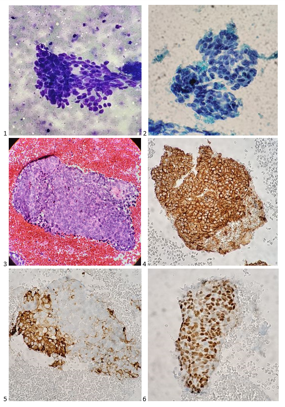
Case 3. A 65 year old female with a history of nevus, OD. Ocular oncology followed up and noted an increase in thickness from 2.76 to 3.00 mm. In light of family history of cutaneous melanoma, treatment is suggested. Clinical diagnosis: ciliary body melanoma.

Images 5-6: Eye, Right, Choroid, Fine Needle Aspiration. Pap-stain.

Final Diagnosis: Scant mildly atypical amelanotic spindle cells, consistent with nevus.

Note: Diagnostic material is very scant. There are a few benign-appearing single spindle cells and one cluster of cohesive spindle cells with no pigment. Prominent nucleoli are not seen. While the cytologic features in the context of clinical presentation is consistent with nevus, the paucity of the sample precludes a definitive diagnosis. There is no diagnostic evidence of malignant melanoma in this sample. Recommend clinical correlation to exclude sampling error.


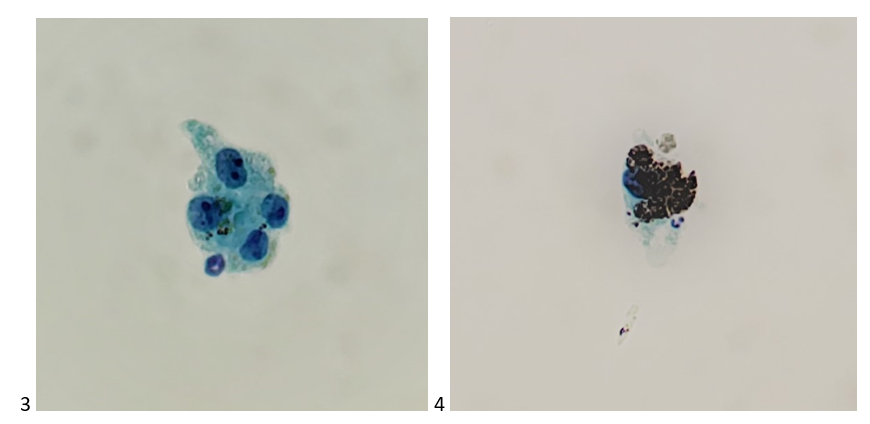
Case 4. A 9 year old female with a history of retinoblastoma, OD; status post IAC (intra-arterial chemotherapy) x5, IVit melphalan x 4, and plaque radiotherapy. She recently developed vitreous hemorrhage (OD). Since the hemorrhage is obscuring the retinal view, vitrectomy is planned.

Image 7: Eye, Right, Vitreous, Fine Needle Aspiration. Pap-stain.

Final Diagnosis: No malignant cells identified. Lymphocytes and histiocytes present.


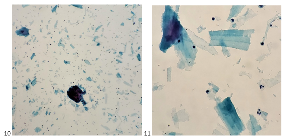
Case 5. An 11 year old female with vitreal retinoblastomas, OU; status post CRD therapy x6; plaque I-125, EBRT, proton beam; IVit Metphalan x2, and PPV (pars plana vitrectomy), OS. She now presents with dense vitreous hemorrhage blocking view since 2019.

Images 8-9: Eye, Left, Vitreous, Fine Needle Aspiration. Pap-stain.

Final Diagnosis: No malignant cells identified. Proteinaceous material and scattered small lymphocytes present.


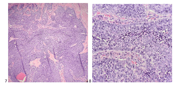
Case 6. A 52 year old female with no cancer history. In 2019, she presented with pain, floaters, decreased visual acuity, and photophobia, OS. She was treated with antibiotic drops and steroids. In 2020, she was noted to have panuveitis with yellow iris nodules. FNA showed necrotic cells inadequate for diagnosis, as well as a negative culture and PCR for HSV, toxoplasmosis, CMV, and VZV. Clinical diagnosis: suspicious for iris lymphoma, OS.

Images 10-11: Eye, Left, Anterior Chamber, Fine Needle Aspiration. Pap-stain.

Final Diagnosis: Mixed inflammatory cells, favor an inflammatory process.

Note: There are neutrophils, lymphocytes, and scattered histiocytes in a background of numerous fragments of an amorphous substance. The cellularity is inadequate for flow cytometric analysis. We performed immunocytochemical stains on cytospin preparations. The lymphocytes are predominantly T-cells showing positive staining for CD45 and CD3 and negative for CD20. There is no diagnostic evidence of malignant lymphoma in this sample. The amorphous substance may represent lens fragments, in which case the possibility of lens-induced uveitis should be considered in the differential diagnosis. Recommend clinical correlation.


As you can tell, benign processes are complicated and require thorough explanations in our pathologic diagnoses, especially when they differ from the clinical impression. The majority of benign ocular FNAs are paucicellular, and we make the most of what we have through optimal preservation and preparation of cellular material. And don’t forget-not everything is cancer. Cyst contents, proteinaceous debris, and inflammatory cells make up a share of cases we see, and it’s okay to diagnosis them as benign. Just keep an eye out.

-Taryn Waraksa, MS, SCT(ASCP)CM, CT(IAC), has worked as a cytotechnologist at Fox Chase Cancer Center, in Philadelphia, Pennsylvania, since earning her master’s degree from Thomas Jefferson University in 2014. She is an ASCP board-certified Specialist in Cytotechnology with an additional certification by the International Academy of Cytology (IAC). She is also a 2020 ASCP 40 Under Forty Honoree.

A 2-For-1 at 61: Mediastinal Cytology Case Studies

For cytologists who perform Rapid On-Site Evaluations (ROSE) on Fine Needle Aspiration (FNA) Biopsies, there is a training platform where you are guided and supported along until you are ready to fly solo. When I was ready to leave that nest, I soared! I saw (and still see) the world through ROSE-colored glasses and fell in love with the responsibility of being competent in assessing adequacy on my own. But I still remember the anxiety of my first solo FNA in diagnostic imaging—a male with a mediastinal mass, 6.2 cm, taking up far too much room in the thorax for him to be asymptomatic. During the timeout, I confirmed the patient’s information and the anatomic site of the biopsy. On the computer I can see the mass on the CT scan image, and then I glanced into the room and saw the patient prone, covered by a sterile drape, and in a much more relaxed state than myself. Diagnostic imaging is likely the coldest department in the hospital, and I was nervously (but thankfully not obviously) sweating. I prep my slides, label everything I need, and head into the procedure room for my first needle pass. I make my smears, rinse my needle into Hanks Balanced Salt Solution, and return to the patient’s side for my second needle pass. I repeat the process and stain my smears. Under the microscope, I see that my slide is saturated with epithelioid cells. Before I actually had the chance to interpret what I was seeing and formulate a differential diagnosis, I went on auto-pilot and thought, “THYMOMA!” “Alright, focus. Don’t get ahead of yourself. It might just be a lucky guess. Look at the second pass,” I said to myself. I move the next slide onto the stage, and it’s even more cellular than before. I’m on a thymoma one-track mind and holding steady. I tell the radiologist that the smears are adequate, but I need 3 more passes from a 25-gauge needle or 2 passes from a 22-gauge needle for my cell block, whichever she prefers, and I also need core biopsies. I received 2 more 22-gauge needle passes, completed my FNA quick evaluation worksheet, checked the core biopsy requisition and container labels, thanked the team, and walked back upstairs to the lab. What an adrenaline rush! Now I just need the seal of approval from my attending pathologist on cytology service for the day, and I get the green light! I presented the case to our cytopathology director, he asked me my thoughts, I shyly suggested a thymoma, and he agreed, told me it was a great sample, and I processed the remainder of my FNA. Phew. I passed, and on an uncommon tumor I briefly studied in school nonetheless.

Here’s that first solo case—an FNA of the Anterior Mediastinum on a 62 year old male patient with no prior cancer history:

Images 1-6. Anterior Mediastinal FNA – 1, DQ-stained smear; 2, Pap-stained smear; 3, H&E cell block section; 4, AE1/AE3 + Immunostain; 5, CK5/6 + Immunostain; 6, p63 + Immunostain.

Cytologic Diagnosis:

– Cytology features highly suggestive of thymic epithelial neoplasm.
Note: We performed immunocytochemical stains on paraffin sections of the cell block. Tumor cells show positive staining for AE1/AE3, CK5/6, CK7, p63, and CD117; and negative staining for CD45, CD5, CD57, TTF-1, synaptophysin, and PAX-8.  The proliferation index by Ki-67 immunostaining is approximately 10%. The combination of morphology and immunoprofile in the context of clinical presentation is consistent with thymoma.

Two months later, the thymus was resected and diagnosed as the following:

– Invasive thymoma, WHO type B3

Images 7-8. Thymus Resection – 7, H&E section 100X; 8, H&E section 400X.

Flash forward 3 years from my initial thymoma case to a CT-guided biopsy of a left-sided mediastinal mass in a 61-year-old male with a history of lymphoma. I went into the procedure with the assumption that I might see a lymphoid transformation to Diffuse Large B Cell Lymphoma (DLBCL) or even Hodgkin’s due to the location. However, on my ROSE, the cells looked similar to but not exactly like the thymoma from 2014. I wanted to go full bore thymic carcinoma with squamoid features. The cells were more disorganized and pleomorphic in appearance than the first thymoma, much more variation in nuclear size. They simply appeared more aggressive. And the lymphocytes sealed the deal on the thymoma diagnosis. I knew it wasn’t DLBCL or Hodgkin’s, but then I started to look more carefully at the lymphocytes. There was something about them too… “LIGHT BULB! GET MORE MATERIAL FOR FLOW! The patient has small lymphocytic lymphoma. It’s likely here too!” Yes, there is a great deal of inner monologue on ROSE’s.

After examining the Diff-Quik slides, Pap-stained slides, and H&E Cell Block sections, I called it like I saw it: “Thymic carcinoma with squamoid features. Atypical lymphoid population, recommend correlation with flow cytometry.”

Images 9-13. Left-sided Mediastinal FNA – 9, DQ-stained smear; 10, Pap-stained smear; 11, H&E cell block section; 12, p63 + Immunostain; 13, CD5 + Immunostain.

The cytology of this case was signed out as:

– Positive for malignant cells.
– Thymic epithelial neoplasm, favor type B3 thymoma.
Note: The specimen contains atypical epithelial clusters admixed with lymphocytes. Immunohistochemical stains performed on cell block sections, with proper positive and negative controls, show that the epithelial tumor cells are positive for pancytokeratin, CK7, p63, CD5, and CD117, while negative for CDX2, TTF-1, GATA3, and WT-1. CD1a and CD57 show faint staining in rare lymphocytes. TDT is negative. CD99 is positive in focal epithelial component and few lymphocytes. The Ki-67 shows proliferative index of focally up to 20% in the epithelial component. The findings support diagnosis if type B3 thymoma (well differentiated thymic carcinoma.)
The flow cytometry report demonstrated a population of CD5 positive monoclonal B-cells.

Three months after the FNA of the mediastinal mass, the patient underwent a radical thymectomy and received the following diagnosis:

– Thymic squamous cell carcinoma arising in a background of thymoma B3 (well differentiated thymic carcinoma). Carcinoma is confined to the thymus. Lymphovascular invasion is identified. Inked surgical resection margin is negative for carcinoma.
– Lymph nodes involved by small lymphocytic lymphoma, no metastatic carcinoma seen.
– The lymphocytes in the tumor are mostly CD3+ T cells. Focal areas show some CD20+ & CD23+ B cells which may represent small lymphocytic lymphoma infiltration.

Images 14-15: Radical Thymus Resection. 14, H&E section 400X; 15, H&E section 600X.

I found this case absolutely fascinating. To be able to diagnosis two entities in one FNA – both a thymic carcinoma and a background of small lymphocytic lymphoma from one sample. There’s something to be said about those rare tumors—after screening classic textbook lung, breast, colon, and pancreatic cancers day in and day out, the infrequently diagnosed tumors (both benign and malignant) are either easily forgotten or forever engrained in the cytology knowledge bank. Fortunately, in both of these cases, thymomas fell into the latter.

Taryn Waraksa, MS, SCT(ASCP)CM, CT(IAC), has worked as a cytotechnologist at Fox Chase Cancer Center, in Philadelphia, Pennsylvania, since earning her master’s degree from Thomas Jefferson University in 2014. She is an ASCP board-certified Specialist in Cytotechnology with an additional certification by the International Academy of Cytology (IAC). She is also a 2020 ASCP 40 Under Forty Honoree.