Hematology Case Study: A 12 Year Old Female with Thrombocytopenia.

Case History

A 12 year old female presented with thrombocytopenia. Previous platelet count performed at a different facility showed a platelet count of <100K.  Patient signs show history of bruising, no history of trauma, intermittent epistaxis.

Family history shows no history of anemia or hypothyroidism from either parent. Incidental finding of hypothyroidism was revealed for this patient when laboratory testing was performed.

gray-platelet-small
Light staining, “gray” platelets.

Laboratory results

DAT: Negative

PT 11.7/INR 1.1

PTT 38.3

Platelet aggregation studies: Decreased response to ADP-Collagen-Epinephrine and Arachidonic Acid. Results of which are consistent with platelet dysfunction due to storage pool defect.

vonWillberand panel shows within range results for Factor 8, vW antigen and vW Ristocetin.

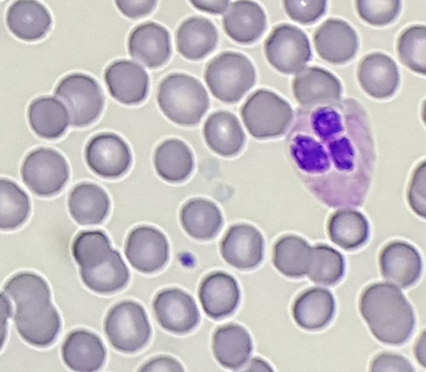
Peripheral blood smear shows light staining (gray) appearance of platelets.

Diagnosis: Gray Platelet Syndrome


Discussion

Gray platelet syndrome (GPS) is an inherited platelet disorder that presents with thrombocytopenia and characteristic pale/gray appearance of platelets under light microscopy. This gray appearance of platelets is due to the absence of alpha granules and their constituents.

According to Gunay-Aygun et al., the diagnosis of GPS requires demonstration of the absence or marked reduction of α-granules in platelets observed by electron microscopy (EM). Megakaryocytes also show decreased α-granules. Platelet dense bodies and lysosomes are unaffected. Alpha granules, the most abundant vesicles in platelets, store proteins that promote platelet adhesiveness and wound healing when secreted during platelet activation. Some α-granule proteins (eg, platelet factor 4 and β-thromboglobulin) are synthesized in megakaryocytes and packed into the vesicles, whereas others are either passively (eg, immunoglobulins and albumin) or actively (eg, fibrinogen) taken up from the plasma by receptor-mediated endocytosis. Proteins synthesized in megakaryocytes are markedly reduced in GPS, whereas other α-granule constituents are less affected. Studies of granule membrane-specific proteins have shown that platelets and megakaryocytes of GPS patients have rudimentary α-granule precursors. Therefore, the basic defect in GPS is thought to be the inability of megakaryocytes to pack endogeneously synthesized secretory proteins into developing α-granules. (Gunay-Aygun et al, 2010).

Most patients who present with GPS are characteristically macrothrombocytopenic and the number of megakaryocytes in the bone marrow appears normal. However platelet survival is reduced. This inability of megakaryocytes to survive is due to the alpha granule deficiency of this disorder therefore leading to thrombocytopenia. Myelofibrosis and splenomegaly is also apparent on patients with GPS but severe hemorrhage is unlikely, bleeding tendencies tend to be mild to moderate for GPS.

Most patients had bleeding symptoms from infancy with the average onset of 2 years of age. Average age of diagnosis is 10-14 years of age; some patients who have Gray Platelet Syndrome have presented with initial diagnosis of ITP (idiopathic thrombocytopenic purpura).

Reference

Gunay-Aygun, M., Zivony-Elboum, Y., Gumruk, F., Geiger, D., Cetin, M., Khayat, M., . . . Falik-Zaccai, T. (2010). Gray platelet syndrome: natural history of a large patient cohort and locus assignment to chromosome 3p. Blood, 116(23), 4990-5001. doi:10.1182/blood-2010-05-286534


-Written in collaboration with Stephanie Foster, BS MLS

ledesma_small

-Carlo Ledesma, MS, SH(ASCP)CM MT(ASCPi) MT(AMT) is the program director for the Medical Laboratory Technology and Phlebotomy at Rose State College in Midwest City, Oklahoma as well as a technical consultant for Royal Laboratory Services. Carlo has worked in several areas of the laboratory including microbiology and hematology before becoming a laboratory manager and program director.

Microbiology Case Study: A 65 year Old Male with Decubitus Ulcer

Case History

A 65 year old male with a history of T7-8 paraplegia and lengthy history of a decubitus ulcer presented for surgical debridement of the wound. An ischial bone biopsy for culture was performed.

Lab Identification

The primary gram stain demonstrated mixed gram positive and gram negative organisms, and the tissue sample from the wound grew Corynebacterium striatum and Bacteroides fragilis. The bone sample grew Enterococcus faecalis and anaerobic gram positive cocci. He was initially treated with ertapenem alone but daptomycin was added to cover enterococcus and he continued this dual regimen. Daptomycin was discontinued due to elevated creatinine kinase levels and he was transitioned to intravenous vancomycin. He completed 42 days of ertapenem and vancomycin. He was transitioned to oral amoxicillin/clavulanic acid and he has not developed new fever, chills, sweats, fatigue or increased drainage from his wound.

corstri1

Image 1. Blood agar plate with gray-white, moist, smooth, non-hemolytic bacterial colonies.

corstri2

Image 2. Chocolate agar plate with gray-white, moist, smooth, bacterial colonies.

The blood and chocolate agar plates grew bacterial colonies while the MacConkey agar had no growth. The gram stain was consistent with Corynebacterium and mass spectrometry identified the organism as Corynebacterium striatum.

Discussion

Corynebacterium striatum is a gram positive bacilli that is part of normal skin and mucosal flora. In immunocompromised patients or through direct inoculation of a sterile site, C. striatum can cause infectious endocarditis, bacteremia, pneumonia, lung abscess, arthritis and chorioamnionitis. Studies have shown that C. striatum also can cause wound infections in patients with underlying disease and previous antibiotic use. Foreign medical devices can also be infected by C. striatum, and removal of the device may be necessary. Vancomycin is used to treat C. striatum due to the variable susceptibility to other antibiotics.


-Mustafa Mohammad, MD is a 3rd year anatomic and clinical pathology resident at the University of Vermont Medical Center.

Wojewoda-small

-Christi Wojewoda, MD, is the Director of Clinical Microbiology at the University of Vermont Medical Center and an Assistant Professor at the University of Vermont.

Microbiology Case Study: A 14 Year Old Female with Neck Swelling

Case History

A previously healthy 14-year-old female presents to the emergency department with three days of progressive facial and neck swelling. The swelling started on the left side. Two days ago she visited her primary care physician where she had negative monospot and mumps IgM testing.  She is fully vaccinated, but was exposed to a mumps outbreak at school.

Discussion

Our patient was diagnosed with mumps by positive RT-PCR from a buccal swab. The mumps virus is a member of the Paramyxoviridae family which includes notable human pathogens parainfluenza, Hendra, and Nipah viruses. Members of this family are enveloped, helical viruses with single-stranded, non-segmented RNA genomes with negative polarity. Mumps is an obligate human pathogen that replicates in the epithelial cells of the upper respiratory tract and subsequently moves to regional lymph nodes. It is spread from person to person via direct contact with respiratory secretions or contact with contaminated fomites. Mumps is a highly contagious disease with as high as 85% of naïve individuals becoming infected after contact with a mumps infected individual. It spreads most efficiently in areas where there is close contact among individuals for prolonged periods of time such as college campuses and close-knit religious communities.

Prior to vaccination for mumps in the 1960s, greater than 150,000 cases of mumps occurred each year in the US. The incubation period for infection is 16-18 days, with the majority of infected persons being asymptomatic or having mild respiratory symptoms. Orchitis causing sterility in post-pubescent males is the main concern of mumps infection but other rare but serious complications include mastitis and oophoritis in females, meningoencephalitis, pancreatitis, and deafness.

Due to sporadic outbreaks of measles since the introduction of the vaccine, the vaccine schedule has been revised from one dose of the MMR (measles, mumps, and rubella) vaccine at age 12-15 months to include another MMR booster at age 4-6 years. We are currently in the middle of yet another outbreak with nearly 6,000 cases of mumps reported to the CDC in 2016 and a high rate of infections reported thus far in 2017 (Figures 1 and 2).

mumps1
Figure 1. Number of cases identified by the CDC in 2017 by state. (Figure courtesy of the CDC Mumps website at https://www.cdc.gov/mumps/outbreaks.html. Content source: National Center for Immunization and Respiratory Diseases [NCIRD], Division of Viral Diseases)
mumps2
Figure 2. Number of cases of mumps per year identified by the CDC.
(Figure courtesy of the CDC Mumps website at https://www.cdc.gov/mumps/outbreaks.html. Content source: National Center for Immunization and Respiratory Diseases [NCIRD], Division of Viral Diseases)


Diagnostic Testing for Mumps

Serological testing for IgM and RT-PCR from a buccal swabs are the mainstay of mumps diagnosis. IgM becomes positive in the first 3-4 days after symptom onset and will remain positive for 8-12 weeks. IgG becomes positive 7-10 days following symptom onset and will remain at high levels for many years and detectable for life. In a vaccinated individuals, IgM testing has less utility as it may be non-reactive or weakly positive following a secondary immune response.

RT-PCR from a buccal swab specimen is the most sensitive test for diagnosis of mumps. It should be performed as soon as a patient is symptomatic, as testing by this method is the most sensitive in the first few days following symptom onset and becomes less sensitive as time goes on.

Urine specimens can be used to isolate mumps in viral culture. Urine is not positive for mumps until greater than 4 days post symptom onset and is less sensitive than PCR performed on the bucal swab. For these reasons, viral isolation from urine is no longer a commonly used test for diagnosis of mumps, although viral culture is still considered the gold standard for mumps conformation.

Resolution

The patient and her family were counseled on the infectious nature of mumps. She was instructed to remain in isolation at home for 6 days after resolution of swelling.


References

  1. Manual of Clinical Microbiology, 11th edition
  2. CDC Mumps Website (www.cdc.gov/mumps/index.html)


Erin McElvania TeKippe, PhD, D(ABMM), is the Director of Clinical Microbiology at Children’s Medical Center in Dallas Texas and an Assistant Professor of Pathology and Pediatrics at University of Texas Southwestern Medical Center.

Leading Lab Safety

The number of medical laboratory scientists is dwindling. Baby Boomers have begun their retirement, and even before that started, there were more job openings than people to fill them. That means more opportunities in the lab world, and in some cases leadership roles are being obtained by less experienced people than in years past. Whether or not one has a long lab history, one aspect of any new leadership position that will be important to grasp is management of the lab safety program.

The first step for a new lab leader is to ensure the existence of a functional laboratory safety program. Do this by looking for specific components of the program, a laboratory safety manual, a safety committee, and lab safety indicators. If these items are in place and functioning as they should, you’re off to a good start.

The laboratory safety manual may be in paper or in an electronic format. It should be separate from the hospital or facility safety manual as there are many lab-specific safety policies and procedures that are required. Maintain document control of these safety policies, ensure they have medical director (or designee) approval, and review these policies in a timely fashion. It is important to remember that while some lab regulatory agencies (like CAP) allow bi-annual policy review, OSHA requires annual reviews. OSHA covers many safety policies in the lab such as the chemical hygiene plan, the exposure control plan and many more.

The laboratory should have a functioning safety committee, no matter the size. If the lab staff is very small, the leader may play a role in the larger hospital or facility safety committee. If the lab is larger, a committee composed of just lab staff is advised. If the hospital or lab is part of a system, the committee should include at least one member from each lab site. The safety committee should meet at least monthly. It is important not to skimp on meetings or cancel them on a regular basis. Let staff know this is a priority for the leadership in the lab. During the meetings provide education, review lab incidents, and raise safety awareness. Train committee members how to perform safety audits, how to develop “safety eyes,” and most especially how to coach each other and their peers in the department.

Another important component of a functioning lab safety program is the use of safety indicators. Much like quality indicators, this safety data can be used to help determine the overall safety culture in the department. A good example indicator includes monitoring the employee exposure and injury rate. By using the laboratory’s OSHA 300 log information, a lab can compare its reportable injury data to national benchmarks. Many safety indicators are typically reactive data (or lagging), but tracking safety meeting attendance can actually serve as a leading indicator for the lab.

Once you’ve assessed the lab’s safety program, the next step a new leader should take is to assess the overall lab safety culture. This can be performed in many ways. One part of performing the assessment is by using your “safety eyes” that was mentioned earlier. Scan the lab visually. What immediate safety issues are seen? What is on the walls of the department? What types of interactions are observed? What is the physical layout? With practice and experience, a leader may be able to do the visual portion of the culture assessment quickly.

Another safety culture indicator tool is a laboratory safety audit. The results of an audit can provide much information about safety practices in the lab such as PPE use, chemical storage, and awareness of fire safety issues. One good model safety audit that can be used is located in the appendix of CLSI’s document Safety in the Clinical Laboratory (GP17-A3). This is a very comprehensive laboratory assessment and it can tell you much about your overall safety culture. As stated before, audit results can be discussed at the lab safety committee meetings, and ideas for improvements can be considered.

Managing the overall lab safety program is a big job, and it is often only one task of many that belongs to a laboratory leader. Change occurs daily in the field of lab medicine, and new leaders are coming aboard. Whether you are new or experienced, however, utilizing these basic first steps will provide a leader with the information needed to identify the safety culture and to understand how the program is operating.


Scungio 1

Dan Scungio, MT(ASCP), SLS, CQA (ASQ) has over 25 years experience as a certified medical technologist. Today he is the Laboratory Safety Officer for Sentara Healthcare, a system of seven hospitals and over 20 laboratories and draw sites in the Tidewater area of Virginia. He is also known as Dan the Lab Safety Man, a lab safety consultant, educator, and trainer.

Diagnostics for Displaced Populations

A few months ago, Sarah Riley discussed diagnosing disease in displaced populations. She says, “… being aware of the problem of limited access to diagnostic laboratory testing in refugee populations is a good start. We need to get a better understanding of the scope of the problem. We should be ready and able to provide specific recommendations for meeting diagnostic needs in these populations including most appropriate diagnostics…”

Dr. Riley’s post has stuck with me, and I wanted to know how I could help. While several organizations work with refugees worldwide, it’s unclear if any dedicate money to laboratories or diagnostics. Thankfully, there’s the ASCP Foundation‘s Global Health Fund. The GHF is dedicated to providing diagnostics, establishing laboratories, and training local personnel in countries where access to pathologists or medical laboratories is inadequate. If this sounds like something you’d like to support, you can do so here.


Swails

-Kelly Swails, MT(ASCP), is a laboratory professional, recovering microbiologist, and web editor for Lab Medicine.


Microbiology Case Study: A 74 Year Old Man’s BAL Specimen

Case History

A laboratory received a bronchioalveolar lavage specimen on a 74 year old male, who is now deceased. The potato flake agar grew white, mucoid colonies, while the Mycosel plate had no growth. The colony was identified as Cryptococcus neoformans by mass spectrometry.

crypto1-1

Image 1. White mucoid colonies on potato flake agar.

Discussion

C. neoformans and C. gattii produce white, mucoid colonies on a variety of agars that usually become visible within 48 hours. Urea and phenoloxidase are positive. L-Canavanine Glycine Bromothymol Blue (CGB) agar helps differentiate C. neoformans colonies from C. gattii, with C. neoformans producing a light green-blue color and C. gattii producing a cobalt blue color. C. neoformans is also described as resembling glass beads on cornmeal agar due the presence of its thick capsule. C. neoformans is generally 5-10 µm in size, however size is variable and they can be increased. Historically, India ink preparation was described to identify the organism due the capsule extruding the ink. Current identification methods include a rapid latex agglutination test for antigen, and mass spectrometry can also be used to identify C. neoformans.

C. neoformans and C. gattii are basidiomycetous, encapsulated yeasts found all over the world. They are commonly found in areas frequented by birds and bats. Patients with recent travel to caves or work in environments that expose them to chickens are at higher risk of infection due to inhalation of C. neoformans. While C. neoformans generally causes pulmonary infections, patients who are immunosuppressed can have disseminated cryptococcosis with CNS involvement. The clinical presentation of cryptococcosis due to the two species is generally indistinguishable. Cryptococcosis can be treated with amphotericin B and flucytosine or fluconazole.


-Mustafa Mohammad, MD is a 3rd year anatomic and clinical pathology resident at the University of Vermont Medical Center.

Wojewoda-small

-Christi Wojewoda, MD, is the Director of Clinical Microbiology at the University of Vermont Medical Center and an Assistant Professor at the University of Vermont.

Hematopathology Case Study: A 7 Year Old Transplant Patient with Neck Swelling

A 7 year old male with a history of restrictive cardiomyopathy status-post orthotopic heart transplant in June, 2010 that was on maintenance doses of tacrolimus and mycophenolate mofetil presented to his primary pediatrician left neck swelling. Starting in January 2017, the patient began with neck pain and swelling in the context of a recent gastrointestinal illness. Per CT report of the neck, a rim enhancing well-defined suppurative level III lymph node measuring 1.4 x 1.2 x 2.1 cm with adjacent soft tissue inflammatory changes extending into the left parapharyngeal space was identified. The patient was subsequently started on antibiotics and was discharged home with some improvement of swelling and pain.

The patient then presented again with continued neck swelling, although painless this time, and the patient’s cardiologist was contacted, who recommended a decrease in tacrolimus dosing. An otolaryngology evaluation was requested and given the concerning findings, the patient was admitted for further work-up, including a biopsy with a lymphoma protocol.

burlym1

burlym2

burlym3
BCL6
burlym4
BCL2
burlym6
EBER


burlym5
Flow Cytometry


Results

Flow cytometry revealed a kappa restricted CD10 positive mature B-cell population.

On biopsy examination, a population of monotonous lymphoid cells that are large in size with round to mildly irregular nuclear contours, open chromatin, and multiple inconspicuous nucleoli are present in a diffuse pattern. Abundant apoptotic bodies and mitotic figures are noted and occasional “starry sky” features are present. By immunohistochemistry, BCL6 highlights the neoplastic lymphocytes while BCL2 highlights background T-cells. EBER is negative.

Overall, despite a negative t(8;14) IGH/MYC translocation, the findings are best considered to be of an EBV-negative post-transplant lymphoproliferative disorder with morphologic features consistent with Burkitt lymphoma.

Discussion

Post-transplant lymphoproliferative disorders (PTLD) are a relatively rare complication in a variety of transplants that occurs in 2-10% of post-transplant patients. Overall, following a solid organ transplant (SOT), PTLD development is 1-5% of recipients with the highest incidence in intestinal and multivisceral transplantations (5-20%). Another factor is EBV status of the recipient, for which those that are EBV-naïve and lack cellular immunity to EBV are susceptible to graft-mediated EBV infection and ultimately developing an increased incidence in early PTLD. This population is overrepresented by pediatric transplant recipients1.

The presentation is highly variable and ranges from benign proliferations to overt lymphoproliferative disorders. Classifications for PTLD include early lesions, which are oligo- or polyclonal proliferations of EBV positive B cells have either a predominant infectious mononucleosis-like proliferation or a plasmacytic hyperplasia form. Polymorphic PTLD is a similar concept to the early proliferative lesions but the host architecture of the native structure is disrupted. Lastly, monomorphic PTLD is an entity that fulfills criteria for a non-Hodgkin lymphoma and is diagnosed according to the criteria of non-transplant associated lymphomas. Within pediatric registry studies, monomorphic PTLD accounts for 35-83% of all PTLD cases. B-cell lymphomas, particularly DLBCL, comprise the vast majority of monomorphic PTLD with plasmacytoma and T-cell lymphoproliferative disorders much less common2.

In this particular case, with the patient having been 7 years post-transplant and negative studies for EBV present, it is not surprising that germinal center phenotypic markers are highly expressed, such as CD10 and BCL6, which has been well elucidated by Jagadeesh, et al. Although not many genetic studies have been performed on post-transplant B-cell lymphomas, regardless of EBV status, there is some data demonstrating trisomies of 9 and/or 11 with translocations 8q24.1 (C-MYC), 3q24 (BCL6), and 14q32 (IGH). Rinaldi et al. noticed a lack of genetic lesions characteristic of postgerminal center derivation, such as gain of chromosome 3 (FOXP1, BCL6, and NFKBIZ) and 18q (BCL2 and NFATC1) together with losses of 6q (PRDM1 and TNFAIP3) in post-transplant DLBCL.  A number of DNA mutations have also been described including genes associated with somatic hypermutation (SHM) such as PIM-1, PAX5, C-MYC, and RhoH/TTF. These particular mutations are also found to be independent of EBV status1.

Overall, post-transplant lymphoproliferative disorders occur in a variety of transplant settings across many age groups and can be dependent on EBV and CMV status as well as the type and degree of immunosuppression. Although many variations take place in PTLD, patients with the monomorphic type are diagnosed according to their non-transplant counterparts. Current perspective includes further analysis of molecular and cellular mechanisms incorporated into research projects, which could better aid in prognostic implications and future therapeutics.

  1. Morscio, et al. “Molecular pathogenesis of B-cell posttransplant lymphoproliferative disorder: What do we know so far?” Clinical and Developmental Immunology 2013.
  2. Mynarek, et al. “Posttransplant lymphoproliferative disease after pediatric solid organ transplantation,” Clinical and Developmental Immunology 2013.


PhillipBlogPic-small

-Phillip Michaels, MD is a board certified anatomic and clinical pathologist who is a current hematopathology fellow at Beth Israel Deaconess Medical Center, Harvard Medical School, Boston, MA. His research interests include molecular profiling of diffuse large B-cell lymphoma as well as pathology resident education, especially in hematopathology and molecular genetic pathology.

Microbiology Case Study: A 58 Year Old Female with Fever, Headache, and Vomiting

Case History

A 58 year old female presented to the emergency department with complaints of a fever (reaching 102.9°F) and headache with associated nausea and vomiting for the past 24 hours. Her past medical history was significant for a resection of a recurrent hemangiopericytoma by the neurosurgery service three weeks prior. The patient also noted clear drainage from this surgical site which had begun 5 days ago. Other symptoms noted at presentation included decreased appetite and dehydration. She denied back & neck pain, photophobia or stroke and seizure-like symptoms. Her vital signs were all within normal limits. On physical exam, a healing surgical wound was noted in the posterior auricular area with clear drainage, but no blood or exudates were visualized.  She had no tenderness when her spine was palpated and neurologic exam showed a left sided facial droop and tongue deviation which were noted previously and attributed to her multiple central nervous system surgeries. Complete blood count (CBC) showed a mild increase in white blood cells and anemia. An external ventricular drain was placed and cerebral spinal fluid (CSF) was sent to the microbiology lab for culture. Blood cultures and a swab from the surgical wound were also collected.

Laboratory Identification

sermar1.png

Image 1. Gram stain of the cytospin CSF showed many acute inflammatory cells and numerous Gram negative bacilli (1000x).

sermar2

Image 2. Growth of large, glossy, reddish-orange colonies on sheep blood agar (image taken after 72 hours of incubation).

sermar3.png

Image 3. Growth of large, deep red colonies on MacConkey agar (image taken after 72 hours of incubation).


Gram stain of the CSF showed numerous acute inflammatory cells and many Gram negative bacilli (Image 1). Culture of the CSF and wound swab showed large, glossy red colonies on sheep blood and MacConkey agars (Images 2 & 3). Analysis of the colony by matrix assisted light desorption ionization time of flight mass spectrometry (MALDI-TOF MS) identified the organism as Serratia marscescens.

Discussion:

Serratia marscescens is a facultative Gram negative bacillus that is a member of the Enterobacteriaceae family. S. marscesens is ubiquitous in the environment and the most frequent and clinically important species in the genus. Although S. marscesens usually doesn’t cause infection in healthy individuals, it is notorious for colonizing and causing infections in hospitalized patients, particularly those who are immunocompromised, in intensive care units (especially intubated patients) and those with indwelling catheters.  While respiratory infection are most common, S. marscesens has also been implicated in numerous other opportunistic infections such as urinary tract infections, wound infections and septicemia. Brain abscesses and meningitis are less common. S. marscesens has been implicated as the cause of outbreaks in hospitals and can often be traced back to pieces of medical equipment including nebulizers, bronchoscopes, laryngoscopes and contaminated solutions. Person to person transmission is also recognized and thought to be predominantly transmitted via direct contact.

In the laboratory, S. marscesens can be identified by its characteristic non-diffusible red pigment, prodigiosin. Care should be taken when interpreting the lactose reaction on MacConkey agar, as the red pigment may be confused with a positive reaction, while S. marscesens is known to be lactose negative.  As a member of the Enterobacteriaceae family, S. marscesens is able to ferment glucose, reduce nitrate to nitrite and has a negative oxidase reaction. A unique feature of this genus is that all Serratia spp. produce three proteolytic enzymes: lipase, gelatinase, and DNase. Commercial systems, including MALDI-TOF MS, are helpful in the identification of S. marscesens as well.

Treatment of Serratia marscescens infections can be difficult due to various antimicrobial resistance mechanisms, such as expression of extended spectrum beta lactamases (ESBLs), AmpC cephalosporinases and carbapenemases, exhibited by the organism. In the case of our patient, she was empirically started on vancomycin and piperacillin-tazobactam and taken to surgery for wound wash out, removal of hardware and repair of CSF leak. Her antibiotics were changed to meropenem and gentamicin. She was discharged to a rehabilitation facility and received meropenem for a total of 6 weeks.


DM

-David Marbury, MD, is a 3rd year Anatomic and Clinical Pathology resident at the University of Mississippi Medical Center.

Stempak

-Lisa Stempak, MD, is an Assistant Professor of Pathology at the University of Mississippi Medical Center in Jackson, MS. She is certified by the American Board of Pathology in Anatomic and Clinical Pathology as well as Medical Microbiology. Currently, she oversees testing performed in both the Chemistry and Microbiology Laboratories. Her interests include infectious disease histology, process and quality improvement and resident education.

The Voice of Sint Maarten

It’s often difficult for a medical student to take time out of their schedule and work on projects in their community. Our free time is often encumbered with the “fire hose” of information that we all need to process and master before we sit for board exams. To be fair, there isn’t any free time per se. It is apparent (in medical school more than any other time I’ve known) that every minute of the time we schedule is, by choice, purposeful or not. With that noted, something exceptional happened this month in a span of three days that I am truly proud of. My “Z-Pack” Zika virus prevention initiative team all came together and tackled three extraordinary events around our Sint Maarten community.

If you’re just joining the Zika-related action, check out the background behind my work as well as some of the major accomplishments, achievements, and noteworthy lessons along the way this past year. My team’s work bridges a gap that exists between public health and the data we laboratorians acquire through diligent research.

The whirlwind of public health outreach events the Z-Pack was able to do were highly productive to the cause:

  • We have bolstered our public health and source reduction message on local radio, television, and print.
  • We have engaged and partnered with innumerable entities within this community and were an integral part of a mainstay annual health fair.
  • We engaged with local community members, not as students, but as public health liaisons fielding in-depth questions and addressing real concerns of the local population.
  • During these episodes, we were able to procure true data which we continue to collect, analyze, and use to formulate new approaches to positive health outcomes.

The first exciting development I listed was the debut into our media campaign. Being invited to the local radio to advertise our work and promote upcoming events was both exciting and reaffirming. In a short interview, I addressed Zika and other virus threats to the island community and discussed epidemiologic data and what it means in the scope of public health. Talking about our work alongside two of my team members and the project manager of the Ministry of Health’s vector control program was a thrill. A fellow team member and I were also fortunate enough to be flagged down by a local cable access television program to promote our work on a short video spot during our presence at the Lion’s Club Annual Health Fair I’ll discuss shortly. These media outlets reminded me of moments back in the laboratory when I had to present data clearly and field questions “on the fly.” Whether it was a staff meeting, educational resource assessment, or CAP inspection response, I couldn’t have been more prepared to handle the translational bridge from data to public view.

voice1.png
Image 1: “Z-Pack” on the radio!

(Listen to the 16 minute radio spot here from PJD2 102.7FM/1300 AM The Voice of Sint Maarten)

I mentioned the Health Fair the local Lion’s Club sponsors each year, with booths that address a plethora of health education outlets from diet/nutrition, to diabetes, to (of course) mosquito reduction.  Partnering with our colleagues in the Ministry of Health we set up several tables in a tented booth and made available all kinds of educational resources for the public. There was a station designated to secondary interventions for combating mosquito risk reduction such as fogging guns and larvicides for standing water areas. I designed some clear-message flyers to distribute to patrons and others passing by our booth and was able to spark some interesting conversations with local community members and business owners who wanted more information—they wanted to distribute and display the same information in their offices and homes. Gaining popularity with the local community, we decided to record those interested parties and give them the title of “official community partners.” Not only will they feel more involved in the process of empowering and advocating for health for their community, but they will be motivated from within! I will say that my absolute favorite part of this health fair was the station our Ministry partners set up which included all their laboratory equipment they use to speciate, quantify, and analyze the local mosquito threat. This, alongside with our friends in local laboratory medicine who were collecting specimens to screen for Zika serologically, made this a very friendly environment for a laboratory professional like myself. You can bet I was happy to talk to visitors about epidemiology and risk reduction over a few microscopes!

voice2.png
Figure 1: Clear-message informational flyers for public patrons to our booths at the health fair.
voice3
Image 2: Health fair snapshots, a fogger gun, and some team building with microscopes.
voice4
Figure 2: Preliminary data processing reveals an improvement in perceptions, attitudes, and behaviors toward Zika virus and overall arbovirus risk reduction.

On a more serious note, I want to speak briefly on the amazing opportunity that our community meeting offered for my team and I to learn some real truths about public health here on the island. With the success of partnering with laboratory services, research work in the field, and participating in a growing media campaign, the Z-Pack arranged a community meeting at a local religious center. Our “community meetings” as proposed in part from our earlier work focus on presenting audience and culturally specific information about reducing arbovirus risks and addressing health within the community. A community liaison connected us to a local Islamic center, where we conducted one of these meetings. Our presentation was received well, and a vigorous discussion followed. Having a partner from the Ministry of Health with us that day provided some clout to our discussions. I drew heavily on my interpersonal skills as a laboratorian when I fielded some really challenging questions from the adult crowd. Concerns in this particular community included specific objections to the effectiveness of the Ministry’s work on reducing mosquito populations, frustration over tourist-heavy areas receiving unfair attention, and true worry over improving health outcomes in a constructive and collaborative way. Taking the time to share their personal experiences was greatly appreciated by my team. Really engaging with the community on an individual level really makes it feel as though we are creating positive change. As a part of our work, data was collected on the effectiveness of our message. Still in its early stages, the data (Figure 4) shows qualitative improvements toward answers in post-presentation surveys which reflect new facts learned, potential for social/behavioral change, and establishment of health risk as a community priority.


ckanakisheadshot_small

Constantine E. Kanakis MSc, MLS (ASCP)CM graduated from Loyola University Chicago with a BS in Molecular Biology and Bioethics and then Rush University with an MS in Medical Laboratory Science. He is currently a medical student at the American University of the Caribbean and actively involved with local public health.

Myers-Briggs Type Indicator

Let me be honest and straightforward: this was not my favorite model when I first learned about it. Until, that is, I went through the certification to become a trainer and I fell head over heels in love, despite it being more complicated and intricate than the other models used and discussed in the Leadership Institute. The MBTI provides a deep understanding of your personality traits, natural skills, and tendencies while highlighting skills you have learned along the way. As an added bonus, this understanding isn’t tied to any life role (work, parent, child, friend, etc.). I, for instance, have a slight preference for extraversion with a lot of introversion tendencies. However, I usually come across as highly extraverted, as I learned to act more extraverted because my sister was very shy growing up and I wanted to balance it out.

The MBTI focuses on your innate personality preference, organized into four dichotomies:

  • Extraversion vs. Introversion (E –I)
  • Sensing vs. Intuition (S – N)
  • Thinking vs. Feeling (T – F)
  • Judging vs. Perceiving (J – P)

Your preferences in each category, when combined, are your type. For example, if I had a preference for Introversion (I), Sensing (S), Feeling (F), and Perceiving (P), my type would be ISFP. This type gives me insights into how I interact with others, process information, come to conclusions, and approach the outside world. Understanding this will allow me to know my strengths and weaknesses as well as those of others. As a leader, applying that knowledge effectively in different situations and with different people is essential.

lotte-small

-Lotte Mulder earned her Master’s of Education from the Harvard Graduate School of Education in 2013, where she focused on Leadership and Group Development. She’s currently working toward a PhD in Organizational Leadership. At ASCP, Lotte designs and facilitates the ASCP Leadership Institute, an online leadership certificate program. She has also built ASCP’s first patient ambassador program, called Patient Champions, which leverages patient stories as they relate to the value of the lab.

Yin and Yang

Who would have thought that our personality is made of contradicting elements?

I truly enjoyed the MBTI course, it was an eye opener of who I am and a trip inwards. Knowing who we really are, our talents, comfort zones and blind spots will help us become better leaders.

So now I know and after all these years (on a personal or professional level) that I am an “ENFP,” these four letters mean that I tend to be extraverted, intuitive, feeling and perceiving. I do agree with the assessment as it reflects who I am and decided after taking the course to put my Middle Eastern Ego aside and not challenge the blind spots.

ENFPs see new possibilities in people, situations, tasks and projects at hand. We tend to have high energy and flexibility. In my line of work, being the Chief Quality Officer at MedLabs Consultancy Group in Amman-Jordan, I find these personal traits very critical to our success as a company to ensure the highest compliance in implementing quality standards throughout our network of laboratories spanning four countries and exceeding 50 in total. Being a people’s person is a great asset in order to touch the hearts, minds and souls of our staff to sustain these quality standards, being 150% convinced rather than simply following the rules. We are trying to “personalize” Quality and Safety, this can only be accomplished through connecting with each staff member and it requires inspiration, a trait that is “built in” ENFPs.

Looking at the blind spots, I find that we tend to get overexcited about projects, juggling many at the same time and loosing track of priorities in the hope of making a difference. Guilty as charged.

I am learning to take one project at a time, see it through completion and start the next one in the pipeline, this gave me and my colleagues a breather and time to reflect if the road that we are taking is indeed the correct one.

So now I am asking myself, what if I did not have the great opportunity to be part of the ASCP Leadership Program and I have missed out on MBTI? What if I did not realize that I am an ENFP? What if I could not appreciate the blind spots?

The simple answer is: I will be a classical leader in it for the title, with little contributions and not much of a positive effect on those who are around me. My job will be stale, with no spirit and dull, so I guess Yin and Yang actually works.

Soudi

-Nael M. Soudi holds a bachelor degree in Microbiology from State University of New York at Plattsburgh (USA). He completed both his Master Degree in Molecular Biology and a postgraduate program in Cytotechnology at Johns Hopkins University (USA). Mr. Soudi is a certified Practitioner in Health Care Quality (CPHQ) and a certified consultant and inspector with the Healthcare Accreditation Council. He is also certified by the International Academy of Cytology (IAC) and the American Society of Clinical Pathologists (ASCP) – Cytology. Mr. Soudi is fully licensed by the American Society of Clinical Pathologists and the College of American Pathologist (CAP) as a Certified Inspector. He is a frequent presenter at regional and international conferences discussing topics in Cytology, leadership, accreditation and healthcare quality.