Hematopathology Case Study: A 68 Year Old Man with Dyspnea on Exertion

Case History

A 68 year old male with no significant past medical history who enjoys long distance cycling presented to an outside emergency department with dyspnea on exertion. Laboratory values at the outside facility showed profound anemia (Hb 10 g/dL) and physical exam revealed lymph adenopathy. The patient was discharged but presented again to his primary care physician with profound dyspnea on exertion, especially after climbing one flight of stairs. Of note, his anemia had worsened with a new Hb of 7 g/dL. For evaluation of the anemia, the patient had a Coomb’s test and it was positive, overall consistent with cold agglutinin disease. For evaluation of the lymphadenopathy, a CT abdomen and chest revealed celiac, portocaval, mesenteric and retroperitoneal lymphadenopathy as well as mild splenomegaly. Due to these findings, the patient presented to Beth Israel Deaconess Medical Center for further evaluation and biopsy of a retroperitoneal lymph node.

A core needle biopsy of a retroperitoneal lymph node was obtained per the recommendation of hematology/oncology.

Diagnosis

AITL-1
H&E, 10X
AITL-2
H&E, 20X
AITL-3
H&E, 50X
AITL-4
CD3
AITL-5
CD20
AITL-6
CD10
AITL-7
CD4
AITL-8
CD7
AITL-9
CD21
AITL-10
Ki-67
AITL-11
EBER ISH
AITL-12
PD1
AITL-13
CXCL13

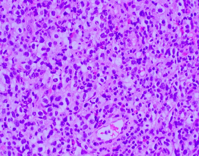
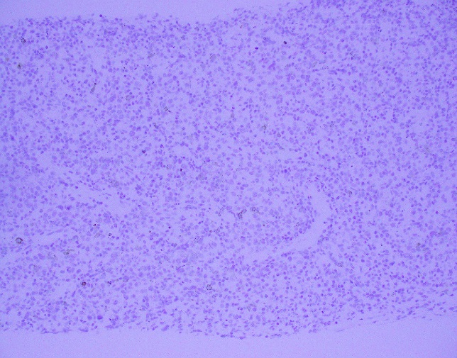
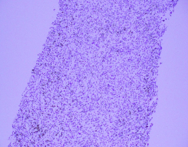
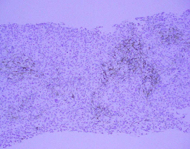
The core needle biopsy material demonstrated a lymphoid population that was polymorphic in appearance with medium to large sized lymphocytes with moderate amounts of pale cytoplasm, irregular nuclei, vesicular chromatin, and some cells with prominent nucleoli. The background cellular population is composed of a mixed inflammatory component including small lymphocytes, scattered neutrophils, eosinophils, and histiocytes.

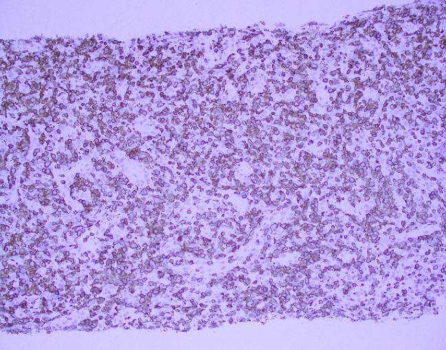
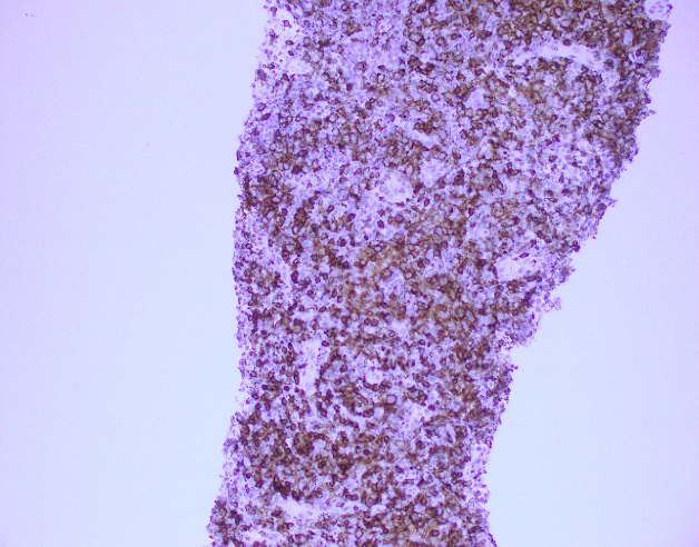
By immunohistochemistry, the medium to large sized cells with pale cytoplasm are positive for CD3, CD2, CD4, and CD5 with complete loss of CD7. CD20 highlights scattered background B-cells. CD21 is positive in disrupted follicular dendritic meshworks. CD10 and BCL6 are negative in neoplastic cells. PD1 is positive in neoplastic cells with a subset co-expressing CXCL13. By Ki-67 immunostaining, the proliferation index is 50-70%. By in situ hybridization for Epstein-Barr virus encoded RNA, a subset of cells are positive.

Overall, with the morphologic and immunophenotypic features present, the diagnosis is that of angioimmunoblastic T-cell lymphoma.

Discussion

Angioimmunoblastic T-cell lymphoma (AITL) is one of the most common types of peripheral T-cell lymphoma and accounts for 15-20% of T-cell lymphoproliferative disorders and 1-2% of all non-Hodgkin lymphomas. Clinical features include presentation with late stage disease with associated generalized lymphadenopathy, hepatosplenomegaly, systemic symptoms, and polyclonal hypergammaglobulinemia. Of note, this patient did have an SPEP that was within normal limits. Other findings, although less common, include effusions and arthritis. Laboratory findings often include cold agglutinins with hemolytic anemias, a positive rheumatoid factor (RF), and anti-smooth muscle antibodies. A hallmark of AITL is the expansion B-cells positive for EBV is seen, which may be an indicator of underlying immune dysfunction. The clinical course is often aggressive with a median survival of less than three years and often succumb to infectious etiologies because of an immune dysregulation.1

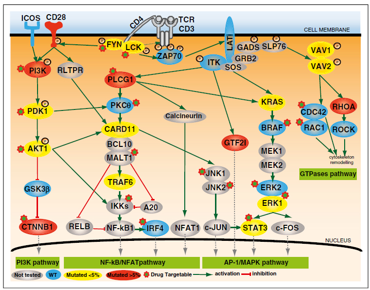
The pathogenesis and relation to other TFH neoplasms of PTCL, NOS is poorly understood. Recent literature indicates dysregulation in key pathways, including the CD28 and TCR-proximal signaling genes, NF-kappaB/NFAT pathway, PI3K pathway, MAPK pathway, and GTPases pathway.2 The complexity of these pathways has long been an issue for TFH lymphoproliferative disorders and has provided insight to potential molecular signatures (see figure 1 adapted from Vallois 2016).

AITL-14
Figure 1 from Vallois 2016

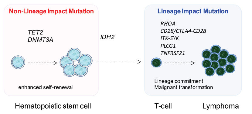
Another recent publication provided additional information regarding molecular insights. Confirmed mutational analyses reveals a high proportion of cases carry a TET2 mutation with less frequent changes in DNMT3A, IDH2, RHOA, and PLCG1. Specifically, RHOA, PLCG1, and TNFRSF21 encode proteins critical for T-cell biology and most likely promote differentiation and transformation into an aggressive clinical course (see figure 2 adapted from Wang 2017).3

AITL-15
Figure 2 adapted from Wang 2017

Overall, AITL is an uncommon TFH cell derived lymphoproliferative disorder characterized by a TFH immunophenotype, expanded and arborizing high endothelial venules, expansion of the follicular dendritic cell meshworks, and EBV positive B-cells in a background of a polymorphic infiltrate. Although it is hypothesized that the underlying mechanism of neoplasia is related to immune dysfunction, new molecular insights have demonstrated that multiple events occur ranging from early molecular changes to later acquired mutations that allow for malignant transformation.

References

  1. Swerdlow, S., et al., WHO Classification of Tumours of Haematopoietic and Lymphoid Tissues, 4th. ed., IARC press: 2008
  2. Vallois, D., et al. “Activating mutations in genes related to TCR signaling in angioimmunoblastic and other follicular helper T-cell-derived lymphomas,” 2016; 128(11): 1490-1502.
  3. Wang, M., et al., “Angioimmunoblastic T cell lymphoma: novel molecular insights by mutation profiling,” 2017; 8(11): 17763-17770.


PhillipBlogPic-small

-Phillip Michaels, MD is a board certified anatomic and clinical pathologist who is a current hematopathology fellow at Beth Israel Deaconess Medical Center, Harvard Medical School, Boston, MA. His research interests include molecular profiling of diffuse large B-cell lymphoma as well as pathology resident education, especially in hematopathology and molecular genetic pathology.

Microbiology Case Study: 42 Year Old Male with Bilateral Lower Extremity Rash

Case History

A 42 year old male presented to the emergency department with a chief complaint of bilateral lower extremity rash. The rash initially began on the dorsum of the patient’s foot, but progressively worsened over the past two weeks. He denied fevers, chills or night sweats. Additionally, he denied any trauma to his legs, burns or exposure to bodies of water. He reported no sick contacts or recent travel and lives at home with his sister and pet dog. His past medical history was significant for squamous cell carcinoma of the oropharynx and alcoholic cirrhosis. On physical exam, he was noted to have extensive cellulitis with the sloughing of skin. Imaging found no acute osteomyelitis or evidence of necrotizing fasciitis. Lab work showed a white count of 17.1 TH/cm2 and elevated ESR and CRP values. He was admitted and started on broad spectrum antibiotics, vancomycin and meropenem. Wound and blood cultures were collected.

Laboratory Identification

pastmult1
Image 1. Gram stain from a positive blood bottle showing Gram negative coccobacilli in pairs (100x oil immersion).
pastmult2
Image 2. Small, grayish colonies grew on blood and chocolate agars after 48 hours incubation in a 35°C incubator with 5% CO2.

The wound culture as well as two blood culture bottles showed growth of the same organism. Gram stain revealed Gram negative coccobacilli that varied from ovoid to short rods. The organism grew on blood and chocolate agars, but not on MacConkey, despite being a Gram negative rod (fastidious pattern). The colonies were non-hemolytic and opaque in appearance. The isolate was positive for catalase, oxidase and indole. MALDI-TOF MS identified the isolates as Pasteurella multocida.

Discussion

Pasteurella spp. are Gram-negative, facultative anaerobic, coccobacilli capable of fermentation. This organism is often found as part of the normal flora of many healthy animals including cats and dogs. P. multocida and P. canis are the most frequently isolated species of the genus. Both of these species are pathogenic in humans. The majority of human infections are wound infections associated with cat (most commonly) & dog bites and scratches. These often result in localized cellulitis and lymphadenitis. Furthermore, rare infections have been reported which include septic arthritis and osteomyelitis, prosthetic joint infection, meningitis, respiratory tract infections, endocarditis, sepsis and bacteremia, and perinatal infections. Systemic infection usually occurs in immunocompromised patients, particularly those with underlying hepatic disease and cirrhosis.

P. multocida is readily recovered by standard media in the clinical microbiology laboratory, growing well on 5% sheep’s blood and chocolate agars, but poorly on MacConkey agar. After overnight incubation on blood agar, small gray colonies with a characteristic musty odor are present. This characteristic musty odor is caused by the production of indole. P. multocida is also oxidase positive and catalase positive. Susceptibility testing for P. multocida from bite wounds in not routinely recommended as these infections most like represent polymicrobial infections and empiric therapy is usually effective. Susceptibility testing should be performed on isolates from normally sterile sites. P. multocida is generally susceptible to penicillin, broad spectrum cephalosporins, tetracyclines, quinolones, trimethoprim-sulfamethoxazole and azithromycin. Resistance has been documented with oxacillin, cephalexin, erythromycin and clindamycin.

In the case of our patient, susceptibility testing was performed by disk diffusion and was susceptible to all the antibiotics listed above with the exception of erythromycin. It was thought he acquired this infection from the family dog licking his feet, with his liver cirrhosis placing him at an increased risk for bacteremia. He was received IV ampicillin/sulbactam for 10 days before being transitioned to an oral regimen for an additional 4 days.


sims

Brooke Sims, MD, is a Cytology Fellow at the University of Mississippi Medical Center.

Stempak

-Lisa Stempak, MD, is an Assistant Professor of Pathology at the University of Mississippi Medical Center in Jackson, MS. She is certified by the American Board of Pathology in Anatomic and Clinical Pathology as well as Medical Microbiology. She is the director of the Microbiology and Serology Laboratories. Her interests include infectious disease histology, process and quality improvement and resident education.

Will Anyone See This Test Result?

We are all aware that there is substantial waste in testing. The mantra of utilization management is “the right test for the right patient at the right time.” This month, I want to focus on the right time. It turns out that many test results are never seen because they arrive after the patient has been discharged. This occurs for both routine and send-out testing. I will examine both.

Turnaround times for send-out testing are generally longer than those for tests performed in house. This means that results for tests ordered toward the end of a hospital stay are likely to be received after the patient has been discharged. Sendout tests are often expensive and, unlike tests performed in house, reducing sendout testing saves the hospital the full charge of the test. The savings can be substantial.

How do you prevent this? A recent article by Fang et al. shows one approach.[1] In this study, conducted at Stanford University, researcher displayed the cost and turnaround time of sendout tests in the computerized provider order entry (CPOE) system and achieved a 26% reduction in orders. I am aware of another hospital that restricts orders of sendout tests when the expected turnaround time is close to the expected remaining length of stay. Consider the graph in Figure 1. The upper panel shows the expected length of stay for a particular patient. The lower panel shows the expected turnaround time for a sendout test. In this case, there is a 62% chance that the test result will arrive after the patient has left the hospital.  Expected discharge dates are routinely kept and it is relatively easy to maintain a database of turnaround times. A hospital could combine these data and set a threshold for orders based on the probability that the result will arrive in time.

Standing orders are another source of waste.  I recently performed an analysis of the test rate as a function of the time until discharge (Figure 2). The test rate was 249 tests per hour for patients who were within 12 hours of discharge and 349 tests per hour for all other patients. It seems odd to me the testing rate in the final 12 hours is 70% of the “normal” testing rate. Further, the distribution of tests in both groups (those about to be discharged vs. all other patients) is very similar (Table 1). The main tests are basic metabolic panels and complete blood counts.  I suspect the majority of the testing within 12 hours of discharge is due to standing orders and the results were not needed for patient care.  The best intervention is less clear in this case because some peri-discharge testing is appropriate and it is difficult to distinguish the appropriate testing from the inappropriate testing. Education is one option. Perhaps the CPOE could raise a flag on orders for patients who are about to be discharged; however, this could be cumbersome and clinicians object to flags and popups that interfere with their workflow. I would be interested in readers’ thoughts on methods to reduce inappropriate peri-discharge testing.

In summary, some results do not reach clinicians in time to affect patient care. This is a source of waste. It is relatively easy to create an intervention to reduce inappropriate sendout testing but more difficult to reduce unnecessary peri-discharge testing.


Reference

  1. Fang DZ, Sran G, Gessner D, Loftus PD, Folkins A, Christopher JY, III, Shieh L: Cost and turn-around time display decreases inpatient ordering of reference laboratory tests: A time series. BMJ Quality and Safety 2014, 23(12):994-1000.


8-2017-fig-1
Figure 1: Comparison of expected length of stay (upper) and turnaround time (lower) for a sendout test.
8-2017-fig-2
Figure 2: Peri-discharge testing
8-2017-tab-1
Table 1: Test patterns stratified by time to discharge. The table shows the percentage of total testing accounted for each group. For example, BMP represents 15% of the total test volume among patients who are within 12 hours of discharge.

Schmidt-small

-Robert Schmidt, MD, PhD, MBA, MS is a clinical pathologist who specializes in the economic evaluation of medical tests. He is currently an Associate Professor at the University of Utah where he is Medical Director of the clinical laboratory at the Huntsman Cancer Institute and Director of the Center for Effective Medical Testing at ARUP Laboratories.


Hematology Case Study: Monocytosis in An Elderly Patient

An 81 year old presented with fatigue and not feeling too well. CBC revealed marked leukocytosis and monocytosis.

  • White cell count: 85.1 K/uL (elevated)
  • Hemoglobin: 8.6 g/dl (decreased)
  • Platelet count: 79 K/uL ( decreased)

Review of peripheral smear revealed leukoerythroblastosis with monocytosis (19.57 K/uL) along with presence of numerous immature monocytoid cells, dysplastic myeloid precursors and 7% blasts, consistent with myelodysplastic/myeloproliferative neoplasm such as chronic myelomonocytic leukemia-1.


cmml1

cmml2


cmml3

For further evaluation of disease progression and/or transformation to acute leukemia, bone marrow evaluation was recommended.

Diagnostic criteria for chronic myelomonocytic leukemia

  1. Persistent peripheral blood monocytosis >1 K/uL
  2. No Philadelphia chromosome or bcr-abl 1 fusion gene
  3. No rearrangement of PDGFRA or PDGFRB
  4. Fewer than 20% blasts in the blood or bone marrow**
  5. Dysplasia in one or more myeloid lineages.


**Blasts include myeloblasts, monoblasts and promonocytes

Prognosis and predictive factors

Survival of patients with CMML is reported to vary from one to more than 100 months, but the median survival time in most series is 2- to 40 months. Progression to AML occurs in approximately 15-30% of cases. A number of clinical and hematological parameters, including splenomegaly, severity of anemia and degree of leukocytosis, have been reported to be important factors in predicting the course of the disease.


Vajpayee,Neerja2014_small
-Neerja Vajpayee, MD, is the director of Clinical Pathology at Oneida Health Center in Oneida, New York and is actively involved in signing out surgical pathology and cytology cases in a community setting. Previously, she was on the faculty at SUNY Upstate for several years ( 2002-2016) where she was involved in diagnostic work and medical student/resident teaching.

Microbiology Case Study: A Routine Sputum Culture on a 20 Year Old Cystic Fibrosis Patient

Case History

A 20 year old woman with cystic fibrosis was routinely screened with bacterial sputum cultures.  The patient reported feeling well and was compliant with her treatment regimen.  She had no history of Nocardia colonization in the past and her last hospitalization was four years prior due to a pulmonary exacerbation in which cultures grew Stenotrophomonas and Pseudomonas.

no1

Image 1: Modified acid fast stain showing two unique traits of Nocardia: long, delicate rods and weak acid fastness.

no2

Image 2: Nocardia colonies on buffered charcoal yeast extract (BCYE) agar.

Discussion

Nocardia are slow growing, aerobic gram negative rods. Nocardia are unique for being weakly acid fast and displaying aerial hyphae.  They are ubiquitous organisms that can cause a variety of infections in immunocompromised individuals. Most often in the United States, nocardiosis causes a lung infection. If left untreated, it can spread to the brain or spinal cord, where up to 44% die [1].

The above patient showed no signs of an infection, making the possibility of colonization by Nocardia more likely.  Cystic fibrosis patients can be colonized by Nocardia and the clinical approach is to treat regardless of the patient’s overall health. However, there is a lack of reporting on whether Nocardia is the cause of an infection when it happens. Host and pathogen interactions are also not well known. Thorn et al. showed that treating cystic fibrosis patients that are colonized with Nocardia with oral antibiotics did not affect their clinical outcome [2]. More studies are needed to be done to see if antibiotics are warranted in circumstances like the above patient.

References:

  1. Nocardiosis. https://www.cdc.gov/nocardiosis/transmission/index.html
  1. Pulmonary nocardiosis in cystic fibrosis. Thorn, Shannon T. et al.. Journal of Cystic Fibrosis, Volume 8 , Issue 5 , 316 – 320


-Angela Theiss is a pathology resident at the University of Vermont Medical Center.

Wojewoda-small

-Christi Wojewoda, MD, is the Director of Clinical Microbiology at the University of Vermont Medical Center and an Associate Professor at the University of Vermont.

Planning Lab Testing for Medical Missions, Part 2

Last month I blogged about key points to consider when preparing to do lab testing in the field. Here I will expand on using point of care testing in medical missions. Point of care testing is easy to use and relatively easy to access, making it very attractive for use in the field or on medical missions. In fact, it is tempting to take these tests and go rogue – it’s not uncommon for point of care diagnostics to be obtained by non-laboratory professionals and tossed in luggage to be used by short-term medical teams. However, this is not in the best interest of the patients or the community. Helping establish point of care testing for medical missions is one very important way that a laboratory professional can get involved in this kind of outreach.

Proper utilization and quality assurance practices are just as critical in the outreach situation as at home in a large lab. Perhaps even more so; for example, in areas with high disease prevalence, false positives and negatives can significantly affect patient care and population health. Under-diagnosis due to false negatives means that those who need treatment might not get it, just as over-diagnosis due to false positives may cause patients to get unnecessary treatment. Unnecessary treatment, especially for infectious diseases, harms the community by contributing to drug resistance.

Most point of care tests, especially lateral flow tests, have built-in controls which lessens the need to run QCs with patient testing. However, it is important to know the limitations of the testing. Sometimes point of care testing systems that are not available in the United States are selected for use in outreach in foreign countries. It’s more likely that an American medical team would be unfamiliar with the tests. A laboratory professional can help establish or at least verify the validity of the tests, including limits of detection and accuracy, before they are deployed. Also, it is often helpful to have the results interpreted for the end user. Little interpretation is needed for the more straightforward qualitative tests that simply give a positive or negative result. Even with these tests, the limit of detection should be available to the provider, especially if this is significantly different from that which the provider is accustomed. Tests that involve titration, such as some of the rapid typhoid and syphilis testing, benefit from having an explanation of what the titers mean clinically available to the end user.

Other tests with results that are prone to confusion are point of care versions of assays more commonly performed in clinical laboratories. Difference in reference intervals for the POCT compared to a conventional test can be particularly confusing. For example, the results of a lateral flow point of care C-reactive protein assay have a different reference interval than results from high-sensitivity C-reactive protein assays used in clinical labs. Using the incorrect reference interval to determine whether a result is normal can lead to over- or under-treatment, which is contrary to the purpose of diagnostic testing. Yet, when using point of care tests in the field, there is not a neat little interpretive comment accompanying the result.

So, how can this be remedied? If the laboratory professional is also on the team, they can be available to provide information as needed. However, if the team is not so fortunate as to have their own laboratory professional, another way to provide the information is to provide a short guide to cheat sheet that briefly explains how to use test results.

Proper utility is also important, especially in areas with high burden of disease or in areas where there is no confirmatory testing. Consider rapid tests for H. pylori. These typically detect antibody to H. pylori, which can be found in up to 70% of asymptomatic populations. The rapid test is of little utility since positive results only indicate the presence of antibody and not necessarily an active infection. Consider using rapid screening tests, such as for HIV, when confirmatory testing is not available. Sometimes a second screening test that employs a different method than the first can be used as a confirmatory test if nothing else is available.

Consider environmental limitations of the testing when selecting tests for use in the field. Many tests are unreliable at extremes of temperature and humidity. This might not always be obvious even when quality controls are used properly. For example, Tang et al (1) showed that the effect of temperatures and humidity similar to what was experienced in Louisiana after Hurricane Katrina on quality control material for a POCT glucose meter system caused significantly depressed results. Also keep in mind that exposure to environmental extremes can reduce the shelf life of POCT and related reagents. If using POCT long term, it is good practice to routinely test a known standard – even on tests with built in quality controls such as the test line on lateral flow tests – to ensure there has not been degradation in quality due to the environment.

Preparing POCT for medical missions is a great way for a laboratory professional to get involved in global health and outreach. From helping to select appropriate tests, to verifying test validity, to teaching proper utilization of testing and providing interpretive guideline, the laboratory professional is a vital part of a medical mission – even if they never leave their lab!

  1. Tang CS, Ferguson WJ, Louie RF, Vy JH, Sumner SL, Kost GJ. Ensuring quality control of point-of-care technologies: effects of dynamic temperature and humidity stresses on glucose quality control solutions. Point of Care 2012;11:147-51.


Sarah Brown Headshot_small

Sarah Riley, PhD, DABCC, is an Assistant Professor of Pediatrics and Pathology and Immunology at Washington University in St. Louis School of Medicine. She is passionate about bringing the lab out of the basement and into the forefront of global health.  

Owning Safety in the Autopsy Suite

The hospital security guard placed the deceased patient into the morgue refrigerator while chatting with his co-worker. They walked away without realizing the door did not close completely. Within the hour the automated temperature recording system sent an alert to the lab on the third floor.

The body had been unclaimed, and it stayed on the bottom shelf in the morgue. No one in the hospital wanted to take ownership of it. After a couple of months, fluids began to fill the shelf where the body was. The environmental services staff refused to clean up the mess since some staff were afraid.

The pathologist wanted to finish the autopsy quickly, so he started before the complete patient chart arrived. When the phone rang in the morgue, the physician on the other end of the phone said he believed the patient may have Creutzfeldt-Jakob Disease (CJD).

Managing safety in the autopsy suite can be difficult, but as these case studies show, it is important. One reason for the struggle is that clear ownership of the area is often not defined. Multiple internal departments and even external agencies may work in the morgue and autopsy suite. Pathologists, medical examiners, research physicians, security personnel, nurses, and organ procurement staff are just some of the various people that may perform tasks in the autopsy suite. This can create some unique and unwanted problems. The laboratory should take the lead in making sure all safety regulations are followed and that other users of the suite comply to avoid any unfortunate mishaps.

The morgue should be treated as a laboratory space, and it should be designed similarly to a BSL-3 laboratory space which includes an anteroom. Warning signs indicating the presence of biological and chemical materials should be placed on entry doors. Whenever work is performed in the area, proper personal protective equipment should be utilized. This PPE may include lab coats, gowns, gloves, respirators, and face protection. Make sure PPE is available in the area at all times. The autopsy space should be adequate, such that procedures may be performed effectively and that items such as knives and saws can be stored and used safely. Ventilation should be adequate (with a recommended minimum 12 air exchanges per hour), and the ambient temperature should be monitored as well.

While other personnel may access the morgue body storage refrigerator, it is often the lab or security departments who monitor the temperature. Since CAP inspectors set specific morgue refrigerator temperature ranges (1.1 to 4.4° Celsius), it can be important to communicate with the people who utilize the unit often. If placing or removing a body takes longer than expected, make sure there is adequate communication so that proper documentation of the temperature outages can be made. If a department other than the lab is responsible for temperature monitoring, make sure it is done correctly so there are no citations during an inspection.

Proper decontamination in the morgue is crucial. Instruments, tables, and counters must be disinfected to remove contamination of bloodborne pathogens. Use a chemical germicide for instrument and surface decontamination such as a 10-percent solution of sodium hypochlorite (or bleach). This intermediate-level disinfection will eliminate most bacteria (including Mycobacterium tuberculosis), and all fungi, and it inactivates viruses such as the hepatitis B virus. Rinsing with water or ethanol after disinfecting will help prevent the pitting of any stainless-steel surfaces.

Dealing with Creutzfeldt-Jakob Disease (CJD) in the autopsy suite requires special safety measures. Procedures should be posted in the area directing staff how to handle tissue and clean up in cases where patients are infected with CJD. The intact brain should be fixed in formaldehyde for one to two weeks before handling or cutting in order to reduce the prion activity. Non-disposable implements used with such patients should be immersed in 1N sodium hypochlorite (NaOH) for one hour before reuse. Surfaces on which autopsies occurred should also be immersed in NaOH for one hour for disinfection purposes.

Chemicals are stored and used in the autopsy suite, and standard safe lab practices should be used. Make sure staff is trained in proper the handling, labeling, and storage of chemicals as well as prepared to handle spills. Spill kits should be available and suitable to the chemicals used in the area. If formaldehyde is used, be sure an appropriate neutralizer is available for spill incidents.

As the most involved and best educated about its dangers, laboratory personnel should take the lead in making sure safety is a priority in the morgue, and educate all who may enter the area. Make sure communication is clear about who will use the suite and when- it’s never good to have someone walk in during an autopsy or organ removal. Use signage when necessary, and be willing to help in any unusual situations, because with a morgue, they definitely will arise. Work together as a team with all who utilize the area, and that ownership of safety will translate into safety for all.


Scungio 1

Dan Scungio, MT(ASCP), SLS, CQA (ASQ) has over 25 years experience as a certified medical technologist. Today he is the Laboratory Safety Officer for Sentara Healthcare, a system of seven hospitals and over 20 laboratories and draw sites in the Tidewater area of Virginia. He is also known as Dan the Lab Safety Man, a lab safety consultant, educator, and trainer.

Chemistry Case Study: Falsely Elevated Methotrexate

High dose methotrexate infusion is widely used in the treatment of malignancies such as leukemia, high risk lymphoma, and osteosarcoma. It can be associated with multiple adverse effects, especially renal toxicity, which could leads to acute kidney injury (AKI), delaying drug elimination and worsening its toxicity. Leucovorin, a reduce folic acid, is commonly used with methotrexate treatment to lessen its toxicity. After administration of methotrexate, serum creatinine and methotrexate concentration should be closely monitored. The levels of serum methotrexate to be associated with a high risk for nephrotoxicity are: 24 h, > 10 μmol/L; 48 h, > 1 μmol/L; 72 h, > 0.1 μmol/L.

In this case, the patient is a 33-yo old male with T-lymphoblastic leukemia in complete remission. He was given consolidation therapy with high dose methotrexate. Leucovorin rescue was given 24 hours after methotrexate administration. Patient’s methotrexate level was at 4.7 μmol/L 3 days postinfusion due to AKI and poor methotrexate clearance. An alternative rescue, glucarpidase (Garboxypeptidase G2), was then given to patient to rapidly lower serum methotrexate level. Glucarpidase cleaves methotrexate molecule to inactive metabolite, DAMPA (2,4-diamino-N-methylpteroic acid). After glucarpidase rescue, patient’s methotrexate level were still remained above the toxic level on the following two days (1.02 μmol/L and 0.68 μmol/L).

In most clinical laboratories, serum methotrexate is measured by immunoassays, and the inactive metabolite of methotrexate after glucarpidase rescue, DAMPA, cross-reacts with immunoassays and interferes the measurement of methotrexate. After glucarpidase treatment, patient’s methotrexate level can be falsely high for 5-7 days, before accurate measurement can be obtained using immunoassays. In this case, the concentrations of methotrexate after glucarpidase rescue were falsely high results due to DAMPA interference. There are laboratory-developed LC-MS methods to detect methotrexate. LC-MS methods are more specific and have no interference from the metabolite, can be used for accurate methotrexate measurement in the case of glucarpidase rescue.


Xin-small

-Xin Yi, PhD, DABCC, FACB, is a board-certified clinical chemist, currently serving as the Co-director of Clinical Chemistry at Houston Methodist Hospital in Houston, TX and an Assistant Professor of Clinical Pathology and Laboratory Medicine at Weill Cornell Medical College.

Microbiology Case Study: An Elderly Man with Abdominal Pain

Case History

The lab received two sets of admission blood cultures from an elderly man with a history of atrial fibrillation, aortic valve replacement six years ago, and idiopathic pancytopenia who presented with abdominal pain and symptoms of decompensated heart failure.

Lab Identification

At 59 hours, one aerobic bottle showed growth of a gram negative bacillus. The organism grew pure yellow-pigmented colonies on the blood and chocolate plates with no growth on the MacConkey plate.

breves4
Gram stain from blood agar plate.

The Verigene microarray was not able to identify the organism against 12 gram negative targets. The organism was identified by MALDI-TOF mass spectrometer as Brevundimonas vesicularis. The organism could not be grown for susceptibility testing.

Discussion

B. vesicularis is an aerobic, glucose non-fermenting, spore non-forming, gram negative bacillus closely related to Pseudomonas species. It will demonstrate slow growth of yellow-pigmented colonies on chocolate and blood agar plates with variable growth on MacConkey agar plates. It has in the past been classified as Corynebacterium and Pseudomonas vesicularis. It has been identified globally in environmental sources such as soil, spring water, and healthcare related water containers. It has been identified as a community-acquired, and increasing a hospital-acquired, pathogen in all age groups, and particularly in immunocompromised patients [1, 2]. It has been most commonly reported as a cause of bacteremia, but has also been implicated as the causative agent in cases of arthritis, meningitis, endocarditis, peritonitis, and urinary tract infection [1]. Interesting in vitro studies have demonstrated that the presence of B. vescularis may facilitate the growth of Legionella [3].

Even in cases of the septicemia in immunocompromised patients the mortality rate from B. vesicularis is relatively low. Strains have demonstrated resistance to most forms of antibiotics though is most consistently susceptible to amikacin and piperacillin-tazobactam [1, 2].


References

  1. Shang ST, Chiu SK, Chan MC, Wang NC, Yang YS, Lin JC, Chang FY. Invasive Brevundimonas vesicularis bacteremia: Two case reports and review of the literature. J Microbiol Immun Inf. (2012) 45: 468-472.
  2. Zhang CC, Hsu HJ, Li CM. Brevundimonas vesicularis bacteremia resistant to trimethoprim-sulfamethoxazole and ceftazidime in a tertiary hospital in southern Taiwan. J Microbiol Immunol Infect. (2012) 45(6): 448-452.
  3. Koide M, Higa F, Tateyama M, Cash HL, Hokama A, Fujita J. Role of Brevundimonas vesicularis in supporting the growth of Legionella in nutrient-poor environments. New Microbiol. (2014) 1:33-39.


-Taylor Goller is a Pathology Student Fellow at University of Vermont Medical Center.

Wojewoda-small

-Christi Wojewoda, MD, is the Director of Clinical Microbiology at the University of Vermont Medical Center and an Associate Professor at the University of Vermont.

Jaffe vs. Enzymatic Method for Serum Creatinine Measurement

The Jaffe and enzymatic methods are the two most common methods for measuring serum creatinine. The Jaffe method is less expensive than the enzymatic assay ($0.30 vs $2.00 per test based on 2014 list prices) but is more susceptible to interferences. Although these tests are not expensive, they are high-volume tests and the savings could be substantial. We were using the enzymatic assay at the University of Utah and estimated that we could save about $50,000 per year by switching to the Jaffe assay; however, we were uncertain whether the Jaffe assay was safe to use due to the potential for interferences. For that reason, we decided to conduct a risk assessment to evaluate the suitability of the Jaffe assay.

Risk is defined as the expected cost of an action. The expected cost has two components: 1) the probability that an event will occur and 2) the consequences or cost of an event:

Risk = prob(event) x cost(event)

The event of interest was misclassification of a patient due to an error in serum creatinine measurement. Nephrologists classify kidney disease based on the estimated glomerular filtration rate which is based on the creatinine value. The distribution of eGFR for patients at our hospital is shown in Figure 1. The dashed lines show decision limits that nephrologists use to classify kidney disease. An eGFR is considered normal or healthy.

We spoke with the nephrologists and learned that they were relatively unconcerned about errors in eGFR in healthy patients (eGFR above 60 ml/min) because there was no potential for harm. Similarly, they felt there was relatively little risk of harm to patients with low eGFRs because these patients are routinely monitored and no major decision would be based solely on a single eGFR measurement. An error in creatinine measurement in a low eGFR patient would be detected by repeat measurements or be inconsistent with other measurements. From the nephrologists’ point of view, the only area of concern was in the region around 60 ml/min.  Patients about 60 ml/min are considered healthy whereas those below 60 ml/min are diagnosed with stage 3a chronic kidney disease. In this zone, an error in serum creatinine could result in a false negative (i.e. observed eGFR greater than 60 ml/min when the true eGFR was less than 60 ml/min). In such cases, a patient may go without care and their disease could progress.  The nephrologists believed that the potential for harm was relatively minor, but potential for harm did exist.

We compared the eGFRs provided by the enzymatic and Jaffee methods to estimate how often patients might be misclassified (Figure 2).1 Focusing on the 60 ml/min decision limit, we found that 17 of 500 (3.4%) of measurements were discordant. Some of these discordant results would be due to imprecision. Discordance due to imprecision would have small differences (bottom of Figure 2) and are unavoidable – they would occur using any method. Discordance due to interference would be expected to have larger differences (top of Figure 2) and could be avoided by using the enzymatic method. We used statistical techniques to estimate the proportion of discordances that were due to interference vs imprecision and found that about 60% of the discordance at the 60 ml/min limit was due to interference. In summary, our risk analysis showed that using the Jaffe method would pose about a 2% rate of avoidable misclassification which presented some potential risk to patients. The nephrologists felt the risk was low but, in theory, disease could unnecessarily progress in a patient with a false negative diagnosis.

Our risk analysis was based on analytical error. We compared magnitude of analytical error to the biological variation in eGFR and found that the analytical error was relatively small in comparison to biological variation (data not shown).  Biological variation was likely to be a more significant cause of misclassification than analytical error.

So, what to do? Was the potential savings of the Jaffe method worth the risk? Some experts recommend against using the Jaffe method. 2-4 On the other hand, most US laboratories use the Jaffe assay. A recent College of American Pathologists proficiency challenge found that 70% of the submitted results were based on the creatinine assay.5

We decided to get the best of both worlds by using BOTH methods. We defined a zone of risk surrounding the 60 ml/min eGFR decision limit (Figure 3). Results in this zone would have some risk of misclassification whereas results outside of the zone would be unlikely to be misclassified using the Jaffee method. All creatinine measurements are initially performed using the Jaffe method. If the result is outside the risk zone, the result is reported. If results fell within the risk zone, they were repeated with the enzymatic method and the results of the enzymatic method are reported. This reflex procedure saves money while avoiding risk. The reflex rate is approximately 15%.

There are circumstances in which one would want to order the best possible test. To that end, we created a special orderable test, based on the enzymatic method, that the nephrologists could use to insure the most accurate results when required. For example, the enzymatic test may be indicated when making decisions regarding biopsies for renal transplant patients. The order volume for the special test has been less than 100 orders per year. 

creat1
Figure 1. Distribution of Estimated Glomerular Filtration rates (eGFR). The distribution is for outpatients at University of Utah for calendar year 2014. The dashed lines indicate decision limits used for classification of chronic kidney disease (15, 30, 45 and 60 ml/min). eGFRs greater than 60 ml/min are considered disease free.
creat2
Figure 2. Discordances in estimated glomerular filtration rate (eGFR) at the 60 ml/min decision limit. The length of each arrow, represents the difference between estimates based on the Jaffe (head) and enzymatic (tail) methods. The dashed line represents two standard deviations of expected imprecision of the difference. Differences greater than 2 standard deviations would most likely be due to analytical interference (loss of specificity).
creat3
Figure 3. Reflex test strategy. The figure shows the distribution of eGFR values for outpatients at the University of Utah.  The dashed lines represent clinical decision limits. The yellow zone represents the range of eGFR values where misclassification could pose a risk to patients. Creatinine is first measured by the Jaffe method. The Jaffe result is reported if the estimated eGFR is outside the yellow zone. If the eGFR is within the yellow zone, the measurement is repeated using the enzymatic method and the result based on the enzymatic method is reported.

References

  1. Schmidt RL, Straseski JA, Raphael KL, Adams AH, Lehman CM. A Risk Assessment of the Jaffe vs Enzymatic Method for Creatinine Measurement in an Outpatient Population. PloS one. 2015;10(11):e0143205.
  2. Cobbaert CM, Baadenhuijsen H, Weykamp CW. Prime time for enzymatic creatinine methods in pediatrics. Clinical Chemistry. 2009;55(3):549-558.
  3. Drion I, Cobbaert C, Groenier KH, et al. Clinical evaluation of analytical variations in serum creatinine measurements: Why laboratories should abandon Jaffe techniques. BMC Nephrology. 2012;13(1).
  4. Panteghini M. Enzymatic assays for creatinine: time for action. Scand J Clin Lab Invest Suppl. 2008;241:84-88.
  5. College of American Pathologists. Chemistry/Therapeutic Monitoring, Participant Survey. 2014.


Schmidt-small

-Robert Schmidt, MD, PhD, MBA, MS is currently an Associate Professor at the University of Utah where he is Medical Director of the clinical laboratory at the Huntsman Cancer Institute and Director of the Center for Effective Medical Testing at ARUP Laboratories.