Microbiology Case Study: Middle Aged Man with Altered Mental Status

Case History

A middle aged male presented to the emergency department with a several day history of altered mental status, insomnia, and lethargy. His family stated that he also had possibly had a seizure. Upon arrival to the emergency department he was febrile to 102°F, hypoxic, but denied shortness of breath, cough, nausea, and vomiting. While in the care of the emergency department he had several witnessed seizures requiring Ativan treatment, so the decision was made to admit the patient for 24-hour EEG monitoring. Past medical history was unremarkable. The patient is a smoker. His hobbies include working as a mechanic and working outdoors in his garden.

Initial brain imaging on MRI demonstrated cortical thickening/gyral swelling with associated signal abnormality within bilateral mesial temporal lobes that is seen with mild associated diffusion abnormality. A lumbar puncture was ordered and his CSF analysis demonstrated normal glucose (73 mg/dL [reference range: 40-80 mg/dL]), normal protein (32 mg/dL [reference range: 15-45 mg/dL]), and normal nucleated cells [reference range: 0-8/mm3]. Bacterial, fungal, and AFB culture as well as PCR for herpes simplex virus and varicella zoster virus were ordered. The CSF bacterial, fungal, and AFB cultures showed no growth of any microorganisms, and PCR results were negative for HSV and VZV. What additional infectious etiologies would you like to test for?

Discussion

Serology studies were ordered which demonstrated positive IgM and IgG antibodies for West Nile virus.

West Nile virus is a member of the Flavivirus genus. It is spread through mosquito bites, and birds are the primary reservoir for this virus. The incubation period for West Nile virus is 4-10 days. Typically, about 80% of those infected with the virus will be asymptomatic. In 20% of cases, the patient will develop a febrile illness, with possible symptoms of headaches, body aches, weakness, joint pain, and fatigue. About 1 in 150 of those infected will develop illness involving the central nervous system. In these cases, symptoms can include high fever, headache, neck stiffness, confusion, seizures, and coma. Death occurs in 10% of those with involvement of the central nervous system. The most important risk factor for death is age with patients over 70 years of age being most at risk.

Diagnosis of West Nile virus generally made by detection of IgM and IgG in the serum and/or CSF. IgM can be detected 3-8 days post symptom onset and remains positive for 2-3 months in the CSF, and occasionally longer in the serum. Diagnosis is made by detection of IgM antibodies or conversion of IgG antibodies, while detection of IgG antibodies in isolation indicates a prior infection. False-positive results can occur in CSF specimens contaminated with blood that is IgM antibody positive. Patients who have been infected with or vaccinated against other flaviviruses can have false-positive serum antibodies. Plaque reduction neutralization test (PRNT) assay can be performed to rule out cross-reactivity with other flaviviruses. RT-PCR testing on CSF is also available for detection of West Nile virus RNA. The sensitivity of CSF RT-PCR is low in immunocompetent patients, but increased in immunocompromised patients due to prolonged viremia.

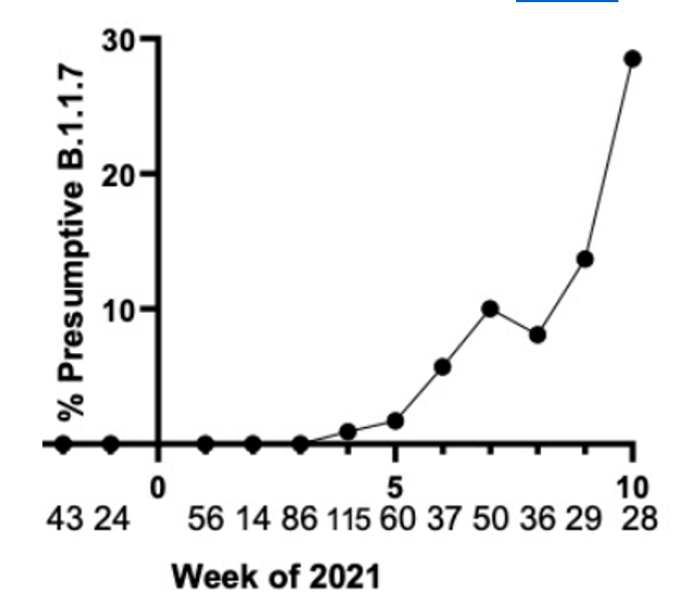
West Nile virus cases could increase in the future as a consequence of climate change. Currently, most cases of West Nile virus occur between June and September and cases have been reported in all 48 lower states. As climate change progresses, average temperatures are expected to increase around the world. As temperature increases, summers become longer, which means a longer season for mosquitoes. Laboratory studies also demonstrate that the virus replicates faster in warmer temperatures. Precipitation patterns are also expected to change in the future. The effect of precipitation is not as clear as that of temperature, but it can still play a role. In areas with more precipitation, increasing amounts of water could provide new breeding grounds for mosquitoes. On the other hand, extra rain can dilute some of the nutrients that the mosquitoes need. In areas with less precipitation, drought conditions can cause rivers to dry up, creating the pools of standing water that give rise to mosquitoes. In addition, as bodies of water become smaller, birds and mosquitoes will be in closer proximity, facilitating faster spread of the virus. We must be prepared for the possibility that West Nile virus cases may be on the rise in the future.

The patient continued to suffer from seizures and required continuous sedation with phenobarbital. For seizure prophylaxis he was given a combination of Keppra and Vimpat, with the dosage adjusted as needed. Throughout his entire hospital course he was monitored on EEG. For the first week of his stay, his EEG results demonstrated seizure like activity, requiring continued use of anti-seizure medication and sedation. After about one week, his seizure activity began to show improvement, and the process of decreasing his medication had begun.

After several days of improvement, his recover was complicated by abdominal compartment syndrome requiring laparotomy. After this, the patient’s seizure activity began to worsen. Within a few days, the patient became hypotensive, and broad spectrum antibiotics were given to protect against infection. Cultures were taken but no infection was identified. The patient ultimately developed shock and passed away.

References

  1. Paz, S. (2015). Climate change impacts on West Nile virus transmission in a global context. The Royal Society. doi:https://royalsocietypublishing.org/doi/10.1098/rstb.2013.0561
  2. Murray, P. R., Rosenthal, K. S., & Pfaller, M. A. (2021). Togaviruses and Flaviviruses. In Medical microbiology. Edinburgh: Elsevier.
  3. Miller, J. Michael, et al. “A guide to utilization of the microbiology laboratory for diagnosis of infectious diseases: 2018 update by the Infectious Diseases Society of America and the American Society for Microbiology.” Clinical Infectious Diseases 67.6 (2018): e1-e94.

-Robert Toelke, MD is a 1st year clinical and anatomic pathology resident at University of Chicago (NorthShore).

-Erin McElvania, PhD, D(ABMM), is the Director of Clinical Microbiology NorthShore University Health System in Evanston, Illinois. Follow Dr. McElvania on twitter @E-McElvania. 

Keeping Abreast of the Times

Hello again everyone and welcome back! Pardon the absolutely cheesy title, but this month I’d like to share parts of a clinical case and discuss some pretty important topics in pathology practice in 2021. While last month’s brief primer on the Cures Act is a definite part of the future of medicine, there are a lot of things developing in our profession—and we should all keep up!

*** Disclaimer: Please note that if/when I share cases or clinical content, they are not current, already signed out, completed and done in compliance full patient privacy guidelines. Always. ***

So let’s dive right in. A rather young male patient was sent to us from an outside facility for mildly painful, uncomfortable bilateral breast lumps. He was noted to have bilateral gynecomastia clinically and was to be evaluated via mammogram and localized needle biopsy. An ultrasound guided core needle biopsy of the left breast was collected from a mass identified on mammography.

Figure 1. Not the typical visual presentation noted on mammography. Biopsy clip marking the location of where the core biopsies were obtained. Male patients can have histologically identical cases of breast carcinoma when compared to females and are often associated with germline mutations, high-risk BRCA1 & 2 status, and Klinefelter syndromes.

Most commonly, when evaluating gynecomastia in a male and considering the diagnostic algorithms including malignancy one worries most about ductal carcinoma: invasive, unspecified type and ductal carcinoma in situ (DCIS) pretty much round off the differential for male breast cancer. Important rule outs include clinical, hormone-related gynecomastia or otherwise relating to germ-line or chromosomal abnormalities (i.e. XXY in Klinefelter), myofibroblastoma, or unrelated distant metastases. So without too much clinical information or history—since this was an outside referral—the differential of potential considerations was pretty short therein.

The biopsy was completed shortly after mammography and was signed out as: fibroadenoma.

Wait. What?

Fibroadenomas are benign and, along with potentially malignant phyllodes tumors, come from mammary tissue, demonstrate biphasic cellular components of benign epithelium (or low-grade stroma).

Translation: to have one of these entities you need mammary tissue, specifically terminal ductal/lobular units to form the necessary architecture changes.

Simpler translation
: fibroadenomas need breast tissue to be fibroadenomas.

Okay, there definitely appears to be breast tissue here to form this entity, however, it is incredibly rare for a male patient (outside of the previously mentioned conditions) to have anatomically functional lobular units. With the other etiologies ruled out, further investigation into the patient’s history was warranted. It was discovered that this patient, although male by indication on referring paperwork, actually identified as female and was on gender affirming hormone therapy for several years. A considerably simple explanation for the confounding biopsy result.

Transgender medicine and transgender pathology considerations have been talked about more in recent years than ever before. While these patients have always existed, societal changes in the last decade alone have brought to light many disparities in medical care and practice habits overall. Here on Lablogatory, there have been between 20-30 mentions of transgender patients since 2018 with entire pieces dedicated to transgender endocrinology, clinical chemistry, etc. Specific examples have included the complicated intersection of transgender care and electronic medical records, various lab values in trans patients, and an excellent overview of laboratory medicine and trans patients by Dr. Jeff SoRelle. There he discusses approaches, literature, and—most importantly—the purposeful vocabulary of transgender medicine as an empowering and informative practice.


Image 2. A 2016 paper in Laboratory Medicine highlights this patient population’s challenges to quality and equity in healthcare and establishes that gatekeepers to data—including us—should consider new ways to create inclusive laboratory medicine practices to maintain our high standard of care. Read it here.

While, this case was rather straightforward after the discovery of the patient’s history of gender affirming therapy, there are still several critical points to highlight here. First, it’s critically important in the setting of medical care to correctly and compassionately include gender identity in patient records—within the caveat of appropriate confidentiality and patient autonomy. Patient care, health management, and diagnostic efforts like ours may rely on a simple biological indication of how this patient presents and experiences their identity with relation to their clinical course. This further strengthens relationships between trans patients and their primary care providers, as well as non-patient-facing specialists like us who often put pieces of the puzzle together without an entire history at our disposal. How does this impact patient care? Simple: a trans male patient may always carry a residual indolent risk of breast cancer, while a trans female patient may need to be included in future mammogram screening. The bottom line should consider treating who you have, and screening with what you have to. Since this patient was sent because of breast lumps, as a male patient a biopsy report of “fibroadenoma” would be exceedingly rare and confounding because of normal physiology outside of specific genetic conditions. But with the right information at the right time, better patient-centered care is possible.

Finally, as a note about proper terminology, vocabulary, and available guidelines in the literature, there is both a paucity of effective transgender medical guidelines as well as a poor grasp on the terminology. Dr. SoRelle does a fantastic job with a short vocabulary outline, but we should all do better. The experiences of our trans patients is challenging to say the least, so as we continue to become stronger and closer advocates for patients in pathology and laboratory medicine, we must become familiar with their experience and advocate for the progress of inclusive medical care.

Trans patients are special, and we are specialists.

Making sure they received the highest level of care alongside all our other patients should be guaranteed. Whether in our clinical chemistry labs or surgical pathology sign outs, we already know how to create an environment of accuracy and enduring inclusion for all our patients—let’s keep it going!

Thank you for reading, I’ll see you next time!

Constantine E. Kanakis MD, MSc, MLS(ASCP)CM is a first-year resident physician in the Pathology and Laboratory Medicine Department at Loyola University Medical Center in Chicago with interests in hematopathology, transfusion medicine, bioethics, public health, and graphic medicine. He is a certified CAP inspector, holds an ASCP LMU certificate, and xxx. He was named on the 2017 ASCP Forty Under 40 list, The Pathologist magazine’s 2020 Power List and serves on ASCP’s Commission for Continuing Professional Development, Social Media Committee, and Patient Champions Advisory Board. He was featured in several online forums during the peak of the COVID pandemic discussing laboratory-related testing considerations, delivered a TEDx talk called “Unrecognizable Medicine,” and sits on the Auxiliary Board of the American Red Cross in Illinois. Dr. Kanakis is active on social media; follow him at @CEKanakisMD.

Microbiology Case Study: A Patient, A Toilet, and a Worm

Case History

A patient presents with a worm they found in the toilet.

Image 1. The offender.
Image 2. Mouth parts of the worm.

Discussion

This is Ascaris lumbricoides, a roundworm. Distinctive morphologic features include tapered ends, mouthparts consisting of three prominent lips (pictured in image 2), and a length of up to 35cm for females. The adults live in the duodenum and proximal jejunum. The eggs have an irregular external mamillated outer shell that gives them a roughened outer surface. Clinically, infection can range from asymptomatic to severe disease, in which the larvae, hatched from ingested eggs, migrate from the small bowel through the circulatory system to the lungs, where they mature in the alveolar capillary bed and cause Ascariasis pneumonitis (Löffler syndrome).

Other diagnostic considerations include Enterobius vermicularis (pinworm), Lumbricus terrestris (earthworm), Trichuris trichiura (whipworm), and the hook worms, Necator americanus and Ancylostoma duodenale.

Enterobius vermicularis, the pinworm, is the most common helminth infection in the United States. Clinically, the classic presentation is a child with pruritus ani. Females measure up to 1.3 cm in length and have a pointed posterior end, and both sexes have lateral alae and a prominent esophageal bulb. The worm in this case is far too large to be a pinworm.

The earthworm, Lumbricus terrestris, is soil-dwelling and non-pathogenic but occasionally encountered in the laboratory for identification purposes. Key morphologic features include a segmented body with no distinctive mouthparts and a clitellum (a mating organ that is a non-segmented portion of the body and often a different color from the rest of the body).

Trichuris trichiura, the whipworm, have a classic whip-like appearance with long, narrow anterior ends that anchor the worm to the large intestine, where they can remain for up to 10 years. Both males and females measure up to 5.0 cm in length, and diagnosis is often made by identification of the eggs, which are football-shaped and have polar plugs at both ends. Clinically, trichuriasis can cause dysentery-type symptoms and, in heavily infected children, can lead to rectal prolapse.

Necator americanus and Ancylostoma duodenale are the hookworms. Adult females measure up to 1.2 cm, and these two species are differentiated by examination of the mouthparts: Necator americanus has cutting plates, while Ancylostoma duodenale has cutting teeth. In addition to the large size difference between hookworms and roundworms, the lamprey-like appearance of these mouthparts is notably different from the “fleshy lips” of Ascaris. Hookworms and roundworms, however, are similar in that their larvae have the ability to migrate through tissue to the blood stream then the lungs, where they can cause Löffler syndrome and are expectorated then swallowed before reaching the small bowel. Unlike Ascaris, the larvae of which hatch from ingested eggs and penetrate the host through the bowel wall to get to the lung capillary beds where they can mature, hookworm larvae hatch outside the body and, on contact with a host (once again, lamprey-style), directly penetrate the skin, enter the circulation, travel to the lungs, then migrate up the bronchial tree to be swallowed. If ingested, Ancylostoma larvae can mature into adults in bowel without needing to migrate through the lungs.

References

  1. Centers for Disease Control and Prevention. “Stool Specimens – Intestinal Parasites: Comparative Morphology Tables.” https://www.cdc.gov/dpdx/diagnosticProcedures/stool/morphcomp.html. Last reviewed May 3, 2016. Accessed April 2, 2021.
  2. “Earthworms.” University of Pennsylvania. https://www.sas.upenn.edu/~rlenet/Earthworms.html. Accessed April 2, 2021.
  3. McPherson, R, and M Pincus. (2011). Henry’s Clinical Diagnosis and Management By Laboratory Methods (22nd Edition, pp. 1218-1220). Philadelphia, PA: Elsevier Saunders.

-Frederick Eyerer, MD is a 3rd year anatomic and clinical pathology resident at the University of Vermont Medical Center.

-Christi Wojewoda, MD, is the Director of Clinical Microbiology at the University of Vermont Medical Center and an Associate Professor at the University of Vermont.

Pathology and laboratory services in low to middle income countries: Challenges and Opportunities for Growth

Successful healthcare systems rely on strong, efficient and reliable pathology and laboratory services. In developed countries like the United States, the role of pathology and the lab in service delivery cannot be overemphasized. Through multidisciplinary tumor board conferences, the central role of pathologists in patient management has become more critical. This reality was further emphasized for me during a tumor board conference, where an oncologist wanted clarification on the difference between invasive ductal carcinoma with mucinous differentiation vs. mucinous carcinoma of the breast. I later learned that the distinction was necessary because of different prognosis and treatment. As pathologists, we work in concert with treating physicians which makes for more efficient and reliable patient care.

However, the situation is not the same in low to middle income countries (LMIC) where the pathology and laboratory workforce is currently suboptimal. One reason that has been attributed to this situation is a lack of awareness of the central role of pathology and lab medicine in developing countries with fragile health care systems. This has led to ineffective policy decisions and inadequate budgetary allocations to the lab, with the attendant catastrophic effects on patient care and outcomes.1

West Africa for example has seen a steady decline in healthcare delivery standards, even though diseases such as Burkitt lymphoma were first described in Africa, followed by the growth of the first human lymphoma/leukemia cell line (the Raji cell line).2 In addition, ‘Cancer in Five Continents’, a publication of the International Agency on Cancer Research, used data from Ibadan-Nigeria and Uganda cancer registries in its early years of publication. Unfortunately, during the years the countries in these regions have not been able to keep up with technologic advances that have since reshaped healthcare service delivery and research.3 Several barriers to sustainable pathology and laboratory services in LMIC have been identified including an inadequate workforce, substandard infrastructure, inadequate education and training, and quality assurance problems.1


Photo by National Cancer Institute on Unsplash

Despite these challenges, there are opportunities to improve healthcare delivery systems in LMIC through effective laboratory and pathology services. One area that needs to be prioritized is the education and training of qualified pathology and laboratory personnel. This can be achieved through cross-cultural competency training and the building of collaborative networks through short term visitor exchange programs. In addition, continuing medical education (CME) opportunities should be made available to training institutions in these countries so they can keep up with modern day standards.

Another opportunity for growth in pathology and lab services in LMIC is through the implementation of accreditation and regulatory programs. These accreditation services should set standards by which lab services operate in these countries to ensure reliable and consistent operations. Such efforts may improve health service deliveries and ultimately improve patient outcomes.

One factor that has been a huge problem in disease prevention in LMIC is lack of adequate screening programs for chronic diseases, including several cancers. In many countries, misplaced priorities, in addition to lack of adequate personnel has been the bane of the healthcare systems. Therefore, policies that promote screening programs in LMIC should be prioritized. This strategy if implemented properly could lead to significant improvements in the healthcare systems, which would ultimately have an impact on patient care.

Furthermore, collaborative healthcare should be prioritized. The care and management of patients should be done collaboratively through clinicians across different specialties with proper communication channels in place. There have been instances where a clinician treating a patient may not have access to laboratory results requested by another clinician, which ultimately impacts the outcome of patients.

The role of efficient and functional laboratory and pathology services in healthcare systems cannot be overemphasized. Systems which lack these services experience catastrophic patient outcomes and until local and international governments prioritize the labs in these low-resource settings, patient outcomes will continue to remain suboptimal.

References

  1. Sayed S, Cherniak W, Lawler M, Tan SY, El Sadr W, Wolf N, Silkensen S, Brand N, Looi LM, Pai SA, Wilson ML, Milner D, Flanigan J, Fleming KA. Improving pathology and laboratory medicine in low-income and middle-income countries: roadmap to solutions. Lancet. 2018 May 12;391(10133):1939-1952. doi: 10.1016/S0140-6736(18)30459-8. Epub 2018 Mar 15. PMID: 29550027.
  2. Pulvertaft JV. Cytology of Burkitt’s Tumour (African Lymphoma). Lancet. 1964 Feb 1;1(7327):238-40. doi: 10.1016/s0140-6736(64)92345-1. PMID: 14086209.
  3. Adeyi OA. Pathology services in developing countries-the West African experience. Arch Pathol Lab Med. 2011 Feb;135(2):183-6. doi: 10.1043/2008-0432-CCR.1. PMID: 21284434.

-Evi Abada, MD, MS is a Resident Physician in anatomic and clinical pathology at the Wayne State University School of Medicine/Detroit Medical Center in Michigan. She earned her Masters of Science in International Health Policy and Management from Brandeis University in Massachusetts, and is a global health advocate. Dr. Abada has been appointed to serve on the ASCP’s Resident’s Council and was named one of ASCP’S 40 under Forty honorees for the year 2020. You can follow her on twitter @EviAbadaMD.

How to Detect COVID-19 Variants of Concern

It’s a little deja-vu writing this title one year after a similar blog post on how to validate a COVID-19 assay at the start of the pandemic. In many ways, the challenges are similar: limited reagents/control material, and rising case counts. At least now, there is increasing support in the way of funding from the federal government that could help with monitoring and surveillance. I’m going to summarize the current methods available for detecting the Variants of Concern and emerging variants.

Whole Genome Sequencing

The principle method used by many is whole genome sequencing. It has the advantage of being able to comprehensively examine every letter (nucleotide) of the SARS-CoV-2 genome (30 kilobases long). At our institution, I’ve been working on the effort to sequence all of our positive specimens. While it is achievable, it is not simple nor feasible at most locations. Limitations include:

  • Financial: must already own expensive sequencers
  • Expertise: advanced molecular diagnostics personnel needed who perform NGS testing
  • Data Analytics: bioinformatics personnel needed to create pipelines, analyze data and report it in a digestible format.
  • Timing: the process usually takes a week at best and several weeks if there is a backlog or not enough samples for a sequencing run to be financially viable.
  • Sensitivity: the limit of detection for NGS is 30 CT cycles, which for us includes only about 1/2- 1/3 of all positive COVID19 specimens.

 Bottom line: WGS is the best at detecting new/ emerging strains or mutations when cost/ time is not a concern.

Mutation Screening

Other institutions have begun efforts to screen for variants of concern by detecting characteristic mutations. For instance, the N501Y mutation in the spike protein is common to the major Variants of Concern (UK B.1.1.7, Brazil P.1, and S Africa B.1.351) and E484K is present in the Brazil (P.1), S Africa (B.1.351) and New York Variant (B.1.526). Thus, several institutions (listed below) took approaches to 1) screen for these mutations and then 2) perform WGS sequentially.

InstitutionMethodTargets
Hackensack Meridian Health (HMH)Molecular Beacon Probes, melting tempN501Y, E484K molecular beacons
Rutgers, New JerseyMolecular Beacon Probes, melting tempN501Y molecular beacons
VancouverProbe + melting curve (VirSNiP SARS-CoV-2 Mutation Assays)N501Y screen + qPCR reflex; Probe, melt curve assay
YaleRT-qPCR probe assayS:144del, ORF1Adel
ColumbiaRT-qPCR probe-assayN501Y, E484K

As you can see, HMH, Rutgers and Vancouver are using assays that use probes specific to characteristic alleles combined with melting temperature curves to detect a mutation induced change. Melting curve analysis is normally performed after qPCR to ensure that a single, correct PCR product is formed. This measure is calculated based on the change in fluorescence that occurs when the fluorescent marker is able to bind to its target DNA. Thus the Tm (melting temperature) is similar to the annealing temperature. In this case where a mutation is present in the probe (DNA fragment) binding site, binding is disrupted and occurs at lower a temperature as seen by the downward shift of 5 degrees Celsius in the graph below.

Figure 1. Schematic showing the melting temperature shift for the HMH designed probe binding normal and mutant (E484K variant) sequences at decreasing concentrations.
Figure 2. Similar shift downward in melting temperature for the Rutgers assay when a wild type probe encounters a mutant vs. WT sequence.

These approaches are quick, but can only perform a 2-3 reactions per well and require much of the same expenses as diagnostic RT-qPCR assays. Most of the studies describe this method as a way of screening for samples to be NGS sequenced, however they will not be as good at detecting emerging strains. For example, the N501Y mutation is not present in the New York nor California variants.

Multiplex RT-qPCR can solve some of these problems. At Columbia and Yale, multiple targets are designed to detect B.1.1.7 (N501Y only at Columbia and S144del + ORF1A del at Yale) vs. Brazil/ S. Africa variants (N501Y & E484K at Columbia and ORF1A only at Yale). As new variants have arrived, we found the New York strain carrying both ORF1A deletion and the E484K mutation. It is now clear there are some hotspot areas for mutation within the SARS-CoV-2 genome, which can complicate interpretations. Therefore, these RT-PCR assays are still useful for screening, but do not replace the need for Whole Genome Sequencing.

Genotyping

Given the overlapping spectrum of mutations, it would be helpful to test several markers all at once in a single reaction. At a certain point, this would effectively “genotype” a variant as well as WGS. The assays above have been limited to 2 targets/ reaction due to limited light detection channels. Therefore, I’ve created a multiplex assay that can be scaled up to include 30-40 targets within a single reaction without the need for expensive probes. This method is multiplex PCR fragment analysis, which is traditionally used for forensic fingerprinting or bone marrow transplant tracking. In this method, DNA of different length is amplified by PCR, then separated by capillary electrophoresis-the same instrument that performs Sanger Sequencing.

Fragment analysis can be performed to detect deletion/ insertion mutations and single nucleotide polymorphisms (SNPs) by allele-specific primers or with restriction enzymes that only cut the WT or Mutant sequence.

I designed the assay to target 3 deletion mutations in B.1.1.7: S:D69_70, S: D144, and ORF1A: D3675_3677. Each deletion has a specific length and if 3/3 mutations are present, then there is 95% specificity for the B.1.1.7 strain. Samples from December to present were tested and in the first batch, I detected the characteristic B.1.1.7 pattern (expected pattern and observed pattern below).

Theoretical picture of what the fragment analysis assay would look like for B.1.1.7. An actual patient sample results below, which showed the expected deletions exactly as predicted:

We have tested and sequenced over 500 positive specimens, and we found increasing levels of the B.1.1.7 strain prevalence up to nearly 30% by the middle of March. All screened B.1.1.7 specimens were validated by WGS. These results and the ability to detect the New York and California variants are detailed in our recent pre-print.

Weekly prevalence of isolates consistent with B.1.1.7 in North Texas.

Implications for future Variant Surveillance

As B.1.1.7 has become the dominant strain, and sequencing efforts are increasing. I would argue that assays should be used for what they are best at. For instance, it could be considered a waste of NGS time and resources to sequence all Variants when >50% are going to be B.1.1.7 if other tests can verify the strain faster for 10-20% of the cost. Instead, I think WGS should be focused on discovering emerging variants for which it is best suited. Across the US, case numbers have been decreasing and the number of specimens testable could be expanded by using a more sensitive PCR assay that could.

References

  1. Clark AE et al. Multiplex Fragment Analysis Identifies SARS-CoV-2 Variants. https://www.medrxiv.org/content/10.1101/2021.04.15.21253747v1
  2. Zhao Y et al. A Novel Diagnostic Test to Screen SARS-CoV-2 Variants Containing E484K and N501Y Mutations. A Novel Diagnostic Test to Screen SARS-CoV-2 Variants Containing E484K and N501Y Mutations | medRxiv
  3. Banada P et al. A Simple RT-PCR Melting temperature Assay to Rapidly Screen for Widely Circulating SARS-CoV-2 Variants. A Simple RT-PCR Melting temperature Assay to Rapidly Screen for Widely Circulating SARS-CoV-2 Variants | medRxiv
  4. Annavajhala MK et al. A Novel SARS-CoV-2 Variant of Concern, B.1.526, Identified in New York. A Novel SARS-CoV-2 Variant of Concern, B.1.526, Identified in New York | medRxiv
  5. Matic N et al. Rapid detection of SARS-CoV-2 variants of concern identifying a cluster of B.1.1.28/P.1 variant in British Columbia, Canada. Rapid detection of SARS-CoV-2 variants of concern identifying a cluster of B.1.1.28/P.1 variant in British Columbia, Canada | medRxiv
  6. Vogels CBF et al. PCR assay to enhance global surveillance for SARS-CoV-2 variants of concern. PCR assay to enhance global surveillance for SARS-CoV-2 variants of concern | medRxiv

Jeff SoRelle, MD is Assistant Instructor of Pathology at the University of Texas Southwestern Medical Center in Dallas, TX working in the Next Generation Sequencing lab. His clinical research interests include understanding how lab medicine impacts transgender healthcare and improving genetic variant interpretation. Follow him on Twitter @Jeff_SoRelle.

Follow the Indicies. It’s Not Always Cold!

One of my favorite things about working in Hematology is handling those “difficult” samples. You know the ones. The one that some techs put aside to work on “later,” or they might decide it’s time to take a break when they see them coming. I love investigating and working on these interesting but perhaps uncooperative samples. At times this involves running samples in different modes, making new slides or albumin smears, and diluting samples. At other times, we investigate a delta or unusual results by checking patient diagnosis and previous results or by calling the care provider for more information and clues to help us resolve the problem.

I’m sure you’ve all seen the sayings “Without the Lab, you’re only guessing” and “Laboratory Professionals get results.” Physicians rely on the lab every day for information used to help diagnose and treat patients. Therefore, our goal is to deliver to the care provider the best possible results in a timely manner. Which means that we don’t just report results because that’s the answer the instrument gave us. With today’s instruments and middleware, we get very accurate and precise results, and about 85% or more of hematology specimens autovalidate. This is important because it leaves us time to work on those specimens with flags, and discrepancies; the ones that need a little more time and attention.

When faced with unusual or conflicting results, we first need to ask ourselves if we are dealing with a spurious sample, interfering substances or true abnormal results. Many labs today use middleware that will give the operator alerts when a sample needs to be investigated. These alerts give us suggestions as to how to handle the specimen but are usually short phrases triggered by certain values or flags and cannot be all encompassing. Operator alerts cannot tell us all the steps we may need to follow to resolve, for example, deltas, platelet clumps, abnormal scattergrams or a possible cold agglutinin. The alerts are great guidelines but it is often necessary to do more. We may need to refer to procedure manuals for SOPS or check instrument manuals or technical bulletins to decide how to handle these specimens. Sometimes we need to be detectives to report the most accurate results. We must review results with a critical eye, use all that “stuff” we learned in school, and be able to make educated decisions based on this investigation.

In my experience, one of the most common troublesome and perhaps misunderstood specimens I see is the one with a “hemoglobin (Hgb) interference” flag. An instrument flag “suspect, turbidity /Hgb interference?” is generally initiated when the MCHC is above a certain value. In our hematology lab, we see this flag when the MCHC is above 37.5 g/dL. What this is telling us is that turbidity may be present in the diluted and lysed sample. This turbidity can interfere with the Hgb detection light path and falsely increase the Hgb. Because the MCH and MCHC are calculated using the Hgb, these parameters are also affected. BUT, an MCHC >37.5 g/dL is not always something that can be or that needs to be corrected. With any parameter 95% of normal values will fall within 2SD of the mean. This means that 5% of normal healthy individuals have MCHC results <32 g/dL or >36 g/dL, and a few may have an MCHC over 37.5 g/dL. An MCHC >37.5 g/dL therefore can indicate a normal specimen, such as in a healthy young male with a Hgb at the high end of the reference range. High MCHCs can also be seen routinely in specimens from patients with spherocytosis or hemoglobinopathies such as Hgb SS, Hgb SC or Hgb C disease. In these conditions the RBCs are hyperdense due to altered surface volume and this leads to a high MCHC.

On our instrument, an MCHC >37.5 g/dL will cause a Hgb/Turbidity flag. An asterisk (*) will appear next to the Hgb, MCH and MCHC. The middleware triggers an operator alert that says “MCHC >37.5. Incubate at 37C for 30 mins. Evaluate for lipemia, icterus, hemolysis, Plasma replacement if indicated, rerun”. So, what’s the first thing to do?? Incubate? Hold on…not so fast. This is one of those instances where hematology is not just black and white. This operator alert is giving us suggestions of how to handle a specimen, but techs need to evaluate the specimen before jumping on the ‘cold’ wagon. Incubating will usually help resolve a cold agglutinin, but won’t help with a sickle cell specimen, or resolve one that’s icteric or lipemic. A grossly hemolyzed sample can give a spurious high MCHC result and, if so, needs to be recollected, not warmed. Putting a specimen that’s hemolyzed or lipemic or icteric in the heating block for 30 or more minutes would only delay reporting of results. My first case example involves a 45 year old female. The MCHC on initial run was 38.1 and the specimen gave a Hgb turbidity flag. The sample was incubated and rerun several times. After 1 hour of incubation, the MCHC was reported as 37.1 with a comment “repeated after warming for 1 hour at 37C”. In this case the patient was a known sickle cell patient. Previous results show that this patient’s MCHC is typically high and previously reported results ranged from 36.1- 37.8 g/dL. When evaluating a specimen with a high MCHC it is important to check the pattern of results. In this case the MCHC was high but the MCV was low. This does not fit the pattern for a cold agglutinin. As noted above, super dense RBCs in sickle cell patients may cause a high MCHC. This specimen was warmed, and even though the MCHC was a bit lower after warming, it would have been acceptable to report the original run MCHC. Checking patient history and previous results, and reviewing the smear for morphology would have allowed these results to be reported in a timely fashion. The operator alert does say “incubate the specimen” but it also says to evaluate. Be sure to check the MCV and MCHC along with patient history before warming specimens that don’t fit the pattern of a cold agglutinin.

Table 1. Case 1 CBC. The patient is a 45 year old known sickle cell patient.

The second example is from a 75 year old male. The CBC flagged Hgb turbidity with an MCHC of 45.8 g/dL. The MCHC >37.5 operator alert triggered Checking the pattern of results for the indicies, the MCHC was very high and the MCV was low. In a specimen with a low or normal MCV and a high MCHC, lipemia, icterus, abnormal proteins or severe leukocytosis can be affecting the Hgb. On evaluation, this sample’s Hgb and Hct did not meet the ‘rule of 3’. The rules of 3 are now generally recognized to be valid only for samples when the RBCs are normal, but the * here is telling us that there is an interference affecting the Hgb. In these cases it is valuable to know what the interference is so we know how to handle the specimen. By spinning down a small aliquot, (or asking chemistry!) we can investigate for lipemia or icterus. The specimen was found to be grossly lipemic. Flagging guidelines for lipemic specimens suggest diluting the specimen 1:5 and rerunning. Alternately, with severely lipemic or icteric samples, plasma replacement procedure may be necessary to correct the results. In this case, a plasma replacement was performed. After a plasma replacement, the WBC, RBC, Hct, MCV and platelet count are reported from the original run. The Hgb interference is what was causing the problem. Thus, when you correct the Hgb you must always correct any indicies that are calculated with the Hgb. The Hgb from the plasma replacement sample is used and the MCH and MCHC are recalculated. Notice that the new lower Hgb value now matches the Hct.

Table 2. Case 2, a 75 year old male with lipemic specimen. Plasma replacement performed. WBC, RBC, Hct, MCV, and Plt were reported from original run. Hgb was reported from plasma replacement sample. MCH and MCHC were recalculated.

Case 3 is a sample from an 80 year old woman. This was an interesting sample because there were multiple things going on here. This patient had an initial result with a high MCHC and MCH, with decreased RBC and Hct. In this patient the initial WBC was 0.64 and the RBC was 0.31. The Hgb of 9.1 /dL was less than the Hct of 3.1 %. MCV was 116 fl and the MCHC was 293.5 g/dL! In specimens with a high MCV and high MCHC we can suspect a cold agglutinin. When the MCV is very high it is because the RBCs are going through the aperture as one big bunch and this is measured as the size of one RBC. Often the Hct is less than the Hgb. Sometimes the RBC and Hct are so low that it causes the MCV to be appear within normal range. On our instrument, a RBC count of <0.5 x10⁶/μL will give a flag “abnormal RBC scattergram” but no other indicies related flags are generated, so we didn’t even get an operator alert to evaluate the MCHC. But, it’s clear there is something very wrong with these results. Warming the sample is used to loosen clumping of RBCs, which lowers the MCV and allows the RBCs to be counted. Make a smear to examine for RBC clumping and look at the sample tube. Many cold agglutinin samples will appear to be ‘grainy’ or have agglutination along the side of the tube. This is the time when we want to incubate the sample. To resolve a cold agglutinin, warming the sample is necessary. Sometime 30 minutes is enough, sometime they need to be incubated longer. Some cold agglutinins are so strong that after incubation a dilution or plasma replacement still needs to be done. Warming this sample did not lower the MCHC. After incubating, I diluted this sample, and also did a plasma replacement to see how results would compare. The new results matched. This sample took a bit more time than others but the cold agglutinin was resolved and we were able to report valid results.

Table 3. CBC results from 80 year old woman with cold agglutinin.
Image 1. Tube from cold agglutinin specimen. Note agglutination in sample along sides of tube.

There are other factors that can affect the Hct or Hgb and cause a high MCHC. Icteric specimens act much like lipemic ones and the Hgb can be corrected with dilution or a plasma replacement. An electrolyte balance can affect the Hct. Abnormal proteins and severe leukocytosis can affect the Hgb. Grossly hemolyzed samples can have a high MCHC. It is important to evaluate the indicies in these samples and correlate the values with previous results and patient history. What concerns me is that I have seen samples being warmed that do not match the indicies patterns for cold agglutinins. I have seen samples from sickle cell patients signed out with a comment “warmed at 37C. Possible cold agglutinin.” I have seen lipemic or icteric samples that are reported out with high MCHCs, erroneously high Hgb or parameters that are not reported at all. While warming these samples may actually lower the MCHC a bit, it still usually remains on the high side and does not give us the clean results that dilution or plasma replacement will. A little extra time looking at the indicies can give us important clues as to how to handle these samples. Doctors use our results every day to make patient care decisions. We need to make sure that we are making decisions every day to give them the best possible results so that patients can get the best care possible.

Table 4. Evaluating high MCHC specimens.

References

  1. Costa, B. M. B., Vellés, M. C., Viana, M. M. F. B., & Rebelo, C. I. M. (2018). Interference of cold agglutinin autoantibodies in erythrogram interpretation: a case report and literature review. Jornal Brasileiro De Patologia e MedicinaLaboratorial54(4). doi: 10.5935/1676-2444.20180043
  2. Sysmex USA. XN-Series Flagging Interpretation Guide. Document Number: 1166-LSS, Rev. 6, March 2021
  3. It’s not Black and White: Unraveling the puzzles of Hematology. Becky Socha MS, BB, MLS(ASCP) Mercy Medical Center, Baltimore, MD
Socha-small

-Becky Socha, MS, MLS(ASCP)CMBBCM graduated from Merrimack College in N. Andover, Massachusetts with a BS in Medical Technology and completed her MS in Clinical Laboratory Sciences at the University of Massachusetts, Lowell. She has worked as a Medical Technologist for over 40 years and has taught as an adjunct faculty member at Merrimack College, UMass Lowell and Stevenson University for over 20 years.  She has worked in all areas of the clinical laboratory, but has a special interest in Hematology and Blood Banking. She currently works at Mercy Medical Center in Baltimore, Md. When she’s not busy being a mad scientist, she can be found outside riding her bicycle.

Cytology Case Study: Little Gland, Big Disease

A 59 year old female initially presented with DCIS, treated by mastectomy and 5 years of adjuvant tamoxifen at another institution. 4 years later, she presented to another hospital with an adrenal mass, uterine fibroids, and an ovarian cyst, where a biopsy and right-sided adrenalectomy confirmed a 10.5 centimeter adrenocortical neoplasm. Margins were close, but negative at <0.1 cm. Microscopically there were areas of necrosis, high nuclear grade, a diffuse growth pattern, and clear cells representing less than 25% of the tumor. A malignancy was favored, but lack of metastasis could not confirm the diagnosis. She presented to the cancer center with stage II adrenal cancer, T2N0M0 and mitotane-induced adrenal insufficiency. Multiple hepatic and pulmonary metastases were subsequently identified and treated with extensive surgery, including a VATS wedge resection, right nephrectomy, hepatic mobilization, lysis of adhesions, dissection of the adrenal vein and vena cava with repair, and resection of an ileal mass.

Palliative radiation therapy targeted the remaining lung nodules, and six cycles of chemotherapy were administered. A CT scan-guided fine need aspiration biopsy was obtained of a 4 centimeter retroperitoneal mass that was suspicious for recurrence on imaging, which cytopathology then confirmed. Taking into consideration the history, my additional differentials included renal cell carcinoma, hepatocellular carcinoma, and plasmacytoma, with metastatic breast cancer as the least likely differential.

Images 1-2. Retroperitoneal mass, right side, FNA-DQ stained smears.
Images 3-4. Retroperitoneal mass, right side, FNA-Pap stained smears.
Images 5-6, Retroperitoneal mass, right side, FNA-H&E cell block sections.

Palliative radiation therapy was then administered to the retroperitoneal and psoas masses, and microwave ablation targeted the segment 3 and segment 6 liver lesions, reducing pain and stabilizing growth, respectively. However, disease continued to slowly progress, so the oncology team sent the retroperitoneal metastasis tissue for molecular testing to assess for potential next lines of therapy. Testing revealed a variant of undetermined significance in MSH6, indeterminate tumor mutational burden, stable MSI, and negative for PDL1. When the case was brought to tumor board, the team recommended ongoing surveillance and palliative therapy (when needed) given the patient’s slowly progressing disease. Often thought of as a rare disease, I’ve examined a fair share of primary and metastatic adrenocortical carcinomas through working at a cancer center. These tiny triangular glands that sit on top of the kidneys have SO much power. Producing and regulating cortisol, aldosterone, and androgenic hormones, the adrenal cortex is an active outer layer. Whether hormonal or neoplastic, it truly is fascinating how a tiny gland could wreak so much large-scale havoc on the human body.

-Taryn Waraksa, MS, SCT(ASCP)CM, CT(IAC), has worked as a cytotechnologist at Fox Chase Cancer Center, in Philadelphia, Pennsylvania, since earning her master’s degree from Thomas Jefferson University in 2014. She is an ASCP board-certified Specialist in Cytotechnology with an additional certification by the International Academy of Cytology (IAC). She is also a 2020 ASCP 40 Under Forty Honoree.

Microbiology Case Study: A 55 Year Old Man with New Onset Neurologic Deficits

Clinical History

A 55 year old male with a 43-pack-year smoking history was transferred to our hospital for evaluation of new onset neurologic deficits including slurred speech, aphasia, and right upper extremity diminished dexterity and neglect. CT chest was remarkable for a mass in the superior segment of the left lower lobe. Needle core biopsy of the lung mass revealed poorly differentiated non-small cell carcinoma. Head MRI demonstrated an enhancing mass in the left frontoparietal junction that was concerning for metastasis from a lung primary. The patient was started on chemotherapy as an outpatient. Follow-up imaging showed growth of the brain mass. A biopsy of the brain mass showed no evidence of metastasis, only “reactive brain with foci of dense mixed inflammation and filamentous bacteria consistent with abscess.”

Image 1. Head MRI demonstrating left frontoparietal mass.

Laboratory Findings

A portion of the brain biopsy was submitted for bacterial smear and culture. The aerobic culture grew chalky white colonies that, when stained with modified acid-fast stain, showed modified acid-fast positive filamentous bacteria, suspicious for Nocardia spp. Bacteria of similar morphology were also seen in the surgical pathology specimen when stained for modified AFB and with GMS.

Image 2. Blood agar plate growing chalky white colonies.
Image 3. Modified acid-fast positive filamentous bacteria at 1000x.

Disscussion

Nocardia is a genus of aerobic, catalase positive, saprophytic bacteria often found in the environment, but that can also be considered as normal flora on skin and in the respiratory tract. Nocardia species are variably acid-fast; for proper identification they must be stained with a modified acid-fast procedure (Fite, Kinyuon), using a weaker decolorizing acid. Nocardia will be negative by traditional acid-fast staining procedures (Ziehl–Neelsen). When Gram stained, Nocardia will appear as branching filamentous gram-positive bacilli with a “beaded” staining pattern (as if a string of beads). 

Multiple species are considered human pathogens, including N. asteroides, N. brasiliensis, N. cyriacigeorgica, N. farcinica, and N. nova. These organisms can cause disease in immunocompromised patients if inhaled or inoculated via trauma. If there is an established pulmonary infection, Nocardia may spread hematogenously, often infecting the brain. 

Central nervous system nocardiosis may occur in any region in the brain and can present with mass effect symptoms without typical infectious symptoms, as in our patient. Prognosis varies based on the extent of disease and the cause of a patient’s immunosuppression. Treatment of CNS nocardiosis usually begins with an induction phase of intravenous TMP-SMX and imipenem for 3-6 weeks or until there is clinical improvement. Once the patient improves, they can be switched to oral therapy with a sulfonamide and/or minocycline and/or amoxicillin-clavulanate.  

References

  • Beaman BL. Nocardia Species : Host-Parasite Relationships. 1994;7(2):213-264.
  • Spelman D. Clinical manifestations and diagnosis of nocardiosis. In: Sexton DJ, Mitty J, eds. UpToDate. UpToDate, Inc.
  • Spelman D. Microbiology, epidemiology and pathogenesis of nocardiosis. In: Sexton DJ, Mitty J, eds. UpToDate. UpToDate, Inc.
  • Spelman D. Treatment of Nocardiosis. In: Sexton DJ, Mitty J, eds. UpToDate. UpToDate, Inc.
  • Tille, Patricia M., PhD, BS, MT(ASCP) Facs. Bailey & Scott’s Diagnostic Microbiology. 14th ed. Elsevier; 2017.

-Michael Madrid, MD is a 1st year Anatomic and Clinical Pathology Resident at the University of Vermont Medical Center.

-Christi Wojewoda, MD, is the Director of Clinical Microbiology at the University of Vermont Medical Center and an Associate Professor at the University of Vermont.

Anticipating Sublimating

Many years ago I worked in a lab that often received dry ice in boxes with our blood product deliveries. The habit in the lab was to dump the excess frozen carbon dioxide into one of our stainless steel sinks. The staff would get excited each time there was a delivery because they liked to run tap water onto the ice to make a “waterfall” of smoke flow onto the floor when they were bored. Before too long, this repeated incorrect placement of dry ice resulted in severe damage to the sink and pipes below. The stainless steel basin cracked and the sink fell down onto the broken pipes below. That particular plumbing is not designed to handle such a low temperature, and the repair was not cheap. Luckily, no one was injured. I thought this was a long-dead practice in labs, but even today I get questions about proper dry ice disposal and am asked whether or not the sink is a good spot for that.

Dry ice sublimates at room temperature. That means it transforms from a solid state directly into a gas. Too much of this gas in a small space will reduce the normal oxygen levels in the area, potentially causing dizziness and asphyxiation. Letting dry ice sublimate in the work place can be a dangerous practice. If you have dry ice to dispose of, the best practice is to set it outside (where other could not have access to it) so it can dissipate into the open air.

Dry ice is often used in the transport of specimens, blood products, and certain lab reagents. The Department of Transportation considers it a dangerous good, and it must be used and labeled specifically if it is to be shipped by land or by air. If dry ice is used in shipping, an additional Class 9 miscellaneous hazard label also must go to the right of the Class 6.2 infectious substance label. In addition to the Class 9 label, the outer box must be labeled with the net quantity of dry ice used.

Another common use of dry ice is with the transport of outreach or clinic lab samples in courier vehicles. Certain samples must be kept frozen for testing, and the use of dry ice provides a convenient method for maintaining the necessary temperatures. Dry ice is placed in a cooler in the courier vehicle, and samples are placed until delivery to the reference laboratory. With that, there are specific safety practices that should be adhered to when using dry ice for this purpose. Couriers are often overlooked when considering safety training, but they are an important piece of the lab sample and testing process. Be sure couriers have complete safety training, including training for the proper handling of dry ice.

Couriers should limit the amount of dry ice placed inside the cooler that will rest in the vehicle. No more than three pounds of dry ice should ever be placed in that cooler. The cooler should never be completely sealed (remember the ice sublimates to gas, and the volume of the gas in the cooler will expand). Also, if dry ice is kept inside of a vehicle, the windows should be left opened, even a tiny bit. There have been incidents where too much dry ice in a closed vehicle has caused a driver to become dizzy or even become unconscious. Obviously, this is a potentially dangerous or even deadly situation and should be avoided completely.

In recent years, the College of American Pathologists (CAP) added new regulations for labs that handle dry ice. These safety rules include the use of appropriate (insulated or cryogenic) gloves and a face shield when handling dry ice. Safety Data Sheets should be available and staff who use dry ice must have documented training. CAP also discusses the need for using dry ice only in well-ventilated areas.

In the laboratory or outreach settings, employees are asked to work with many dangerous substances, bloodborne pathogens, chemicals, and sometimes dry ice. Inherently, these departments are not safe, but OSHA requires that employees be able to work safely in those places, and it can be done. Proper training and oversight of safety are the keys to ensuring your employees can collect, transport, and process lab samples in such a way in which all involved in these processes are kept safe.

Dan Scungio, MT(ASCP), SLS, CQA (ASQ) has over 25 years experience as a certified medical technologist. Today he is the Laboratory Safety Officer for Sentara Healthcare, a system of seven hospitals and over 20 laboratories and draw sites in the Tidewater area of Virginia. He is also known as Dan the Lab Safety Man, a lab safety consultant, educator, and trainer.

Lab Inspections in a COVID World

The current pandemic has highlighted the importance of the laboratory in the delivery of healthcare. Patients and families depend on the laboratory to delivery accurate and timely results.  Regulations have been written to ensure laboratories meet society’s expectations.  Medical laboratories are one of the most highly regulated industries requiring biennial inspections by accrediting agencies. Despite operating under the COVID-19 testing pressures, laboratories still need to be inspection-ready.

New Inspection Process

As a result of social-distancing mandates and state-level restrictions, laboratories need to adjust to a new inspection environment. The College of American Pathologists (CAP) is temporarily allowing virtual inspections and has created information on its website about a few laboratories’ experiences with the virtual inspection process.

The CAP has also shared some expectations laboratories should be aware of when discussing inspection aspects.

Currently, in states where there are travel restrictions with quarantine requirements, a greater than 5% positivity rate, or where the institutions have travel/visitor restrictions, the laboratory medical director may choose to have a virtual inspection. However, the laboratory should be aware that they will still be required to have an in-person on-site inspection within 4-6 months if virtually inspected.

In addition, the laboratory director and the inspection team must both agree to perform a virtual inspection.

Virtual Aspects

Laboratories should take into consideration some of the aspects of a virtual inspection. In-person inspections for many small to medium laboratories often consist of inspectors being on-site for only one day. Conversely, virtual inspections can be weeks or even a month in duration depending on the laboratory’s size, the number of specialties, and the inspectors’ availability.

Virtual inspections also require a lot of document handling. Laboratories utilizing manual worksheets, quality control and troubleshooting logs will need to upload these documents for review. The CAP has created a secure website for this purpose, but it still requires personnel to scan each document individually.

Technical Priorities

There is also the risk of technical issues hampering the virtual process. Laboratories must have reliable Wi-Fi, electronic communication devices (laptops, tablets, cameras) and have personnel comfortable with the challenges inherent in managing multiple requests simultaneously. Having a dedicated IT person for an inspection is a great but difficult to get asset.

Mult-day Inspection

If there are no limiting COVID restrictions, laboratories may still opt for an in-person inspection.

Some inspection teams (in agreement with the laboratory medical director) have modified the in-person inspection process so that it is conducted over a 3-4 day time period. In this process, only a few inspectors come on each day to inspect specific disciplines. Usually, one inspector will return the next day to provide some continuity to the inspection process.

Instead of an intense one-day process, spreading an in-person inspection out to 3-4 days allows the team and facility to practice social distancing, reduces the level of stress, and gives the laboratory more time to provide evidence or have a deficiency changed to “corrected on-site.”

Competency Note

Laboratories need to be reminded that regardless of COVID, the requirements for competency still apply. New hires must still have semi-annual competencies performed at the required frequencies, and the laboratory must be able to provide competency documentation during an inspection.  There are no exceptions to the competency mandate.

Conclusion

It is expected laboratory administrators and managers may have a bit of angst regarding the uncertainty that comes with a new inspection process affecting the entire laboratory. Amid the COVID crises, the laboratory has been tasked to deliver high-quality results efficiently. Laboratories across the nation have met the COVID challenge and are able to adapt to the demands inspections require. Virtual inspections are just another example of the laboratory adapting to meet its regulatory and accrediting requirements.

-Darryl Elzie, PsyD, MHA, MT(ASCP), CQA(ASQ), has been an ASCP Medical Technologist for over 30 years and has been performing CAP inspections for 15+ years. Dr. Elzie provides laboratory quality oversight for four hospitals, one ambulatory care center, and supports laboratory quality initiatives throughout the Sentara Healthcare system.