A Pathology Emergency

Hi everybody! Welcome back. Thanks for following along last month’s update on Zika epidemiology and clinical lab crossovers. This time I’ve got a story to tell…

This is my last month of medical school! And, as such, I decided to go out with a bang and finish up with my last rotation in Emergency Medicine at The Brooklyn Hospital Center. It was a fantastic month! One would think that EM and Path are two very distant specialties, but they are more alike than you might realize. That could be a whole separate article but consider this: managing critical situations, ensuring fast-paced accurate response times, engaging in high-stakes algorithms, and making sure mistakes are caught early. Sounds to me like there’s lots of overlap…remember my discussion on high reliability organizations or the critical role interdisciplinary medicine plays in creating good patient outcomes? All things aside, all clinicians have a critical role to play, but what happens when you put an (almost) pathologist in an emergency room?

Basically, you get me having a fun four weeks—I used to be an EMT and help teach EMS courses, so I do like this stuff. But something else happened this month that really made this experience special…

Image 1. Typically, med students have minor roles to play in real-life critical codes, but some of our duties include managing monitor attachments for vital signs, securing peripheral IV access, obtaining emergency labs, and other supportive measures while the rest of the code team manages…well, the resuscitation efforts. Source: Life in the Fast Lane.

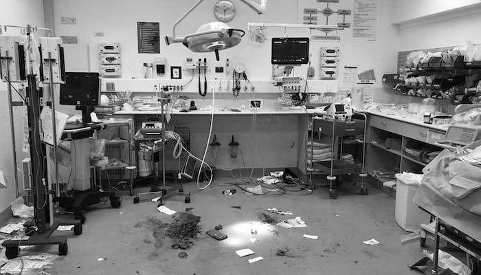
Saturday, July 27th. I got to sleep in because I was on the night shift for four days. No big deal. When I finally got to the hospital, there was pandemonium. Extra ambulances in the loading bay, a couple squad cars outside, a stab wound victim in the trauma bay, lots of noise and folks everywhere—what was routine hospital stuff somehow seemed like I was in the middle of filming an actual episode of ER. (I’m obviously partial to particular shows…okay, maybe Chicago Med?) When I report to my team, I learn that the computers have been down. All day. No electronic health records, no charting, no histories, no internet to look up guidelines/recommendations on UpToDate—and most tragically: no lab results.

Ok. This is it. I’m on the other (read: clinical) side of an awful downtime shift. I’ve experienced plenty of downtime in the lab, but this night I took a deep breath, reminded myself its going to be okay, and did my best to label things right. But a problem appears that’s more serious than labeling type and screens the right way without a computer: results are backlogged for hours! I’m talking no blood gases, no lactic acids, no pregnancy confirmations! I overheard senior residents and my attending that night talk about how the lab is struggling and they didn’t have enough people to figure out this downtime debacle.

This was a moment. It’s not often med students get to be literally useful in any clinical situation but after high-speed thinking about it, I interjected and made my elevator pitch:

“Dr. X, Dr. Y – I’ve got several years of hospital lab experience and lots of background in managing crises and downtime situations, if you want I’ll head over to the lab and see if I can help this situation at all, at least for the ER…”

There was a short pause. Then an enthusiastic wave of approval with hands waving me to go help out our laboratorian colleagues. Please note: the instances where tidbits of knowledge as a medical laboratory scientist prove useful as a medical student on rounds are far and few between for their ability to really captivate a group of doctors who identify themselves far from any lab medicine; so, this was a win. Explaining the importance of order of draw, or why sensitivity goes down when you don’t adequately fill blood cultures, or why peripheral smears should come with some interdisciplinary caveats aren’t quite as sexy as an emergency room, on metaphorical fire, with no one but you knowing anything about how labs work.

So, I ran on over to the laboratory, fully intending to do what I could to help in my unofficial just-a-friendly-neighborhood-med student capacity. That’s when I met Jalissa Hall!

I walked into the main lab area and asked if I could talk to the supervisor, thinking I would just explain my experience and offer what I could to their staff who I’m sure were buried in downtime SOPs and make sure I got critical results back to my team in the ER—a win-win! When I asked who was in charge, a very busy Ms. Hall walked out from behind the chemistry section and said, “you can talk to me. What’s going on?” I’m sure she thought I was there to complain, seemingly like many other clinicians were, but I stopped and gave her the same elevator speech I delivered moments ago with the postscript: “what can I do for you?” I remember she stopped, thought about if for roughly 10 seconds, and presented me with her situation briefing:

  • Computers have been down since roughly 05:00 am
  • There’s a computer virus that had all servers shut down indefinitely
  • There’s no communication between the hospitals EHR and the labs LIS
  • Moreover, no patient information is coming across to the analyzers (MRNs, specimen IDs, etc.)
  • There are 4-5 critical units (ER, OR, ICU, OB, NICU) that require STAT results
  • Clinicians have been coming to the lab all day looking for informal results reporting
  • The limited lab staff has had to manually print results on paper and work to match them with barcodes, specimens, and manual requisitions before releasing results
Image 2. Jalissa Hall, MLS(ASCP) (left) and a very tired me (right) after a great night of solving lab-related communication problems! Anyone else need an emergency room pathologist? Sounds like a new clinical specialty/fellowship to me…

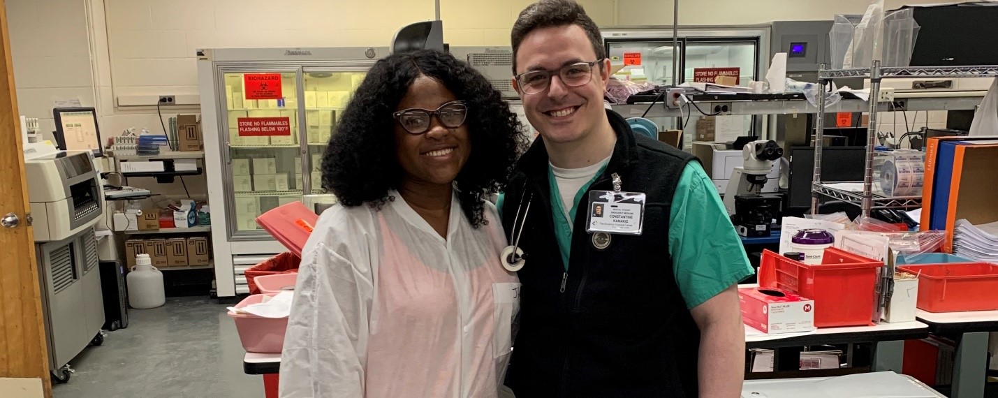
Deal. I know I can’t jump on the analyzers because New York is one of the states that requires clinical laboratory licensure (which I do not hold). In my informal survey I noted three medical lab scientists (including Ms. Hall), someone in specimen processing, and someone in blood bank. Basically, in order to make sure the lab could operate at peak performance with what they had, I helped alleviate the “paper problem” for them at least for the ER specimens. I matched requisitions with instrument raw data, made copies for downtime recording, delivered copied results to the ER, rinsed, lathered, and repeated—for eight hours! I obviously had to toe the line for the ER results, but there were other nurses and doctors who came in for the other areas’ results. No one worked more than the folks in that lab that night, and no one more so than Jalissa. After things cooled down a bit, I got the chance to connect with her and talk about her career and asked if she had anything to share with all of you—she definitely did.

Lablogatory family: please meet Jalissa Hall, MLS (ASCP)!
(Responses paraphrased because, honestly it was late, and downtime was busy, and we were tired, ok?)

Jalissa has been working for about five years as a generalist, with two jobs—like most of us have done. She works at The Brooklyn Hospital Center as a generalist and at NYU Hospital Lab in their hematology section. She is a graduate from the excellent MLS program at Stony Brook University in NY. She’s got ambitious career goals that are aimed at climbing as high as she can in laboratory medicine, and she’s got the enthusiasm and work ethic to match! I got the chance to ask her some real questions, during a real down-time crisis. This is what she had to say:

What made you go into laboratory medicine?

JH: I really want to help people. I love the behind-the-scenes aspect of being a medical laboratory scientist, but I think sometimes it can be too behind the scenes…

What did you think of tonight’s downtime issues?

JH: …it could have gone better. There seems to have been some panic, people kept walking in and shuffling the papers around. I tried my best to organize by floor, have two copies of each result (one for us and one to send upstairs), and requisitions match orders, but it was difficult. We have a downtime protocol, but we just couldn’t keep up with the volume and extent of how long it’s been down for. There’s really been no help outside the lab to work with us during this time so it’s a challenge.

What could have happened better?

JH: No outside help meant no room to breathe. On the inside, supervisors off duty tonight called staff in but none were available to come in. We don’t have an on-call person. We’re understaffed or short-staffed like so many labs out there; it’s problematic.

How is this going to look tomorrow?

JH: It’s not looking good, haha! Morning draw is definitely going to have a hard time. Catching up with these backlogs is one thing, but if orders can’t come across the LIS we’ll have to address that problem for sure. We’ve got a great staff though, so I’m sure it’s going to be fine.

What would be your “top tips” for all our fellow laboratorians reading this?

JH: First and foremost, being driven matters. If you want to get ahead, if you want to excel and climb high within an organization or in our profession, you have to work hard and keep working toward your goals.

Pro-tip #1: One of the biggest issues is “vertical cooperation.” Basically, some call it administration-buy-in, but it means administration working with employees in the lab to make the best decisions for our patients. If employees are burned out or if there aren’t enough resources to effectively perform our responsibilities it creates risks! It all comes down to patients, and making sure we’re in the best position to deliver diagnostic data for them means considering all aspects of lab management.

Pro-tip#2: If we want to fix the workforce shortages our labs regularly experience, we have to increase our efforts in advocacy within our profession. Having programs increase awareness of this job as a profession increases the pull and interest of potential new partners to work with. My school did it, other schools do this; increasing the number of programs that expose students to career opportunities in lab medicine would address our short-staffing problems everywhere!

Pro-tip #3: TELL OTHERS ABOUT OUR PROFESSION! I talked about our role being too behind the scenes…well the way to fix that is professional PRIDE! Own our accomplishments, share our role, advocate for our recognition, celebrate our peers!

Pro-tip #4: The future is not scary. Lots of folks shy away from tech advancement, fearing that automation and other developments mean losing jobs—it doesn’t. Why can’t today’s lab scientists become tomorrows experts on automation, LIS software, and other aspects of our cutting-edge field?

It was a pleasure to meet Jalissa and even better to work alongside her and learn about her passions and goals within the field we both care about! It was particularly special for me to be able to use my knowledge and experience to really contribute to my clinical team and bring laboratory medicine to the forefront where it doesn’t often shine!

Image 3. In a fantastic book I read recently, the authors of You’re It: Crisis, Change, and How to Lead When it Matters Most talk about leadership as a moment—a moment where you step up to a situation because you have skills and experiences which make you uniquely qualified to serve in a role which aims at a positive outcome. I had a small version of that in front of my attending (important for evaluations in medical school of course), but that downtime night was Jalissa’s “you’re it” moment for sure! (Source: Google)

Signing off from any new clinical rotations because this guy’s done with his medical school clerkships! Now I’ve gotta knock out some board exams and go on some residency interviews…wish me luck! I’ll check in with you next month after the 2019 ASCP Annual Meeting in Phoenix, Arizona—hope to see some of you there!

See you all next time and thanks for reading!

–Constantine E. Kanakis MSc, MLS (ASCP)CM graduated from Loyola University Chicago with a BS in Molecular Biology and Bioethics and then Rush University with an MS in Medical Laboratory Science. He is currently a medical student actively involved in public health and laboratory medicine, conducting clinicals at Bronx-Care Hospital Center in New York City.

The Disaster Risk Assessment

There are multiple types of risk assessments required when managing a laboratory safety program. OSHA’s Bloodborne and Airborne pathogens standards require assessing the risk of employees’ exposure to particular lab hazards. Risk assessments can be used to determine whether or not to add an emergency eyewash station, and all lab chemicals need to be assessed for the hazards they pose. These are just some assessments that are needed, and there are particular steps to take when performing them. But what about the lab emergency management plan? Should the lab perform a risk assessment for that? The answer is yes, although the terminology used may be different. To prepare a disaster readiness plan for the lab, the risk assessment that is needed is known as a Hazard Vulnerability Analysis (HVA).

The Centers for Medicare & Medicaid Services (CMS) requires that all healthcare facilities use an “all-hazards” approach when considering emergency preparedness and planning. While some laboratories may be included with the facility-wide disaster plan, the lab should absolutely have its own plan with specific instructions that apply directly to the department. That means the lab should also consider an all-hazards approach.

It may seem daunting to try to consider every possible disaster that could occur in the department, but that is not exactly what the directive from CMS dictates. An all-hazards approach means that emergency plans should be scalable or flexible so that it can be used for many types of disasters. The plan should focus on the lab’s ability to continue to offer services, especially those deemed critical, as a disaster situation unfolds.

The first step to the plan creation is the risk assessment- the Hazard Vulnerability Analysis. The HVA can be a table that lists all of the potential types of disaster; natural, man-made, facility-specific, etc. List as many as you can think of, and be sure to include specific disasters that may be particular to your locale (earthquakes, blizzards, etc.). Rate each disaster type by probability, severity of impact, and level of readiness of the lab to respond. Using that data, you can calculate the risk percentage for each emergency type.

One other requirement imposed by CMS is that facilities must include emerging infectious diseases as one potential type of hazard class. With the advent of particular diseases in the past years like Ebola, Zika, and certain influenza types, it is important to consider how an outbreak would affect lab operations and staffing. The risk level of infectious diseases may vary as incidents and outbreaks occur in particular geographic regions or if pandemics arise.

The HVA should be reviewed and updated as necessary each year. Things change that can affect what is on your HVA list. The addition of a nearby airport might make you consider adding airline disaster to the HVA. A change in weather patterns could occur as well. In 2011 a surprise earthquake in Virginia made state facilities re-look at their HVA list of possible emergency situations. Also, the actual list of disasters might not change, but there may be a change in the potential of a particular incident occurring.

If your lab or facility has not yet performed the HVA risk assessment, there is no need to panic. There are several model HVA tools available on line that can be used. As with any risk assessment, be sure to keep documentation readily available, review it each year, and make sure staff are trained about not only the HVA process, but in how to use the emergency management plan as well. There is a great amount of work that can go into preparing for a disaster, and training and drills for your staff will help to facilitate a smoother activation of the plan when the real emergency situation occurs.

Dan Scungio, MT(ASCP), SLS, CQA (ASQ) has over 25 years experience as a certified medical technologist. Today he is the Laboratory Safety Officer for Sentara Healthcare, a system of seven hospitals and over 20 laboratories and draw sites in the Tidewater area of Virginia. He is also known as Dan the Lab Safety Man, a lab safety consultant, educator, and trainer.

When Gender Goes Awry in Electronic Health Records

For most people working in laboratory medicine, their first encounter with transgender patients likely arose from an issue involving the Electronic Health Record (HER). For me, I was called into the reference lab, because an abnormally high estradiol result was found by the referring lab. They were concerned this might be coming from a hormone secreting tumor, but inspection of the patient’s record revealed they had been taking higher than recommended doses of their feminizing hormones.

Today I will share stories from issues that arise in EMR when gender doesn’t equal sex. While these may not specifically happen to all of you, I hope they can be informative or help you anticipate future problems.

Transgender issues came up at one of our institutions when providers were getting dozens of messages in their in-baskets about new flagged lab results for multiple patients. This is very annoying, because they have to address each of these messages or they are out of compliance with the hospital. An investigation revealed that all of the patients involved were transgender patients. In order to get estradiol, sold as oral contraception pills, the pharmacy had to administratively change their sex in the EHR for approval, then change it back.

This moved their corresponding reference ranges out of sync, which triggered a new results flag. Changing the sex back triggered other flags and more messages. This was finally resolved after a committee was convened and several meetings occurred, but no one would have anticipated this type of issue arising from a simple action to get patients their medicine.

Sometimes transgender patients have their sex changed legally. If an EHR only includes one sex entry instead of gender and sex assigned at birth, then certain lab errors may prevent processing of important samples. The pregnancy test for a transgender man could be auto-rejected. This can be an issue even for providers in front of the patient as was recently reported in a case to the NEJM about a transman who was mistaken as obese instead of pregnant and miscarried their child.

Similarly, a prostate biopsy from a transgender woman could be auto-rejected by a surgical pathology system as an inappropriate specimen type for the patient. Even further, an EHR could fail to prompt a provider from making a prostate cancer risk assessment in a transgender woman, which could result in improper screening.

I would recommend that EHR includes three separate fields (sex assigned at birth, gender, and legal sex) to fully recognize transgender patients and provide optimal personalized healthcare to them.

References

  1. Gupta S, Imborek KL, Krasowski MD. Challenges in Transgender Healthcare: The Pathology Perspective. Lab Med. 2016 Aug; 47(3):180-188.
  2.  Stroumsa D, Roberts EFS, Kinnear H, Harris LH. The Power and Limits of Classification – A 32-YearOld Man with Abdominal Pain. N Engl J Med. 2019 May 16;380(20):1885-1888. doi:10.1056/NEJMp1811491.

-Jeff SoRelle, MD is a Chief Resident of Pathology at the University of Texas Southwestern Medical Center in Dallas, TX. His clinical research interests include understanding how the lab intersects with transgender healthcare and improving genetic variant interpretation.

Global Health Narratives Interview Series: Meet Dr. Kumarasen Cooper.

Kumarasen Cooper, MD, PhD completed his medical training from his home country in South Africa and his PhD at Oxford. He now works as a surgical pathologist at the University of Pennsylvania and is responsible for leading the initiative to engage the pathology department in the Botswana-UPenn partnership through the Perelman School of Medicine Center for Global Health. He has over 260 publications and has lectured in 5 continents. Despite this busy schedule, Dr. Cooper devotes two separate months of the year to work in Botswana’s only academic pathology department, where he pours his energy into helping the department advance.

I met Dr. Cooper through email when I heard about the work he was doing in Africa. He generously agreed to come visit my department to give an excellent Grand Rounds lecture on his experiences working in Global Pathology, and he led a much-appreciated resident slide session of unusual and difficult cases from his work in Botswana. Humility and grace envelop Dr. Cooper despite his brilliant accomplishments. He also proved to be incredibly generous with a refusal of his speaker honorarium, in exchange for an agreement that we would collect pathology textbooks to send to the under-supplied residency program in Botswana. I’m excited to share the inspiring work that he does through the Botswana-UPenn partnership with all of you today, as I think this program could be used as a model for all institutes to involve their pathology departments in global health opportunities.

Q: What began your interest in global health?

A: I was born, raised, and completed my medical training in South Africa. I spent 15 years working as a Pathologist and served as the Chair of Pathology in Johannesburg until I was recruited to the US to work as Vice-Chair at the University of Vermont. I knew when I left Africa that I would always come back, and that I could use what I learned abroad to give back in some way. I wasn’t sure in what form that would take at the time, but I knew there was work that still needed to be done. This was also influenced by my visits to the pathology departments in many different countries over the years…I was able to gain a sense of the ‘haves and have-nots’, and so developed a strong feeling that I needed to give back.

Q: How did you hear about the Botswana-University of Pennsylvania (BUP) partnership and was pathology an active part in that already?

A: When I first discovered the partnership, I thought that this may be an avenue for me to participate in global pathology. At the time, the pathology department was not involved in any of the ongoing BUP projects, though other clinical departments at UPenn were. After my initial assessment of the Botswana pathology department and its resources in April of 2016, I was able to identify ways that I could help. Together with the Director of BUP, I approached the Chairman of my department with the proposal, and we started the pathology partnership program in October of that year. Since then, I travel to Botswana twice a year for one month at a time, and each time I take 1-2 residents from UPenn along with me.

Q: Can you describe the pathology department in Botswana?

A: To serve a population of just over 2 million people, Botswana has only one academic pathology department, a College of the University of Botswana (UB) School of Medicine, which consists of six pathologists who are all from other countries. There are currently no Botswana pathologists working in the department. There are about six technicians working in the laboratory, all of whom were trained internationally. The laboratory receives around 7,000 surgical specimens yearly, plus cytology, and autopsy. They work with an extremely limited panel of immunostains that are not routinely used but are spared for the rare case that cannot be diagnosed with morphology alone.

The residency program is still very new. There are six residents in the program at the present time, and the program is designed so that they will spend the first two years in Botswana and then they will continue their final years of training in South Africa. I look forward with anticipation to the first Botswana trained pathologists in the country.

Q: What is your role when visiting Botswana?

A: We try to help with everything we can. I sign out cases with the residents during the time I am there, and I teach the residents using these cases every day. The UPenn residents that I bring with me are eager to teach as well, so they deliver didactics regularly also. We all participate in tumor boards and the FNA clinic. We each take on projects that we can partner with them to tackle…things like improving turnaround time, quality improvement, and SOP preparations.  We also work on developing academic programs, grossing templates and manuals (A UPenn pathology PA spent two weeks working in Botswana on this project), synoptic reports, cancer guidelines…anything they need I try to help them with.

Q: How are the UPenn pathology residents given credit in their home program to join you?

A: As of this year, the BUP pathology program is now offered as one of the official electives that residents are allowed to choose from. They are able to use elective time and their travel expenses are paid for by a resident travel grant.

Q: In your role as supervisor of the UPenn residents, what do you see the residents gaining from the experience?

A: The residents that have come with me to Botswana are very compassionate and are eager to contribute in any way they can. Experiencing pathology in Botswana, where people are trying to achieve so much with so little resources, it makes the UPenn residents even more grateful for all of the resources they have available to them. They also have the opportunity to not only learn from the unusual cases that present in Botswana, but also the opportunity to contribute their own unique set of skills – some have focused on teaching autopsy technique, others give enthusiastic  and detailed lectures, and one gave a talk about successful study techniques. [For more information about the resident experience, one can read more about it in the UPenn blog here: https://pathology.med.upenn.edu/department/blogs/residency-matters/penns-pathology-residency-program-reaches-botswana]

Q: How do you see the BUP pathology partnership affecting the trainees in Botswana? What changes have you seen since you started working with them?

A: The residents in Botswana really appreciate the partnership that we have formed.  I have seen the residents develop so much since working with them. At first, they were reserved and now they actually request lectures on topics they feel they could improve on. They are still very humble and respectful, but I have encouraged them to be advocates for themselves. They have really embraced their program and I’m very proud of them. We have a deep appreciation for each other and are proud of what we have achieved together.

We’ve also started hosting Botswana residents at UPenn for a one month rotation so they have the opportunity to supplement their training even further. We fly them to the US, house them, and include them in our residency training program for the month. They have the opportunity to sit in on sign-outs, shadow grossing and autopsy, attend conferences, and be exposed to the advanced testing that we routinely perform in the US.

Q: How do you see the pathology partnership growing in years to come?A: I’m currently helping them find placements in South Africa or possibly partnering with private laboratories to help expose the residents to a greater diversity and volume of cases. As the program continues to grow, we look forward to seeing the fruits of the partnership for many years to come.


-Dana Razzano, MD is a Chief Resident in her third year in anatomic and clinical pathology at New York Medical College at Westchester Medical Center and will be starting her fellowship in Cytopathology at Yale University in 2020. She was a top 5 honoree in ASCP’s Forty Under 40 2018 and was named to The Pathologist’s Power List of 2018. Follow Dr. Razzano on twitter @Dr_DR_Cells.

Hey, What’s the Buzz on Zika?

Hello everyone and welcome back!

Last month, it was as fun to write about hematology peripheral smear differentials as it was to address the importance of interdisciplinary collaboration. I found myself in a unique position both as a medical student as well as a former medical laboratory scientist in what was a great clinical training rotation in hematology/oncology. Now, with just one rotation left until the end of my medical school journey, I want to take you on a look back at some of the very first posts I made here on Lablogatory and update you on the intersectional, collaborative topic that I shared with you almost two years ago: Zika!

Image 1. ASCP’s official professional society partner, The Pathologist. I’ve been getting them in my mailbox since the official partnership was announced. It’s an excellent platform for laboratory professionals across scopes to discuss relevant topics in pathology. I was particularly excited to see Zika make an appearance last month! (Source: The Pathologist [online] https://thepathologist.com/diagnostics/our-powers-combined)

In a recent digital article on ASCP’s partner, The Pathologist, author and staff editor Michael Schubert wrote about the connectivity between public health, epidemiologic research, laboratory medicine, and clinical patient outcomes. He examined the effectiveness and accuracy of Zika testing availability in commercially available assays and spoke with a leading virologist in the field from Berlin. You may recall one of those “ancient” posts I made about Zika, where I was part of a research team that used the same methodology! Combined immunoglobulin-specific assays, arbovirus detection in the heat of a public health epidemic’s epicenter, and lab medicine that complimented my concurrent immunology class in med school—what more could you ask for?

And, since the last tagged Lablogatory Zika update I can see was by Dr. Sarah Riley in February of 2017, here’s my update! Dr. Riley’s post was a fantastic summary of the Zika epidemic, its troublesome diagnostic assessments, and the recommendations and plans of organizations like the World Health Organization (WHO). She was, and still is, right—the “struggle is still real’ when it comes to Zika testing. Curious about what it was like during the 2016 epidemic? Who was doing testing, what kind of testing, and what was the lab data climate? Well…it feels like it’s time for a…

*** FLASHBACK ***

An Arbovirus Abroad

Hey! My inaugural post! It was fun to go back and see the data from the work then (Spoilers: epidemiologic updates are on your horizon). We were just getting started to take an assessment of the situation and address it as a public health concern. My then Caribbean location was a great place to study Zika trends coming from Brazil, Puerto Rico, and Florida. As a snapshot, at that time (Dec 2016) there were a purported almost 2,000 cases, however less than a fifth of those cases were serologically confirmed by lab testing. Before the recommendations to move toward RT-PCR, most labs in the region were requesting commercially available screening tests for IgG/IgM assays.

Image 2a. These were the (then) suspected Zika viral infection cases per epidemiological week, Pan-American Health Organization (PAHO) and World Health Organization (WHO) 2016. My wife and I are included in these statistics—that mosquito virus rash is awful!
Image 2b. Remember that spoiler I promised above? Well here’s the updated WHO epidemiologic data for confirmed Zika cases in the region we worked in. Seems like the mosquitoes…buzzed off. (Source: WHO)

Healthy Me

How do you reach people when you’ve got compelling public health lab data that translates to possible prevention of infection and spread of disease? Easy: go to where the people are and engage them when and where they’re comfortable. One of the overarching themes in public health is mitigating barriers to change by way of utilizing social humility. This a certainly a type of interdisciplinary collaboration because if we’re the experts on IgM and IgG trends in testing confirmations, the public are the experts in social determinants of health within their communities.

Image 3. Want to make sure a message gets home to every family? Bug their kids about Zika bugs in fun, educational ways. That’s me delivering one of my “Healthy Me” presentations to children, October 2016.

Laboratory Data and Global Health Security

As my team and I were busy preparing SOPs, conducting a new project aimed at improving local health literacy and source reduction, securing IRB approval, and collecting data about the residents of Sint Maarten to correlate with local Health Ministry projections, one of the officials—who now serves as a regional director for PAHO—took our work to the Global Health Security Agenda Summit. Talk about motivation! In and out of the lab, I worked with teams who were getting some fantastic work done on the ground with respect to mosquito-borne virus research.

Image 4. IgM and IgG seroprevalence of Zika virus (along with other Arboviruses i.e. West Nile, Chikungunya, Dengue, and Yellow Fever etc.) within the community around my medical school. We used commercially available IgG and IgM assays from Germany with great success. Internal controls and known cases were fantastic ways to include internal validation.

IRBs and Public Heath Pathology

For those of us who work in laboratory medicine, it’s easy to talk about the best way to test, detect, and treat an epidemiologic threat—it’s even exciting when it’s a current threat. But to really be successful, you’ve got to collaborate with those outside of the lab, and often this means thinking “outside the box.” Public health is different from lab medicine in that while lab-work is based around results, testing, and organized data-driven decisions, success in public health is highly determined by community buy-in in the form of partnerships!

Figure 1. There’s a method to the community “buy-in” concept. With a foundation in evidence-based practices, any project aimed at improving public health outcomes must include some critical components like clear objectives, attainable goals, sustainability, and effective (and constant) re-evaluation.

*** FLASH … FORWARD? ***

So, after my time in Sint Maarten, I came to New York City to rotate through my clinical clerkships. And, if you’ve seen some more recent post-Zika posts on this website, you know they’ve been going great! Within a few months of being here, my wife brought back some swag from a training session she attended. (Side note: she’s a graduate-level nurse, working in the public health non-profit sector with vulnerable populations in the inner city—she’s too busy to blog.) After months of both of us working and learning about Zika and public health initiatives in the Caribbean, we were greeted by this fantastic toolkit from the New York State Department of Public Health!

Image 5. Empowering a large number of patients with highly variable demographics is challenging. The NYS DOH distributed “Prevention Kits” for Zika Virus which included: Zika Virus educational materials in 8 languages, pamphlets on reducing mosquito activity, travel related information for pregnant women, 2 larvicide pellets with instructions for using larvicide, picaridin insect repellent, and condoms.
Image 6. That’s us! My wife Kathryn and I presenting on the importance of Disaster Planning and Implementation of Preparedness Programs at the 2019 Caribbean Conference of Disaster Medicine. Disasters are bad on their own, but think about what happens months after flooding, hurricanes, or destruction—transmittable diseases. And that includes standing-water-borne mosquito viruses!

The take home message: collaboration is key, both inside and out of the lab. Schubert’s piece in The Pathologist created a fantastic dialogue in addressing the clinical needs for interdisciplinary collaboration. The best testing means finding out exactly where the needs are and using data-driven decisions to implement change or action. In the lab, that means constantly working for higher quality and better patient outcomes in every test, result, report, and (mosquito) byte of data. In the field, it means the same thing, but instead of metrics like sensitivity, specificity, and TAT it’s about cultural humility, attainable goals, and dynamic timing.

Thanks for reading! Hope most of our national heat wave spared you, but if it didn’t remember: don’t keep standing water around, wear light loose clothing, and use appropriate insect repellent!

See you next time!

–Constantine E. Kanakis MSc, MLS (ASCP)CM graduated from Loyola University Chicago with a BS in Molecular Biology and Bioethics and then Rush University with an MS in Medical Laboratory Science. He is currently a medical student actively involved in public health and laboratory medicine, conducting clinicals at Bronx-Care Hospital Center in New York City.

An Introduction to Laboratory Regulations: Part 1

Everyone who works in a laboratory knows that there are certain rules and regulations to be followed to ensure accuracy in testing, and the safety of both the patient and testing personnel. With all the acronyms floating around (CLIA, FDA, CAP, CMS, TJC) it can get confusing to keep track of who controls what, and which rules apply to your specific lab. In the first installment of this 3-part series on regulations, we’ll review the different federal agencies responsible for oversight and moderation of the laboratory. In part 2 we’ll go further in-depth to demystify testing complexity (waived, non-waived, PPM) and why it’s important to know the correct classification for the tests you perform. Lastly, we’ll review the optional accreditations available to labs, and how accreditation differs from certification.

CLIA

CLIA refers to the Clinical Laboratory Improvement Amendments of 1988. These amendments were drafted to the Public Health Services Act, in which the federal program was revised to include certification and oversight of clinical laboratory testing. Although there have been two additional amendments made after 1988 (1997, 2012), the law still continues to be cited as CLIA ’88 as it is named within legislation.

These CLIA regulations helped to establish quality standards for all U.S. laboratory testing performed on human specimens (except for research) for the purpose of assessment of health, or the diagnosis, prevention, or treatment of disease. The regulations cover all aspects of testing including general laboratory requirements, quality monitors, pre-analytics, analytic performance, post-analytics, and personnel requirements.

In addition to setting the basic ground rules for performing quality laboratory testing, the CLIA regulations also require clinical laboratories to be certified by their state as well as the Center for Medicare & Medicaid Services (CMS) before accepting human samples for diagnostic testing. Laboratories can obtain multiple types of CLIA certificates, based on the kinds of diagnostic tests they perform. In order for laboratories to receive payments from Medicare or Medicaid, laboratories must be properly certified for the testing they are performing and billing for.

There are 3 federal agencies responsible for enforcing the CLIA regulations: The Food & Drug Administration (FDA), Center for Medicaid Services (CMS) and the Center for Disease Control and Prevention (CDC). Each agency has a unique role in assuring quality laboratory testing.

CMS

The Centers for Medicare & Medicaid Services (CMS) is the federal agency responsible for ensuring that the CLIA standards are upheld and enforced. Their responsibilities include the following:

  • Issuing laboratory certificates
  • Collecting user fees
  • Conducting inspections and enforcing regulatory compliance
  • Approving private accreditation organizations (such as CAP) for performing inspections, and approves state exemptions
  • Monitoring laboratory performance on Proficiency Testing (PT) and approving PT programs
  • Publishing CLIA rules and regulations

FDA

The Food & Drug Administration (FDA) is primarily responsible for reviewing and approving new tests, instruments, and equipment used in diagnostic laboratories. They also perform the following tasks:

  • Categorize tests based on complexity
  • Review requests for Waiver by Application from manufacturers
  • Develop rules/guidance for CLIA complexity categorization

CDC

The Center for Disease Control and Prevention (CDC) responsibilities include the following tasks:

  • Provide analysis, research, and technical assistance
  • Develop technical standards and laboratory practice guidelines, including standards and guidelines for cytology
  • Conduct laboratory quality improvement studies
  • Monitor proficiency testing practices
  • Develop and distribute professional information and educational resources
  • Manage the Clinical Laboratory Improvement Advisory Committee (CLIAC)

To summarize, CLIA establishes the rules and guidelines that laboratories must follow to ensure they are providing accurate laboratory results. Federal agencies then work together to support the CLIA amendments and enforce compliance. All certified laboratories will be subject to inspection by regulatory agencies to ensure compliance with the rules. In some cases, your local state Department of Health (DOH) or accrediting agency may be more stringent or have additional requirements to be followed – always go with the stricter requirement to ensure compliance with all agencies.

Coming up next we’ll review how the FDA decides the complexity of each test, and how this designation will affect the CLIA rules to be followed.

References

  1. Electronic Code of Federal Regulations: https://www.ecfr.gov/cgi-bin/text-idx?SID=1248e3189da5e5f936e55315402bc38b&node=pt42.5.493&rgn=div5
  2. Interpretive Guidelines for Laboratories: https://www.cms.gov/regulations-and-guidance/legislation/clia/interpretive_guidelines_for_laboratories.html

†


-Kyle Nevins, MS, MLS(ASCP)CM is one of ASCP’s 2018 Top 5 in the 40 Under Forty recognition program. She has worked in the medical laboratory profession for over 18 years. In her current position, she transitions between performing laboratory audits across the entire Northwell Health System on Long Island, NY, consulting for at-risk laboratories outside of Northwell Health, bringing laboratories up to regulatory standards, and acting as supervisor and mentor in labs with management gaps.

Blood Bank Case Study: “What’s Your Type?”

The general public doesn’t always know a lot about laboratory testing in general, but most people know a little about blood types, even if it’s what they have learned from TV! Blood types do seem to come up in casual conversation. We might hear a conversation about blood type after someone has donated blood, or between family members comparing notes, who ask “What’s your type?” Yet, even with medical technologists, there can still be some confusion about blood types and blood typing, particularly if one has not worked in Blood Bank in many years. I recently received an email from a colleague who had a few questions about blood types, as she has not worked in Blood Bank for over 40 years. I always tell my students that no question is a bad question, and indeed, she asked some very good questions, which I will address with this case study.

  • What blood type is listed on a patient’s chart if they type “O Du”?
  • What blood type is recorded on a donated unit of blood typed “O Du”?
  • What type of blood does an “O Du” patient receive?
  • Can an “O Du” patient have a transfusion reaction if they are transfused with O positive blood? Would she need to receive O negative blood in a transfusion?
  • Does an “O Du” patient need to receive RhoGAM if she pregnant and her husband is Rh positive?

If you have ever wondered or can’t remember details about any of these questions, you’re in the right place. So, what’s new, if anything, with blood types?

Landsteiner discovered the ABO blood group system in 1901, and identified A, B and O blood types, using experiments performed on blood from coworkers in his laboratory. The discovery of the codominant AB blood type soon followed, but it was not until around 1940 that the Rh blood group was first described. In 1946, Coombs and coworkers described the use of the antihuman globulin (AHG) to identify weak forms of Rh antibodies in serum. For us old blood bankers, the original name for this test was the Coombs’ test. (You will still find physicians ordering a Coombs’ test!) The current and proper name for this is the direct antibody test (DAT), which is used to detect in vivo sensitization of RBCs. AHG can also be used to detect in- vitro sensitization of RBCs using the 2 stage indirect antibody test (IAT).

Since Landsteiner’s work, we have not discovered any new blood groups that are part of the routine blood type. The ABO and Rh blood groups are still the most significant in transfusion medicine, and are the only groups consistently reported. However, we currently recognize 346 RBC antigens in 36 systems.1 Serological tests determine RBC phenotypes. Yet, today we can also determine genotype with family studies or molecular testing. This case study and 2 part blog reviews some terminology in phenotyping, some difficulties and differences encountered, and explores the possibility of RHD genotyping to assess a patient’s true D status.

Our case study involves a 31 year old woman who is newly married. She is not currently pregnant, has never been pregnant, is not scheduled for surgery but has had a prior surgery 15 years ago, and has never received any blood products. She and her husband recently donated blood and, as first time blood donors, just got their American Red Cross (ARC) blood donor cards in the mail. The husband noted that his card says that he is type O pos. The woman opens her card, and, with a puzzled look on her face, says “My card says I’m an O Pos, too. There must be a mistake.” She knows she has been typed before and checks her MyChart online. Sure enough, her blood type performed at a local hospital is listed in her online MyChart as O negative. She further checks older printed records and discovers that 15 years ago, before surgery, she was typed at a different hospital as “O Du”. She is very upset, wondering how she can have 3 different blood types. She is additionally concerned because they are planning to have children and recalls being told that because she is Rh negative, that she would need Rhogam. Is she Rh negative or positive, and what does Du mean? Will she need Rhogam when pregnant? She has many questions and calls the ARC donor center for an explanation.

What blood type is listed on a patient’s chart if they type “O Du”?

What is happening here, what is this woman’s actual blood type, and what testing can be done to ensure accuracy in Rh typing? From the patient reports, it appears that this woman has what today we call a “weak D.” Du is an older terminology that should no longer be used, and that has been replaced by the term “weak D.” But, why does she have records that show her to be an O neg, a type O, Du (today, this would be written O weak D), and now, a card from ARC stating she is O pos?

RhD negative phenotypes are ones that lack detectable D antigen. The most common Rh negative phenotype results from the complete deletion of the RHD gene. Serologic testing with anti-D is usually expected to produce a strong 3+ to 4+ reaction. A patient with a negative anti-D at IS and at IAT would be Rh negative. If the patient has less than 2+ strong reaction at immediate spin (IS), but reacts at IAT, they would be said to have a serologically weak D.1 Historically, weak D red blood cells (RBCs) are defined as having decreased D antigen levels which require the IAT for detection. Today’s reagents can detect many weak D types that may have been missed in the past, without the need for IAT. However, sometimes IAT is still necessary to detect a weak D. When this is necessary is dependent on lab SOPs and whether this is donor testing or patient testing. The reported blood type of this patient also depends on the SOPs of the laboratory that does the testing. And, the terminology used for reporting is also lab dependent. It is not required by AABB to test patient samples for weak D (except for babies of a mother who is D negative). There is also no general consensus as to the terminology to be used in reporting a weak D. Some labs would result this patient as O negative, weak D pos. Some labs may result O pos, weak D pos. Others may show the individual reactions but the resulted type would be O pos. Labs who do not perform weak D testing would report this patient as O, Rh negative. The following chart explains why this patient appears to have 3 types on record.

Figure 1. Tube typing results of same patient from different labs with different SOPs.

What blood type is recorded on a donated unit of blood typed “O Du?”

AABB Standards for Blood Banks and Transfusion Services requires all donor blood to be tested using a method that is designed to detect weak D. This can be met through IAT testing or another method that detects weak D. If the test is positive, the unit must be labeled Rh positive. This is an important step to prevent alloimmunization in a recipient because weak D RBCs can cause the production of anti-D in the recipient. This also explains why the ARC donor card this patient received lists her type as O pos.

What type of blood does an “O Du” patient receive?

Historically, weak D red blood cells (RBCs) were defined as having decreased D antigen levels which require the IAT for detection. A patient who is serologic weak D has the D antigen, just in fewer numbers. This type of weak D expression primarily results from single-point mutation in the RHD gene that encodes for a single amino acid change. The amino acid change causes a reduced number of D antigen sites on the RBCs. Today we know more about D antigen expression because we have the availability to genotype these weak D RBCs. More than 84 weak D types have been identified, but types 1, 2, and 3 represent more than 90% of all weak D types in people of European ethnicity.2 An Rh negative patient has no D antigen and should, under normal circumstances, only receive Rh negative blood. Yet, there has been a long history of transfusing weak D patients with Rh positive RBCs. 90% of weak D patients genotype as Type 1, 2 or 3 and may receive Rh positive transfusions because they rarely make anti-D. 2

It is now known that weak D can actually arise from several mechanisms including quantitative, as described above, position effect, and partial D antigen. Molecular testing would be needed to differentiate the types, but, with the position effect, the D antigen is complete and therefore the patient may receive Rh positive blood with no adverse effects. On the other hand, a partial D patient may type serologically as Rh negative or Rh positive and can be classified with molecular testing. It is important to note that these partial D patients are usually only discovered because they are producing anti-D. If anti-D is found, the patient should receive Rh negative blood for any future transfusions.

Thus, 3 scenarios can come from typing the same patient. With a negative antibody screen, and because 90% of weak D patients have been found to be Type 1, 2 or 3 when genotyped, many labs do not routinely genotype patients and will report the blood type as Rh pos and transfuse Rh pos products. However, depending on the lab medical director and the lab’s SOPs, these same patients may be labeled Rh neg, weak D and receive Rh negative products. There is no general consensus on the handling and testing of weak D samples. The 3rd scenario is that many labs do not test for weak D in patients at all, and a negative D typing at IS would result in reporting the patient as Rh neg, with no further testing. In this case, the patient would be transfused with Rh negative products.

Can an “O Du” patient have a transfusion reaction if they are transfused with O positive blood? Would she need to receive O negative blood in a transfusion?

This question was covered somewhat in the above discussion. Policies regarding the selection of blood for transfusion are lab dependent, dictated by the lab medical director, and are based on the patient population, risk of developing anti-D, and the availability or lack of availability of Rh negative blood products. Anti-D is very immunogenic. Less than 1 ml of Rh pos blood transfused to an Rh negative person can stimulate the production of anti-D. However, not all patients transfused with Rh positive blood will make and anti-D. As discussed above, 90% of weak D patients are types 1, 2 or 3, would be unlikely to become alloimmunized to anti-D. If a weak D patient with a negative antibody screen receives a unit of D pos RBCs, there is a very small possibility that they are a genotype who could become alloimmunized to the D antigen and produce anti-D. However, as stated above, the majority of weak D patients can be transfused with D positive RBCs. Thus, with few exceptions, from a historical perspective, one can safely classify the weak D as D positive.

This question gets a little trickier when dealing with females of childbearing age. We particularly want to avoid giving Rh positive blood to females to avoid anti-D and the complications of Hemolytic Disease of the Fetus and Newborn. Therefore, when dealing with these patients, lab policies and physicians tend to be more conservative in their approach to transfusion. The consequences, however, in males and older females are less serious and these patients could be given Rh positive blood if there exists a shortage of Rh negative units. Any patient who becomes alloimmunized to the D antigen, would thereafter be transfused with Rh negative products.

Does an “O Du” patient need to receive RhoGAM if she pregnant and her husband is Rh positive?

This, again, would be up to the medical director, the lab’s SOPs or the patient’s physician. Depending on lab practice, the lab may or may not perform weak D testing. If the lab does not perform weak D and results this patient as Rh neg, the patient would get Rhogam. If the lab does do weak D testing and finds a weak D phenotype, the decision whether or not to give Rhogam would be up to lab practices and the practitioners involved. The lab’s policy on terminology used in resulting the type may also reflect the decision whether or not to give Rhogam. This brings up a lot of questions in the lab because we know that a patient who would not make anti-D would not need Rhogam. So, what is the best course of action? Read my next blog to learn more about troubleshooting and resolving D typing discrepancies!

From the discrepancies in reported type in this individual, and putting all the pieces of the puzzle together, we can conclude that this patient is a weak D phenotype. However, the type reported and the terminology used varies from lab to lab. Molecular testing is available, yet most labs are still using serological testing for blood types for both donors and patients. This is based on several factors within the lab setting. Stay tuned for my next Blood Bank blog exploring D typing discrepancies and the financial aspects of performing genotype on pregnant patients to clarify Rh type.

-Becky Socha, MS, MLS(ASCP)CM BB CM graduated from Merrimack College in N. Andover, Massachusetts with a BS in Medical Technology and completed her MS in Clinical Laboratory Sciences at the University of Massachusetts, Lowell. She has worked as a Medical Technologist for over 30 years. She’s worked in all areas of the clinical laboratory, but has a special interest in Hematology and Blood Banking. When she’s not busy being a mad scientist, she can be found outside riding her bicycle.

Think S.P.I.L.L.E.D.

Large biological and chemical spills are not a common occurrence in the laboratory. That’s a good thing, but when they do occur, they can create a very dangerous situation. It is vital that lab staff know how to handle such events even though they may not be commonplace.

Some laboratories differentiate between large and small spills. They may have an emergency number to call for a hazardous spill response team. Other smaller facilities simply don’t have that in place. Either way, it’s important for laboratory professionals to know they are the experts about the biological and chemical materials they use, and they need to be in charge as the experts when a spill situation needs to be managed.

Most laboratory spills can be managed using a standardized step-wise process known as the S.P.I.L.L.E.D. procedure. I don’t usually ask lab staff to memorize the acronym, but having the information contained on a poster with the lab spill kits can make a clean-up procedure go smoothly.

S = Secure the Site – Make sure no one walks through the area where a spill has occurred. It could be a dangerous situation if a hazardous chemical is spilled, and you would never want someone slipping in the area or tracking the spilled material to another area.

P = Protect Yourself – Arm yourself with the appropriate Personal Protective Equipment (PPE). In a lab spill event, this would mean using a lab coat, gloves, and face protection to prevent accidental splashes.

I = Inspect the Spill – Look to see what was spilled. If it is a hazardous chemical, is there a concern about fumes? Obtain a Safety Data Sheet to see if section 6 will give any special information about handling the accidental release or spill of that chemical. Consider other spill concerns such as broken glass or possible ignition sources if flammable material is involved.

L = Lay Down a Barrier – If the spill is large and spreading, lay down spill pillows or booms designed to contain a flow of liquids. Surround the spill area with these materials. Sometimes, the use of an emergency shower can create the need for a barrier to be made.

L = Lay Down Absorbents – No matter the size of the spill, the next step is to place any absorbent powders, granules or clean-up pads to soak up the spilled material. If the absorbent is also a neutralizer, make sure you allow the necessary time for neutralization to occur.

E = Extract the Mess – Use implements to pick up the materials used for stopping and absorbing the spill.

D = Dispose of the Waste – Properly dispose of all materials involved with the spill clean-up. If there was glass involved, be sure to use a sharps container.  Biohazard material should go into an appropriate container, and chemical waste materials may need to be disposed of separately for pick-up by a chemical waste vendor.

Lab staff should be able to access spill control materials quickly, and the necessary items should be stored in a location designated by signage. Perform an inventory of spill supplies and make sure there are adequate materials that could handle spills of the biohazards and chemicals stored and used in the department. Be sure items in the spill kit are not expired, and if there is no expiration date for absorbent powders, check them at least annually for effectiveness.

All laboratory staff need to have complete spill clean-up training. Give information about the types and locations of spill kits and how to handle various types of spills that can occur. Once that training is done, it will become important to perform spill drills in the department. Drills can be performed a number of different ways, but a common method involves having a “victim” spill water onto the floor and claim the material splashed into their eyes. Watch from a distance to see how the staff reacts. Do they provide appropriate first aid? Do they inspect the container label? Do they access the correct clean-up supplies and facilitate cleaning efficiently? Make notes of how the drill went, discuss them with the staff, and repeat the drills until all staff are comfortable with a spill situation. Biological and chemical spills should not be a common occurrence in the lab. When they do occur, however, the situation can become serious quickly, and a fast and effective clean-up needs to occur. Because these events are rare, it becomes important to provide regular spill training and drills so staff can remain ever-ready to handle them.

Dan Scungio, MT(ASCP), SLS, CQA (ASQ) has over 25 years experience as a certified medical technologist. Today he is the Laboratory Safety Officer for Sentara Healthcare, a system of seven hospitals and over 20 laboratories and draw sites in the Tidewater area of Virginia. He is also known as Dan the Lab Safety Man, a lab safety consultant, educator, and trainer.

The Paperwork of Transgender Care

I don’t think anyone enjoys filling out the paperwork at a doctor’s office. For transgender individuals, this can be an experience that ranges from irksome to offensive. Most intake forms don’t allow for expression of their gender identity. Furthermore, confusion on gender and sex can create real confusion and healthcare failures in several places that laboratory medicine encounters a transgender individual.

Arguably the first place the lab encounters a transgender patient is via the phlebotomist. These professional collectors of blood must confirm two patient identifiers, which are often name and date of birth. The “name” used is the legal name. Using a transgender person’s “dead name” (name given at birth) represents a gender they do not want to be associated with and can be a very offensive experience. “Isn’t it obvious that name is not what I look like?”

While names can be legally changed, this happens with varying difficulty and legal cost in different states. A solution is to improve training of phlebotomists to explain the necessity of confirming a legal name so lab results are properly matched to the patient. Additionally, front-desk intake workers should be similarly trained to interact with transgender patients when recording demographic information. This can be aided by electronic health records (EHR) becoming more flexible and inclusive of the gender diversity.

Traditionally, EHR would only include one field for SEX: M or F.

Several in the laboratory community have asked how many different gender options should be included? Facebook included up to 71 options in 2017. That’s a big step up from the 2 traditional EHRs are built around.

The World Professional Association for Transgender Health (WPATH) executive committee in 2011 outlined the recommended fields to include in EHR: preferred name, sex assigned at birth, gender, and pronoun preference. EHRs are evolving and can be flexible depending on the user requirements. At my program, we use EPIC at 3 different different sites (children’s, county and university hospitals) and each has a different version.

From what I’ve seen preferred name is an easy addition and would not interfere with functions of the EHR or Laboratory Information Systems (LIS), which is the Lab’s version of EHR.

If the field for sex assigned at birth is different from gender, then it would clear up any confusion about whether the person is transgender and then they should be addressed by the pronouns matching the gender. While there is a spectrum of genders, only transgender males and transgender females are of a high enough prevalence to have medically relevant recommendations. Plus, if a system at least starts here, they could expand further as necessitated by their population.

EHR could include preferred pronouns, but I haven’t seen this implemented in an EHR yet. Ideally, you would just use the pronouns that match the intended appearance of the individual (ma’am to someone wearing a dress, etc.).

Lastly, I think Legal sex should be added to the EHR as well. One of our hospitals has this and it makes several processes easier such as processing hormone medication.

Legal (or administrative) sex, sex assigned at birth, and gender data fields provide the clearest and simplest picture of a patient and should be a minimum for labs making recommendations for changes to HER.

Next month I will describe in greater detail the issues that can arise in the lab when gender or sex are entered incorrectly in the system for transgender patients and how this can negatively affect care delivery.

References

  1. Deutsch MB, Green J, Keatley J, Mayer G, Hastings J, Hall AM, World Professional Association for Transgender Health EMR Working Group. Electronic medical records and the transgender patient: recommendations from the World Professional Association for Transgender Health EMR Working Group. J Am Med Inform Assoc. 2013 Jul-Aug; 20(4):700-3.
  2. Gupta S, Imborek KL, Krasowski MD. Challenges in Transgender Healthcare: The Pathology Perspective. Lab Med. 2016 Aug; 47(3):180-188.

-Jeff SoRelle, MD is a Chief Resident of Pathology at the University of Texas Southwestern Medical Center in Dallas, TX. His clinical research interests include understanding how the lab intersects with transgender healthcare and improving genetic variant interpretation.

Not Starbucks but the DMV

I merrily wait in line at Starbucks for my iced cappuccino with soy milk, pay $5+ for $0.25 worth of goods poured into my $14.00 souvenir mug, and walk out the door with my head held high, joyous with the privileges of conspicuous consumption. My server was super-cheery and the brief exchange we had was so pleasant—they really love me!  I need that high because I am off to the Department of Motor Vehicles (DMV) for a driving-related task and know–just know–that there will be an incredibly long line at the end of which sits a disgruntled government employee who doesn’t care if I show up or not. Their motivation to help us is non-existent. “Why would anyone ever work here?” I ask, sipping my delicious beverage.

Today, a doctor called someone in the United States (US) and told them the biopsy taken from their leg earlier this week has come back as invasive cancer. A bit distraught and nervous, the patient called up a nationally recognized cancer center, from which they only live a few miles, and on the end of the line is a caring, pleasant voice who informs them they can be seen today! The valet parking is gorgeous, the building is gleaming with glass and steel, and every face they see as they journey from check-in to clinic is smiling, compassionate, and sincere. Their nurse and then doctor are both genuine people with their patient’s best interest in mind, and they carefully and completely explain what has been found, what needs to be done, and how they are going to get through all of this together. As they depart, the receptionist grabs them for a brief moment to return their private insurance card and waves at them as they depart, adding, “We will see you soon!”

Today, someone in Africa went back to the hospital—an 8-hour journey from their home—where their biopsy was performed a month ago, hoping to get the result. After several people searched multiple offices and inquired with several people, the result is found and brought to them, a single piece of paper. Payment is required before they can receive the biopsy results. They have brought money with them, which they gathered from three neighbors, their brother, and by selling some chickens, and pays for the report. They read the report and, at the bottom, notices that it says additional testing is needed. Confused, they ask for help and a pathologist comes to find them. Respectfully, the pathologist explains that additional testing is needed, which is not available in the hospital despite the pathologist’s strong desire to have it, but they can send the biopsy to a lab elsewhere to do the testing which will cost about 3 times what they just paid for the primary report. They happen to have enough and pay the amount requested. The report will be back in about a month. Two months later, they have returned to the hospital for the 4th time and the report is now available. The testing that was done simply confirms that the primary diagnosis is accurate. They go to the oncology clinic on the same campus and sit in the waiting area with 3 dozen other people. They sleep at the clinic overnight outside with about a dozen people. The following afternoon, they are finally seen and the oncologist reviews the report. He notes that if the patient had come to the clinic as soon as they had the biopsy result three months ago, a simple surgery would have cured them of this lesion. But now, because they waited so long, there is only chemotherapy available which is expensive and, the oncologist reports, doesn’t actually work very well for this tumor.

Before you shed a tear for this terrible situation (while I sip my cappuccino and a nurse begins someone’s chemotherapy in a shiny, brightly lit, and expansively windowed infusion unit not far away), we have to ask ourselves what is really going on? First and foremost, this is an allegory to make a few points but the situation is repeated over and over again every day in the US and Africa. However, as a simple, superficial explanation, the person with cancer in the US is receiving their cancer therapy from Starbucks and the person in Africa had to go to the DMV.

Cancer care in the United States is almost entirely in the private sector, dispersed among the 1500 cancer treatment facilities, of which 70 are comprehensive cancer centers.[i] Based on the US population, the expected cancer rate, 100% detection, and 240 working days for a given cancer center, there are on average only 5 new patients per day per cancer center. Is that why one can often get that appointment right away in a major cancer center or is it really a concierge customer service effort? A standard private insurance plan for which I pay, for example, $250 per month and my employer pays $1300 per month is accepted by cancer centers and results in small co-pays for multiple appointments, which can be covered with a Flexible Spending Account (FSA) or Health Savings Account (HSA). On insurance statements after appointments, some of the services received cost thousands of dollars but the patient portion was only, say, a hundred dollars, again, which may be paid with FSA/HSA. It’s so great that we have insurance because the insurance company is bearing the brunt of costs. But are they?

In the United States, 79% of facilities providing health care are private, a mix of non-profit and for-profit.[ii] But 64% of all healthcare in the United States is paid for by the US government through Medicare, Medicaid, the Veterans Administration (VA) system, and Children’s Health Insurance Program (CHIP).[iii],[iv] Since almost every cancer care facility is private (or, stated another way, “not free”), that means that for every one of us at the cancer center getting treatment, for which we and our employer are paying through insurance, there are two people getting the same treatment at the same high-level quality of care for which the government is paying. Those other deductions from our paychecks for Medicare and Medicaid (which everyone pays, regardless of how old, as long as they are employed and regardless of their own health insurance plan) are going towards the 64% coverage. The point is not that the US healthcare system is expensive. The point is that there is a lot of revenue and resource being put into the healthcare system and, thus, there is a high-quality product or experience that is available.

If we look at any low GINI index country and compare their GDP with the US GPD and compare their spending on healthcare as a % of GDP, we don’t even need to do the math to see that there is very little money per person available in the system for any type of healthcare. The challenge in low-resourced settings (by which it is meant low-resourced patients in low-resources locations) is both a lack of funding available to provide healthcare services along with a lack of “stuff” to provide those services. We can invoke the law of supply and demand to try and argue that the people can rise up and demand more healthcare facilities and “someone” will meet that supply. In the US, this results in the Starbucks model. In a low-resourced setting who has the incentive to meet that supply? Where does the government get the money from to create such a system? What private corporation is going to start a healthcare program that provides universal coverage regardless of what you can pay?

The answer is really quite simple. This model of healthcare is insufficient for cancer and isn’t going to work for all patients. Moreover, the Starbucks model is not really applicable, sustainable, nor equitable. When we go to Starbucks for their coffee, to some degree, our choice of Starbucks is because of the a) flavor of the coffee, b) cost of the coffee, c) perception of the coffee, and/or d) convenience of the coffee. We could always choose Dunkin’, Peet’s, Tim Horton’s (maybe let’s not go there for this analogy), or Green Mountain coffee at a different location. There is variation in pricing and convenience. There is variation in the condiments we can use to doctor our coffee. An economy and series of markets exist which allow Starbucks to gather resources from dozens of other companies to provide your coffee. But, ultimately, we are all buying coffee which has caffeine which has a desired effect. We can go to a free AA meeting or to a soup kitchen and get some pretty basic coffee if we don’t have the money to pay. The point is we have choices and we can pay a high price, a low price, or no price and we get coffee.

The Starbucks model does work for a certain sector of the population but not everyone. Since vast majority of cancer care in the US is private, the Starbucks model falls down because we don’t actually have any free options as a society and “low-cost healthcare” is not typically appealing to most Americans with cancer because they have their mortality at stake (no one wants cancer nor does anyone want to die from cancer). In fact, desperation in the face of cancer is what makes the US one of the only places in the developed world where people go bankrupt trying to be treated for cancer. The ultimate inequity is that cancer care is “pay to play” in the US and there essentially aren’t safety nets for any populations that can’t pay (homeless) or are living below a certain income threshold (i.e., the ~10% of Americans without healthcare plus a large percentage with insufficient insurance).[v]

Please remember, these are human beings and they didn’t choose to get cancer (there is no demand for cancer… there is only demand for cancer care!). Since they didn’t have a choice in the disease they have to be burdened with, why is there an expectation that they should pay for the treatment? Moreover, if a patient has a stage I cancer, easily surgically removed and cured vs. a Stage III cancer requiring months of various therapies at a very high cost, how do we ethically explain an increased cost for a worst state of disease? It’s really an inverse quality spectrum and we make patients pay more for getting a lot less. We pay for insurance in case we ever do get cancer (or other major disease). It’s a risk reduction or risk aversion pre-payment. Like we do with our car or our house or our boat. Those last three things we choose to have (and are luxuries). We don’t get to choose to have health. It’s just an inherent part of being human so holding someone accountable for it because they didn’t have the resources to “prepare for the worst” is really the wrong attitude. Our healthcare system isn’t perfect but there are gaps that could be easily filled if resources are allocated efficiently to meet the whole populations needs—that’s the benefit of having a large resource supply into the system. We just have to find the operational efficiency to make the costs work.

However, when we remove the luxuries of insurance, Medicare, and Medicaid and other payments systems from the health sector or, worse, simply assume the government’s role is to provide healthcare 100% free to all citizens in a resource-limited or resource-constrained setting, we suddenly have an untenable situation. The economy and tax-base are not there to create the resources. We find overworked, underpaid, and undersupplied medical staff working in crowded conditions. For single entity care (e.g., HIV, tuberculosis, malaria), vertical programs have made great strides in combatting these diseases even in some of the poorest countries in the world. But cancer is anything but simple with the complexity of cross-discipline collaboration, spectrum of disease, range of treatments, and inherent costs creating huge gaps in the delivery of cancer care. Economic and physical infrastructure for the provision of care is what is needed to meet this challenge. Our current Starbucks model in the US would be extremely difficult to replicate in a low-resourced setting due to the lack of infrastructure. However, when this infrastructure is assessed, planned for, and implemented, cancer care can be delivered in these settings at a significantly lower cost per patient. Adding infrastructure implementation high-quality private facilities and public-private partnerships creates a way forward to pump resources into the system and insure that no patient is left behind. To round out this allegory, AAA locations (a commercial car-servicing company) in various parts of the US allow one to renew your driver’s license with them, rather than the DMV. I did this once, it was VERY fast, friendly, and efficient. This type of public-private partnership worked for me and I believe it will work for cancer if we are willing to try.

References

[i] NCI-designated Cancer Center. https://en.wikipedia.org/wiki/NCI-designated_Cancer_Center  Retrieved May 21, 2019.

[ii]  “Fast Facts on US Hospitals”. Aha.org. Retrieved December 1, 2016.

[iii] Himmelstein DU, Woolhandler S (March 2016). “The Current and Projected Taxpayer Shares of US Health Costs”. American Journal of Public Health. 106 (3): 449–52. doi:10.2105/AJPH.2015.302997. PMC 4880216. PMID 26794173. Government’s share of overall health spending was 64% of national health expenditures in 2013

[iv] ^ Leonard K (January 22, 2016). “Could Universal Health Care Save U.S. Taxpayers Money?”. U.S. News & World Report. Retrieved July 12, 2016.

[v] https://www.kff.org/uninsured/fact-sheet/key-facts-about-the-uninsured-population/

milner-small

-Dan Milner, MD, MSc, spent 10 years at Harvard where he taught pathology, microbiology, and infectious disease. He began working in Africa in 1997 as a medical student and has built an international reputation as an expert in cerebral malaria. In his current role as Chief Medical officer of ASCP, he leads all PEPFAR activities as well as the Partners for Cancer Diagnosis and Treatment in Africa Initiative.