Surgical Pathology Case Study: An Enlarging Neck Mass with A Non-Diagnostic FNA

Case History

The patient is a 41 year old male with a history of smoking who presents with a tender, slowly growing mass on the angle of the left mandible for the past 1 to 1.5 years. The patient also complains of otalgia, but no dysphagia or weight loss. A computed tomography (CT) scan was performed, which demonstrated a 3.2 x 2.6 cm enhancing mass in the superficial lobe of the left parotid gland with no significantly enlarged lymph nodes and a patent Stensen’s duct (Image 1). A fine needle aspirate (FNA) was performed that showed acinic and ductal cells, but was not diagnostic. The decision was made to take the patient to surgery in order to perform a parotidectomy.

Image 1. CT scan demonstrating the mass in the left parotid gland (red arrow)

Diagnosis

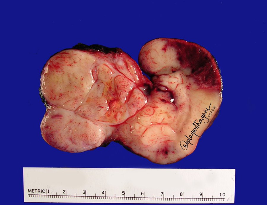
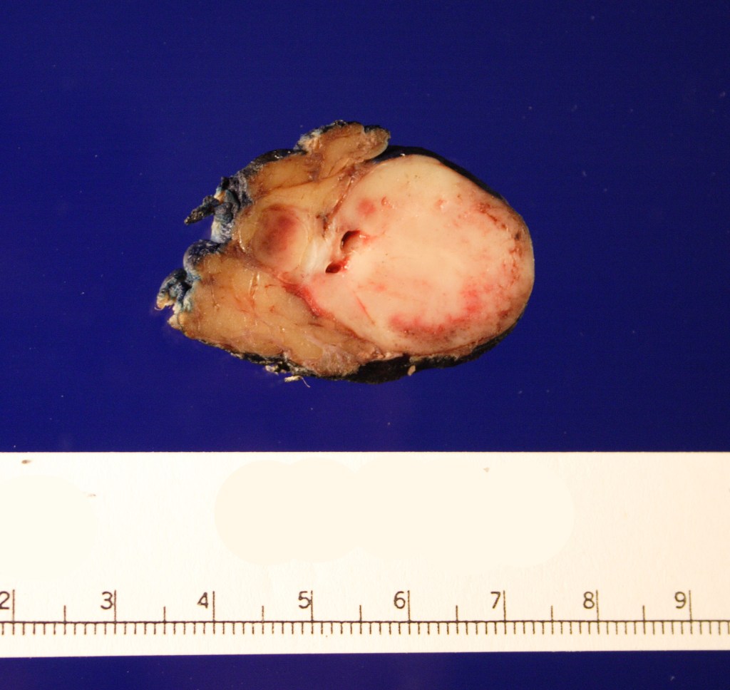
Received in the surgical pathology laboratory for intraoperative consultation was a 0.3 x 0.2 x 0.2 cm biopsy of the left superficial lobe parotid gland mass. The tissue was frozen, stained and read out as an “acinic cell neoplasm”. Following the frozen diagnosis, the main specimen was received for routine processing, weighing 19.0 gm and measuring 4.0 x 4.0 x 3.0 cm. The specimen was unoriented and entirely inked black. It was serially sectioned revealing a 2.5 x 2.5 x 2.4 cm tan, friable, well-circumscribed mass and surrounding tan-brown, beefy-appearing parotid tissue (Image 2). A second tan-brown nodule measuring 1.5 x 1.0 x 0.8 cm abuts the larger tan mass. Representative sections of the larger mass are submitted in cassettes 1-8, and the smaller nodule is entirely submitted in cassettes 9 and 10.

Image 2. Cut surface of the parotid gland demonstrating the well-circumscribed tan mass

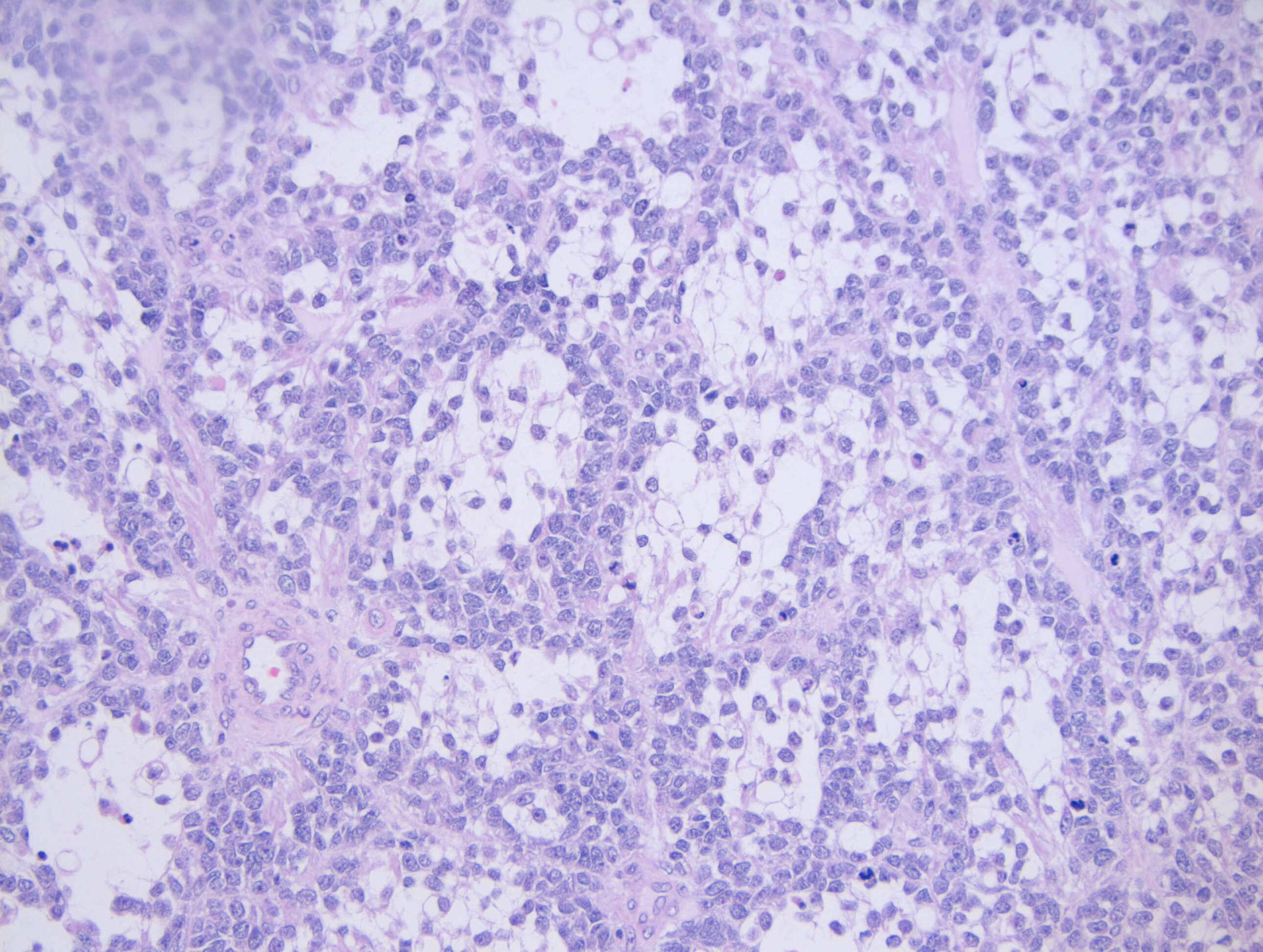
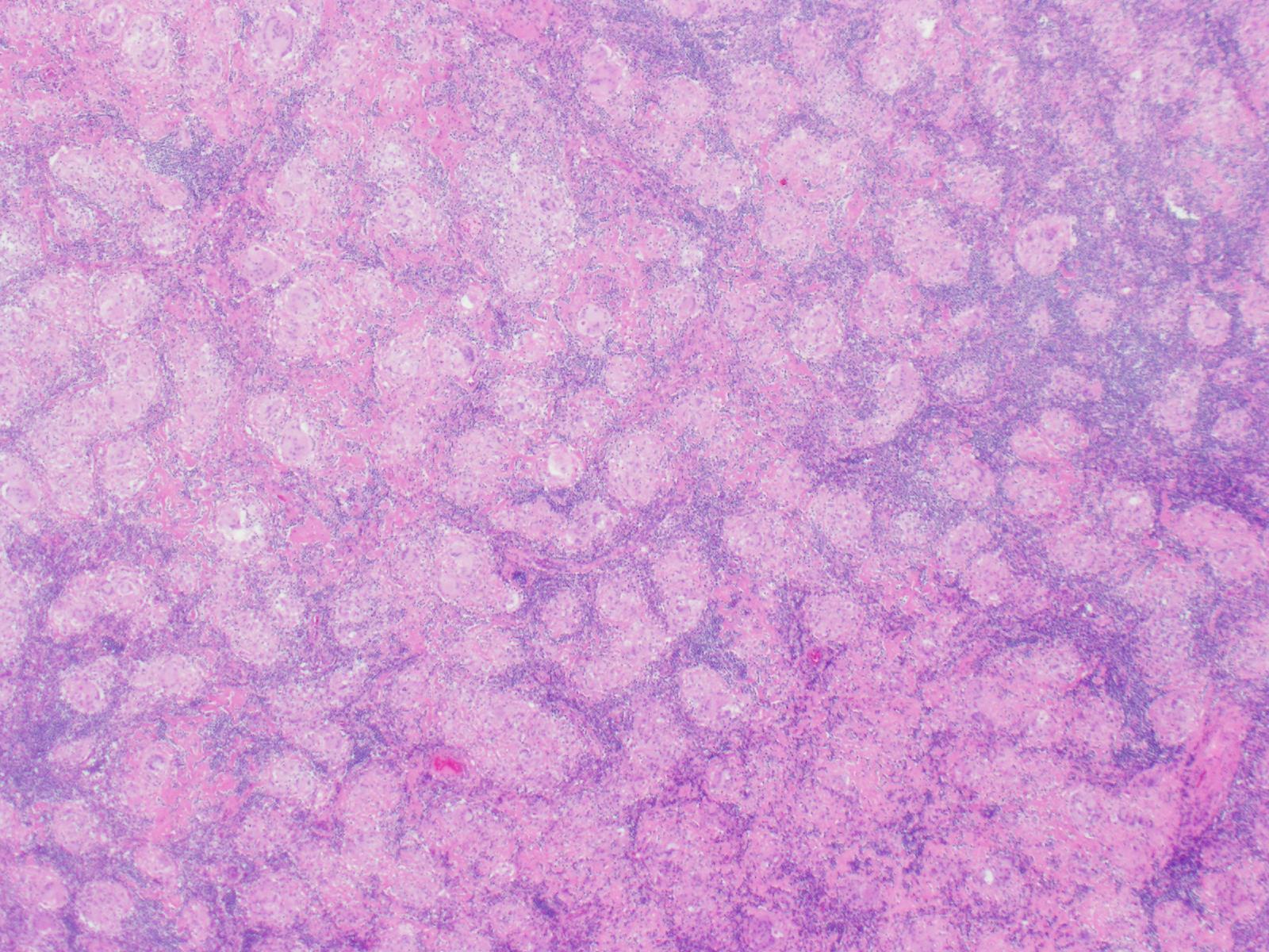
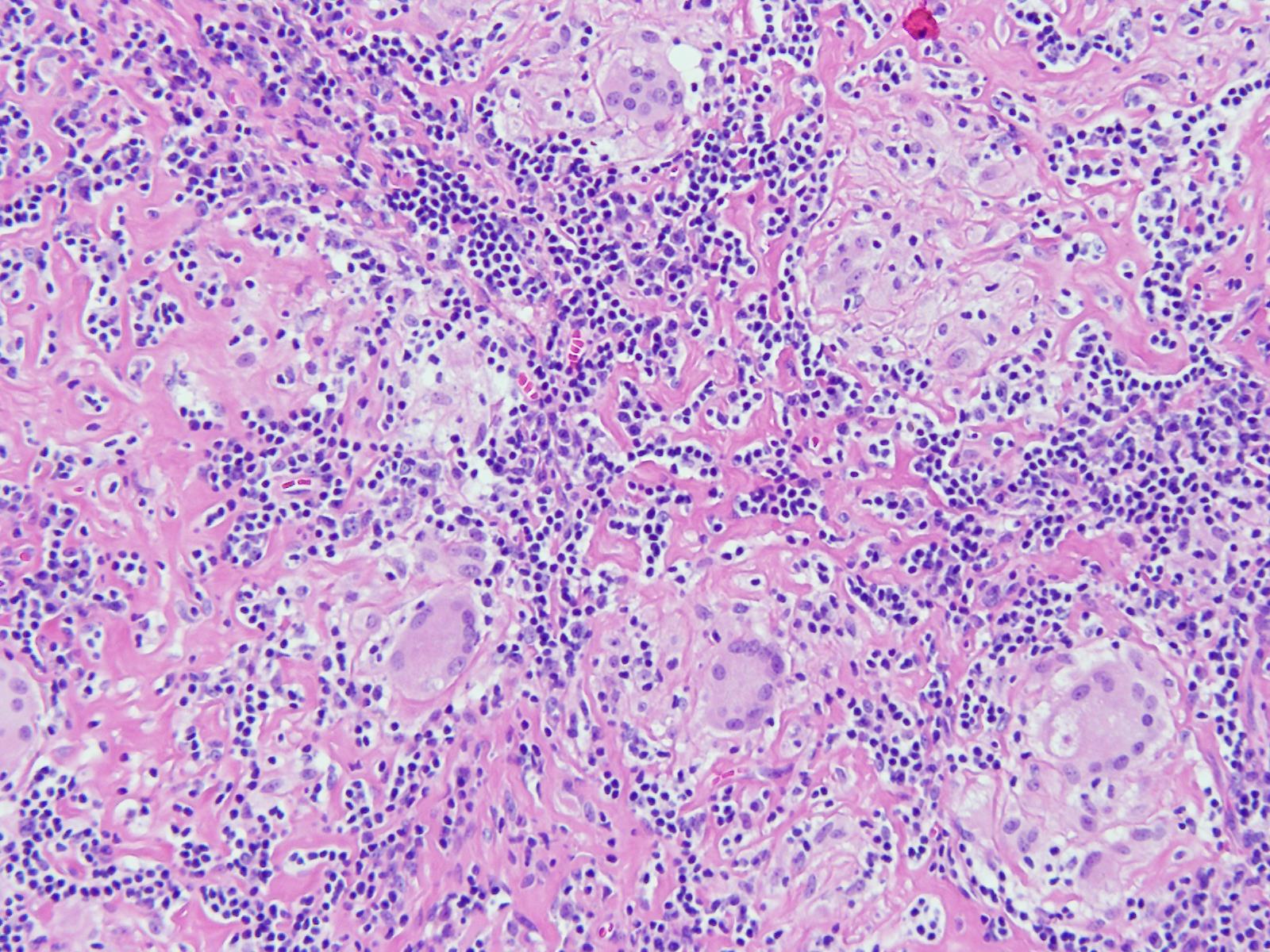
Microscopy demonstrates a low-grade, very well differentiated tumor consistent with an acinic cell carcinoma with complete inked surfaces (i.e. the mass has not been transected and excision appears complete). There is a small focus of capsular “disruption”/parenchymal hemorrhage, which most likely corresponds to the area sampled for intraoperative consultation. In addition, there are two separate benign periparotid lymph nodes.

Discussion

Acinic cell carcinoma (ACC) is a rare tumor of the parotid gland, representing 2 to 4% of all primary parotid gland neoplasms. It is the second most common childhood salivary gland malignancy behind mucoepidermoid carcinoma, but has been found throughout the age range. There is a gender predilection, as ACC is found in females more than males in a 3:2 ratio. One of the first cases of ACC dates back to 1892, in which the tumor was diagnosed as being a “blue dot tumor”, thought to be called this due to the intracytoplasmic zymogen granules.

Clinically, ACC presents as a slowing growing mass in the salivary glands, most commonly in the parotid gland. Other symptoms are not commonly found until late in the diagnosis, and include pain, facial nerve palsy, and nodal disease. There have also been cases of ACC that arise in the minor salivary glands. Unlike minor salivary gland carcinomas that arise in the palate, ACC of the minor salivary glands will mostly be found in the buccal mucosa and upper lip.

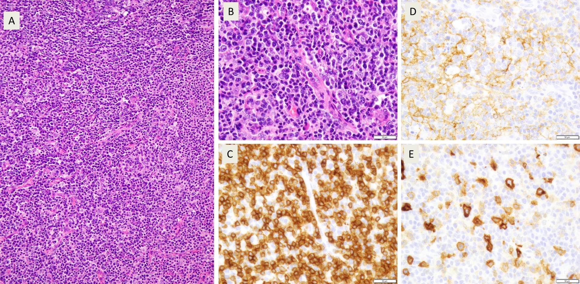
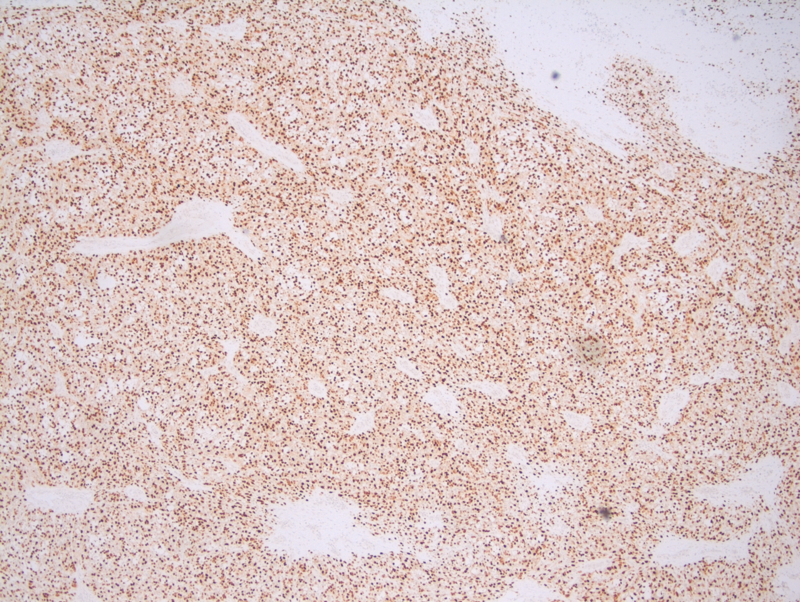
Grossly, ACC presents as a round, well-circumscribed to variably encapsulated mass with a rubbery, gray-tan, solid to cystic cut surface, commonly with areas of hemorrhage and necrosis. Histologically, the mass will be composed of acinar type cells with basophilic granular cytoplasm, clear cells with glycogen or mucin, intercalated ducts, non-specific glandular cells and a few mitotic figures. ACC is defined by the World Health Organization as a malignant epithelial neoplasm of the salivary glands in which at least some of the neoplastic cells demonstrate serous acinar cell differentiation, which is characterized by zymogen secretory granules, and can also include salivary ductal cells (Image 3). It is common for sections taken of ACC to show microscopic invasion of the capsule with nests of tumor cells outside the capsule. There are four histologic patterns that were described by Abrams et al in 1965 that are still applicable today: solid, microcystic, papillary cystic and follicular. Immunohistochemical stains, if needed, will be positive for keratin, alpha-1-antichymotrypsin and alpha amylase. It can be difficult to distinguish ACC from normal acini or benign salivary gland tumors (leading to a false negative result) on cytology due to the absence of any hallmark malignancy features such as necrosis and pleomorphism, but centrally placed large nuclei, distinct nucleoli, binucleated cells, and ill-defined cell borders can help make this distinction. The same caution applies to aspirates because if the tumor is cystic, it may be interpreted as being hypocellular and deemed to be a benign salivary cyst.

Image 3. Photomicrograph demonstrating the zymogen granules within the cytoplasm of the cells

Imaging by ultrasound, CT and magnetic resonance imaging (MRI) can prove to be worrisome as similar with cytology, the scans can demonstrate a mass with benign features, and thus a more favorable diagnosis. On ultrasound, ACC will appear lobular, well-defined, hypoechoic and poorly vascularized. Ultrasound can be useful help to determine the size and location of the mass, as well to help with ultrasound guided fine needle biopsies. On CT, the mass will appear non-specific with limited heterogenous enhancement but can be used to demonstrate the relationship of the mass to the facial nerve, and to identify any distant metastases. On MRI, ACC can have a nonspecific intensity pattern similar to benign salivary gland neoplasms, but low T1 and T2 signals can help suggest vascularity, fibrosis and calcification within the mass. In addition, MRI can help in assessing the parotid gland, stylomastoid foramen, and any possible facial nerve invasion or perineural invasion.

Risk factors for the development of ACC include radiation exposure and familial predisposition. Risk factors for the development of salivary gland tumors, but not necessarily ACC, include radiation exposure, the use of iodine 131 in the treatment of thyroid disease (isotope is concentrated in the salivary glands), and working with materials in certain industries, such as those that use asbestos and rubber manufacturing, metal in the plumbing industries, and woodworking in automobile industries.

Complete surgical excision is considered the primary treatment option, with postoperative radiotherapy in cases of incomplete removal, recurrence, undifferentiated ACC, positive margins, and cervical lymph node metastasis. Removal of the facial nerve may be necessary in T3 and T4 cases, as well as a possible neck dissection. As of now, ACC has been considered chemo-resistant, and treatment with chemotherapy is not suggested. Around 35% of tumors will recur, and that percentage rises to 80-90% if the tumor is incompletely excised. ACC has a 5 year survival rate of 90%, a 10 year survival rate of 88%, and there have even been of cases of recurrence occurring up to 30 years after the initial procedure. If metastasis was to occur, although rare, the spread tends to be more hematogenous than lymphatic, with the most common sites being the lungs and bones.

References

  1. Al-Zaher N, Obeid A, Al-Salam S, Al-Kayyali BS. Acinic cell carcinoma of the salivary glands: a literature review. Hematol Oncol Stem Cell Ther. 2009;2(1):259-64.
  2. Bury D, Dafalla M, Ahmed S, Hellquist H. High grade transformation of salivary gland acinic cell carcinoma with emphasis on histological diagnosis and clinical implication. Pathol Res Pract. 2016;212(11):1059-1063. DOI: 10.1016/j.prp.2016.08.005.
  3. Rosero DS, Alvarez R, Gambó P, et al. Acinic Cell Carcinoma of the Parotid Gland with Four Morphological Features. Iran J Pathol. 2016;11(2):181–185.
  4. Vander Poorten V, Triantafyllou A, Thompson LD, et al. Salivary acinic cell carcinoma: reappraisal and update. Eur Arch Otorhinolaryngol. 2016;273(11):3511-3531. DOI: 10.1007/s00405-015-3855-7
  5. Zahra Aly F. Acinic Cell Carcinoma. Pathology Outlines. http://www.pathologyoutlines.com/topic/salivaryglandsaciniccell.html. Revised April 30, 2019. Accessed August 23, 2019.

-Cory Nash is a board certified Pathologists’ Assistant, specializing in surgical and gross pathology. He currently works as a Pathologists’ Assistant at the University of Chicago Medical Center. His job involves the macroscopic examination, dissection and tissue submission of surgical specimens, ranging from biopsies to multi-organ resections. Cory has a special interest in head and neck pathology, as well as bone and soft tissue pathology. Cory can be followed on twitter at @iplaywithorgans.

Hematology Case Study: Thrombocytopenia in a 4 Year Old Child

A 4 year old child was brought to the pediatrician by her mother with a complaint of new onset of severe bruising on her legs. The mother could not recall any falls or bumps that would have caused the bruising. On exam, the physician also noted mucosal bleeding in the oral cavity. Questioning revealed that the patient had experienced flu like symptoms several weeks earlier. The physical exam was normal except for the bleeding. There was no family history of bleeding disorders. A CBC was ordered.

Reported CBC Results

WBC, RBC, Hgb, Hct, RBC indicies normal

Platelet count 26 x 103/μL

IPF 22% (reference range IPF% 1.0-7.0%) The physician evaluated the results, noting the normal CBC but decreased platelet count. The above results also show the immature platelet fraction (IPF), an additional Advanced Clinical Parameter reported from the Sysmex XN hematology analyzer. A low platelet count, as seen in this patient, will reflex a fluorescent platelet count (PLT-F). The impedance count (PLT-I) can be falsely increased if small RBCs or fragments are counted as platelets. On the other hand, in an optical platelet count, when measuring platelets by size (PLT-O), large platelets can be missed, giving a falsely low count. In this case there was a low platelet count and an instrument flag for an abnormal platelet scattergram. The PLT-F, on the other hand, uses a platelet specific dye which eliminates interference seen with other methods. The fluorescent dye labels the RNA, and forward scatter is used to determine size while side fluorescence is used to measure RNA content. With gating set based on cell volume and RNA content, the PLT-F can be measured. Therefore, the reflexed and more reliable PLT-F was the reported count.

Figure 1. PLT-F scattergram. The PLT-F channel measures forward scatter (FSC) on the Y axis and side fluorescence (SFL) on the X axis.1

Additionally, when there is an abnormal scattergram or a low platelet count, the IPF% and IPF# are also reported. The immature platelet fraction is a measure of the youngest platelets, or reticulated platelets. These are the first circulating platelets, right out of the bone marrow. An increased IPF indicates an increase in platelet production, yet this child’s platelet count was very low. This suggests that the thrombocytopenia may be due to excessive destruction of platelets; the bone marrow was actively making platelets, but they were being destroyed, causing the low platelet count.

Figure 2. Platelet scattergrams from a healthy individual with a normal IPF (a) and a patient with a high IPF (b). Mature platelets appear as blue dots, green dots represent the IPF with increased cell volume and higher fluorescence intensity compared to mature platelets.1

Diagnosis

Immune Thrombocytopenia- ITP.

Primary immune thrombocytopenia (ITP), formerly known as idiopathic thrombocytopenic purpura or immune thrombocytopenic purpura, is one of the most common bleeding disorders of children. In most cases, it presents with sudden onset of bruising and petechiae in an otherwise healthy child, with normal WBC and hemoglobin. ITP is an autoimmune bleeding disorder in which the immune system makes anti-platelet antibodies which bind to platelets and cause destruction. Even though the exact cause of ITP remains unknown, it is recognized that it can follow a viral infection or live vaccinations. While there are some similarities between pediatric ITP and ITP in adults, in children this tends to be an acute disease which is self-limiting and resolves itself in several weeks, with no treatment. However, in a small number of children, the disorder may progress to a chronic ITP. In contrast to ITP in children, a chronic form is more commonly seen in adults. It is usually a diagnosis of exclusion, does not follow a viral illness and requires treatment.

This patient recovered in a few weeks. One month after the initial episode, her PLT was 174 x 103/μL and her IPF% was 6.0%

Conclusion

An IPF reported with a CBC, in combination with a low platelet count, is fast, inexpensive, and can be extremely beneficial in aiding in a timely diagnosis. As the child’s platelet count recovered, the IPF% returned to normal range. ITP can therefore be monitored with a CBC. Thus, the IPF can be used not only to help diagnose but also as an indicator of remission.

References

  1. Sysmex America, 2019. www.sysmex.com/us. Used with permission
  2. Arshi Naz et al. Importance of Immature platelet Fraction as a predictor of immune thrombocytopenic purpura. Pak J Med Sci 2016 Vol 32 No 3:575-579
  3. Briggs,C. Assessment of an immature plateletfraction (IPF) in peripheral thrombocytopenia. Br J Haematol 2004Jul;126(1):93-9
  4. Sysmex White Paper. The role of the ImmaturePlatelet Fraction(IPF) in the differential diagnosis of thrombocytopenia. www.sysmex.com/us
  5. D-Orazio, JA, Neely, J, Farhoudi,N. ITP in children: pathophysiology and current treatment approaches.J Pediatr Hematol Oncol.2013 Jan;35(1): 1-13

-Becky Socha, MS, MLS(ASCP)CM BB CM graduated from Merrimack College in N. Andover, Massachusetts with a BS in Medical Technology and completed her MS in Clinical Laboratory Sciences at the University of Massachusetts, Lowell. She has worked as a Medical Technologist for over 30 years. She’s worked in all areas of the clinical laboratory, but has a special interest in Hematology and Blood Banking. When she’s not busy being a mad scientist, she can be found outside riding her bicycle.

The Fundamental Attribution Error

As laboratory safety professionals, we know that an important part of the job is the ability to coach other lab team members when unsafe situations are observed. To coach someone is to confront a coworker about an issue for the sake of safety-theirs, yours, or that of a patient. Those coworkers may be fellow lab employees, supervisors, managers, or even physicians. The word “confront” might sound strong, particularly to those who may be uncomfortable with these types of encounters, but this coaching is an important and valuable skill. 

Coaching your peers is no easy task, and it takes practice to be able to do it well. I recently walked into a laboratory that was unfamiliar to me, and I saw a technologist working at the bench with no lab coat, no gloves, and no face protection. At first I thought, “that would never happen in a one of my labs,” and then, “the lab safety culture here is terrible.”

I learned I was wrong on both counts, and the incident reminded me of the necessity to stop and think before forming an opinion or even speaking about a lab safety issue. I provide training often about how to coach staff who are acting unsafely while in the lab, and I have learned that how a coaching moment will go depends largely on what is in the head of the coach before he or she speaks. It is important to remember that if someone acts in a manner that displeases or disappoints you, there are several possible sources of influence acting on that person.

Psychologists have coined it the “Fundamental Attribution Error.” Humans who are disappointed usually think the other person has committed the wrong intentionally or because they are not intelligent. Neither of these conclusions is ever correct, and that thought process usually leads to a coaching session that will not be successful.

Take the scenario I mentioned above, for example. What is your gut reaction when you see someone working in a lab without PPE? Maybe that lab tech just found out a relative had passed away and they were waiting for someone to relieve them, or maybe there were no lab coats or gloves available in their size. The possibilities are endless, so you need to train yourself to be calm first and to ask questions to learn what is really happening without making assumptions. It’s more difficult to do than one would think.

The success of a safety coaching moment is determined in your head before you even speak. You have the power to make it a positive event. It is true that some people just will not accept it well no matter what we do (a reminder to ourselves to always be ready to accept coaching), but by and large a successful event starts in the mind of the person who is coaching for safety.

When you see a lab safety problem, it is vital that you confront the person. However, before you do so, ask yourself, “why would a rational person behave this way? What am I not seeing here?” If you start with that, your coaching for safety will be much more successful, and you will see a positive change in your overall lab safety culture.

Dan Scungio, MT(ASCP), SLS, CQA (ASQ) has over 25 years experience as a certified medical technologist. Today he is the Laboratory Safety Officer for Sentara Healthcare, a system of seven hospitals and over 20 laboratories and draw sites in the Tidewater area of Virginia. He is also known as Dan the Lab Safety Man, a lab safety consultant, educator, and trainer.

Hematopathology Case Study: A 69 Year Old Male with Weight Loss and Generalized Lymphadenopathy

Case History

The patient is a 69 year old male who presented to the hospital with a 3-month history of drenching night sweats, weight loss, fatigue, and generalized lymphadenopathy. He also endorsed a very itchy rash all over his body. He denied smoking. There was no other relevant social or family history.

Physical examination confirmed diffuse lymphadenopathy, hepatosplenomegaly and a mild diffuse skin rash. Notably, there was a 2.5 cm level-1 lymph node palpated in the left neck. This was subsequently biopsied.

Biopsy

Biopsy of the level-1 neck lymph node revealed a 2.3 x 1.5 x 1.2 cm mass pink-tan and firm mass. Sectioning revealed a glossy white-tan cut surface. H&E staining revealed a polymorphic lymphocytic infiltrate of in the interfollicular zones. The infiltrating lymphocytes ranged from small to large cells with abundant cytoplasm, eosinophils, and plasma cells. There was also a notable increase in the number of high endothelial vessels lined by lymphocytes with irregular nuclear borders and clear cytoplasmic zones.

Image 1. Polymorphic infiltrate of small, mature appearing lymphocytes (A), with prominent blood vessels and clear cytoplasm (B). Most of these cells were CD3 positive T cells (C) with expanded CD21 positive FDC meshworks (D) and scattered CD30 positive immunoblasts (E)

Further characterization by immunohistochemical staining showed the majority of the interfollicular cells to be CD3 and CD5 expressing T cells. These were a mix of CD4 and CD8 positive cells but with marked CD4 predominance. CD7 appeared positive in a smaller population of T-cells compared to CD3 (consistent with loss of this pan-T-cell marker). Varying numbers of the interfollicular cells were positive for CD10, BCL-6, CXCL-13, and PD-1 with a strong positivity for ICOS, phenotypically consistent with an expansion of Tfh (T-follicular helper cell) cells.

Interspersed between the T cells were numerous CD20 positive cells with prominent nucleoli that also revealed CD30 positivity. CD21 staining revealed expanded follicular dendritic cell meshworks. EBER ISH was positive in a rare subset of cells. Kappa and lambda ISH showed an increased number of polytypic plasma cells.

Flow Cytometry showed the presence of a small population of T-cells that were CD4 positive but CD3 negative. There was no evidence of B-cell clonality. TCR-G PCR was positive.

A final diagnosis of Angioimmunoblastic T-cell lymphoma (AITL) was rendered.

Discussion

AITL is a relatively rare neoplasm of mature T follicular helper cells, representing about 1-2% of all non-Hodgkin lymphomas. It is; however, one of the more common subtypes of peripheral T-cell lymphomas, accounting for 15-30% of this subgroup. The condition was first reported in 1974 in Lancet as a non-neoplastic abnormal immune reaction1. It was first recognized as a distinct clinical entity in in 1994 in the Revised European American Lymphoma Classification2. The disease shows a geological preference to Europe (28.7%) over Asia (17.9%) and North America (16%). AITL occurs primarily in middle aged and elderly individuals and shows a slight predominance of males over females.

The disease has a strong association with EBV infection, but the neoplastic T-cells are almost always EBV negative, creating an interesting question of EBV’s function in the etiology of AITL. AITL most often presents late in the disease course with diffuse systemic involvement, including hepatosplenomegaly, lymphadenopathy and other symptoms such as rash with pruritis and arthritis. Lab findings include cold agglutinins, rheumatoid factor and anti-smooth muscle antibodies. There also tends to be immunodeficiency secondary to the neoplastic process. The clinical course of AITL is variable, but the prognosis is poor, with the average survival time after diagnosis being < 3 years. The histological features and genetic findings have not been found to impact clinical course.

Microscopically, AITL presents with either partial or total effacement of the normal lymph node architecture with perinodal infiltration. The cells of AITL are small to medium-sized lymphocytes with clear to pale cytoplasm, distinct cell membranes and very minimal cytological atypia. These cells often congregate around the high endothelial venules. The T-lymphocytes are present in a largely polymorphous inflammatory background of other lymphocytes, histiocytes, plasma cells and eosinophils. There are 3 overlapping sub-patterns of AITL. The first of these is similar to a reactive follicular hyperplasia, and can only be distinguished from normal hyperplasia by use of immunohistochemical stains to differentiate the neoplastic cells from normal reactive cells. The second pattern has retained follicles, but they show regressive changes. The third pattern has completely or sub totally effaced. These three patterns seem to be on a spectrum with one another, given that progression from the first to the third pattern has been seen on consecutive biopsies in the same patient.

Cytologically, AITL cells express pan-T-cell markers including CD2, CD3 and CD5 and the vast majority are CD4 positive. CD3 may be quantitatively decreased or absent by flow cytometry. There are a variable number of CD8 positive T-cells. The tumor cells also show the immunophenotyping of normal T follicular helper cells including CD10, CXCL13, ICOS, BCL6 and PD1 in 60-100% of cases. CXCL13 and CD10 are the most specific, whereas PD1 and ICOS are the most sensitive.

References

  1. Horne, C., Fraser, R., & Petrie, J. (1974). Angio-Immunoblastic Lymphadenopathy With Dysproteinemia. The Lancet, 304(7875), 291. doi:10.1016/s0140-6736(74)91455-x
  2. Harris, N.l. “A Revised European-American Classification of Lymphoid Neoplasms: a Proposal from the International Lymphoma Study Group.” Current Diagnostic Pathology, vol. 2, no. 1, 1994, pp. 58–59., doi:10.1016/s0968-6053(00)80051-4.
  3. Swerdlow, Steven H. WHO Classification of Tumours of Haematopoietic and Lymphoid Tissues. International Agency for Research on Cancer, 2017.
  4. “Angioimmunoblastic T Cell Lymphoma.” Pathology Outlines – PathologyOutlines.com, http://www.pathologyoutlines.com/topic/lymphomanonBAITL.html.

-Zachary Fattal is a 4th year medical student at the Central Michigan University College of Medicine. He is pursuing a career in pathology and has a special interest in hematopathology, cytopathology and blood bank/transfusion medicine. You can follow him on Twitter @Paraparacelsus.

Kamran M. Mirza, MD, PhD, MLS(ASCP)CM is an Assistant Professor of Pathology and Medical Education at Loyola University Health System. A past top 5 honoree in ASCP’s Forty Under 40, Dr. Mirza was named to The Pathologist’s Power List of 2018. Follow him on twitter @kmirza.

Global Health Narratives Interview Series: Meet Dr. Adebowale Adeniran

Adebowale Adeniran, MD is a surgical pathologist and cytopathologist currently practicing at Yale University and serves as the Director of Cytopathology there. He completed his medical school training in Nigeria and moved to the United States to complete a residency and fellowship.

I am fortunate enough to know him as my future attending, as I will be joining the cytopathology fellowship program at Yale in 2020. I also know him through attending last year’s Friends of Africa meeting at USCAP, where he gave a presentation about the status of pathology services in Africa. His points were compelling and he spoke with passion and heart about the issue. He is a true global health advocate and I was delighted to have the chance to talk with him about the work that he is doing in Africa and learn more about the USCAP Friends of Africa group. Read on to be inspired by his commitment to global health and learn how you can also get involved!

Q: How did you recognize the need to contribute to improving pathology services in Africa?

A: Being from Nigeria and having worked there for a short time as a House Officer, I knew that there were improvements to be made in the healthcare delivery system, but I hadn’t thought of improving pathology services specifically. It wasn’t until I was in my second fellowship at Memorial Sloan Kettering that I had the opportunity to meet Dr. Brian West. He told me about the USCAP Friends of Africa group in which he was an active part and had been since the start. He was involved in education initiatives and would routinely travel to Africa to give lectures and educational seminars.

I went to the next Friends of Africa meeting at the annual USCAP meeting and was able to speak to others doing similar work to Dr. West. This inspired me to also get involved and have been participating in the group ever since. I learned that pathologists practicing back home in Nigeria, and in most other countries in Sub Saharan Africa, face challenges in practicing pathology that we don’t have in the US. It only takes seeing the situation once to realize the great need there is. There are a range of problems, from outdated equipment, to supply shortages, to all of the things that we take for granted like consistent access to electricity and water supply. In general, governments tend to be apathetic to funding healthcare and especially pathology services, which results in compromised patient care with very few pathologists to read cases, long turnaround times, and limited diagnoses. The training programs are usually working with few or old textbooks and limited exposure to advanced testing modalities. You see these problems and your heart bleeds; you feel compelled to get involved and give back.

Q: What is the mission of the USCAP Friends of Africa?

A: The organization has evolved and expanded over the years to increase their outreach to Sub Saharan Africa with the aim of improving pathology services there. The main leaders in the group, Drs. Adekunle Adesina, Patrick Adegboyega, Kunle Adesokan, and Jaiye Ogunniyi-Thomas have made big strides since the start and pathology has come a long way because of it. The group is supported by the USCAP Foundation and they work to distribute free educational materials to pathologists and training programs. They also work with the East and West African divisions of the IAP in developing and hosting teaching projects called “Schools of Pathology”, which are special yearly meetings. They are usually around a weeklong of intensive teaching and mentoring, and they will be held in different countries in West and East Africa to equalize the opportunities for people to participate. Pathologists from across the regions travel to be a part of it.

Q: What ways have you found to contribute to improving pathology services in Africa?

A: For the last five or six years, I’ve worked most frequently in Nigeria in my medical school alma mater, where I travel back yearly to give lectures and teach residents with slide sessions. It’s also a good opportunity for me to review any difficult cases with the department and offer an outside consultation. I also send journals and reading materials they don’t have access to otherwise. I’ve also had opportunity to work with three other medical schools in the area in similar ways.

Volunteering with USCAP Friends of Africa, I participated in last year’s School of Pathology meeting that was held in Lagos, Nigeria. This was the first time that I was able to teach in that program and it was a very good experience.

Q: In what ways can the pathology community get involved with global health?

A: One very simple and easy way to contribute is to give a donation to the USCAP Foundation Global Partners. Every year since 2015, they sponsor pathologists from low and middle income countries to travel to the USCAP meeting through a scholarship, the Global Partners Travel Award. This supports those who often don’t have easy access to attending academic conferences and who cannot afford the travel cost and meeting registration fees to travel to USCAP.

Another is by attending the USCAP Friends of Africa meeting at the annual USCAP meeting and signing up for the many ways you can volunteer your time and expertise. Anyone who has the desire and ability to go and teach, organize slide sessions, or collaborate on research projects, has the opportunity to do so through this organization. These things go a long way and are really appreciated.

Donations of textbooks, supplies, and equipment such as cryostats are also needed. Developing the laboratory services in these countries is really needed and I would encourage those who can to set up private pathology laboratories to help meet the need.

Academic institutions in the US can offer ways of enhancing training opportunities for African pathologists and trainees by offering short- or long-term exchange programs. This helps to bridge the gap between practiced based learning in resource limited vs. US institutions.

-Dana Razzano, MD is a Chief Resident in her third year in anatomic and clinical pathology at New York Medical College at Westchester Medical Center and will be starting her fellowship in Cytopathology at Yale University in 2020. She was a top 5 honoree in ASCP’s Forty Under 40 2018 and was named to The Pathologist’s Power List of 2018. Follow Dr. Razzano on twitter @Dr_DR_Cells.

A Pathology Emergency

Hi everybody! Welcome back. Thanks for following along last month’s update on Zika epidemiology and clinical lab crossovers. This time I’ve got a story to tell…

This is my last month of medical school! And, as such, I decided to go out with a bang and finish up with my last rotation in Emergency Medicine at The Brooklyn Hospital Center. It was a fantastic month! One would think that EM and Path are two very distant specialties, but they are more alike than you might realize. That could be a whole separate article but consider this: managing critical situations, ensuring fast-paced accurate response times, engaging in high-stakes algorithms, and making sure mistakes are caught early. Sounds to me like there’s lots of overlap…remember my discussion on high reliability organizations or the critical role interdisciplinary medicine plays in creating good patient outcomes? All things aside, all clinicians have a critical role to play, but what happens when you put an (almost) pathologist in an emergency room?

Basically, you get me having a fun four weeks—I used to be an EMT and help teach EMS courses, so I do like this stuff. But something else happened this month that really made this experience special…

Image 1. Typically, med students have minor roles to play in real-life critical codes, but some of our duties include managing monitor attachments for vital signs, securing peripheral IV access, obtaining emergency labs, and other supportive measures while the rest of the code team manages…well, the resuscitation efforts. Source: Life in the Fast Lane.

Saturday, July 27th. I got to sleep in because I was on the night shift for four days. No big deal. When I finally got to the hospital, there was pandemonium. Extra ambulances in the loading bay, a couple squad cars outside, a stab wound victim in the trauma bay, lots of noise and folks everywhere—what was routine hospital stuff somehow seemed like I was in the middle of filming an actual episode of ER. (I’m obviously partial to particular shows…okay, maybe Chicago Med?) When I report to my team, I learn that the computers have been down. All day. No electronic health records, no charting, no histories, no internet to look up guidelines/recommendations on UpToDate—and most tragically: no lab results.

Ok. This is it. I’m on the other (read: clinical) side of an awful downtime shift. I’ve experienced plenty of downtime in the lab, but this night I took a deep breath, reminded myself its going to be okay, and did my best to label things right. But a problem appears that’s more serious than labeling type and screens the right way without a computer: results are backlogged for hours! I’m talking no blood gases, no lactic acids, no pregnancy confirmations! I overheard senior residents and my attending that night talk about how the lab is struggling and they didn’t have enough people to figure out this downtime debacle.

This was a moment. It’s not often med students get to be literally useful in any clinical situation but after high-speed thinking about it, I interjected and made my elevator pitch:

“Dr. X, Dr. Y – I’ve got several years of hospital lab experience and lots of background in managing crises and downtime situations, if you want I’ll head over to the lab and see if I can help this situation at all, at least for the ER…”

There was a short pause. Then an enthusiastic wave of approval with hands waving me to go help out our laboratorian colleagues. Please note: the instances where tidbits of knowledge as a medical laboratory scientist prove useful as a medical student on rounds are far and few between for their ability to really captivate a group of doctors who identify themselves far from any lab medicine; so, this was a win. Explaining the importance of order of draw, or why sensitivity goes down when you don’t adequately fill blood cultures, or why peripheral smears should come with some interdisciplinary caveats aren’t quite as sexy as an emergency room, on metaphorical fire, with no one but you knowing anything about how labs work.

So, I ran on over to the laboratory, fully intending to do what I could to help in my unofficial just-a-friendly-neighborhood-med student capacity. That’s when I met Jalissa Hall!

I walked into the main lab area and asked if I could talk to the supervisor, thinking I would just explain my experience and offer what I could to their staff who I’m sure were buried in downtime SOPs and make sure I got critical results back to my team in the ER—a win-win! When I asked who was in charge, a very busy Ms. Hall walked out from behind the chemistry section and said, “you can talk to me. What’s going on?” I’m sure she thought I was there to complain, seemingly like many other clinicians were, but I stopped and gave her the same elevator speech I delivered moments ago with the postscript: “what can I do for you?” I remember she stopped, thought about if for roughly 10 seconds, and presented me with her situation briefing:

  • Computers have been down since roughly 05:00 am
  • There’s a computer virus that had all servers shut down indefinitely
  • There’s no communication between the hospitals EHR and the labs LIS
  • Moreover, no patient information is coming across to the analyzers (MRNs, specimen IDs, etc.)
  • There are 4-5 critical units (ER, OR, ICU, OB, NICU) that require STAT results
  • Clinicians have been coming to the lab all day looking for informal results reporting
  • The limited lab staff has had to manually print results on paper and work to match them with barcodes, specimens, and manual requisitions before releasing results
Image 2. Jalissa Hall, MLS(ASCP) (left) and a very tired me (right) after a great night of solving lab-related communication problems! Anyone else need an emergency room pathologist? Sounds like a new clinical specialty/fellowship to me…

Deal. I know I can’t jump on the analyzers because New York is one of the states that requires clinical laboratory licensure (which I do not hold). In my informal survey I noted three medical lab scientists (including Ms. Hall), someone in specimen processing, and someone in blood bank. Basically, in order to make sure the lab could operate at peak performance with what they had, I helped alleviate the “paper problem” for them at least for the ER specimens. I matched requisitions with instrument raw data, made copies for downtime recording, delivered copied results to the ER, rinsed, lathered, and repeated—for eight hours! I obviously had to toe the line for the ER results, but there were other nurses and doctors who came in for the other areas’ results. No one worked more than the folks in that lab that night, and no one more so than Jalissa. After things cooled down a bit, I got the chance to connect with her and talk about her career and asked if she had anything to share with all of you—she definitely did.

Lablogatory family: please meet Jalissa Hall, MLS (ASCP)!
(Responses paraphrased because, honestly it was late, and downtime was busy, and we were tired, ok?)

Jalissa has been working for about five years as a generalist, with two jobs—like most of us have done. She works at The Brooklyn Hospital Center as a generalist and at NYU Hospital Lab in their hematology section. She is a graduate from the excellent MLS program at Stony Brook University in NY. She’s got ambitious career goals that are aimed at climbing as high as she can in laboratory medicine, and she’s got the enthusiasm and work ethic to match! I got the chance to ask her some real questions, during a real down-time crisis. This is what she had to say:

What made you go into laboratory medicine?

JH: I really want to help people. I love the behind-the-scenes aspect of being a medical laboratory scientist, but I think sometimes it can be too behind the scenes…

What did you think of tonight’s downtime issues?

JH: …it could have gone better. There seems to have been some panic, people kept walking in and shuffling the papers around. I tried my best to organize by floor, have two copies of each result (one for us and one to send upstairs), and requisitions match orders, but it was difficult. We have a downtime protocol, but we just couldn’t keep up with the volume and extent of how long it’s been down for. There’s really been no help outside the lab to work with us during this time so it’s a challenge.

What could have happened better?

JH: No outside help meant no room to breathe. On the inside, supervisors off duty tonight called staff in but none were available to come in. We don’t have an on-call person. We’re understaffed or short-staffed like so many labs out there; it’s problematic.

How is this going to look tomorrow?

JH: It’s not looking good, haha! Morning draw is definitely going to have a hard time. Catching up with these backlogs is one thing, but if orders can’t come across the LIS we’ll have to address that problem for sure. We’ve got a great staff though, so I’m sure it’s going to be fine.

What would be your “top tips” for all our fellow laboratorians reading this?

JH: First and foremost, being driven matters. If you want to get ahead, if you want to excel and climb high within an organization or in our profession, you have to work hard and keep working toward your goals.

Pro-tip #1: One of the biggest issues is “vertical cooperation.” Basically, some call it administration-buy-in, but it means administration working with employees in the lab to make the best decisions for our patients. If employees are burned out or if there aren’t enough resources to effectively perform our responsibilities it creates risks! It all comes down to patients, and making sure we’re in the best position to deliver diagnostic data for them means considering all aspects of lab management.

Pro-tip#2: If we want to fix the workforce shortages our labs regularly experience, we have to increase our efforts in advocacy within our profession. Having programs increase awareness of this job as a profession increases the pull and interest of potential new partners to work with. My school did it, other schools do this; increasing the number of programs that expose students to career opportunities in lab medicine would address our short-staffing problems everywhere!

Pro-tip #3: TELL OTHERS ABOUT OUR PROFESSION! I talked about our role being too behind the scenes…well the way to fix that is professional PRIDE! Own our accomplishments, share our role, advocate for our recognition, celebrate our peers!

Pro-tip #4: The future is not scary. Lots of folks shy away from tech advancement, fearing that automation and other developments mean losing jobs—it doesn’t. Why can’t today’s lab scientists become tomorrows experts on automation, LIS software, and other aspects of our cutting-edge field?

It was a pleasure to meet Jalissa and even better to work alongside her and learn about her passions and goals within the field we both care about! It was particularly special for me to be able to use my knowledge and experience to really contribute to my clinical team and bring laboratory medicine to the forefront where it doesn’t often shine!

Image 3. In a fantastic book I read recently, the authors of You’re It: Crisis, Change, and How to Lead When it Matters Most talk about leadership as a moment—a moment where you step up to a situation because you have skills and experiences which make you uniquely qualified to serve in a role which aims at a positive outcome. I had a small version of that in front of my attending (important for evaluations in medical school of course), but that downtime night was Jalissa’s “you’re it” moment for sure! (Source: Google)

Signing off from any new clinical rotations because this guy’s done with his medical school clerkships! Now I’ve gotta knock out some board exams and go on some residency interviews…wish me luck! I’ll check in with you next month after the 2019 ASCP Annual Meeting in Phoenix, Arizona—hope to see some of you there!

See you all next time and thanks for reading!

–Constantine E. Kanakis MSc, MLS (ASCP)CM graduated from Loyola University Chicago with a BS in Molecular Biology and Bioethics and then Rush University with an MS in Medical Laboratory Science. He is currently a medical student actively involved in public health and laboratory medicine, conducting clinicals at Bronx-Care Hospital Center in New York City.

Surgical Pathology Case Study: A 6 Year Old Patient with Sudden Onset Abdominal Pain and a Worrisome Mass on Imaging

Case History

The patient is a 6 year old who developed abdominal pain 2 days prior to admission. The patient was in school when the abdominal pain began, resulting in the patient doubling over in pain. The pain resolved within 1 hour, however, because the initial presentation was an unremitting abdominal pain, the patient was taken to an outside hospital for evaluation. There was no vomiting, diarrhea, or constipation. On physical exam, the patient was very tender to palpation in the right lower quadrant and was unable to tolerate deep palpation. A computed tomography scan was subsequently ordered which showed a large mass in the pelvic peritoneum. The patient was admitted to surgery for an exploratory laparotomy, with resection of the pelvic mass.

Diagnosis

Received fresh in the Surgical Pathology laboratory is a 162.5 gm, 10.2 x 7.5 x 4.0 cm lobulated, ovoid mass of pink-tan, rubbery tissue that appears encapsulated by a thin translucent membrane. The margins are inked black and the specimen is serially sectioned revealing glistening, gray-tan soft tissue with focal areas of yellow discoloration and softening. Along one edge of the specimen, there is a 4.0 x 1.5 cm rim of dark red-brown, rubbery tissue (Figure 1). Portions of the fresh specimen are submitted in glutaraldehyde for electron microscopy if needed, RPMI for cytogenetics, and are snap-frozen as well. Touch preparations are also made and gross photographs are taken. Representative sections are submitted as follows:

Cassette 1-7:    Sections of mass including inked capsule

Cassette 8-10:   Representative sections from central portion of mass including areas of softening and discoloration

Cassette 11-13: Additional representative sections of the mass

Image 1. Cut surface of a gray-tan mass with yellow areas of discoloration and hemorrhage around periphery.

Histologically, the mass is composed of sheets and nests of small round cells along thin fibrous septa, giant multinucleated cells, and rare strap cells. Necrosis less than 5%. The margins are positive, although the specimen is unoriented. Venous and lymphatic invasion is absent. Immunohistochemical (IHC) stains are ordered and the results are listed below:

Positive IHC stains: Myogenin, desmin, CD56 and Bcl-2

Negative IHC stains: S-100, keratin AE1/AE3, CAM 5.2, SMA, CD99, Fli-1, WT-1, and EMA

In addition to the IHC stains, a portion of tissue was sent for cytogenetics testing, which showed a chromosomal translocation at t(2;13)(q35;q14). Based on the histologic appearance, IHC stains, and cytogenetic testing, the specimen was signed out as an alveolar rhabdomyosarcoma with a pathologic stageof pT2b, N0, MX.

Following the diagnosis, the patient was placed on a chemotherapy regimen of Vincristine, Adriamycin, Etoposide and Cytoxan, as well as radiation therapy.

Discussion

Rhabdomyosarcoma is the most common malignant soft tissue tumor in children and is the most common malignant solid tumor in children after neuroblastoma and Wilms tumor, accounting for 5-10% of all childhood tumors. 90% of these tumors occur in patients under the age of 25, and approximately 70% occur in children under 10 years of age. The most common locations of rhabdomyosarcoma are in the head and neck region, followed by the genitourinary system, extremities and then torso.

The 2013 World Health Organization classification of skeletal muscle tumors divided rhabdomyosarcoma into four types based on histology:

  1. Embryonal rhabdomyosarcoma (botryoides and anaplastic variant)
  2. Alveolar rhabdomyosarcoma (solid and anaplastic variant)
  3. Pleomorphic rhabdomyosarcoma
  4. Spindle cell/sclerosing rhabdomyosarcoma

Alveolar rhabdomyosarcoma (ARMS) accounts for approximately 20-30% of all rhabdomyosarcoma tumors, with no genetic predisposition. Although it is most common in teenagers, ARMS affects all ages. Most patients will present with a painless soft tissue mass, but based on the size and location of the mass, it may cause mass effect. A quarter of patients will have metastasis at the time of diagnosis, most commonly to the bone marrow, bones, and lymph nodes.

Grossly, ARMS presents as a solid, well-defined mass with a fleshy, tan-gray cut surface. Histologically, it is composed of small, blue, round cells and occasional round to spindle shaped rhabdomyoblasts. When compared to embryonal rhabdomyosarcoma, the rhabdomyoblasts in ARMS are slightly larger. ARMS is broken down into two subtypes: the classic subtype and the solid subtype. In the classic subtype, the tumor is composed of nests of cells that adhere to the edges of fibrous septa, resembling pulmonary alveoli (hence the name alveolar rhabdomyosarcoma). Multinucleated giant cells with a peripherally located nuclei may also be present. In the solid subtype, there will be nests and sheets of neoplastic cells that are separated by thin fibrovascular septa, but will not form in the classic alveolar pattern (Image 2).

Image 2. 20x photomicrograph demonstrating the neoplastic cells lining up along thin fibrous septa, giving the appearance of pulmonary alveoli

Due to the various appearances of rhabdomyosarcoma, it has become important to integrate immunohistochemical (IHC) stains and molecular testing into the diagnosis. The most common IHC stains that are used to determine the rhabdomyoblastic differentiation of a sarcoma is through the use of Myogenin and Myogenic differentiation 1 (MyoD1) stains, in which both stains will be positive in rhabdomyosarcoma. These two stains can be furthered used to help narrow down a diagnosis of ARMS because if more than 50% of the neoplastic cells express Myogenin, this is highly suggestive of a diagnosis of ARMS (Figure 3). In ARMS, the MyoD1 will have a variable expression. Additional positive IHC stains for ARMS can include: desmin, P-cadherin, and bcl-2.

Image 3. Myogenin IHC stain demonstrating a strong, homogenous expression

To go along with IHC stains, molecular testing has been shown to be affective with determining the type of rhabdomyosarcoma. There have been two translocations that have been identified in ARMS. The first is at t(2;13)(q35;q14), which results in a fusion of the PAX3 gene with the FOXO1 gene (previously known as the FKHR gene). This translocation is present in 60% of all ARMS cases, and has been found to occur mostly in older children and younger adults. The second translocation is at t(1;13)(p36;q14), which results in a fusion of the PAX7 gene with FOXO1, and is present in approximately 20% of all ARMS cases. The remaining 20% are fusion negative, and are associated with the solid subtype histologically. There is early preliminary data that shows a less aggressive disease course in patients with the PAX7-FOXO1 fusion, compared to those with the PAX3-FOXO1 fusion.

In order to determine the best treatment course, patients who are diagnosed with rhabdomyosarcoma are divided into a low risk, intermediate risk or high risk group based on the pathologic stage, clinical stage and clinical group. The pathologic stage is determined using the Pretreatment TNM Staging System that was set forth by the Intergroup Rhabdomyosarcoma Study (IRS) group (not the same as the TNM staging system put out by the American Joint Committee on Cancer) below:

The clinical stage is then determined using the TNM staging above and the Pretreatment Clinical Staging System below that is also put out by the IRS group:

In the above Clinical Staging System, a favorable site is defined as occurring in the orbit, biliary tract, head and neck region (excluding parameningeal) and genitourinary region (excluding prostate and bladder). Any other site not listed is considered unfavorable. Next, a clinical group is assigned based on the extent of the disease using the Clinical Grouping System below, which again is put out by the IRS group:

Lastly, based on the clinical stage and clinical group determined above, the patient is assigned a risk group of either low risk, intermediate risk, or high risk using the Children’s Oncology Group guidelines listed below:

When compared to embryonal rhabdomyosarcoma, which is the most common type of rhabdomyosarcoma, ARMS has a worst prognosis. The IRS group clinical group and stage can help to predict the overall outcome of the patient, with the standard treatment regimen composed of surgery, radiation therapy and chemotherapy.

References

  1. Dziuba I, Kurzawa P, Dopierala M, Larque A, Januszkiewicz-Lewandowska D. Rhabdomyosarcoma in Children – Current Pathologic and Molecular Classification. Pol J Pathol. 2018;69(1):20-32. doi:10.5114/pjp.2018.75333
  2. Liu H, Zhao W, Huang M, Zhou X, Gong Y, Lu Y. Alveolar rhabdomyosarcoma of nasopharynx and paranasal sinuses with metastasis to breast in a middle-aged woman: a case report and literature review. Int J Clin Exp Pathol. 2015;8(11):15316–15321. Published 2015 Nov 1.
  3. Owosho AA B Ch D, Huang SC Md, Chen S Mbbs, et al. A clinicopathologic study of head and neck rhabdomyosarcomas showing FOXO1 fusion-positive alveolar and MYOD1-mutant sclerosing are associated with unfavorable outcome. Oral Oncol. 2016;61:89–97. doi:10.1016/j.oraloncology.2016.08.017
  4. Ozer E. Alveolar Rhabdomyosarcoma. Pathology Outlines. http://www.pathologyoutlines.com/topic/softtissuealvrhabdo.html. Revised March 26, 2019. Accessed July 26, 2019.
  5. Rudzinski ER, Anderson JR, Hawkins DS, Skapek SX, Parham DM, Teot LA. The World Health Organization Classification of Skeletal Muscle Tumors in Pediatric Rhabdomyosarcoma: A Report From the Children’s Oncology Group. Arch Pathol Lab Med. 2015;139(10):1281–1287. doi:10.5858/arpa.2014-0475-OA
  6. Rhabdomyosarcoma Staging and Clinical Risk Groups. Stanford Medicine Surgical Pathology Criteria. http://surgpathcriteria.stanford.edu/srbc/rhabdomyosarcoma/staging.html. Accessed August 10, 2019

-Cory Nash is a board certified Pathologists’ Assistant, specializing in surgical and gross pathology. He currently works as a Pathologists’ Assistant at the University of Chicago Medical Center. His job involves the macroscopic examination, dissection and tissue submission of surgical specimens, ranging from biopsies to multi-organ resections. Cory has a special interest in head and neck pathology, as well as bone and soft tissue pathology. Cory can be followed on twitter at @iplaywithorgans.

When Gender Goes Awry in Electronic Health Records

For most people working in laboratory medicine, their first encounter with transgender patients likely arose from an issue involving the Electronic Health Record (HER). For me, I was called into the reference lab, because an abnormally high estradiol result was found by the referring lab. They were concerned this might be coming from a hormone secreting tumor, but inspection of the patient’s record revealed they had been taking higher than recommended doses of their feminizing hormones.

Today I will share stories from issues that arise in EMR when gender doesn’t equal sex. While these may not specifically happen to all of you, I hope they can be informative or help you anticipate future problems.

Transgender issues came up at one of our institutions when providers were getting dozens of messages in their in-baskets about new flagged lab results for multiple patients. This is very annoying, because they have to address each of these messages or they are out of compliance with the hospital. An investigation revealed that all of the patients involved were transgender patients. In order to get estradiol, sold as oral contraception pills, the pharmacy had to administratively change their sex in the EHR for approval, then change it back.

This moved their corresponding reference ranges out of sync, which triggered a new results flag. Changing the sex back triggered other flags and more messages. This was finally resolved after a committee was convened and several meetings occurred, but no one would have anticipated this type of issue arising from a simple action to get patients their medicine.

Sometimes transgender patients have their sex changed legally. If an EHR only includes one sex entry instead of gender and sex assigned at birth, then certain lab errors may prevent processing of important samples. The pregnancy test for a transgender man could be auto-rejected. This can be an issue even for providers in front of the patient as was recently reported in a case to the NEJM about a transman who was mistaken as obese instead of pregnant and miscarried their child.

Similarly, a prostate biopsy from a transgender woman could be auto-rejected by a surgical pathology system as an inappropriate specimen type for the patient. Even further, an EHR could fail to prompt a provider from making a prostate cancer risk assessment in a transgender woman, which could result in improper screening.

I would recommend that EHR includes three separate fields (sex assigned at birth, gender, and legal sex) to fully recognize transgender patients and provide optimal personalized healthcare to them.

References

  1. Gupta S, Imborek KL, Krasowski MD. Challenges in Transgender Healthcare: The Pathology Perspective. Lab Med. 2016 Aug; 47(3):180-188.
  2.  Stroumsa D, Roberts EFS, Kinnear H, Harris LH. The Power and Limits of Classification – A 32-YearOld Man with Abdominal Pain. N Engl J Med. 2019 May 16;380(20):1885-1888. doi:10.1056/NEJMp1811491.

-Jeff SoRelle, MD is a Chief Resident of Pathology at the University of Texas Southwestern Medical Center in Dallas, TX. His clinical research interests include understanding how the lab intersects with transgender healthcare and improving genetic variant interpretation.

Hematopathology Case Study: A 36 Year Old Woman with an Incidental Neck Mass

Case History

A 36 year old female underwent thyroidectomy for multinodular goitre that led to the fortuitous discovery of a neck mass. The neck mass specimen submitted comprised two lymph nodes measuring 2.2 cm and 1.3 cm in the greatest dimensions, with a fleshy tan cut surface.

Biopsy Findings

H&E stained sections revealed numerous non-necrotizing granulomas effacing and replacing normal lymph node architecture. These consisted of pale epithelioid histiocytes and Langhans type of giant cells. The granulomas lacked a peripheral rim of lymphocytes. AFB and GMS stains were negative for microorganisms

Diagnosis

A diagnosis of non-necrotizing granulomatous lymphadenitis was rendered noting that in the correct clinical context the findings could represent sarcoidosis.

Discussion

Granulomatous inflammation is a special type of chronic inflammatory response characterised by the formation of discrete collections of histiocytes called granulomas. Activated histiocytes appear as epithelioid cells with round to oval nuclei, often with irregular contours and abundant granular eosinophilic cytoplasm with indistinct cell borders. They may coalesce to form multinucleated giant cells. When found in the lymph node, the reaction pattern is called granulomatous lymphadenitis. It can be caused by a variety of different conditions, and therefore, requires thorough workup to come to a conclusive diagnosis.

On the basis of presence or absence of necrosis, granulomatous lymphadenitis can be classified as necrotizing or non-necrotizing. Additionally, the presence of an abscess, usually central, indicates a suppurative lymphadenitis.

Non-necrotizing granulomatous lymphadenitis:

Sarcoidosis lymphadenitis is the prototype of non-necrotizing granulomatous lymphadenitis. It shows the presence of discrete granulomas without a peripheral rim of lymphocytes, called “naked granulomas”. The early phase shows follicular hyperplasia and sinus histiocytosis, followed by appearance of epithelioid cell nodules toward the end of this phase. The peak phase shows well-demarcated granulomas composed of epithelioid cells with scattered multinucleated giant cells observed throughout the lymph node. Granulomas may occasionally coalesce. In the late phase, increased collagen fibers result in fibrosis and hyalinization. There are no neutrophils and it is uncommon to find small foci of central necrosis. Numerous inclusions such as asteroid, Schaumann, or Hamazaki-Wesenberg bodies can be seen. In this case, we observed well-demarcated granulomas throughout the lymph node, typical of the peak phase without any caseous necrosis or suppuration.

Other causes of granulomatous lymphadenitis can be ruled out as follows.

Sarcoid-like lymphadenitis: It shows a similar pattern of non-necrotizing lymphadenitis like sarcoidosis. However, classically sarcoid like reaction shows scattered small epithelioid granulomas with sparsely arranged epithelioid cells. The border of the granulomas is usually obscure. The CD4:CD8 ratio ranges from 0.8 to 2.25 while in sarcoidosis, it is >3.5. These findings help distinguish sarcoid-like lymphadenitis from sarcoidosis.

Sarcoid-like adenitis may be seen in numerous conditions such as carcinoma, Toxoplasmosis, fungal infections, tuberculosis, immunocompromised states, pneumoconiosis etc. The fact that tuberculosis and fungal infections can present with a non-necrotizing granulomatous lymphadenitis highlights the importance of performing fungal (PAS & GMS) and AFB (Ziehl Neelson) stains in non-necrotizing lymphadenitis as well. In this case, the granulomas had distinct borders, numerous epithelioid cells, no organisms were identified on special stains, nor was there any history of immune compromise; ruling out a sarcoid-like reaction.

Berylliosis: The lymph node picture in Berylliosis is identical to that of sarcoidosis. We may even see asteroid bodies or Schaumann bodies. A diagnosis can be established by eliciting a history of chronic exposure to Beryllium. Beryllium lymphocyte proliferation test (BeLPT) is a test that measures Beryllium sensitization and is very specific for Beryllium exposure. There was no known history of exposure to Beryllium in this case.

Toxoplasmosis: A classic triad of follicular hyperplasia, small granulomas composed of epithelioid cells within and around hyperplastic follicles and, monocytoid B cell hyperplasia, is observed in toxoplasmosis lymphadenitis. This case did not show follicular hyperplasia, ruling out toxoplasmosis.

Necrotizing granulomatous lymphadenitis

Even though we did not find any necrosis in this case, yet, it is worthwhile to review briefly the various causes of necrotizing lymphadenitis.

  • Non-suppurative

Tuberculosis: Histology of a tuberculous lymph node is characterised by central caseous necrosis surrounded by an epithelioid cell layer. The outermost layer is comprised of lymphocytes and fibrosis. Plasma cells are not observed. Diagnosis can be established by performing an AFB stain that demonstrates acid fast rod shaped bacteria in the areas of necrosis. Organisms can also be detected by PCR.

BCG lymphadenitis: About 0.7 to 2.3% of BCG vaccinated children may develop BCG lymphadenitis that is smaller than tuberculous lymphadenitis. Early phase shows follicular hyperplasia and sinus histiocytosis. Later, there is development of micronodules of epithelioid granulomas without necrosis and epithelioid cell granulomas with central caseous necrosis. Langhans giant cells are rare.

Fungal infections: Fungal infections by Histoplasma, Cryptococcus, coccidiodomycosis, pneumocystis may also cause a necrotizing granulomatous inflammation. There are numerous neutrophils, and fungal structures can be seen. GMS and PAS can be used in cases where it is difficult to the find the fungal elements on H&E.

  • Suppurative

Tularemia: There are three forms of histological changes, Abscess form, showing abscess with central necrosis and mononuclear cells, Abscess-granulomatous form with granulomas with central necrosis, which form large lesions with central abscesses, and granulomatous form with caseating necrosis at the centre of the granulomas.

Cat Scratch disease: Similar to tularemia, there are three phases of histologic presentation, an early phase of follicular hyperplasia, intermediate phase of microabscess, and a late phase of granulomatous inflammation. Monocytoid B cell clusters are observed close to the abscess.

Conclusion

Sarcoidosis is usually diagnosed by excluding other causes of granulomatous inflammation, as we did in this case. Characteristic non-necrotizing, discrete granulomas were seen throughout the lymph node. The age of the patient and female gender epidemiologically support the diagnosis. This case reflects an example work up of a granulomatous lymphadenitis that is a morphologic presentation of myriad diseases.

-Swati Bhardwaj, MD has a special interest in surgical pathology and hematopathology. Follow her on Twitter at @Bhardwaj_swat.

Kamran M. Mirza, MD, PhD, MLS(ASCP)CM is an Assistant Professor of Pathology and Medical Education at Loyola University Health System. A past top 5 honoree in ASCP’s Forty Under 40, Dr. Mirza was named to The Pathologist’s Power List of 2018. Follow him on twitter @kmirza.

Global Health Narratives Interview Series: Meet Dr. Kumarasen Cooper.

Kumarasen Cooper, MD, PhD completed his medical training from his home country in South Africa and his PhD at Oxford. He now works as a surgical pathologist at the University of Pennsylvania and is responsible for leading the initiative to engage the pathology department in the Botswana-UPenn partnership through the Perelman School of Medicine Center for Global Health. He has over 260 publications and has lectured in 5 continents. Despite this busy schedule, Dr. Cooper devotes two separate months of the year to work in Botswana’s only academic pathology department, where he pours his energy into helping the department advance.

I met Dr. Cooper through email when I heard about the work he was doing in Africa. He generously agreed to come visit my department to give an excellent Grand Rounds lecture on his experiences working in Global Pathology, and he led a much-appreciated resident slide session of unusual and difficult cases from his work in Botswana. Humility and grace envelop Dr. Cooper despite his brilliant accomplishments. He also proved to be incredibly generous with a refusal of his speaker honorarium, in exchange for an agreement that we would collect pathology textbooks to send to the under-supplied residency program in Botswana. I’m excited to share the inspiring work that he does through the Botswana-UPenn partnership with all of you today, as I think this program could be used as a model for all institutes to involve their pathology departments in global health opportunities.

Q: What began your interest in global health?

A: I was born, raised, and completed my medical training in South Africa. I spent 15 years working as a Pathologist and served as the Chair of Pathology in Johannesburg until I was recruited to the US to work as Vice-Chair at the University of Vermont. I knew when I left Africa that I would always come back, and that I could use what I learned abroad to give back in some way. I wasn’t sure in what form that would take at the time, but I knew there was work that still needed to be done. This was also influenced by my visits to the pathology departments in many different countries over the years…I was able to gain a sense of the ‘haves and have-nots’, and so developed a strong feeling that I needed to give back.

Q: How did you hear about the Botswana-University of Pennsylvania (BUP) partnership and was pathology an active part in that already?

A: When I first discovered the partnership, I thought that this may be an avenue for me to participate in global pathology. At the time, the pathology department was not involved in any of the ongoing BUP projects, though other clinical departments at UPenn were. After my initial assessment of the Botswana pathology department and its resources in April of 2016, I was able to identify ways that I could help. Together with the Director of BUP, I approached the Chairman of my department with the proposal, and we started the pathology partnership program in October of that year. Since then, I travel to Botswana twice a year for one month at a time, and each time I take 1-2 residents from UPenn along with me.

Q: Can you describe the pathology department in Botswana?

A: To serve a population of just over 2 million people, Botswana has only one academic pathology department, a College of the University of Botswana (UB) School of Medicine, which consists of six pathologists who are all from other countries. There are currently no Botswana pathologists working in the department. There are about six technicians working in the laboratory, all of whom were trained internationally. The laboratory receives around 7,000 surgical specimens yearly, plus cytology, and autopsy. They work with an extremely limited panel of immunostains that are not routinely used but are spared for the rare case that cannot be diagnosed with morphology alone.

The residency program is still very new. There are six residents in the program at the present time, and the program is designed so that they will spend the first two years in Botswana and then they will continue their final years of training in South Africa. I look forward with anticipation to the first Botswana trained pathologists in the country.

Q: What is your role when visiting Botswana?

A: We try to help with everything we can. I sign out cases with the residents during the time I am there, and I teach the residents using these cases every day. The UPenn residents that I bring with me are eager to teach as well, so they deliver didactics regularly also. We all participate in tumor boards and the FNA clinic. We each take on projects that we can partner with them to tackle…things like improving turnaround time, quality improvement, and SOP preparations.  We also work on developing academic programs, grossing templates and manuals (A UPenn pathology PA spent two weeks working in Botswana on this project), synoptic reports, cancer guidelines…anything they need I try to help them with.

Q: How are the UPenn pathology residents given credit in their home program to join you?

A: As of this year, the BUP pathology program is now offered as one of the official electives that residents are allowed to choose from. They are able to use elective time and their travel expenses are paid for by a resident travel grant.

Q: In your role as supervisor of the UPenn residents, what do you see the residents gaining from the experience?

A: The residents that have come with me to Botswana are very compassionate and are eager to contribute in any way they can. Experiencing pathology in Botswana, where people are trying to achieve so much with so little resources, it makes the UPenn residents even more grateful for all of the resources they have available to them. They also have the opportunity to not only learn from the unusual cases that present in Botswana, but also the opportunity to contribute their own unique set of skills – some have focused on teaching autopsy technique, others give enthusiastic  and detailed lectures, and one gave a talk about successful study techniques. [For more information about the resident experience, one can read more about it in the UPenn blog here: https://pathology.med.upenn.edu/department/blogs/residency-matters/penns-pathology-residency-program-reaches-botswana]

Q: How do you see the BUP pathology partnership affecting the trainees in Botswana? What changes have you seen since you started working with them?

A: The residents in Botswana really appreciate the partnership that we have formed.  I have seen the residents develop so much since working with them. At first, they were reserved and now they actually request lectures on topics they feel they could improve on. They are still very humble and respectful, but I have encouraged them to be advocates for themselves. They have really embraced their program and I’m very proud of them. We have a deep appreciation for each other and are proud of what we have achieved together.

We’ve also started hosting Botswana residents at UPenn for a one month rotation so they have the opportunity to supplement their training even further. We fly them to the US, house them, and include them in our residency training program for the month. They have the opportunity to sit in on sign-outs, shadow grossing and autopsy, attend conferences, and be exposed to the advanced testing that we routinely perform in the US.

Q: How do you see the pathology partnership growing in years to come?A: I’m currently helping them find placements in South Africa or possibly partnering with private laboratories to help expose the residents to a greater diversity and volume of cases. As the program continues to grow, we look forward to seeing the fruits of the partnership for many years to come.


-Dana Razzano, MD is a Chief Resident in her third year in anatomic and clinical pathology at New York Medical College at Westchester Medical Center and will be starting her fellowship in Cytopathology at Yale University in 2020. She was a top 5 honoree in ASCP’s Forty Under 40 2018 and was named to The Pathologist’s Power List of 2018. Follow Dr. Razzano on twitter @Dr_DR_Cells.