Logistics, Meetings, and Evacuations

Almost perfectly timed during my classroom-to-clinicals transition period from American University of the Caribbean School of Medicine (AUC), the 2017 ASCP Annual Meeting in Chicago was an excellent opportunity to get back to my lab roots. The Annual Meeting is always an excellent opportunity to expand lab-related knowledge, learn some new clinical skills, and easily network with colleagues. Professional societies like ours are highly dependent on the partnership and collaboration of our fellow scientists, sponsors, and clinicians. I have been fortunate enough to attend two of these meetings as an award recipient bookending my pre-clinical years in medical school. In 2015, I attended the ASCP Annual Meeting in Long Beach, California as a Regional Member of the Year. This year’s Annual Meeting was held in my hometown of Chicago, where I’m proud to say I attended as a 2017 ASCP Top 40 Under Forty honoree! But these meetings are more than just conferences with awards ceremonies—at every ASCP meeting I reconnected with old friends in workshops, shook hands with our great ASCP leadership, and collaborated with colleagues in roundtables or other sessions. There is a place for all of us in laboratory medicine. Our respective insights bring something valuable to the final outcomes of improved patient care. Nowhere is this more evident than the Annual Meeting.

annualmeeting1
Image 1. ASCP Annual Meeting Chicago 2017, Top 40 Under Forty, and ASCP CEO Dr. Blair Holladay

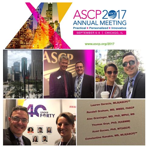
Among the endless list of educational events and sessions, I especially enjoyed being a collaborator in round table discussions including “The Benefits of Data Integration in Clinical Decisions.” I truly enjoy these roundtables and wish I could have done more of them! Topics included effective feedback, utilization and evaluations, education, and global health initiatives—all of which I’m sure I’ve written about in one way or another this past year. Seeing some of the content in my online Lab Management University (LMU) modules applied in real-life situations was reaffirming. Attending sessions and meeting renowned experts from informatics to hematopathology was exciting. The keynote speakers were captivating. Dr. Birx’s discussion on PEPFAR and global health initiatives clearly piqued my interests, and Drs. Caliguiri and Pritchard’s lectures on analytics and resources spoke directly to my work in cancer research. And don’t forget: it’s all worth continuing education credit—can’t beat that.

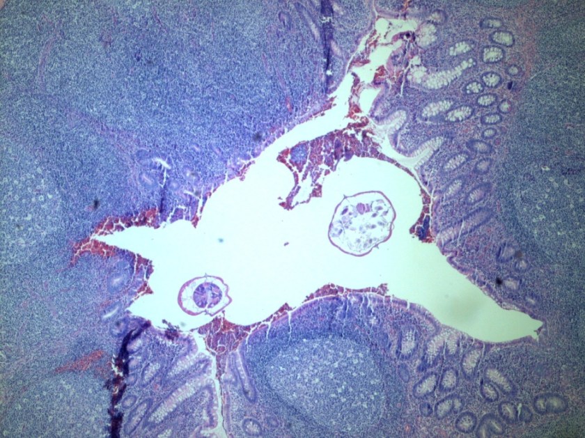
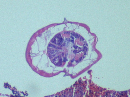
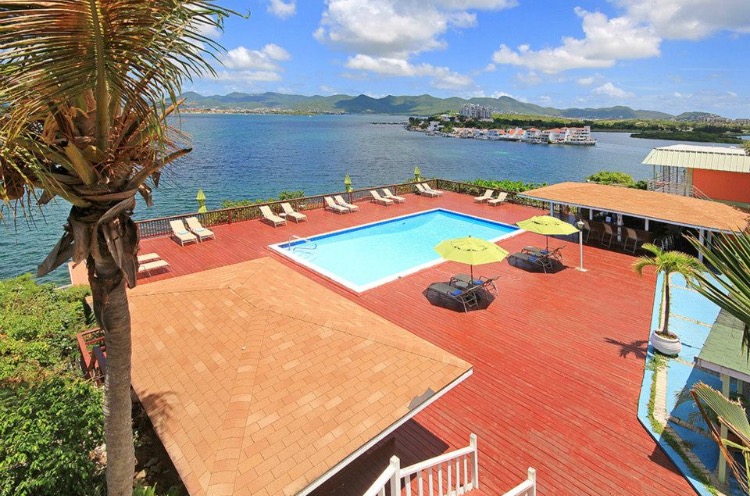
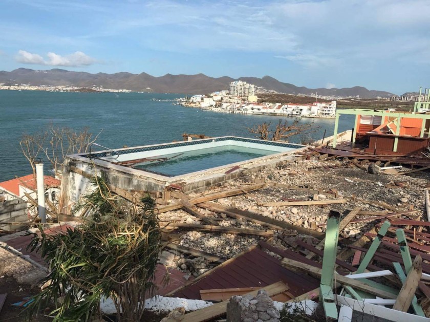
I would just like to simply thank ASCP again for all the work that goes into these meetings. I know from experience that planning large events involves quite a bit of logistics. And in managing these events ASCP truly provides an excellent environment to collaborate and learn. What brought this appreciation for logistics to the forefront was a disaster that had unfolded in the week prior to the meeting. On the island of Sint Maarten, the location of my medical school and my home for two years, was absolutely decimated by hurricane Irma. The school managed to withstand for the most part intact and acted as a shelter for students, faculty, and family. While being sheltered from that storm, endless homes, apartments, and business were destroyed. Taking nothing but a suitcase or two to campus ended up the only possessions many people in the AUC community had left. The school and its administration did a spectacular job creating a stable, safe, and even comfortable environment for students and their families while evacuation efforts were organized. While AUC managed to get students off the island via military assistance and/or charter flights, evacuees were taken to the Chicago suburbs. Right after the ASCP Annual Meeting I began having conversations with contacts in the Chicago Department of Public Health and Emergency Preparedness to offer assistance. I provided contact data, relayed satellite telephone numbers to the right contact points, and provided relevant information regarding demographics, health, and needs. Both my wife (a trained nursing leader) and myself were happy to be involved with connecting critical points in this process. All the students and their families were accounted for and taken care of in the Chicago suburbs, and were later moved to the new school location relatively unscathed. Logistics from a distance can be difficult, especially when it’s behind-the-scenes. A lot of lab decisions are made that way, and ultimately we do our best for our patients.

annualmeeting2
Image 2. Resort and villas halfway between my apartment and AUC campus on Sint Maarten before the hurricane.


annualmeeting3
Image 3. Resort and villas halfway between my apartment and AUC campus on Sint Maarten after the hurricane.

The take home message: be flexible, be humble, and be helpful. If we want to improve patient outcomes, we need to work with evidence-based approaches matched with intelligent compassion. As laboratorians, we apply our scientific approach to critical life-saving algorithms. This was no exception. Lessons discussed at the Annual Meeting between networking colleagues and official sessions are accurate. Tap into your resources, keep an active and dynamic network, know what you can do and what you cannot, and always try to help. That’s what makes a good laboratorian, a good clinician, a good friend, and hopefully a good physician.

Thanks for reading! If you’re interested in donating to disaster relief for anyone affected by this year’s violent hurricane season in Sint Maarten, visit www.rotarysxm.org.


ckanakisheadshot_small

Constantine E. Kanakis MSc, MLS (ASCP)CM graduated from Loyola University Chicago with a BS in Molecular Biology and Bioethics and then Rush University with an MS in Medical Laboratory Science. He is currently a medical student at the American University of the Caribbean and actively involved with local public health.

Hematopathology Case Study: A 56 Year Old Male with an Enlarged Lymph Node

Case History

A 56-year-old male with a past medical history significant for HIV currently on HAART presented to his primary care physician with an isolated enlarged left inguinal lymph node. In the context of his immunocompromised state, the patient was sent for a core needle biopsy of the lymph node to further elucidate the etiology of the isolated lymphadenopathy.

Diagnosis

luetiche20x
H&E, 20x
luetiche50x
H&E, 50x
leutiche100x
H&E, 100x
luetictrep
Treponema immunoperoxidase

The core needle biopsy demonstrated multiple suppurative granulomata with a mixed inflammatory background including abundant plasma cells. The plasma cells are also found to surround small blood vessels. A Treponema immunostain was performed which highlighted the spirochetes. Overall, the diagnosis is that of luetic lymphadenitis.

Discussion

Syphilitic infections can cause isolated lymphadenopathy, especially in the inguinal lymph nodes. The morphologic features of luetic lymphadenitis include interfollicular plasmacytosis, capsular fibrosis, endarteritis, and occasionally sarcoid-like granulomata with rare cases demonstrating suppurative features. The differential diagnosis includes rheumatoid arthritis associated lymphadenopathy but a key histologic difference is that the capsular fibrosis of luetic lymphadenitis will have an infiltrate of lymphocytes and plasma cells while RA associated lymphadenopathy traditionally does not. Immunohistochemistry for Treponema organisms also serves to confirm the diagnosis. It is important to keep in mind the patient’s clinical history when interpreting the biopsy was as well as the differential for interfollicular plasmacytosis with capsular fibrosis.


PhillipBlogPic-small

-Phillip Michaels, MD is a board certified anatomic and clinical pathologist who is a current hematopathology fellow at Beth Israel Deaconess Medical Center, Harvard Medical School, Boston, MA. His research interests include molecular profiling of diffuse large B-cell lymphoma as well as pathology resident education, especially in hematopathology and molecular genetic pathology.

Microbiology Case Study: A 2 Week Old Female with Eye Discharge

Case History

A 2 week old African American female presented to the pediatric emergency department (ED) with erythema, swelling and copious mucopurulent discharge from the right eye. One week earlier, her Mom noted similar symptoms in the left eye which spontaneously resolved. Mom denied fever, irritability, lethargy, rash, and respiratory or urinary symptoms. The baby was born at term through a spontaneous vaginal delivery with no complications. Mom received regular prenatal care and all screening tests were negative. The baby received erythromycin eye ointment at birth prior to initial discharge. Complete blood count showed a slight leukocytosis (WBC 15.7 TH/cm2) and cerebral spinal fluid (CSF) values were unremarkable. A complete sepsis work up was performed with blood, CSF, eye swabs and urine sent for bacterial cultures. Given the high suspicion for a sexually transmitted infection, an eye swab was also collected for Neisseria gonorrhoeae and Chlamydia trachomatis polymerase chain reaction (PCR). Herpes simplex virus PCR from the CSF was also performed. The patient was started on IV ampicillin, cefotaxime and oral erythromycin in the ED.

Lab results

neiss1
Image 1. The eye swab showed growth of glistening, grey bacterial colonies on sheep blood and chocolate agars after 48 hours incubation at 35°C in 5% CO2.
neiss2
Image 2. Gram stain of the bacterial colonies showing uniform Gram negative diplococci.

The organism was positive for both catalase and oxidase and identified by matrix-assisted light desorption ionization- time of flight (MALDI-TOF) as Neisseria meningitidis. The health department also confirmed the identification. PCR of the eye swab was negative for Neisseria gonorrhoeae and Chlamydia trachomatis. Bacterial cultures from the blood, CSF and urine were all negative.

Discussion

Neisseria meningitidis is an encapsulated Gram negative diplococcus (Image 2) that is usually transmitted through large droplet secretions from the oropharynx from colonized individuals. It can cause invasive meningococcal disease, which can present as meningitis (high fever, stiff neck, and headache), acute sepsis or a combination of both. Waterhouse Friderichsen-syndrome can result in severe dissemination forms of the disease and is characterized by petechial hemorrhages, involvement of the adrenal glands, and disseminated intravascular coagulopathy (DIC). Rarely, N. meningitidis can cause acute bacterial conjunctivitis (1.5 % – 2.5% of cases). Local complications, including corneal ulcers or a more systemic disease, may occur as well.

N. meningitidis produces multiple virulence factors that help cause disease and evade human immune defense mechanisms. The polysaccharide capsule represents the major virulence factor and is also the basis of meningococcal serotyping. Twelve different capsular serotypes can be distinguished, with serotypes A, B, C, W, X, and Y accounting for most invasive disease worldwide. Other virulence factors include pili, which helps the bacteria attach to host surfaces, and IgA protease, an enzyme that cleaves IgA and allows the bacteria to escape the humoral portion of the immune system.

In the laboratory, N. meningitidis grows well on both blood and chocolate agars after 24 hours of incubation (Image 1) and it is positive for both catalase and oxidase. Traditionally, sugar fermentation was used to differentiate Neisseria species from one another. N. meningitidis ferments both glucose and maltose whereas N. gonorrhoeae is only capable of fermenting glucose. Currently, more rapid identification methods (MALDI-TOF, PCR and sequencing) are being increasingly used in most laboratories for a faster and more accurate identification of Neisseria species. The work up of suspected N. meningitidis isolates must be performed using BSL 2 standards, as aerosols created during mobilization from culture plates or performance of biochemical testing has been known to cause invasive disease in laboratory workers.

In general, N. meningitidis is susceptible to penicillin and cefotaxime, but susceptibility testing by disk or gradient diffusion is recommended. Both rifampin and ciprofloxacin can be used for chemoprophylaxis in close contacts of the patient and healthcare & laboratory workers. In addition, a number of meningococcal vaccines are available in the United States (US) and the Centers for Disease Control & Prevention (CDC) recommends vaccinating all adolescents and people at high risk for infection (college students, military recruits, those who had a splenectomy and patients with complement deficiencies). The most common vaccine is a quadrivalent polysaccharide-protein conjugate vaccine which covers serotypes A, C, W and Y. Recently in 2014, the Food and Drug Administration (FDA) approved Trumenba, a vaccine effective against serotype B, which a common serotype causing invasive disease in the US.


AS-small

-Akram Shalaby, MD, is a first year anatomical and clinical pathology resident at the University of Mississippi Medical Center.

Stempak

-Lisa Stempak, MD, is an Assistant Professor of Pathology at the University of Mississippi Medical Center in Jackson, MS. She is certified by the American Board of Pathology in Anatomic and Clinical Pathology as well as Medical Microbiology. She is the director of the Microbiology and Serology Laboratories. Her interests include infectious disease histology, process and quality improvement and resident education.

Team Dynamics

Teams are one of the most discussed work units. We throw “teamwork” and “team ethic” around during job interviews and performance reviews. When we apply for jobs, we highlight our teamwork capabilities on our resumes. Teams are indeed essential to productive work environments because they are the vital learning units in an organization. In other words, when teams learn, the entire organization learns.

But what constitutes a team exactly? Simply put, teams are a group of two or more people that have a shared goal. Not only that, they are committed to the team process and use team language (“we” instead of “I”) when discussing accomplishments and failures. Teams also focus on learning, whether that learning comes from outside information, success, or failure. Finally, teams possess a strong sense of commitment and accountability.

Teams that consistently perform above expectations are called high-performance teams. Everyone wants to be a part of a high-performance team, but how do they happen? These teams consistently have one trait in common: experiencing and working through conflict. Conflict is one of THE best things that can happen to a team, because when handled and resolved well, teams learn, grow, and function better as a unit.

Each person has different preferences for their role on a team. Everyone gravitates toward one these five team roles: Creator, Advancer, Refiner, Executor, and Flexer. The Creator focuses on generating ideas; the Advancer communicates the ideas; the Refiner challenges ideas; the Executor implements the ideas; and the Flexer assumes any of the other four roles based on the needs of the team. High-performing teams have members who are in their preferred role where they can excel and are sustained because those roles give them energy. It is our job as leaders to find jobs, tasks, and team roles where others can flourish. Without energized people, leaders will not be able to create high-performing teams or high-performing organizations.

lotte-small

-Lotte Mulder earned her Master’s of Education from the Harvard Graduate School of Education in 2013, where she focused on Leadership and Group Development. She’s currently working toward a PhD in Organizational Leadership. At ASCP, Lotte designs and facilitates the ASCP Leadership Institute, an online leadership certificate program. She has also built ASCP’s first patient ambassador program, called Patient Champions, which leverages patient stories as they relate to the value of the lab.


The team dynamics module gave me a great insight into my tendencies and an understanding on effectively getting the best out of teams.

When I received my assessment results, I learned I am an “advancer.” I tend to focus on execution and I pay attention to team interactions. One weakness of this profile, though, is how it potentially interacts with creators and refiners. If one is unaware of the valuable differences in prospective creators and refiners bring to the table, an advancer may get frustrated working with them in a team.

I came to realize I should be sure to include a refiner on the team, since having only executors or advancers could mean bypassing the analysis piece. I also gained new prospective on how I perceive creators. While in the past I may have discounted them as scattered or unrealistic, I learned this stems from the fact I like concrete ideas. Embracing innovation is essential to advancement and this is where creators excel.

The sections about communication and team roles were enlightening. How creators can easily get bored with discussions that are too concrete, and how executors are uninterested in theoretical discussions. On the flip side of things, creators need to partner with advancers, refiners and executors to bring about innovation. Advancers rely on refiners and refiners can benefit from the enthusiasm and networking of the advancers. I use these important concepts now in meetings and when I try to put teams together for a given project.

Although this may come with time, leadership and team member selection are paramount to foster the trust and respect and to facilitate free expression of ideas and sharing of information.

wareth-small

-Laila Osama Abdel Wareth, MBBCh, FCAP, MRCPC, EMHCA is the Chief of Clinical Pathology for the Pathology & Laboratory Medicine Institute at the Cleveland Clinic Abu Dhabi in the United Arab Emirates.


Microbiology Case Study: An 8 Year Old with Acute Appendicitis

Case History

An 8-year-old female presented to an outside hospital with appendicitis-like clinical symptoms and underwent a laparoscopic appendectomy. Gross examination of the appendix (7.2 cm in length x 0.5 cm in diameter) wall was unremarkable and the lumen contained a minimal amount of hemorrhage. The specimen was entirely submitted for microscopic evaluation.

entver1

Image 1. Cross section of appendix containing two intra-luminal helminths (H & E stain).

entver2

Image 2. Cross section of female Enterobius vermicularis containing eggs (H & E stain).

entver3

Image 3. Cross section of male Enterobius vermicularis (H & E stain).

Discussion

Enterobius vermicularis (human pinworm) is an intestinal nematode (roundworm) with a worldwide distribution that is most prevalent among school-age children. Cross sections of the nonsegmented, cylindrical worms demonstrate a well-developed digestive tract, reproductive system, and two lateral alae (Images 1-3). E. vermicularis has two sexes and Image 1 demonstrates that the male is smaller than the female. Humans are directly infected upon ingestion of E. vermicularis eggs (fecal-oral route of transmission). The eggs then hatch and immature worms undergo maturation within the human gastrointestinal tract (Image 1). Eggs are shed in stool and the typical E. vermicularis eggs (Image 2) are thick-shelled with one flattened aspect, described as “D-shaped”. Patients with the infection are commonly asymptomatic or may complain of perianal pruritus. Rarely, patients present with abdominal pain secondary to E. vermicularis-associated acute appendicitis (1).

Reference

  1. Arca MJ, Gates RL, Groner JI, Hammond S, Caniano DA. 2004. Clinical manifestations of appendiceal pinworms in children: an institutional experience and a review of the literature. Pediatr Surg Int 20(5):372-5.


-Adina Bodolan, MD is a 1st year anatomic and clinical pathology resident at the University of Vermont Medical Center.

Wojewoda-small

-Christi Wojewoda, MD, is the Director of Clinical Microbiology at the University of Vermont Medical Center and an Associate Professor at the University of Vermont.

Bringing it Home

A recent report from the Centers for Disease Control (CDC) found that twenty-four laboratory workers were infected with a strain of Salmonella typhimurium, an enteric pathogen. The infections were reported in sixteen states across the country. Of those infected, six were hospitalized with symptoms such as diarrhea, fever, and severe abdominal cramps. Luckily, there were no deaths reported. These infections occurred in various teaching and clinical laboratories. The worst part? This could have been avoided.

When interviewed, some of those who became ill said they remembered specific exposure events. Many others who were unsure of how they became exposed described unsafe behaviors in the laboratory. Those victims admitted to working in the lab setting without lab coats or gloves, and many reported not washing their hands before leaving the department.

If you’re a laboratory leader, you very likely work during the day shift. Hopefully, when management is on site, staff is compliant with safety. If not, you may need to examine your safety program and leadership style. Do you enforce safety regulations in the lab? Do you lead by example? Do you don PPE when you pick up the phone or use a computer in the lab?

If safety seems to be good during the day, you may want to make a visit during the off-shifts. Depending on the level of safety culture, there may be anything happening from solid safe practices to open eating and drinking in the department. I know that was the norm in many labs 25 years ago, but those unsafe practices and safety violations should now be ancient history. Unfortunately, that is not the case, and that is one reason we have bacterial infection outbreaks in our laboratories.

An experienced lab auditor will tell you it is not difficult to assess the lab safety culture in a department, even on inspection day. I once entered a lab as part of an accreditation inspection team, and I watched as the lab staff struggled to find gloves. Even though they knew the inspection was imminent, they could not hide the fact that glove use was not the norm for them in that lab. A complete lab safety audit can reveal a number of inappropriate practices such as improper PPE use, gum chewing, cell phone use, and many others.

The National Institute for Occupational Safety and Health (NIOSH) has educated workers for years about hazard and exposure control. The “Hierarchy of Controls” is an excellent model to use in the laboratory setting, although certain facts about it may be surprising. The first and best two controls to remove hazards are elimination and substitution. Of course, these are not always possible in the lab setting. While there are substitutes for hazardous chemicals, the inherently dangerous specimens that are handled cannot be replaced or removed.

Engineering controls create physical barriers between the hazard and the employee. Biological Safety Cabinets (BSCs) and Chemical Fume Hoods are powerful engineering controls. Administrative and Work Practice controls are the safety policies and actual practices that help prevent infection. Written safety procedures are designed to change the way people work, and standard work practices include not eating or drinking in the lab setting and practicing hand hygiene when necessary.

The final control for infection prevention is Personal Protective Equipment (PPE). In the hierarchy, PPE is considered the last resort for staff protection. Since the lab hazard cannot be eliminated, and since humans commit errors with procedures, that final method of protection must be utilized. Lab coats, gloves and face protection need to be used at all times when working in the laboratory. Without it, the worker is at great risk for exposure- and that is what happened in the labs where the Salmonella infections occurred. Each of the controls that should be in effect in the lab were bypassed, and there were consequences.

It is always better to read about incidents that occur in other laboratories rather than have to report them about your own. When I hear of such stories, I always look at my own labs to see if such an event could occur there. What opportunities exist in my lab safety program? What about yours? Be sure to learn from these unfortunate events and keep your own staff safe.

The personal (and probably painful) part of the infection outbreak was that these laboratory workers were infected on the job, and then they brought it home. The CDC report says nothing about infections being spread to family members or friends, but it certainly could have happened. If there are weaknesses in your lab safety program, what could your staff be bringing home? What infections or diseases could be spread because of unsafe work practices? Now is the time to take the lead for your safety program before such an event can occur. Bring safety home for your staff. Teach them and lead them so that the unsafe practices of the past turn into practices that keep everyone healthy into the future.


Scungio 1

Dan Scungio, MT(ASCP), SLS, CQA (ASQ) has over 25 years experience as a certified medical technologist. Today he is the Laboratory Safety Officer for Sentara Healthcare, a system of seven hospitals and over 20 laboratories and draw sites in the Tidewater area of Virginia. He is also known as Dan the Lab Safety Man, a lab safety consultant, educator, and trainer.

Guest Post: Drone Transport of Specimens

On a hot afternoon in late September 2016 the Johns Hopkins Medical Drones team drove to a flight field in the Arizona desert with 40 vacutainer tubes filled with human blood obtained from volunteers. The individually wrapped tubes sat in two custom-designed white plastic cooler boxes which had wires coming out of one end, ventilation holes at the other, and ran off the drone’s battery power. We carefully placed one of the boxes on the drone, stood back, and flew the samples around for 260 kilometers in what seemed like an unending series of concentric circles. Great. But why would doctors be involved in this exercise?

For the last 3 years, the Johns Hopkins medical drones team has examined the stability of human samples transported via drone. Our approach has been similar for each study. Get two sets of samples, fly one on the drone, then take both sample sets back to laboratory for analysis to see if there are any changes. However, until this study in Arizona we had only flown these samples up to about 40km, in mild weather, and for up to 40 minutes at a time. A request to set up a drone network in a flood-prone area of a country in Southwestern Africa made us realize that we needed to repeat the stability tests in warmer weather and for longer flights. This drone network would serve clinics that were up to 50 km away from each other, therefore requiring round-trips of at least 100km. Once we received this request it became clear pretty quickly that our previous tests flying for to 40km were not good enough for an aircraft that would have to fly in a hot environment between several clinics that were each 50km away from each other.

After the 3-hour 260km flight, we took both sets of samples back to the Mayo Clinic laboratories in Scottsdale, Arizona and performed 19 different tests on the samples. Each pair of samples was compared to check for differences between the flown and not-flown sample sets. Although results from sample pairs were similar for 17 of the 19 tests, small differences were seen in Glucose and Potassium, which do also vary in other transport methods. We suspect the differences seen in this test arose because the not-flown samples were not as carefully temperature controlled as the flown samples in the temperature-controlled chamber. This study (which is the longest flight of human samples on a drone to date) shows that drones can be used for blood samples even for long flights in hot conditions. However, the temperature and other environmental variables must be well-controlled to keep the blood stable.


TimAmukele-small

-Dr. Timothy Amukele is an Assistant professor in the Department of Pathology at the Johns Hopkins School of Medicine and the Director of Clinical Laboratories at Johns Hopkins Bayview Hospital. He is also the Medical Director of two international research laboratories in Uganda and Malawi. He has pioneered the use of unmanned aerial systems (colloquially known as drones) to move clinical laboratory samples.

Jeff Street-small

-Jeff Street is an unmanned systems engineer and pilot at the Johns Hopkins School of Medicine with more than 10 years of experience in the development of new and innovative vehicles. He is leading the Johns Hopkins aircraft development efforts for a wide range of medical cargo applications.


Microbiology Case Study: Specimen Referral from a 20 Month Old Male

Case History

A 20 month old male presented to an outside hospital with symptoms unknown to our laboratory. That laboratory sent us the specimen recovered from a diaper (Image 1).

asc1
Image 1.

Discussion

The nematode Ascaris lumbricoides is one of the most common helminth infections in the United States. It can grow to be 20-35 cm long. Infection occurs when an egg is ingested, usually in a small child eating dirt contaminated with human feces. When the larvae hatch they penetrate the duodenal wall. From there, the larvae go into the blood stream and eventually end up in the pulmonary circulation where the larvae grow in the alveoli.  In about three weeks, the larvae are coughed up from the lungs and swallowed.  The worms then mature in the jejunum (primarily).  Infection most often shows no symptomatology. If symptoms are present, they can range from mild abdominal discomfort to intestinal blockage and even cough as the worms migrate to the lungs [1].

Diagnosis can be made by examining concentrated stool for knobby-coated, bile-stained eggs that are oval [2].  However, some of the adult worms can pass with the feces.

References

  1. https://www.cdc.gov/parasites/ascariasis/index.html
  2. Murray PR, Rosenthal KS, Pfaller MA. Medical Microbiology, Seventh Edition. Elsevier Health Sciences; 2012.


-Angela Theiss, MD is a 1st year anatomic and clinical pathology resident at the University of Vermont Medical Center.

Wojewoda-small

-Christi Wojewoda, MD, is the Director of Clinical Microbiology at the University of Vermont Medical Center and an Associate Professor at the University of Vermont.

Here’s to Fresh STARTs: Sustainable Transitions And Reachable Targets

From the title, you might be able to tell that I’ve been busy on Lab Management University (LMU). Going through the online modules and lessons in the LMU certificate program I mentioned this past May, I’ve been able to hone several skills in interpersonal communication, resource management, and project leadership. (A worthwhile investment through ASCP which I highly recommend!) Another thing that’s kept me quite busy over the last two years has been Zika-virus and mosquito-related public health initiatives both inside and outside the laboratory. In a recent blog post, I discussed the correlation between measuring success in projects like these just as one would with common lab-centric goals. And, as a conclusion to that hard work, this will be my last directly-related Zika/public health post. Transitioning to the second half of medical school, I’ll be leaving behind a true grassroots project that not only reached countless people but has the promise to be sustainable after my departure from the island of Sint Maarten back to the states.

As with many times in life, I would say fresh starts are a welcome chance to reflect and grow upon things you might have learned or goals you might be closer to finishing. What has been made clear to me in my time working through classrooms, cases, exams, and projects is that the “jargon” we use as laboratorians is definitely worth its weight. It isn’t full of hollow charges for metrics and goals; it’s about real data and real solutions. Having the ability to apply my prior experiences in laboratory medicine throughout medical school—both inside and outside the classroom—has been an invaluable benefit. The general principles that guided my last blog post reflected simple goals (i.e. turnaround time compared with public health metrics) which utilize fundamental models of data collection, adjustment, and success. The essential model I described a few months ago is now a mainstay of a project that will continue to improve public health statistics slowly as time goes on.

8-28-1

The model as it stands now consists of clear steps to identify problems which require interventions, highlight gaps in current practice, data collection from literature exists currently, collaboration with partners in a community of trust, and continuing those partnerships as improvements are made incrementally over time. The model has been repeated and successfully modified for these last two years from on-campus blood testing with procedural write-ups and data evaluation, to teaching school-aged children about mosquito prevention, to partnering with local government officials and having your projects adopted into their portfolio, and visiting individuals in their homes to discuss health and prevention.

To keep it brief, I’ve had an amazing experience here being able to lead and contribute to a wonderful and impactful project such as this. It has become increasingly clearer to me throughout this work that the values and skills programs like LMU teach are directly parallel with successes in various clinical settings. From the bedside to the laboratory to public health in the field, the lessons of how to effectively engage people and solve problems are critical. My time in laboratories before medical school, the daily grind of classwork here, and the projects I’ve been able to write about have all given me the space to try these skills on real situations—and hopefully that will make me the best clinician I can be after medical school is completed. Check out my “highlight reel” of partnerships, workshops, and active management in Zika prevention below.

Be sure to check back here next time, I’ll be writing from my hometown as I’ll explore ASCP’s Annual Meeting in Chicago, IL this coming September and report back on why it’s important to network and stay involved with our great professional community. Thanks for reading!

8-28-2

8-28-3


ckanakisheadshot_small

Constantine E. Kanakis MSc, MLS (ASCP)CM graduated from Loyola University Chicago with a BS in Molecular Biology and Bioethics and then Rush University with an MS in Medical Laboratory Science. He is currently a medical student at the American University of the Caribbean and actively involved with local public health.