Hematopathology Case Study: A 45 Year Old Man with Cytopenias and High Ferritin

Case history

A 45 year-old man presented with vomiting and diarrhea for 5 days. Laboratory studies demonstrated anemia and thrombocytopenia, an elevated ferritin level (23,772 ug/L) and methemoglobinemia. Chest roentgenography revealed cardiomegaly. A follow-up ECHO showed a desreased ejection fraction of 15%. Work-up confirmed viral myocarditis and G6PD deficiency as the cause of the cardiac findings and methemoglobinemia respectively. His clinical condition deteriorated despite therapy: he developed acute kidney and liver failure and had worsening cytopenias. A bone marrow biopsy was performed.

Histomorphological findings

image-121
Bone marrow aspirate smear (100x)
BM17-228 HLH 100x-2
Bone marrow aspirate smear (100x)
CPC Core biopsy 20x
Bone marrow core biopsy (H&E, 20x)
CPC Core biopsy 40x
Bone marrow core biopsy (H&E, 40x)
CPC Core biopsy 100x
Bone marrow core biopsy (H&E, 100x)

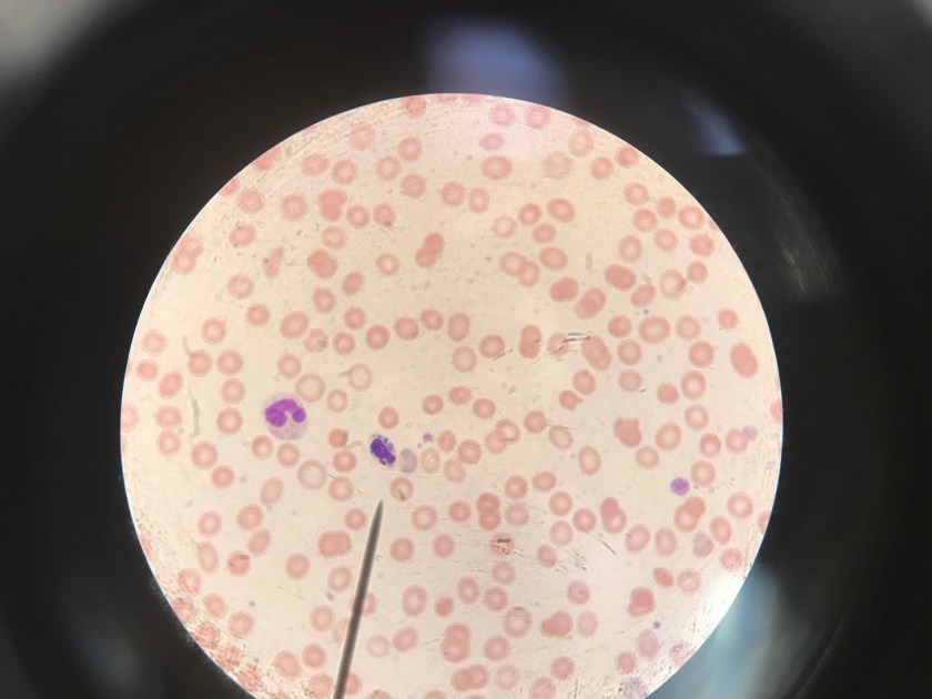
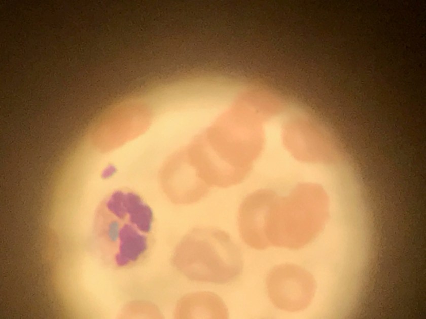
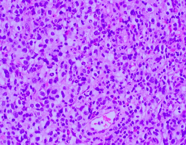
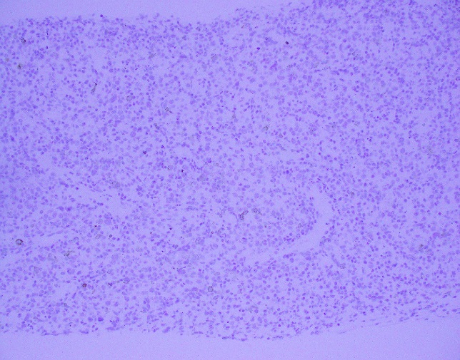
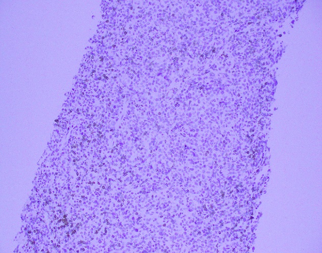
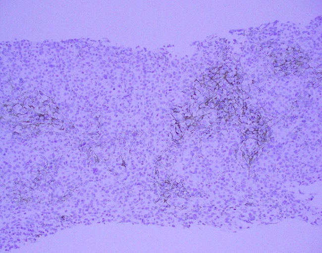
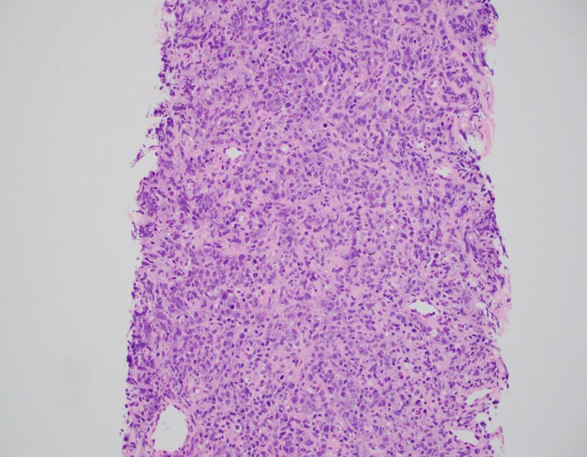
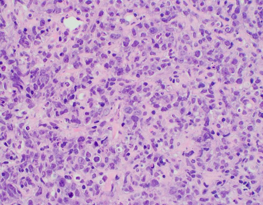
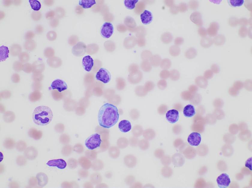
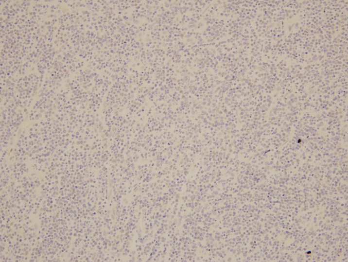
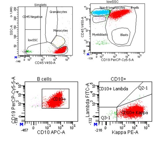
Evaluation of the peripheral blood (not pictured) confirmed a macrocytic anemia with marked anisopoikilocytosis including schistocytes, polychromasia, nucleated red blood cells, absolute neutrophilia, monocytosis and thrombocytopenia. The marrow aspirate smear demonstrated appropriate maturation in all cell lines. Scattered hemophagocytic histiocytes (pictured above) were noted. The bone core biopsy was high-normocellular for age with progressive trilineage hematopoeisis. Scattered histiocytes with internalized erythroid cells and debris were visualized. There was no increase in blasts. Flow cytometry analysis performed on the bone marrow aspirate did not show a significant increase in blast population. Gating on the lymphocytes did not show a B-cell monoclonal population or T-cell abnormality by markers assayed.

Diagnosis

High-normocellular marrow with progressive trilineage hematopoeisis and prominent hemophagocytic histiocytes.

Overall the patient met the clinical criteria (see below) for Hemophagocytic Lymphohistiocytosis (HLH); with fever (≥38.5 C), splenomegaly, bicytopenia, presence of hemophagocytic histiocytes in bone marrow and high ferritin level (>500ng/mL).

A clinical diagnosis of HLH was rendered.

Discussion

HLH is an uncommon hematologic disorder that is often fatal.  The underlying pathogenesis involves an exaggerated but ineffective inflammatory response of excessive macrophage and T-cell activation, and impairment of natural killer (NK) and cytotoxic T-cell function. HLH has familial and acquired forms. Secondary, or acquired HLH can be associated with infections (especially viral etiologies), underlying malignancy (particularly lymphomas and leukemias), and medications used for systemic lupus erythematosus. Clinically, autoimmune disease-associated HLH overlaps significantly with macrophage activation syndrome (MAS).

HLH is a clinical diagnosis that can be established with molecular testing or by meeting five of eight clinical and laboratory diagnostic criteria according to the HLH-2004 guidelines.

HLH-2004: Revised diagnostic guidelines for HLH10

The diagnosis HLH can be established if one of the two criteria below is met:

  1. A molecular diagnosis consistent with HLH (i.e., reported mutations found in either PRF1 or MUNC13-4); or
  2. Diagnostic criteria for HLH are fulfilled (i.e., at least five of the eight criteria listed below are present:
    • Persistent fever
    • Splenomegaly
    • Cytopenias (affecting ≥2 of 3 lineages in the peripheral blood):
      • Hemoglobin <90g/L (in infants <4 weeks: <100g/L)
      • Platelets <100 x 109/L
      • Neutrophils <1.0 x 10⁹/L
    • Hypertriglyceremia and/or hypofibrinogenemia:
      • Fasting triglycerides ≥3.0 mmol/L (i.e., ≥ 265mg/dl)
      • Fibrinogen ≤1.5 g/L
    • Hemophagocytosis in bone marrow* or spleen or lymph nodes, no evidence of malignancy
    • Serum ferritin ≥ 500µg/L (i.e., 500 ng/ml)
    • Low or absent NK cell activity (according to local laboratory reference)
    • Increased serum sIL2Rα (according to local laboratory reference)

*In hematopathology, the finding of relevance is the presence of hemophagocytic histiocytes in the marrow or other biopsies organs. While debris-laden histiocytes are commonly noted in marrow aspirates, the findings of engulfed erythroid cells is warranted to call a ‘hemophagocytic’ histiocyte.

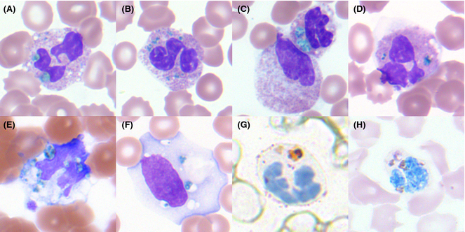
Often a bone marrow biopsy will be performed in cases where there is clinical suspicion for HLH. This serves to try and visualize the hemophagocytic activity, and to rule out other diseases with similar clinical presentations as HLH. The pathologic evaluation of HLH is tricky, since there is no established criteria for quantitation of hemophagocytic histiocytes in a bone marrow aspirate. Furthermore, hemophagocytosis is not specific to HLH and can be seen in other conditions such as: post-blood transfusion, chemotherapy, sepsis and major operations. Published data shows that the presence of hemophagocytosis has a sensitivity of 83% and a specificity of only 60% in diagnosing HLH.

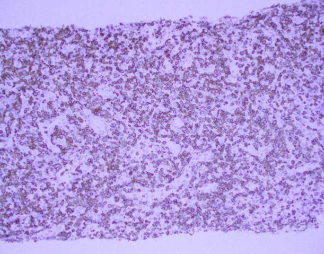
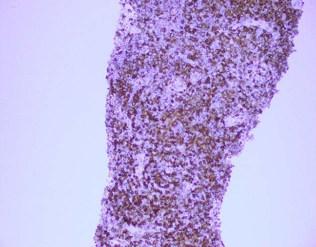
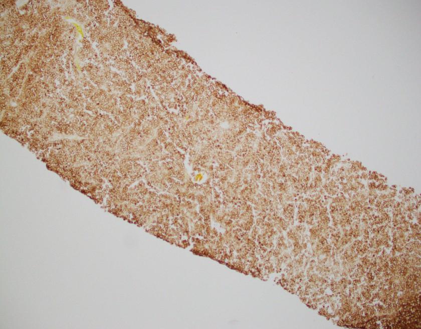
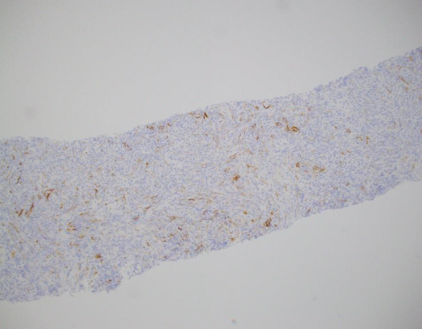
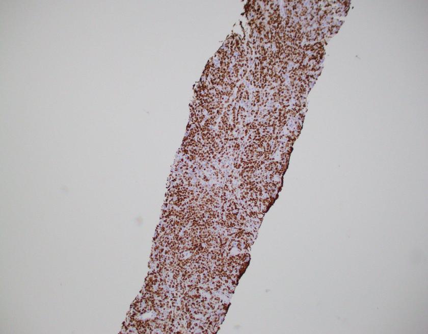
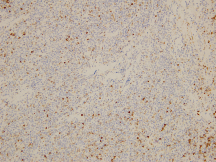
What about immunohistochemical staining for the histiocytes? While IHC can help outline histicoytic cells, unfortunately, quantitation of hemophagocytic histiocytes in the core biopsy or clot sections with the aid of CD68 immunostains does not correlate well with disease probability either.

Overall, the nonspecificity of hemophagocytosis in the marrow, even when present in high amounts, should remind both pathologists and clinicians that an isolated finding of hemophagocytosis lacks specificity and does not necessarily suggest HLH when the clinical presentation and laboratory findings are not compatible with the diagnosis. However, there still remains value in bone marrow biopsy examination in cases where clinical suspicion for HLH is high; in order to exclude other marrow processes; and in the rare case where there may not have been clinical suspicion of HLH but the presence of hemophagocytic histiocytes can raise that differential.

For additional images of hemophagocytic histiocytes check out these amazing picture tweets by Dr. Kate Dannheim (@KDannheimMD) https://twitter.com/KDannheimMD/status/933128799818002432) and Dr. Bharat Ramlal (@BeRaad87): https://twitter.com/HHPathology/status/926087105381531648


References

  1. Ho C, Yao X, Tian L, et al. Marrow Assessment for Hemophagocytic Lymphohistiocytosis Demonstrates Poor Correlation with Disease Probability. Am J Clin Pathol. 2014 Jan;141(1):62-71
  2. Hunter JI et al. HLH-2004: Diagnostic and therapeutic guidelines for hemophagocytic lymphohistiocytosis. Pediatr Blood Cancer. 2007 Feb;48(2):124-31.


AIK

-Ayse Irem Kilic is a 1st year anatomic and clinical pathology resident at Loyola University Medical Center. Follow Dr. Kilic on twitter @iremessa.

Mirza-small

-Kamran M. Mirza, MD PhD is an Assistant Professor of Pathology and Medical Director of Molecular Pathology at Loyola University Medical Center. He was a top 5 honoree in ASCP’s Forty Under 40 2017. Follow Dr. Mirza on twitter @kmirza.

Hematology Case Study: Crystal of Death

Case History

A 56 year old female presented with symptoms of sepsis. During surgery, patient bleed profusely and received blood products. However, the patient expired.

Laboratory Findings

  • WBC: 18.8 x 10⁹/L
  • Hemoglobin: 5.6 g/dL
  • Lactate: 8.3 mmol/L
  • AST: 1485
  • ALT: 1625

The blood smear was reviewed for these white blood cell inclusions:

green-incl-1
Image courtesy of Georgia McCauley, PhD, MT(AMT)
green-incl-2.jpg
Image courtesy of Georgia McCauley, PhD, MT(AMT)

Discussion

The image presented reveals to be the “blue green crystal of death”. Medical literature has documented an association between acute hepatic failure and coarse, bright-green neutrophilic inclusions. Upon identification of these unique inclusions patients have been reported to have poor outcomes and usually die within 24-72 hours (Haberichter KL, 2017). The exact nature of these inclusions has yet to be determined; it is postulated that they arise from lipofusion-like substance. Bright green inclusions in neutrophils have been reported as a sign of impending patient death (Hodgson, 2015).

Refractile bright-green irregular inclusions within neutrophils have been reported as a marker of impending patient death. In the three reported cases, death occurred within 2 d of recognition of the inclusions (Harris et al2009; Jazaerly & Gabali, 2014). Disease associations with green neutrophil inclusions included acute liver failure secondary to acetaminophin overdose, lactic acidosis with multisystem organ failure subsequent to trauma (Harris et al2009) and Escherichia coli-associated septic shock (Jazaerly & Gabali, 2014). Harris et al (2009) suggested that the inclusions were related to blood-borne bile products.

green-incl-3.jpg
Image 3 source: Source: Hodgson, 2015

These findings on the peripheral smear should be reported and considered a critical finding. Laboratory professionals and hematologists should acknowledge these inclusions; patients are noted to be seriously ill at the time of detection of neutrophil inclusions and have an ominous 24-72 hour survival period.

References

  1. Haberichter, K. L., & Crisan, D. (2017). Green Neutrophilic Inclusions and Acute Hepatic Failure: Clinical Significance and Brief Review of the Literature. Annals of Clinical & Laboratory Science47(1), 58-61.
  2. Harris, V.N., Malysz, J.& Smith, M.D. (2009) Green neutrophilic inclusions in liver disease. Journal of Clinical Pathology, 62, 853–854.
  3. Hodgson, T. O., Ruskova, A., Shugg, C. J., McCallum, V. J., & Morison, I. M. (2015). Green neutrophil and monocyte inclusions–time to acknowledge and report. British journal of haematology170(2), 229-235.
  4. Jazaerly, T.& Gabali, A.M. (2014) Green neutrophilic inclusions could be a sign of impending death! Blood, 123, 614.


ledesma_small

-Carlo Ledesma, MS, SH(ASCP)CM MT(ASCPi) MT(AMT) is the program director for the Medical Laboratory Technology and Phlebotomy at Rose State College in Midwest City, Oklahoma as well as a technical consultant for Royal Laboratory Services. Carlo has worked in several areas of the laboratory including microbiology and hematology before becoming a laboratory manager and program director.

Hematopathology Case Study: A 56 Year Old Male with an Enlarged Lymph Node

Case History

A 56-year-old male with a past medical history significant for HIV currently on HAART presented to his primary care physician with an isolated enlarged left inguinal lymph node. In the context of his immunocompromised state, the patient was sent for a core needle biopsy of the lymph node to further elucidate the etiology of the isolated lymphadenopathy.

Diagnosis

luetiche20x
H&E, 20x
luetiche50x
H&E, 50x
leutiche100x
H&E, 100x
luetictrep
Treponema immunoperoxidase

The core needle biopsy demonstrated multiple suppurative granulomata with a mixed inflammatory background including abundant plasma cells. The plasma cells are also found to surround small blood vessels. A Treponema immunostain was performed which highlighted the spirochetes. Overall, the diagnosis is that of luetic lymphadenitis.

Discussion

Syphilitic infections can cause isolated lymphadenopathy, especially in the inguinal lymph nodes. The morphologic features of luetic lymphadenitis include interfollicular plasmacytosis, capsular fibrosis, endarteritis, and occasionally sarcoid-like granulomata with rare cases demonstrating suppurative features. The differential diagnosis includes rheumatoid arthritis associated lymphadenopathy but a key histologic difference is that the capsular fibrosis of luetic lymphadenitis will have an infiltrate of lymphocytes and plasma cells while RA associated lymphadenopathy traditionally does not. Immunohistochemistry for Treponema organisms also serves to confirm the diagnosis. It is important to keep in mind the patient’s clinical history when interpreting the biopsy was as well as the differential for interfollicular plasmacytosis with capsular fibrosis.


PhillipBlogPic-small

-Phillip Michaels, MD is a board certified anatomic and clinical pathologist who is a current hematopathology fellow at Beth Israel Deaconess Medical Center, Harvard Medical School, Boston, MA. His research interests include molecular profiling of diffuse large B-cell lymphoma as well as pathology resident education, especially in hematopathology and molecular genetic pathology.

Hematopathology Case Study: A 68 Year Old Man with Dyspnea on Exertion

Case History

A 68 year old male with no significant past medical history who enjoys long distance cycling presented to an outside emergency department with dyspnea on exertion. Laboratory values at the outside facility showed profound anemia (Hb 10 g/dL) and physical exam revealed lymph adenopathy. The patient was discharged but presented again to his primary care physician with profound dyspnea on exertion, especially after climbing one flight of stairs. Of note, his anemia had worsened with a new Hb of 7 g/dL. For evaluation of the anemia, the patient had a Coomb’s test and it was positive, overall consistent with cold agglutinin disease. For evaluation of the lymphadenopathy, a CT abdomen and chest revealed celiac, portocaval, mesenteric and retroperitoneal lymphadenopathy as well as mild splenomegaly. Due to these findings, the patient presented to Beth Israel Deaconess Medical Center for further evaluation and biopsy of a retroperitoneal lymph node.

A core needle biopsy of a retroperitoneal lymph node was obtained per the recommendation of hematology/oncology.

Diagnosis

AITL-1
H&E, 10X
AITL-2
H&E, 20X
AITL-3
H&E, 50X
AITL-4
CD3
AITL-5
CD20
AITL-6
CD10
AITL-7
CD4
AITL-8
CD7
AITL-9
CD21
AITL-10
Ki-67
AITL-11
EBER ISH
AITL-12
PD1
AITL-13
CXCL13

The core needle biopsy material demonstrated a lymphoid population that was polymorphic in appearance with medium to large sized lymphocytes with moderate amounts of pale cytoplasm, irregular nuclei, vesicular chromatin, and some cells with prominent nucleoli. The background cellular population is composed of a mixed inflammatory component including small lymphocytes, scattered neutrophils, eosinophils, and histiocytes.

By immunohistochemistry, the medium to large sized cells with pale cytoplasm are positive for CD3, CD2, CD4, and CD5 with complete loss of CD7. CD20 highlights scattered background B-cells. CD21 is positive in disrupted follicular dendritic meshworks. CD10 and BCL6 are negative in neoplastic cells. PD1 is positive in neoplastic cells with a subset co-expressing CXCL13. By Ki-67 immunostaining, the proliferation index is 50-70%. By in situ hybridization for Epstein-Barr virus encoded RNA, a subset of cells are positive.

Overall, with the morphologic and immunophenotypic features present, the diagnosis is that of angioimmunoblastic T-cell lymphoma.

Discussion

Angioimmunoblastic T-cell lymphoma (AITL) is one of the most common types of peripheral T-cell lymphoma and accounts for 15-20% of T-cell lymphoproliferative disorders and 1-2% of all non-Hodgkin lymphomas. Clinical features include presentation with late stage disease with associated generalized lymphadenopathy, hepatosplenomegaly, systemic symptoms, and polyclonal hypergammaglobulinemia. Of note, this patient did have an SPEP that was within normal limits. Other findings, although less common, include effusions and arthritis. Laboratory findings often include cold agglutinins with hemolytic anemias, a positive rheumatoid factor (RF), and anti-smooth muscle antibodies. A hallmark of AITL is the expansion B-cells positive for EBV is seen, which may be an indicator of underlying immune dysfunction. The clinical course is often aggressive with a median survival of less than three years and often succumb to infectious etiologies because of an immune dysregulation.1

The pathogenesis and relation to other TFH neoplasms of PTCL, NOS is poorly understood. Recent literature indicates dysregulation in key pathways, including the CD28 and TCR-proximal signaling genes, NF-kappaB/NFAT pathway, PI3K pathway, MAPK pathway, and GTPases pathway.2 The complexity of these pathways has long been an issue for TFH lymphoproliferative disorders and has provided insight to potential molecular signatures (see figure 1 adapted from Vallois 2016).

AITL-14
Figure 1 from Vallois 2016

Another recent publication provided additional information regarding molecular insights. Confirmed mutational analyses reveals a high proportion of cases carry a TET2 mutation with less frequent changes in DNMT3A, IDH2, RHOA, and PLCG1. Specifically, RHOA, PLCG1, and TNFRSF21 encode proteins critical for T-cell biology and most likely promote differentiation and transformation into an aggressive clinical course (see figure 2 adapted from Wang 2017).3

AITL-15
Figure 2 adapted from Wang 2017

Overall, AITL is an uncommon TFH cell derived lymphoproliferative disorder characterized by a TFH immunophenotype, expanded and arborizing high endothelial venules, expansion of the follicular dendritic cell meshworks, and EBV positive B-cells in a background of a polymorphic infiltrate. Although it is hypothesized that the underlying mechanism of neoplasia is related to immune dysfunction, new molecular insights have demonstrated that multiple events occur ranging from early molecular changes to later acquired mutations that allow for malignant transformation.

References

  1. Swerdlow, S., et al., WHO Classification of Tumours of Haematopoietic and Lymphoid Tissues, 4th. ed., IARC press: 2008
  2. Vallois, D., et al. “Activating mutations in genes related to TCR signaling in angioimmunoblastic and other follicular helper T-cell-derived lymphomas,” 2016; 128(11): 1490-1502.
  3. Wang, M., et al., “Angioimmunoblastic T cell lymphoma: novel molecular insights by mutation profiling,” 2017; 8(11): 17763-17770.


PhillipBlogPic-small

-Phillip Michaels, MD is a board certified anatomic and clinical pathologist who is a current hematopathology fellow at Beth Israel Deaconess Medical Center, Harvard Medical School, Boston, MA. His research interests include molecular profiling of diffuse large B-cell lymphoma as well as pathology resident education, especially in hematopathology and molecular genetic pathology.

Hematology Case Study: Monocytosis in An Elderly Patient

An 81 year old presented with fatigue and not feeling too well. CBC revealed marked leukocytosis and monocytosis.

  • White cell count: 85.1 K/uL (elevated)
  • Hemoglobin: 8.6 g/dl (decreased)
  • Platelet count: 79 K/uL ( decreased)

Review of peripheral smear revealed leukoerythroblastosis with monocytosis (19.57 K/uL) along with presence of numerous immature monocytoid cells, dysplastic myeloid precursors and 7% blasts, consistent with myelodysplastic/myeloproliferative neoplasm such as chronic myelomonocytic leukemia-1.


cmml1

cmml2


cmml3

For further evaluation of disease progression and/or transformation to acute leukemia, bone marrow evaluation was recommended.

Diagnostic criteria for chronic myelomonocytic leukemia

  1. Persistent peripheral blood monocytosis >1 K/uL
  2. No Philadelphia chromosome or bcr-abl 1 fusion gene
  3. No rearrangement of PDGFRA or PDGFRB
  4. Fewer than 20% blasts in the blood or bone marrow**
  5. Dysplasia in one or more myeloid lineages.


**Blasts include myeloblasts, monoblasts and promonocytes

Prognosis and predictive factors

Survival of patients with CMML is reported to vary from one to more than 100 months, but the median survival time in most series is 2- to 40 months. Progression to AML occurs in approximately 15-30% of cases. A number of clinical and hematological parameters, including splenomegaly, severity of anemia and degree of leukocytosis, have been reported to be important factors in predicting the course of the disease.


Vajpayee,Neerja2014_small
-Neerja Vajpayee, MD, is the director of Clinical Pathology at Oneida Health Center in Oneida, New York and is actively involved in signing out surgical pathology and cytology cases in a community setting. Previously, she was on the faculty at SUNY Upstate for several years ( 2002-2016) where she was involved in diagnostic work and medical student/resident teaching.

Molecular Perspectives of Diffuse Large B-cell Lymphoma

Case

A 100 year old female was seen for follow-up for her hypertension, mild renal impairment, and fatigue. The patient also stated a three week duration of pain in the area of the right upper quadrant that radiates to her back. No other symptoms or concerns were expressed.

An abdominal CT was performed which showed a 6.6 x 2.1 cm soft tissue mass in the right posterior chest wall that also encases the 11th rib. Given the concern for a malignant process, a core needle biopsy was obtained for histology only.

b-cell1
H&E, 20x
b-cell2
H&E, 50x
b-cell3
CD20
b-cell4
CD10
b-cell5
BCL6
b-cell6
MUM1
b-cell7
Ki-67

The H&E stained sections show a diffuse infiltration of atypical lymphoid cells that are large in size with irregular nuclear contours, vesicular chromatin, and some with prominent nucleoli. Frequent apoptotic bodies and mitotic figures were seen. By immunohistochemistry, CD20 highlights the infiltrating cells, which are positive for BCL2, BCL6, and MUM1 (major subset). CD10 is negative within the atypical lymphoid population. CD3 highlights background T-cells. Ki-67 proliferation index is approximately 70%. EBER ISH is negative.

Overall, the findings are consistent with diffuse large B-cell lymphoma, NOS with a non-GCB phenotype by the Hans algorithm.

Discussion

Diffuse large B-cell lymphoma (DLBCL) is the most common B-cell lymphoma in adults comprising 30%-40% of new adult lymphomas. Approximately 50% of patients will be cured, even in advanced cases; however, those that fail conventional therapy ultimately succumb to their illness.1 Up to 30% of patients have refractoriness or relapse after initial therapy with rituximab based regimens, particulary R-CHOP (ritixumab, cyclophosphamide, doxorubicin, vincristine, and prednisone).

In the era of new molecular techniques and in the context of the heterogeneous nature of DLBCL, it has become important to accurately assess cell of origin (COO) as this has prognostic implications. With the seminal paper from Alizadeh and colleagues, gene expression profiling (GEP) by a microarray platform produced the concept of germinal center (GCB) versus activated B-cell (ABC) types of DLBCL.2 In the context of prognosis and R-CHOP therapy, the GCB type has a 3 year PFS of 75% as opposed to the ABC type that has a 3 year PFS of 40% (P<.001).3 Although GEP analysis is considered the ideal modality for determining COO, however, given the constraints of most modern hematopathology practices, surrogate immunohistochemical algorithms were developed to aid in COO determination. Of the multiple algorithms, the Hans algorithm is the most widely used and accepted for IHC determination of COO.

b-cell8
Adapted from Hans et al., Blood, 2004

The COO determination has revealed multiple genetic alterations that are shared between the GCB and ABC phenotype while distinct changes have been identified in each type. Molecular mechanisms at play include, but are not limited to, histone modification, blocks to terminal differentiation, cell cycle activation, PI3K/AKT signaling activation, mTOR pathway activation, as well as a multitude of other signaling cascades. A common shared dysregulated pathway between GCB and ABC types include mutations in CREBBP and EP300, which is in approximately 30% of DLBCL cases and slightly enriched in the GCB group. Mutations/deletions in these genes result in inactivation and alter histone modification subsequently thought to contribute to acetylation of BCL6, which is a key regulatory protein in lymphomagenesis. Up to 33% of DLBCL have mutations in MLL2, which has a broad effect on chromatin regulation and epigenomic alteration. Approximately 35% of DLBCL cases with up to two- to three-fold increase in ABC type cases have genetic alterations in BCL6, particularly chromosomal rearrangements and mutations in the 5’ sequence. Pasqualucci et al also described other factors that lead to BCL6 inactivation, including mutations in MEF2B and FBXO11.4

ABC type DLBCL often displays canonical pathway activation of NF-ƙB signaling, which ultimately promotes survival, proliferation, and inhibition of apoptosis. This potentially is a result of alterations in the CBM signalosome (CARD11, BCL10, and MALT1) with up to 10% of ABC-DLBCL cases having a mutation in CARD11. Another modality of ABC activation is through the B-cell receptor signaling pathway in which 20% of cases harbor a CD79A or CD79B mutation.  Interestingly enough, recurring mutations in MYD88 occur in ~30% of ABC-DLBCLs, which results in upregulation of NF-kB and Janus kinase-signal transducers. Other important genetic alterations include involvement by signaling pathways of spleen tyrosine kinase (SYK), PI3K, Bruton tyrosine kinase (BTK), and protein kinase C-β (PKC-β).

GCB type DLBCL often expresses CD10, LMO2, and BCL6 and has a less understood and distinct pathway when compared to ABC-DLBCL. The most common alterations include t(14;18) IGH-BCL2 (30-40%), C-REL amplification (30%), EZH2 (20%) and PTEN mutations (10%). These changes are almost never seen in ABC-DLBCL.

b-cell9.png
Adapted from Pasqualucci et al., Semin Hematol, April 2015

Although the findings in GCB and ABC type DLBCL are described, they are not absolute and multiple studies done by whole exome sequencing (WES) and whole genome sequencing (WGS) have elucidate further complexities and genetic changes. In 2015, data from Novak and colleagues revealed CNAs and mutations that were associated EFS, which also underscored the important 24 month milestone for survival.5 Morin et al in 2013 described 41 novel genes in DLBCL which demonstrated just how complex and heterogeneous DLBCL truly is (see figure below).6

b-cell10.png
Adapted from Morin et al., Blood, 2013.

As common as DLBCL is, there is much to be understood not only for lymphomagenesis, but for correct classification and risk stratification. Many targeted therapies have been designed and are in trials at the moment, but given the nature of DLBCL and its heterogeneity, more work on the molecular front is needed. Modalities for assessing COO are currently on the market but are not widely used. Perhaps COO determination by IHC may be an antiquated method, but it is currently the standard by which most pathologists practice. Overall, DLBCL in all its forms is not a uniform entity that can easily be defeated, but requires thought and diligence in achieving a cure.


  1. Lohr, JG et al. “Discovery and prioritization of somatic mutations in diffuse large B-cell lymphoma (DLBCL) by whole-exome sequencing,” Proc Natl Acad Sci USA. 2012; 109(10): 3879-3884
  2. Alizadeh AA, et al. “Distinct types of diffuse large B-cell lymphoma identified by gene expression profiling,” Nature 2000, 403:503-11
  3. Sehn, L and Gascoyne, R “Diffuse large B-cell lymphoma: optimizing outcome in the context of clinical and biologic heterogeneity,” Blood. 2015;125(1):22-32
  4. Pasqualucci, L and Dalla-Favera, Riccardo, “The Genetic Landscape of Diffuse Large B Cell Lymphoma,” Semin Hematol. 2015 April; 52(2): 67-76
  5. Novak, AJ et al. “Whole-exome analysis reveals novel somatic genomic alterations associated with outcome in immunochemotherapy-treated diffuse large B-cell lymphoma,” Blood Cancer Journal (2015) 5
  6. Morin, R et al. “Mutational and structural analysis of diffuse large B-cell lymphoma using whole-genome sequencing,” 2013;122(7):1256-1265


PhillipBlogPic-small

-Phillip Michaels, MD is a board certified anatomic and clinical pathologist who is a current hematopathology fellow at Beth Israel Deaconess Medical Center, Harvard Medical School, Boston, MA. His research interests include molecular profiling of diffuse large B-cell lymphoma as well as pathology resident education, especially in hematopathology and molecular genetic pathology.

Hematology Case Study: An 80 Year Old Male with History of CLL

Case History

80 year old male patient with history of CLL presented to the emergency room with cough and not feeling well. He was diagnosed with CLL 4 years ago; had been asymptomatic and hence had not received any treatment. CBC done in the emergency room revealed a markedly elevated WBC count of 136 K/uL, decreased hemoglobin of 6.4 g/dl and mildly decreased platelet count at 131 K/uL.

Examination of peripheral blood smear revealed marked lymphocytosis (114.91 K/uL). Majority of the lymphocytes were small with round to oval nuclei. Few larger cells with morphology consistent with prolymphocytes were also noted (overall <5%). Further there was increased polychromasia and spherocytes were easily identified. The patient’s blood type was A positive and the antibody screen was positive. Direct antiglobulin test was positive (IgG) and the antibody identification panel was consistent with the presence of a warm autoantibody. His bilirubin and LDH were both elevated at 3.1 g/dl and 574 U/L, respectively.

The findings were consistent with warm immune mediated hemolysis.

aiha-cll
Image 1. Prolymphocyte, smudge cell, and abundant lymphocytes.

Discussion

Autoimmune hemolytic anemia (AIHA) due to the presence of warm agglutinins is mostly always due to the presence of IgG antibodies that react with protein antigens on the red blood cell (RBC) surface at body temperature.

Underlying causes or conditions that may be associated with AIHA include the following:

  • Preceding viral infections (usually in children).
  • Typical AIHA due to the presence of warm agglutinins has been described in patients with HIV infection.
  • Autoimmune and connective tissue diseases (eg, systemic lupus erythematosus, autoimmune lymphoproliferative syndrome).
  • Immune deficiency diseases, such as common variable immunodeficiency.
  • Malignancies of the immune system (eg, non-Hodgkin lymphoma, chronic lymphocytic leukemia [CLL], with a higher incidence in those treated with purine analogs).
  • Prior allogeneic blood transfusion, hematopoietic cell transplantation, or solid organ transplantation

The incidence of autoimmune hemolytic anemia (AIHA) in patients with CLL is difficult to determine with certainty. As many as one-third of patients with CLL may develop AIHA over the course of their illness unrelated to treatment modality. The prevalence increases with disease stage, from a rate of approximately 4 percent in Binet stage A to 10 percent in stages B and C. The incidence of AIHA may be higher following purine analog treatment.

Vajpayee,Neerja2014_small

-Neerja Vajpayee, MD, is the director of Clinical Pathology at Oneida Health Center in Oneida, New York and is actively involved in signing out surgical pathology and cytology cases in a community setting. Previously, she was on the faculty at SUNY Upstate for several years ( 2002-2016) where she was involved in diagnostic work and medical student/resident teaching.

Hematology Case Study: A 12 Year Old Female with Thrombocytopenia.

Case History

A 12 year old female presented with thrombocytopenia. Previous platelet count performed at a different facility showed a platelet count of <100K.  Patient signs show history of bruising, no history of trauma, intermittent epistaxis.

Family history shows no history of anemia or hypothyroidism from either parent. Incidental finding of hypothyroidism was revealed for this patient when laboratory testing was performed.

gray-platelet-small
Light staining, “gray” platelets.

Laboratory results

DAT: Negative

PT 11.7/INR 1.1

PTT 38.3

Platelet aggregation studies: Decreased response to ADP-Collagen-Epinephrine and Arachidonic Acid. Results of which are consistent with platelet dysfunction due to storage pool defect.

vonWillberand panel shows within range results for Factor 8, vW antigen and vW Ristocetin.

Peripheral blood smear shows light staining (gray) appearance of platelets.

Diagnosis: Gray Platelet Syndrome


Discussion

Gray platelet syndrome (GPS) is an inherited platelet disorder that presents with thrombocytopenia and characteristic pale/gray appearance of platelets under light microscopy. This gray appearance of platelets is due to the absence of alpha granules and their constituents.

According to Gunay-Aygun et al., the diagnosis of GPS requires demonstration of the absence or marked reduction of α-granules in platelets observed by electron microscopy (EM). Megakaryocytes also show decreased α-granules. Platelet dense bodies and lysosomes are unaffected. Alpha granules, the most abundant vesicles in platelets, store proteins that promote platelet adhesiveness and wound healing when secreted during platelet activation. Some α-granule proteins (eg, platelet factor 4 and β-thromboglobulin) are synthesized in megakaryocytes and packed into the vesicles, whereas others are either passively (eg, immunoglobulins and albumin) or actively (eg, fibrinogen) taken up from the plasma by receptor-mediated endocytosis. Proteins synthesized in megakaryocytes are markedly reduced in GPS, whereas other α-granule constituents are less affected. Studies of granule membrane-specific proteins have shown that platelets and megakaryocytes of GPS patients have rudimentary α-granule precursors. Therefore, the basic defect in GPS is thought to be the inability of megakaryocytes to pack endogeneously synthesized secretory proteins into developing α-granules. (Gunay-Aygun et al, 2010).

Most patients who present with GPS are characteristically macrothrombocytopenic and the number of megakaryocytes in the bone marrow appears normal. However platelet survival is reduced. This inability of megakaryocytes to survive is due to the alpha granule deficiency of this disorder therefore leading to thrombocytopenia. Myelofibrosis and splenomegaly is also apparent on patients with GPS but severe hemorrhage is unlikely, bleeding tendencies tend to be mild to moderate for GPS.

Most patients had bleeding symptoms from infancy with the average onset of 2 years of age. Average age of diagnosis is 10-14 years of age; some patients who have Gray Platelet Syndrome have presented with initial diagnosis of ITP (idiopathic thrombocytopenic purpura).

Reference

Gunay-Aygun, M., Zivony-Elboum, Y., Gumruk, F., Geiger, D., Cetin, M., Khayat, M., . . . Falik-Zaccai, T. (2010). Gray platelet syndrome: natural history of a large patient cohort and locus assignment to chromosome 3p. Blood, 116(23), 4990-5001. doi:10.1182/blood-2010-05-286534


-Written in collaboration with Stephanie Foster, BS MLS

ledesma_small

-Carlo Ledesma, MS, SH(ASCP)CM MT(ASCPi) MT(AMT) is the program director for the Medical Laboratory Technology and Phlebotomy at Rose State College in Midwest City, Oklahoma as well as a technical consultant for Royal Laboratory Services. Carlo has worked in several areas of the laboratory including microbiology and hematology before becoming a laboratory manager and program director.

Hematopathology Case Study: A 7 Year Old Transplant Patient with Neck Swelling

A 7 year old male with a history of restrictive cardiomyopathy status-post orthotopic heart transplant in June, 2010 that was on maintenance doses of tacrolimus and mycophenolate mofetil presented to his primary pediatrician left neck swelling. Starting in January 2017, the patient began with neck pain and swelling in the context of a recent gastrointestinal illness. Per CT report of the neck, a rim enhancing well-defined suppurative level III lymph node measuring 1.4 x 1.2 x 2.1 cm with adjacent soft tissue inflammatory changes extending into the left parapharyngeal space was identified. The patient was subsequently started on antibiotics and was discharged home with some improvement of swelling and pain.

The patient then presented again with continued neck swelling, although painless this time, and the patient’s cardiologist was contacted, who recommended a decrease in tacrolimus dosing. An otolaryngology evaluation was requested and given the concerning findings, the patient was admitted for further work-up, including a biopsy with a lymphoma protocol.

burlym1

burlym2

burlym3
BCL6
burlym4
BCL2
burlym6
EBER


burlym5
Flow Cytometry


Results

Flow cytometry revealed a kappa restricted CD10 positive mature B-cell population.

On biopsy examination, a population of monotonous lymphoid cells that are large in size with round to mildly irregular nuclear contours, open chromatin, and multiple inconspicuous nucleoli are present in a diffuse pattern. Abundant apoptotic bodies and mitotic figures are noted and occasional “starry sky” features are present. By immunohistochemistry, BCL6 highlights the neoplastic lymphocytes while BCL2 highlights background T-cells. EBER is negative.

Overall, despite a negative t(8;14) IGH/MYC translocation, the findings are best considered to be of an EBV-negative post-transplant lymphoproliferative disorder with morphologic features consistent with Burkitt lymphoma.

Discussion

Post-transplant lymphoproliferative disorders (PTLD) are a relatively rare complication in a variety of transplants that occurs in 2-10% of post-transplant patients. Overall, following a solid organ transplant (SOT), PTLD development is 1-5% of recipients with the highest incidence in intestinal and multivisceral transplantations (5-20%). Another factor is EBV status of the recipient, for which those that are EBV-naïve and lack cellular immunity to EBV are susceptible to graft-mediated EBV infection and ultimately developing an increased incidence in early PTLD. This population is overrepresented by pediatric transplant recipients1.

The presentation is highly variable and ranges from benign proliferations to overt lymphoproliferative disorders. Classifications for PTLD include early lesions, which are oligo- or polyclonal proliferations of EBV positive B cells have either a predominant infectious mononucleosis-like proliferation or a plasmacytic hyperplasia form. Polymorphic PTLD is a similar concept to the early proliferative lesions but the host architecture of the native structure is disrupted. Lastly, monomorphic PTLD is an entity that fulfills criteria for a non-Hodgkin lymphoma and is diagnosed according to the criteria of non-transplant associated lymphomas. Within pediatric registry studies, monomorphic PTLD accounts for 35-83% of all PTLD cases. B-cell lymphomas, particularly DLBCL, comprise the vast majority of monomorphic PTLD with plasmacytoma and T-cell lymphoproliferative disorders much less common2.

In this particular case, with the patient having been 7 years post-transplant and negative studies for EBV present, it is not surprising that germinal center phenotypic markers are highly expressed, such as CD10 and BCL6, which has been well elucidated by Jagadeesh, et al. Although not many genetic studies have been performed on post-transplant B-cell lymphomas, regardless of EBV status, there is some data demonstrating trisomies of 9 and/or 11 with translocations 8q24.1 (C-MYC), 3q24 (BCL6), and 14q32 (IGH). Rinaldi et al. noticed a lack of genetic lesions characteristic of postgerminal center derivation, such as gain of chromosome 3 (FOXP1, BCL6, and NFKBIZ) and 18q (BCL2 and NFATC1) together with losses of 6q (PRDM1 and TNFAIP3) in post-transplant DLBCL.  A number of DNA mutations have also been described including genes associated with somatic hypermutation (SHM) such as PIM-1, PAX5, C-MYC, and RhoH/TTF. These particular mutations are also found to be independent of EBV status1.

Overall, post-transplant lymphoproliferative disorders occur in a variety of transplant settings across many age groups and can be dependent on EBV and CMV status as well as the type and degree of immunosuppression. Although many variations take place in PTLD, patients with the monomorphic type are diagnosed according to their non-transplant counterparts. Current perspective includes further analysis of molecular and cellular mechanisms incorporated into research projects, which could better aid in prognostic implications and future therapeutics.

  1. Morscio, et al. “Molecular pathogenesis of B-cell posttransplant lymphoproliferative disorder: What do we know so far?” Clinical and Developmental Immunology 2013.
  2. Mynarek, et al. “Posttransplant lymphoproliferative disease after pediatric solid organ transplantation,” Clinical and Developmental Immunology 2013.


PhillipBlogPic-small

-Phillip Michaels, MD is a board certified anatomic and clinical pathologist who is a current hematopathology fellow at Beth Israel Deaconess Medical Center, Harvard Medical School, Boston, MA. His research interests include molecular profiling of diffuse large B-cell lymphoma as well as pathology resident education, especially in hematopathology and molecular genetic pathology.

Sickle Cell Anemia

Sickle cell disease is a blood cell disorder that affects a lot of Americans, especially those of African descent. The disease is a result of a genetic defect that changes the structure of hemoglobin. This alteration in hemoglobin causes normally round red cells to become sickle in shape as well as sticky and deformed. As a result, these deformed red cells block perfusion of red blood cells into circulation and vital organs and affects the vasculature resulting in severe pain, organ damage and sometimes stroke.

Management of sickle cell disease includes blood transfusion, hydorxyurea and pain management. The long-term administration of hydroxyurea to stimulate the production of fetal hemoglobin is a form of therapy that provides relief in that fetal hemoglobin essentially protects the red cells from sickling. The transfusion of red cells lowers the amount of sickle hemoglobin levels.

Transplantation of hematopoietic stem cells from HLA-identical siblings can be curative in several nonmalignant hematologic disorders, including aplastic anemia, β-thalassemia major, congenital immunodeficiency disorders, and certain inborn errors of metabolism. Pilot studies of bone marrow transplantation for the treatment of young patients with symptomatic sickle cell disease have demonstrated eradication of the underlying disease with low transplantation-related mortality (Bhatia and Walters, 2007).

The process for successful bone marrow transplant to cure sickle cell involves administration of chemotherapy or immunosuppressive drugs to eradicate all the cells.  Now, doctors have developed a more successful regimen where patients take immunosuppressive drugs with a low dose body irradiation, a treatment much less harsh than chemotherapy. Next, donor cells from a healthy and tissue-matched sibling are transfused into the patient. Stem cells from the donor produce healthy new blood cells in the patient, eventually in sufficient quantity to eliminate symptoms. In many cases, sickle cells can no longer be detected. Patients must continue to take immunosuppressant drugs for at least a year.

In the reported trial, published online in the journal Biology of Blood & Marrow Transplantation, physicians from the University in Illinois Chicago transplanted 13 patients, 17 to 40 years of age, with a stem cell preparation from the blood of a tissue-matched sibling. In a further advance of the NIH procedure, the physicians successfully transplanted two patients with cells from siblings who matched but had a different blood type (Parmet, 2015)

Stem cell transplantation has proven itself to provide cures for a lot of hematologic malignancies, now it is successfully finding its way to cure other hematologic abnormalities, including sickle cell anemia.  This continued advancement in medicine will provide relief to a lot of patients who are suffering from sickle cell disease.

References

  1. Bhatia, M., & Walters, M. C. (2007). Hematopoietic cell transplantation for thalassemia and sickle cell disease: past, present and future. Bone Marrow Transplantation, 41(2), 109-117. doi:10.1038/sj.bmt.1705943
  1. Parmet, S. (2015). Adults with sickle cell disease cured with stem cell transplants. Retrieved March 25,2017, from https://news.uic.edu/cure-for-sickle-cell


ledesma_small

-Carlo Ledesma, MS, SH(ASCP)CM MT(ASCPi) MT(AMT) is the program director for the Medical Laboratory Technology and Phlebotomy at Rose State College in Midwest City, Oklahoma as well as a technical consultant for Royal Laboratory Services. Carlo has worked in several areas of the laboratory including microbiology and hematology before becoming a laboratory manager and program director.