Case History
A 56 year old female presented with symptoms of sepsis. During surgery, patient bleed profusely and received blood products. However, the patient expired.
Laboratory Findings
- WBC: 18.8 x 10⁹/L
- Hemoglobin: 5.6 g/dL
- Lactate: 8.3 mmol/L
- AST: 1485
- ALT: 1625
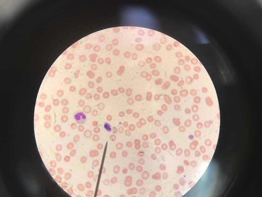
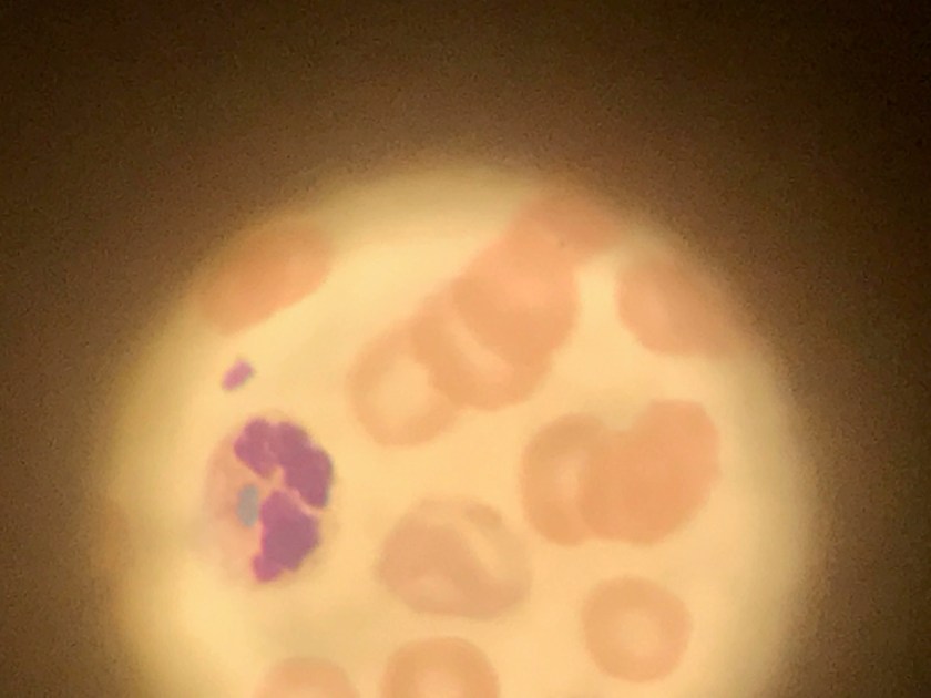
The blood smear was reviewed for these white blood cell inclusions:
Discussion
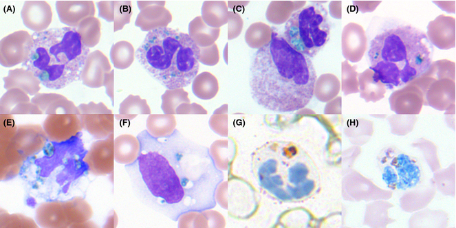
The image presented reveals to be the “blue green crystal of death”. Medical literature has documented an association between acute hepatic failure and coarse, bright-green neutrophilic inclusions. Upon identification of these unique inclusions patients have been reported to have poor outcomes and usually die within 24-72 hours (Haberichter KL, 2017). The exact nature of these inclusions has yet to be determined; it is postulated that they arise from lipofusion-like substance. Bright green inclusions in neutrophils have been reported as a sign of impending patient death (Hodgson, 2015).
Refractile bright-green irregular inclusions within neutrophils have been reported as a marker of impending patient death. In the three reported cases, death occurred within 2 d of recognition of the inclusions (Harris et al, 2009; Jazaerly & Gabali, 2014). Disease associations with green neutrophil inclusions included acute liver failure secondary to acetaminophin overdose, lactic acidosis with multisystem organ failure subsequent to trauma (Harris et al, 2009) and Escherichia coli-associated septic shock (Jazaerly & Gabali, 2014). Harris et al (2009) suggested that the inclusions were related to blood-borne bile products.
These findings on the peripheral smear should be reported and considered a critical finding. Laboratory professionals and hematologists should acknowledge these inclusions; patients are noted to be seriously ill at the time of detection of neutrophil inclusions and have an ominous 24-72 hour survival period.
References
- Haberichter, K. L., & Crisan, D. (2017). Green Neutrophilic Inclusions and Acute Hepatic Failure: Clinical Significance and Brief Review of the Literature. Annals of Clinical & Laboratory Science, 47(1), 58-61.
- Harris, V.N., Malysz, J.& Smith, M.D. (2009) Green neutrophilic inclusions in liver disease. Journal of Clinical Pathology, 62, 853–854.
- Hodgson, T. O., Ruskova, A., Shugg, C. J., McCallum, V. J., & Morison, I. M. (2015). Green neutrophil and monocyte inclusions–time to acknowledge and report. British journal of haematology, 170(2), 229-235.
- Jazaerly, T.& Gabali, A.M. (2014) Green neutrophilic inclusions could be a sign of impending death! Blood, 123, 614.

-Carlo Ledesma, MS, SH(ASCP)CM MT(ASCPi) MT(AMT) is the program director for the Medical Laboratory Technology and Phlebotomy at Rose State College in Midwest City, Oklahoma as well as a technical consultant for Royal Laboratory Services. Carlo has worked in several areas of the laboratory including microbiology and hematology before becoming a laboratory manager and program director.
