Microbiology Case Study: A 28 Year Old Female with Cough.

Case History

A 28 y/o female with a past medical history of chronic eosinophilic pneumonia, chronic persistent asthma, and elevated IgE status post Xolair therapy presented with a cough. She is a former smoker and a former IV drug user. She has been having a productive cough since March and has not improved despite multiple courses of antibiotic therapy. She coughs mostly in the morning and describes her sputum as thick and greenish. She does not have any associated fevers and does not feel that her rescue inhalers help much. She was given a course of doxycycline for 10 days, and sputum was sent for culture.

Laboratory Identification

pasmul1
Image 1: Gram stain showed many polys, moderate mixed gram positive and gram negative organisms. Sputum culture was reported out as mixed gram negatives.
pasmul2
Image 2: Chocolate and blood agar plates of the mixed gram positive and gram negative organisms.

One of the gram negative rods was identified by the MALDI-ToF as Pasteurella multocida.

Discussion

The genus Pasteurella consists of multiple identified species with the one most commonly seen in the clinical setting as Pasteurella multocida. As a genus, they are typically gram-negative straight bacilli that are nonmotile, oxidase-positive, catalase-positive, nitrate reducing, and ferment glucose. They will grow on blood and on chocolate agars, but importantly will not grow on MacConkey. Their colony morphology on blood agar is generally convex, smooth, and nonhemolytic.

Infections with Pasteurella are classically associated with animal bites, such as from a dog or cat. However, prior cases in the literature have shown that pulmonary infection with Pasteurella can be associated with other chronic pulmonary diseases such as COPD (1). The choice for using doxycycline is supported in the literature and was specifically discussed in a prior case with improvement (2).

References:

  1. Klein NC. et al. Pasteurella multocida pneumonia. Semin Respir Infect 1997; 12 (1): 54-56.
  2. Bhat S. et al. A case of lower respiratory tract infection with canine-associated Pasteurella canis in a patient with chronic obstructive pulmonary disease. J Clin Diagn Res 2015; 9 (8): DD03-DD04.


-Jeff Covington, MD, PhD, is a 1st year anatomic and clinical pathology resident at the University of Vermont Medical Center.

Wojewoda-small

-Christi Wojewoda, MD, is the Director of Clinical Microbiology at the University of Vermont Medical Center and an Associate Professor at the University of Vermont.

Why is it Important to Learn About Generations?

Understanding and appreciating different generations is critical for effective and productive teams, departments, and companies. Currently, there are five different generations in the workplace: Traditionalists, Baby Boomers, Generation X, Generation Y/Millennials, and Generation Z. A wide variety of experiences exist between these generations. For example, most traditionalists grew up without television, while almost all Generation Z’ers have a cell phone. If we look deeper, however, we can see commonalities between Traditionalists and Gen Z; both grew up during economic strife (The Great Depression and the Great Recession, respectively). Understanding each other’s views and values will allow different generations to increase their appreciation of one another. This, in turn, will lead to better communication and collaboration because people are now talking from a sense of appreciation and acknowledgement. When people feel heard, understood, and valued, they are more likely to invest time and energy into their projects and jobs and they are more likely to stay at an organization. Truth is, we need people of all generations to make organizations effective. You want the “getting the job done” attitude of the Traditionalists, the teamwork skills of Baby Boomers, the self-reliance of X’ers, the multitasking abilities of Millennials, and the entrepreneurship of Generation Z. Combined, these qualities create a powerful workforce that is able to handle any challenge that comes its way.

It is important to remember that learning can, and should, go both ways: newer generations can pay attention to the older generation’s lessons and knowledge, while older generations can learn a lot from the younger ones (and not just about how to use technology). Each generation has its own unique perspective, challenges, and contributions, and we can all grow by listening to and learning from people who are different than us. Generational diversity is one way to strengthen your team.

lotte-small

-Lotte Mulder earned her Master’s of Education from the Harvard Graduate School of Education in 2013, where she focused on Leadership and Group Development. She’s currently working toward a PhD in Organizational Leadership. At ASCP, Lotte designs and facilitates the ASCP Leadership Institute, an online leadership certificate program. She has also built ASCP’s first patient ambassador program, called Patient Champions, which leverages patient stories as they relate to the value of the lab.


gen-graph1

The differences are many and yet so few.  This is stated so clearly by Gretchen Gavett when she wrote in the Wall Street Journal:

“Baby Boomers, Gen Xers, Millennials, the Gen Z up-and-comers – we all want the same things, (income, sure, but also purpose, and to feel valued) just in slightly different ways. The challenge is to look past the stereotypes and listen to one another so that good work gets done efficiently and humanely.”        

Let’s begin with the GI Generation. The youngest of this generation are in their early 90’s so they are almost non-existent in the workplace.  They are our oldest living generation and were born at the beginning of the 19th century. Most of the soldiers during WWII came from this generation.

Traditionalists make up 2% of the current workforce which is the smallest percentage. However, they represent the institutional memory of a workplace. They know and remember the organization’s past and founding goals. Typically born between 1927 and 1945, they went through their formative years during the Great Depression and its aftermath.

Baby Boomers are currently the largest generation at approximately 77 million people in the United States. (Generation Y runs a close second.) Born between the years of 1946 and 1964, they are the post-World War II generation. The Baby Boomers represent about 29% of the workforce; that number is declining by the day.

Generation X is bookended by the two largest generations, Baby Boomers and Generation Y. They are born between 1965 and 1980. They make up approximately 23% of the workforce.

Generation Y, also known as the Millenials, are born between 1981 and 2000. The Millenials are currently about 42% of the workforce, which makes them the largest working generation.  They have their own values and characteristics (as do the other generations) their numbers make them a force to be reckoned with. 

Generation Z is our newest generation.  They’re currently around 4% of the workforce and growing.  They grew up during the great recession after the early 2000’s.  We are learning about what the Generation Z’s value and their characteristics as each day passes.

The challenge we all face: how can we connect, communicate, and collaborate most effectively in the workplace and outside of the workplace?

Source: https://hbr.org/2009/10/are-you-ready-to-manage-five-g

Stakenas-small

-Catherine Stakenas, MA, is the Senior Director of Organizational Leadership and Development and Performance Management at ASCP. She is certified in the use and interpretation of 28 self-assessment instruments and has designed and taught masters and doctoral level students.  


A Candida Comeback?

Hello again everyone! And special thanks to the readers who read, commented, shared, and reached out to me from my last post “A Serious Aside,” talking about physician burnout and health worker suicide. Numerous people had so much to say in support of this topic—and it’s well deserved—sharing their personal stories and relating their own connects, so I truly appreciate it.

This time, how about something different? In the past few months, I’ve been working through my clinical rotations at a major community hospital in New York City, in the Bronx. A CDC-sponsored screensaver image keeps appearing at terminals throughout floors, services, and clinics; and it directly addresses healthcare professionals to monitor hygiene practices to eliminate Candida infections. I’ll have to admit—innocuous stuff—I’ve been seeing health-message PSA-like screensavers at work for years, about a myriad of topics. Who hasn’t seen those? “Keep beds out of the hallways,” “Protect you and your patients from MRSA,” “Make sure lab requisitions are filled out properly…” the list is endless. But having seen my aforementioned screensaver about Candida one too many times, I had to find out what this was about. You might have thought that, since I spent time working in an HIV clinic, this was a simple PSA for those patients otherwise immunocompromised. Right? Nope.

This particular PSA from the CDC warns about Candida auris, a true blue (or pinkish gold, rather) member of everyone’s favorite budding, germ-tube positive, yeast family. C. auris has been in literature for roughly the past decade. Having etiologic origins in southeast Asia and spreading west through the Middle-East, all throughout Africa, and even the UK, this bug has caught the eyes of epidemiologists around the world. Two years ago, the CDC1 and Public health England2 issued warnings about this pathogen, its multi-drug resistance, and its virulence in healthcare-associated infections. Last fall, the NY State Department of Health published their official update for guiding clinicians and laboratory staff.3 In this report, they discussed infection control, prevention, and detection limitations.

canaur1
Image 1. CDC screensaver on hospital computers. Due to increased incidence of reported cases, epidemiologic data suggest prevention measures would benefit patients. C. auris is associated with healthcare-related infections and can live in the environment for an extended period of time.
canaur2
Image 2. NY State Department of Health, Report on C. auris. Informing clinicans and laboratory staff about epidemiology, prevention, detection in the laboratory, and associated implications of limitations and multidrug resistance.

So what’s so scary about C. auris? The two most challenging features of this emerging pathogen are its multi-drug resistance and its relatively difficult identification.

This yeast has been shown to show resistance to many antifungal/antimicrobial agents including fluconazole, voriconazole, amphotericin-B, echinocandins, and even flucytosine. Even more concerning is that nearly half of the C. auris strains collected in research done in Asia, Africa, and South America demonstrated multi-drug resistance patterns to two or more combination therapies. These are most of our first-line standard of care therapies for invasive candidiasis in patients!

canaur3.png
Image 3. UpToDate recommendations summary for candidemia and invasive candida infections.

There are various other recommendations regarding therapies to C. auris specifically, as its potential for resistance are known, but infection control along with empiric therapy seem to be the current standard.

The major risk factors for C. auris infections include the relative status of individual patients: intensive care, acute renal failure, immunocompromised status, localized or systemic infections, and colonization. Simply being hospitalized is an associated risk. On my current service of patients I’m part of a nephrology/medicine team. There are several chronic infection, ESRD, immunocompromised, or otherwise applicable patients to these risk stratifications. No wonder we’ve got those screensavers!

Concerns for identifying C. auris take us back to the lab. Detecting this bug is not as simple as a couple microscopic morphologies and a yeast API strip—sorry to my old mycology professors. C. auris based on chemical tests like these can produce confounding results. Even VITEK identification (unless you’re running Vitek 2 with Biomerieux software) or culture growth can yield non-specifics like C. haemulonii or Saccharomyces cerevisiae. C. auris has a very high salt and temperature tolerance, and with no particular morphologic identifiable features, it remains a challenging identification. It can be grown on dulcitol agar or CHROMagar, but you do not get clear results. What’s the way to get the ID then? Ultimately MALDI-TOF, PCR, and molecular testing is the answer. There are already available C. auris sequences you can obtain for in-house validation if you’re using MALDI already. And when it comes to susceptibility, fear not: as far as I’ve been able to read E-Tests still work.

canaur4.png
Image 4. Definitely not a definitive CHROMagar result.

I was very impressed with MALDI when I was working in Chicago, and a community hospital I was with just finished validating when I left for medical school. I am glad to see it again with this emergent pathogen, and it definitely demonstrates the next wave of instrumentation. Extremely rapid and very accurate.

The variable drug susceptibility, virulence, and ability to thrive in the environment actively threaten those with long inpatient stays. This microorganism is treated with standard precautions and infection control measures. Currently NY leads the nation by far in purported cases of C. auris. So … please wash your hands. A lot. I know I am.

Thanks! See you next time!

References

  1. Centers for Disease Control and Prevention. Clinical Alert to U.S. Healthcare Facilities – Global Emergence of Invasive Infections Caused by the Multidrug-Resistant Yeast Candida auris. https://www.cdc.gov/fungal/diseases/candidiasis/candida-auris-alert.html
  2. Public Health England. Candida auris identified in England. https://www.gov.uk/government/publications/candida-auris-emergence-in-england/candida-auris-identified-in-england
  3. NY State Department of Health https://www.health.ny.gov/diseases/communicable/c_auris/docs/c_auris_update_for_lab_staff.pdf


ckanakisheadshot_small

Constantine E. Kanakis MSc, MLS (ASCP)CM graduated from Loyola University Chicago with a BS in Molecular Biology and Bioethics and then Rush University with an MS in Medical Laboratory Science. He is currently a medical student at the American University of the Caribbean and actively involved with local public health.

Hematopathology Case Study: A 45 Year Old Male with Mediastinal Mass

Case History

A 45 year old male underwent a chest MRA for aortic dilation due to his history of an aneurysmal aortic root. Upon imaging, an incidental anterior mediastinal mass was seen that measured 4.0 cm. In preparation for an upcoming cardiac surgery, the patient underwent a thymectomy with resection of the mass. The sample is a section from the mediastinal mass.

Diagnosis

HVCD-HE-2x
H&E, 2x
HVCD-HE-4x
H&E, 4x
HVCD-HE-lollipop
H&E, 10x. Green Arrows: “lollipop” germinal centers
HVCD-HE-twinning
H&E, 10x. Red arrow: focal “twinning” of germinal centers

Sections show an enlarged lymph node with several follicles demonstrating atrophic-appearing germinal centers which are primarily composed of follicular dendritic cells. These areas are surrounded by expanded concentrically arranged mantle zones. Focal “twinning” of germinal centers is present. Additionally, prominent centrally placed hyalinized vessels are seen within the atrophic germinal centers giving rise to the “lollipop” appearance.

By immunohistochemistry, CD20 highlights B-cell rich follicles while CD3 and CD5 highlight abundant T-cells in the paracortical areas. CD10 is positive in the germinal centers while BCL2 is negative. CD21 highlights expanded follicular dendritic meshwork. CD138 is positive in a small population of plasma cells and are polytypic by kappa and lambda immunostaining. HHV8 is negative. MIB1 proliferation index is low while appropriately high in the reactive germinal centers.

Overall, taking the histologic and immunophenotypic findings together, the findings are in keeping with Castleman’s disease, hyaline vascular type. The reported clinical and radiographic reports suggest a unicentric variant.

Discussion

Castleman’s disease comes primarily in two varieties: localized or multicentric. The localized type is often classified as the hyaline vascular type (HVCD). Demographically, it’s a disease of young adults but can be found in many ages. The most common sites for involvement are the mediastinal and cervical lymph nodes.

The classic histologic findings of HVCD involve numerous regressed germinal centers with expanded mantle zones and a hypervascular interfollicular region. The germinal centers are predominantly follicular dendritic cells and endothelial cells. The mantle zone gives a concentric appearance, often being likened to an “onion skin” pattern. Blood vessels from the interfollicular area penetrate into the germinal center at right angles, giving rise to another food related identifier, “lollipop” follicles. A useful diagnostic tool is the presence of more than one germinal center within a single mantle zone.

The differential diagnosis of HVCD includes late stage HIV-associated lymphadenopathy, early stages AITL, follicular lymphoma, mantle cell lymphoma, and a nonspecific reactive lymphadenopathy. A history of HIV or diagnostic laboratory testing for HIV would exclude the first diagnosis. AITL usually presents histologically as a diffuse process but atypia in T-cells with clear cytoplasm that co-express CD10 and PD-1 outside of the germinal center are invariably present. EBER staining may reveal EBV positive B immunoblasts in early AITL, which would be absent in HVCD. The most challenging differential would include the mantle zone pattern of mantle cell lymphoma. Flow cytometry revealing a monotypic process with co-expression of cyclin D1 on IHC would further clarify the diagnosis.1

Overall, unicentric Castleman’s disease is usually of the hyaline vascular type. Surgical resection is usually curative in these cases with an excellent prognosis.2


References

  1. Jaffe, ES, Harris, NL, Vardiman, J, Campo, E, Arber, D. Hematopathology. Philadelphia: Elsevier Saunders, 2011. 1st ed.
  2. Ye, B, Gao, SG, Li, W et al. A retrospective study of unicentric and multicentric Castleman’s disease: a report of 52 patients. Med Oncol (2010) 27: 1171.


PhillipBlogPic-small

-Phillip Michaels, MD is a board certified anatomic and clinical pathologist who is a current hematopathology fellow at Beth Israel Deaconess Medical Center, Harvard Medical School, Boston, MA. His research interests include molecular profiling of diffuse large B-cell lymphoma as well as pathology resident education, especially in hematopathology and molecular genetic pathology.

Microbiology Case Study: A 59 Year Old Female with Fevers, Weakness, and Altered Mental Status

Case History

A 59 year old African American female presented to the emergency department with fevers, weakness, fatigue and altered mental status. Her past medical history was significant for hypertension, diabetes mellitus (type 2) with end stage renal disease and a recent cerebrovascular accident the month prior. Her surgical history included a mitral valve repair surgery three years ago and a renal transplant two years ago. Current medications included prednisone, mycophenolate and tacrolimus immunosuppressive agents. Physical examination was unremarkable except for a temperature of 101°F and she was oriented to person, place and time. Pertinent labs included a WBC count of 13.2 TH/cm2, microcytic anemia, and a creatinine of 1.51 mg/dL. Due to previous cardiac surgery, a transesophageal echocardiograph (TEE) was performed and showed a large vegetation (1.6 x 1.5 cm) on the mitral valve.  A diagnosis of endocarditis was made and the patient was started on broad-spectrum antibiotics & taken to surgery for a mitral valve replacement. Multiple blood cultures were negative to this point. Portions of the mitral valve were submitted to surgical pathology and the microbiology laboratory for bacterial, fungal and AFB cultures.

Laboratory identification

Surgical pathology received an aggregate of tan-yellow, fibrous tissue fragments (3.1 x 1.5 x 1.1 cm). Histologic assessment showed a confluent mass containing abundant narrow, septate hyphae consistent with a fungal infection (Image 1). No definitive pigment was identified. Grocott’s methenamine silver (GMS) stain also highlighted the narrow hyphae with numerous septations (Image 2). In the microbiology laboratory, a darkly pigmented mold grew after 5 days of incubation on Sabouraud dextrose agar (Image 3). Lactophenol cotton blue prep showed pigmented, curved conidia with 2-3 transverse septations consistent with Curvularia spp (Image 4). All blood cultures were finalized as no growth after 5 days. Fungitell was found to be greater than 500 pg/ml and Aspergillus galactomannan was negative (<0.5).

curve1.jpg
Image 1. Sections of mitral valve tissue showed a confluent mass of abundant hyphal elements (H&E, 4x).
curve2.jpg
Image 2. Special stains of the fungal mass highlighted narrow hyphae with numerous septations and acute angle branching (GMS, 4x).
curve3.png
Image 3. A darkly pigmented mold grew of Sabouraud dextrose agar after 5 days of incubation at 25°C.
curve4.jpg
Image 4. Many pigmented, curved conidia with multiple transverse septations were seen (lactophenol cotton blue prep, low power).

Discussion

Curvularia spp. belong to a heterogeneous group of dematiaceous or black molds. The presence of pigment in this category of molds is due to melanin in the hyphae. Dematiaceous molds are ubiquitous in nature and can occasionally cause human infections.  These molds have a characteristic dark appearance on fungal media that is often dark gray, brown or black in color. In addition, when the reverse of the plate or slant is examined, the under surface is also pigmented. Based on their growth rate, the dematiaceous fungi are divided into the fast growers, such as Curvularia, Bipolaris and Alternaria spp., which are mature in 5-7 days. The second group is slow growers that take between 7-25 days to fully mature. Examples of slow growers include Phialophora, Exophila/Wangiella, Cladosporium, Fonsecaea and Rhinocladiella spp.

Most commonly, dematiaceous molds infections usually present as phaeophyphomycosis, chromoblastomycosis or mycetomas. These three entities are cutaneous or subcutaneous mycoses that are obtained by traumatic implantation but vary from one another based on clinical features and histologic features of the mold in tissues. They are most frequently cause infection in male agricultural workers in rural areas of tropical or subtropical climates.  These infections are indolent in nature but can lead to significant morbidity over time, as they are difficult to treat effectively.

In addition to the above superficial infections, Curvularia spp. has also be known to cause keratitis, sinusitis and wound infections. In immunosuppressed individuals, disseminated infections with spread to the lungs and brain have been documented. Endocarditis due to Curvularia spp. is quite rare with very few cases previously reported in the literature. On those documented, Curvularia spp. infections tend to have a predilection for prosthetic heart valves or occur after cardiac surgery. Diagnosis of infective endocarditis is difficult as symptoms are indolent and blood cultures do not have a high yield. Therefore, culture of the vegetation may be the only way to make a diagnosis.

In the microbiology laboratory, Curvularia spp. will grow on routine fungal media as a darkly pigmented mold in a relatively short time. On lactophenol cotton blue prep, Curvularia spp. produce large conidia that usually contain 4 cells that are divided by transverse septations. The conidia take on a curved appearance due to swelling of the subterminal cell, which is often the largest and most deeply pigmented. If identification is necessary beyond the genus level, panfungal PCR assays followed by sequencing of ribosomal genes may be useful in providing a species level diagnosis from fresh or paraffin embedded tissue.

For localized infections, surgical treatment alone may be adequate in some cases.  In infections that are extensive or if there is dissemination, treatment with newer triazoles, such as posaconazole or voriconazole, have shown a broad spectrum of activity against dematiceous molds. Amphotericin B is also another effective option. While susceptibility testing can be performed on clinically significant Curvularia spp. infections, interpretative breakpoints have not been defined and clinical correlation is lacking.

In the case of our patient, she remained on a ventilator following surgery and with the identification of mold on histology, she was started on micafungin. She was switched to amphotericin B after the mold was classified as Curvularia spp. Her condition did not improve despite therapy and she died 3 weeks after surgery.


-Azniv Azar, MD, is a fourth year anatomical and clinical pathology resident at the University of Mississippi Medical Center.

Stempak

-Lisa Stempak, MD, is an Assistant Professor of Pathology at the University of Mississippi Medical Center in Jackson, MS. She is certified by the American Board of Pathology in Anatomic and Clinical Pathology as well as Medical Microbiology. She is the director of the Microbiology and Serology Laboratories. Her interests include infectious disease histology, process and quality improvement, and resident education.

When Rapid Blood Culture Identification Results Don’t Correlate, Part 2: Contamination

All laboratories are prone to contamination events. Blood products, analyzers, reagents, media, etc. all have the potential to be contaminated. If you are a molecular microbiologist, then you have to worry about not only bacterial, but also nucleic acid contamination. 

The Issue

The topic of my blog last month focused on discrepant results between blood culture and PCR. Traditional blood culture workflow involves correlating the Gram stain result to what grows in culture. Nowadays, many laboratories are also performing PCR on positive blood cultures. Because we know PCR is more sensitive, it may be easy for some to justify discrepancies. Let’s image that gram positive cocci in clusters were observed in the Gram stain, the PCR detected Staphylococcus and Enterococcus DNA, but only S. aureus grew in the culture. Where did the Enterococcus come from and where did it go? It was not observed in the Gram stain and it didn’t grow in cultures, so was it “real”? Possibly. It could be a contaminant or it could be real, just present in low numbers. It’s difficult to say without having to invest more effort.

When this type of situation occurs in my laboratory, three things happen. First, we review the data. For example, if the Gram stain is discrepant, then we review the Gram stain or perform an acridine orange stain (in the case of positive PCR, but negative culture). If it’s the PCR, then we would make sure that a result entry error did not occur, etc. Second, we add the comment, “clinical correlation needed”. We have found little value in going back to the blood culture bottle and trying to recover the missing organism because in most cases when we look hard enough, using selective agar and other strategies, we do find the organism from the PCR results buried among overgrowth. Therefore, our approach is to let the clinician know that they must use other clinical data to aid in their diagnosis. Third, we document all discrepant blood culture PCR results; which includes an automatic notification to the doctoral director.

Next, let’s imagine that two more blood cultures (from different patients) become positive all within a relatively short period of time from the first discrepant result noted above. gram negative bacilli are observed in one culture and the other displays gram positive bacilli. PCR detects Enterococcus DNA in both cases. What are the odds of that happening? Not good. Something strange is going on!

The Solution

A contamination investigation needs to immediately occur. The two likely sources of contamination are 1) the PCR assay or 2) the blood culture bottles. To determine whether the issue is due to amplicon or target contamination of the PCR assay, we need to identify which instruments reported the Enterococcus. Was it a single instrument or were different instruments involved? Our laboratory performs routine “swipe” tests of the environment as part of our quality control, which allows us to monitor contamination. Swipe tests may also be performed 1) after a known contamination event (i.e., spill due to cracked or leaky product) to ensure that decontamination was properly carried out, 2) to investigate increased positivity rates, or 3) follow up on unusual results, such as the scenario outlined above.

PCR may be performed on a random sampling of uninoculated bottles to determine whether the issue is due to contamination of the blood culture media. If the contamination is high density, this may be useful; however if it is low density, then all bottles you test may still be negative. If the contamination is due to bacterial DNA, then Gram stain or culture will not be useful, hence the need for PCR. It is important to note that the presence non-viable organisms and/or nucleic acids (at levels that can be detected by PCR) is a known limitation noted in the package insert of some blood culture media and PCR manufacturers. If contamination is suspected, then immediately file a report with the manufacturer. Be sure to document lot numbers and expiration dates so that they may alert other customers.
The Conclusion

Human error contributes to the majority of discordant laboratory results. However, errors in interpretation and result entry/clerical errors are only part of the problem. Contamination events only complicate matters. If the test volume is significant, then the number of discordant results should be quickly realized, especially if there truly is a contamination issue. It is important to have a process in place to help reconcile contamination events as quickly as possible as they have the potential to majorly impact operations and patient care.


References

  1. https://labmedicineblog.com/2018/02/20/when-rapid-blood-culture-identification-results-dont-correlate-part-1-clinical-correlation-needed/


Martinez Headshot-small 2017

-Raquel Martinez, PhD, D(ABMM), was named an ASCP 40 Under Forty TOP FIVE honoree for 2017. She is one of two System Directors of Clinical and Molecular Microbiology at Geisinger Health System in Danville, Pennsylvania. Her research interests focus on infectious disease diagnostics, specifically rapid molecular technologies for the detection of bloodstream and respiratory virus infections, and antimicrobial resistance, with the overall goal to improve patient outcomes.

Synergistic Decision Making

Contrary to common belief, the group is NOT as strong as the weakest link. Instead, a group is as strong as its capacity to compensate for the weakest link. We have all experienced this when, for example, a colleague does not do their share for a presentation or project. This does not mean that the project or presentation fails; it means that other team members will compensate and do additional work that was initially assigned to the unproductive team member. The group thus does not sink to the level of the unproductive member. Instead, it rises to the level of how well others can do that members’ job.

When teams reach synergy, they reach a high level of effectiveness and productivity. In order to find out if your team is synergistic, this course conducts a simulation. The team-building simulation, designed by Human Synergistics International, revolves around some type of emergency situation: people are stranded in the desert, a tsunami is coming, they are surrounded by incoming bush fire, there is a severe snowstorm on the way, or people are stranded on a float plane in the middle of the subarctic. Through a video story, participants of this course are introduced to their situation and then asked to rank available items in order of importance. This is first done individually and then with a group while being observed by one person who is assessing their discussion. Once the correct ranking is revealed, participants will see the difference between their individual and group scores and they receive insights about how effectively they worked together.

Understanding the challenges of a team and how to move ineffective behaviors to productive ones is essential for team synergy. This course follows the Human Synergistics circumplex, explained in more detail in the Organizational Savvy and Reacting to Change course blog. In short, this circumplex indicates which behaviors are constructive, passive/defensive or passive/aggressive. Awareness of the constructive and ineffective behaviors will increase a team’s synergy. The idea behind this model is that when a team adopts constructive behavior, their collaborative results will produce greater results than the sum of their individual efforts. These groups are not as strong as their weakest link, nor are they as strong as their capacity to compensate for the weakest link. Rather, these groups are as strong as their syngeristic capacity.

lotte-small

-Lotte Mulder earned her Master’s of Education from the Harvard Graduate School of Education in 2013, where she focused on Leadership and Group Development. She’s currently working toward a PhD in Organizational Leadership. At ASCP, Lotte designs and facilitates the ASCP Leadership Institute, an online leadership certificate program. She has also built ASCP’s first patient ambassador program, called Patient Champions, which leverages patient stories as they relate to the value of the lab.


“Doctor! We need your help STAT … in Antarctica!”

As a pathologist based in Denver, Colorado, I can easily say this is not a statement I ever expected to hear. Because of my sub-specialty expertise in surgical and cytopathology, and my role as chairman of the pathology department in a tertiary care facility, it was not unusual for colleagues, staff and administrators to stop by my office or to phone me for a matter in need of immediate attention. The conversation would usually start with, “Doctor Sirgi, we need your help as soon as possible with …”. I always welcomed these opportunities to assist with whatever matter needed attention, knowing full well the ultimate beneficiary of these calls would be a patient or an anxious family member. However, I could not hide my surprise when I heard the second part. “You need me where?!” I asked, thinking I had misheard the latter part of the phrase. It turns out my assistance really was immediately needed in Antarctica!

That moment in June 1999 I learned the headquarters of Antarctic Support Associates (ASA) is based in Englewood, Colorado (a suburb of Denver). ASA is contracted by the National Science Foundation to provide science support to the United States Antarctic Program (USAP), based at the Amundsen-Scott South Pole Station (ASSPS). The ASA director told me they had received a desperate call from the scientific team deployed in the South Pole informing them their only medical doctor on site, Dr. Jerri Nielsen, had discovered a breast lump worrisome for cancer during self-palpation. Considering Antarctica was in deep winter, with outside temperatures hovering around negative 85 degrees Fahrenheit evacuating the doctor for medical tests and treatment was completely impossible. This was a full-fledged “Houston we have a problem!” kind of situation.

As soon as I arrived at ASA, a videoconference was established with the afflicted doctor and a few non-medical scientists on site via satellite link-up; the first order of business was to understand the elements of the problem and offer a potential course of action. We only had a few precious minutes of satellite connection before lost of signal. We learned the following:

  • The doctor had self-detected a sizable breast mass of hard consistency.
  • Nobody around her had any experience at performing a biopsy or fine needle aspiration, let alone surgery.
  • There was no laboratory facility or expertise to offer pathology examination, should a sample be obtained.
  • There was no mammography or ultrasound equipment adequate for the evaluation of a breast mass.
  • There was no adequate medication, should a diagnosis of malignancy be established.

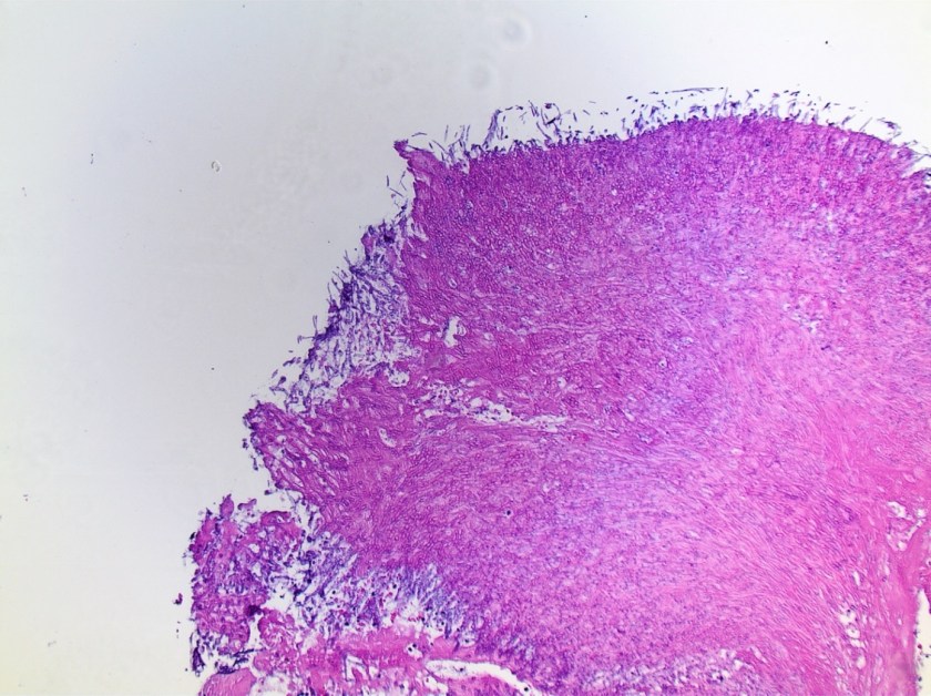
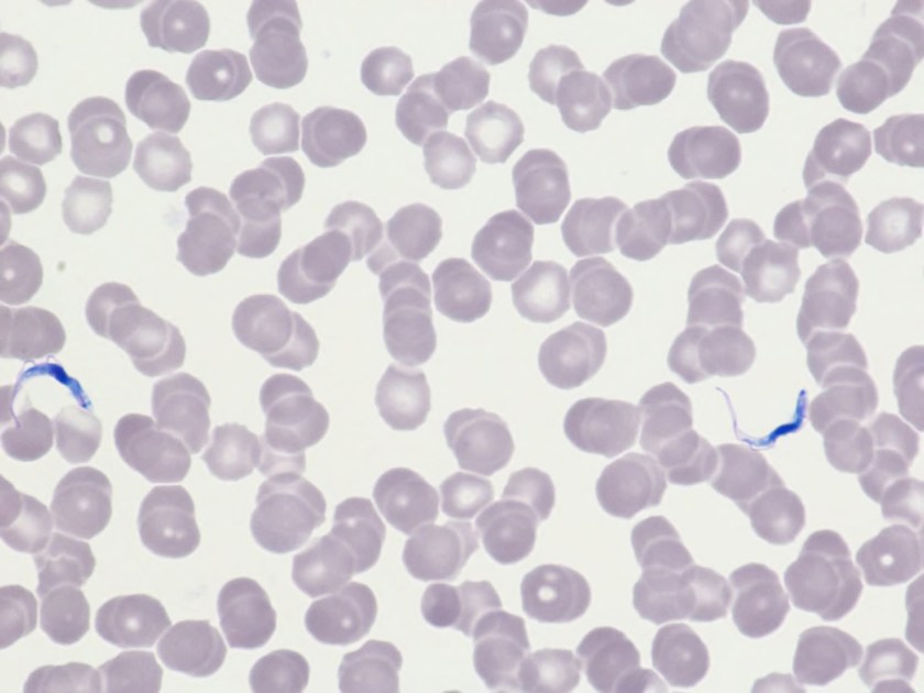
With the possibility (or the wishful thinking) that we could still be dealing with a benign lesion, I recommended that we first focus our efforts on securing a diagnosis. Luckily, the rudimentary equipment available to Dr. Nielsen included needles, glass slides, Giemsa stain, an antiquated microscope (with no camera attachment), and a medium resolution digital camera borrowed from scientists working in another area of the research facility. I explained in detail to Dr. Nielsen and her team of worried volunteers how to use these seemingly unrelated pieces of material and equipment. Keep in mind that all this happened at a time when digital pathology was still in its infancy (if not fetal stage), and a hefty dose of DIY had to be improvised on the spot.

I had brought a needle, an orange, a couple of glass slides, and three jars filled with the fluids needed for a quick staining of the material obtained. Dr. Nielsen had herself and her crew of non-medical scientists. I demonstrated how to perform a fine needle aspiration, smear the material obtained on a glass slide, and how to properly stain it for microscopic examination.

These were but the very first steps of a long journey toward obtaining a diagnosis. Considering Dr. Nielsen had no expertise in the examination of pathology material, she needed to follow steps completely unfamiliar to her in order for me (and other experts mobilized around the country) to establish a diagnosis:

  • Perform a medical procedure she had never performed before … on herself!
  • Prepare smears of the material aspirated from the mass
  • Have those smears stained
  • Use a microscope to identify areas of cellularity on the slides obtained
  • Use a camera to take pictures of these areas
  • Load the pictures in an email
  • Transmit an email “heavy in data” across the planet, on a very slow satellite linked connection

Dr. Nielsen performed the procedure on herself the next day. The pictures I received a day later were impossible to interpret because the slides had been improperly stained; areas photographed had abundant red blood cells but no breast epithelial cells to evaluate. The team was understandably quite discouraged when they received our feedback. I sent them an email commending them on their efforts and further guiding them on:

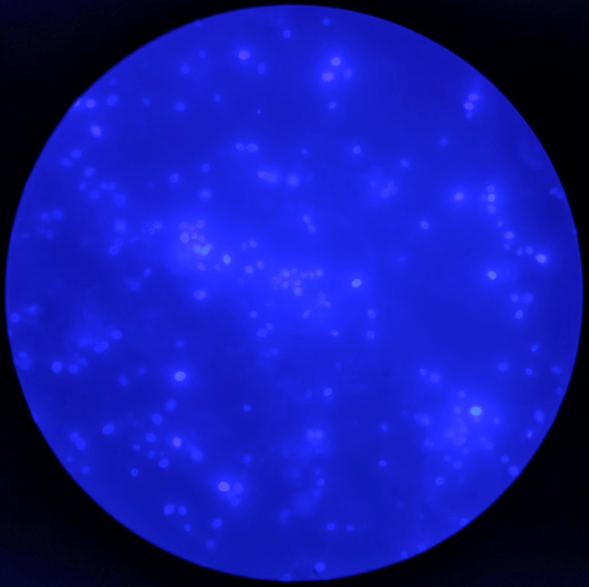
  • Troubleshooting the staining process
  • Focus on the best areas to take pictures, using a breast cytopathology atlas as a visual aide

Their second attempt was much improved and allowed us to unequivocally establish a diagnosis of malignancy affecting Dr. Nielsen’s breast. Reaching a diagnosis was good; however, the tragic reality still remained that the patient had cancer and it was completely impossible to evacuate her from her current location.

The “home team” (anybody not based on the other end of the world) immediately started mobilizing resources from different areas of expertise to:

  • Get Dr. Nielsen the treatment she needed while stuck in Antarctica
  • Get Dr. Nielsen out of the South Pole as soon as meteorological conditions allowed

The following immediate priorities were then identified and acted upon:

  • Per the oncologists consulted, adequate chemotherapy could not be started in the absence of knowing the tumor’s biomarkers status
  • To establish this status, better tissue was needed for further immunohistochemical testing
  • Each medical specialty involved with the rescue effort made recommendations for the type of equipment and material that needed to be transported to the South Pole (including specialized medical atlases, ultrasound equipment, newer microscopes equipped with high resolution digital cameras, regular and immunohistochemical stains with appropriate easy to use instructions, various chemotherapy drugs for different treatment possibilities).

The equipment, with duplicate units of everything sent, was placed in crates and flown to the US Air Force base in New Zealand. Ace pilots volunteered to drop the equipment over the Amundsen-Scott South Pole Station, despite terrible weather conditions, zero visibility over the drop zone, and no chance of landing or refueling during the mission. Ultimately, a couple of attempts were necessary to successfully drop the needed equipment over the area. The station personnel worked for hours in negative 85 degrees Fahrenheit temperatures and near zero visibility to collect the dropped material, much of it severely damaged, and transport the surviving equipment back to the base.

Treatment began, the tumor was stabilized, and Dr. Nielsen returned to the U.S., where she continued treatment as soon as weather allowed it. Unfortunately, she succumbed to her illness several months later.

What started as a “Dr. Sirgi, we need your help STAT … in Antarctica” developed into a medical rescue mission of monumental proportion. Ordinary people from different walks of life and medical expertise worked synergistically to develop on-the-fly life-saving solutions that had never been tried before. In the end:

  • A heroic doctor performed diagnostic procedures on herself and braved all kinds of challenges in an attempt to survive.
  • A staff of scientists with limited to no medical experience rose to the occasion to act as capable and devoted medical assistants.
  • Physicians and medical technologists from around the country, who were previously strangers, synergistically worked together to coordinate efforts to save a colleague who was trapped in some of the harshest conditions in the world.
  • Administrators of the Antarctic Support Associates (ASA) organization worked day and night to secure any and all expertise and needed equipment for the rescue mission.
  • Air Force pilots voluntarily risked their lives to rescue a fellow human being.

No one involved woke up on that first day thinking they would be called for such a noble endeavor. All parties involved were ordinary citizens, and every single one tapped into his or her infinite leadership potential to collaborate with colleagues in order to resolve an almost impossible situation. Although There were many links of uncertain strength in this effort due to lack of experience or expertise, the common resolve and demonstrated leadership of all players involved created an indestructible chain of potential and led ultimately to the mission’s resounding success.


Sirgi pic-small

-Karim E. Sirgi, MD, MBA is board certified in anatomic and clinical Pathology, with additional board certification in cytopathology. He is active as an independent healthcare consultant, and is the current president of the CAP Foundation. Additional biographical information can be accessed at www.karimsirgimd.com

Hematology Case Study: The Race to Save a 48 Year Old Man from a Rare Disease

A 48-year-old Caucasian male presented to a Baltimore Emergency Room complaining of fever, chills, and aches. He stated he had not been feeling well for the past week. His symptoms had progressed rapidly over the last 3 days to include night sweats, nausea and excessive somnolence. History taken in the ER revealed the patient had returned 10 days prior from a Safari in Botswana and Zambia. The patient was admitted to the ICU, in shock, with a BP of 75/50. Even though the patient had taken anti-malarial medication, the doctors suspected malaria. Blood was sent to the lab for a blood parasite exam and treatment for malaria was started while the doctors waited for the confirmation.

In the Hematology laboratory, technologists perform microscopy of thick and thin blood smears to look for malarial parasites. The thin smear is a typical Wright Giemsa stained wedge smear, and the thick smears are prepared and stained so that the red blood cells are lysed, and the sample is concentrated, making examination easier. Thorough, careful examination of the thick smear is aimed to identify whether a particular parasite is present, but they require a long drying period and take several hours to prepare and read. Thin smears can detect the parasites but also permit identification of particular species of malaria. While the thick smears were drying the technologist examined the thin smear.

The technologist who examined this patient’s thin smears saw parasites (image 1) under her microscope. She consulted with a supervisor and pathologist to confirm, and the patient’s doctor was notified that the patient did not have malaria, but instead, had Trypanosoma! This was an exciting find in the laboratory, as there have been only 40 cases seen in the US in the past 50 years.

tryp1
Image 1. This slide shows the parasite, in dark blue. The parasite causes
African trypanosomiasis, also known as sleeping sickness
(Courtesy of Greater Baltimore Medical Center).

The race for diagnosis and treatment did not stop there, as there are 2 types of African trypanosomiasis, or African sleeping sickness, and effective and appropriate treatment must be started in a timely fashion. Both types look identical on a blood smear and both are considered universally fatal, if not treated. West African trypanosomiasis and East African trypanosomiasis are caused by the tsetse fly, which only lives in rural Africa. The patient stated he did remember being bitten by tsetse flies, and because there had been such a short span of time between being bitten and the onset of symptoms, doctors concluded that the patient had the rarer and fast-acting East African trypanosomiasis, which can kill within months.

Epidemiologists at CDC were contacted, who then consulted other infectious disease specialists at CDC. There are 2 treatments depending the stage of the disease. Surinam is the first line of defense, but melarsoprol, which is arsenic-like and very toxic, must be used if the parasites have reached the central nervous system. Because of the urgent need to start treatment, emergency shipments of both drugs were flown to Baltimore. The patient was started on Surinam to reduce the number of parasites in his blood to a level low enough to allow a spinal tap to be performed. After confirming that the CSF showed no signs of the parasite, treatment with surinam was continued and the patient was discharged a week later and has made a full recovery.

Because of the excellent work done by the medical technologists who made the first discovery, the speed with which the critical calls were made, the actions of the doctors involved, and the cooperation of the CDC, this patient received his first dose of Surinam a little over 24 hours after his blood was sent to the lab. This case shows the importance of a thorough medical and travel history in differential diagnosis. It also illustrates the importance of the competency evaluations and surveys in which all laboratory professionals are required to participate. None of the technologists, doctors or scientists involved had ever actually seen a case of African Trypanosomiasis, they had only read about it in books and seen it on competency assessments.

This case is based on an actual case from 2016. My coworker, Gail Wilson, was the technologist who first saw the Trypanosoma on the slides. Many thanks to Gail for her keen eye and attention to detail!

tryp2
Image 2: L&R: Trypanosoma brucei in thin blood smears stained with Giemsa. Center: A close up of a tsetse fly. Credit: DPDx


References 

  1. Jon E. Rosenblatt Barth Reller Melvin P. Weinstein.pages 1103-1108, Laboratory Diagnosis of Infections Due to Blood and Tissue Parasites Clinical Infectious Diseases, Volume 49, Issue 7, 1 October 2009; retrieved March 2018 from https://academic.oup.com/cid/article/49/7/1103/316703
  1. Ivo Elliott, Trupti PatelJagrit Shah, and Pradhib Venkatesan. West-African trypanosomiasis in a returned traveller from Ghana: an unusual cause of progressive neurological decline BMJ Case Rep. 2014; 2014: bcr2014204451. Published online 2014 Aug 14.doi: 1136/bcr-2014-204451; retrieved March 2018 from https://www.ncbi.nlm.nih.gov/pmc/articles/PMC4139564/
  1. Lena H. Sun. Medical Detectives raced to save a man from a rare, ‘universally lethal’ disease; retrieved March 2018 from https://www.washingtonpost.com/news/to-your-health/wp/2016/12/22/medical-detectives-raced-to-save-a-man-from-a-rare-universally-lethal-disease/?utm_term=.16d7b136bc47
  1. Parasites – African Trypanosomiasis (also known as Sleeping Sickness). Retrieved March 2018 from https://www.cdc.gov/parasites/sleepingsickness/
  1. DPDx- Laboratory Identification of parasites of Public Health Concern; retrieved March 2018 from https://www.cdc.gov/dpdx/


Socha-small

-Becky Socha, MS, MLS(ASCP)CM BB CM graduated from Merrimack College in N. Andover, Massachusetts with a BS in Medical Technology and completed her MS in Clinical Laboratory Sciences at the University of Massachusetts, Lowell. She has worked as a Medical Technologist for over 30 years. She’s worked in all areas of the clinical laboratory, but has a special interest in Hematology and Blood Banking. When she’s not busy being a mad scientist, she can be found outside riding her bicycle.


Microbiology Case Study: A 52 Year Old Man with End Stage Renal Disease

Case History

A 52-year-old man with multiple medical issues including a history of end stage renal disease on hemodialysis, chronic pancreatitis status post distal pancreatectomy, intravenous drug use through dialysis catheter, and multiple types of bacteremia presented with chills and abdominal pain. Labs on admission included a white blood cell count of 28.64 k/cmm, hemoglobin 8.8 g/dL, and platelets 581 K. He was diagnosed with a pancreatitis flare and admitted for pain management, with further labs drawn. After one day, he felt much better and was discharged with a pending blood culture to follow up on. At 61 hours, one bottle flagged positive with yeast seen on gram stain.

Laboratory findings

cryptlaur1
Image 1: potato flake agar with creamy tan-white colonies.
cryptlaur2.png
Image 2: calcofluor white fluorescent stain showing round yeast forms.

The organism was identified as Cryptococcus laurentii via MALDI-ToF MS. A follow-up fungal culture was negative, however, repeat blood culture grew Stenotrophomonas maltophilia. His tunneled catheter was removed, and two days later the patient required urgent interventional radiology access for dialysis. He completed a two-week course of ceftazidime and was discharged. 

Discussion 

Cryptococcus laurentii is a very rare fungal pathogen. It is a psychrophilic organism, growing ideally at 15 °C, and is the most common yeast found in tundra.1 Major reservoirs include soil, food, and pigeon excrement.2 C. laurentii usually causes infection in immunocompromised hosts, although rare incidents of infection in immunocompetent patients have been reported. Reported manifestations have included fungemia, meningitis, peritonitis, pneumonia, pyelonephritis, keratitis, and skin infection.3

Cryptococcus laurentii is a urease-positive organism. Gram stain shows large budding yeasts without hyphae. The yeast grows on routine agar as whitish-yellow creamy colonies and on birdseed agar as whitish or greenish colonies. Staining with calcofluor highlights encapsulated yeast forms. Molecular diagnosis can be accomplished by ribosomal RNA sequencing of the internal transcribed spacer and D1/D2 regions. Treatment in most cases has been with fluconazole, although in one case of peritoneal dialysis catheter-related peritonitis, voriconazole was used due to low fluconazole susceptibility.4

References

  1. Molina-Leyva A, Ruiz-Carrascosa JC, Leyva-Garcia A, Husein-Elahmed H. Cutaneous Cryptococcus laurentii infection in an immunocompetent child. International Journal of Infectious Diseases. 2013;17(12). doi:10.1016/j.ijid.2013.04.017.
  2. Johnson, L. B., Bradley, S. F. and Kauffman, C. A. Fungaemia due to Cryptococcus laurentii and a review of non-neoformans cryptococcaemia. Mycoses. 1998;41: 277–280. doi:10.1111/j.1439-0507.1998.tb00338.x
  3. Furman-Kuklińska K, Naumnik B, Myśliwiec M. Fungaemia due to Cryptococcus laurentii as a complication of immunosuppressive therapy – a case report. Advances in Medical Sciences. 2009;54(1). doi:10.2478/v10039-009-0014-7.
  4. Asano M, Mizutani M, Nagahara Y, et al. Successful Treatment of Cryptococcus laurentii Peritonitis in a Patient on Peritoneal Dialysis. Internal Medicine. 2015;54(8):941-944. doi:10.2169/internalmedicine.54.3586.


-Prajesh Adhikari, MD is a 3rd year anatomic and clinical pathology resident at the University of Vermont Medical Center.

Wojewoda-small

-Christi Wojewoda, MD, is the Director of Clinical Microbiology at the University of Vermont Medical Center and an Associate Professor at the University of Vermont.

Is It Possible to Have Coexistence of Hepatitis B Surface Antigen and Antibody?

Hepatitis B surface antigen (HBsAg) is the serologic hallmark of acute Hepatitis B virus (HBV) infection. It can be detected in serum using immunoassays a few weeks after HBV infection, and normally disappears after 4-6 months in recovered patients (1). Antibodies against HBsAg (anti-HBs) appears as a response from the host immune system, and these antibodies neutralize HBV infectivity and clear circulating HBsAg (2). Anti-HBs generally persist in life, indicating recovery and immunity from HBV infection.

Some of us may simply assume that the presence of anti-HBs should always associated with the loss of HBsAg. However, it is possible to see concurrent anti-HBs and HBsAg in patients. In fact, coexistence of HBsAg and anti-HBs is not rare, and has been reported in 10 to 25 percent of HBV chronic carriers in previous studies (3-4).  The underlying mechanism is not fully understood but several reports explained it as HBsAg mutants escaping the immune system (2-4). HBsAg mutants are believed to arise under the selective pressure from the host immune system, or from vaccinations (4-6).

“a” determinant in HBsAg is one of the main target of anti-HBs. It has been reported that mutations in the “a” determinant of the surface gene (S-gene) result in amino acid substitutions in HBsAg, and reduce the binding of anti-HBs to HBsAg, leading to immune escape (4). The first HBV mutant was reported by Zanetti et al in 1988 as G145R mutation. In their report, infants born to HBsAg carrier mothers developed breakthrough infections despite receiving HBIG and HBV vaccine at birth (5). Since this report, several other HBsAg mutations have been reported (4, 6).

Currently, there is no easily available assay to diagnose individuals who are suspected of harboring HBsAg escape mutants. Moreover, mutated HBsAg may leads to false negativity in some serologic assays, leading to a missed diagnosis of chronic HBV infection (6). Another concern is the potential risk of transmission to others, as vaccination does not provide protection from these mutated viruses (8); this is especially important in liver transplant recipient and newborns from HBsAg positive mothers.

References

  1. Lok A, Esteban R, Mitty J. Hepatitis B virus: Screening and diagnosis. UpToDate. Retrieved Feb 2018 from https://www.uptodate.com/contents/hepatitis-b-virus-screening-and-diagnosis#H3
  2. Liu W, Hu T, Wang X, Chen Y, Huang M, Yuan C, Guan M. Coexistence of hepatitis B surface antigen and anti-HBs in Chinese chronic hepatitis B virus patients relating to genotype C and mutations in the S and P gene reverse transcriptase region. Arch Virol 2012;157:627–34.
  3. Colson P, Borentain P, Motte A, Henry M, Moal V, Botta-Fridlund D, Tamalet C, Gérolami R. Clinical and virological significance of the co-existence of HBsAg and anti-HBs antibodies in hepatitis B chronic carriers. Virology 2007;367:30–40.
  4. Lada O, Benhamou Y, Poynard T, Thibault V. Coexistence of hepatitis B surface antigen (HBs Ag) and anti-HBs antibodies in chronic hepatitis B virus carriers: influence of “a” determinant variants. J Virol. 2006 Mar;80(6):2968-75.
  5. Zanetti AR, Tanzi E, Manzillo G, Maio G, Sbreglia C, Caporaso N, Thomas H, Zuckerman AJ. Hepatitis B variant in Europe. 1988 Nov 12; 2(8620):1132-3.
  6. Leong J, Lin D, Nguyen M. Hepatitis B surface antigen escape mutations: Indications for initiation of antiviral therapy revisited. World J Clin Cases 2016;4:71.
  7. Colson P, Borentain P, Motte A, Henry M, Moal V, Botta-Fridlund D, Tamalet C, Gérolami R. Clinical and virological significance of the co-existence of HBsAg and anti-HBs antibodies in hepatitis B chronic carriers. 2007;367:30–40.
  8. Thakur V, Kazim S, Guptan R, Hasnain S, Bartholomeusz A, Malhotra V, Sarin S. Transmission of G145R mutant of HBV to an unrelated contact. J Med Virol 2005;76:40–6.


Xin-small

-Xin Yi, PhD, DABCC, FACB, is a board-certified clinical chemist, currently serving as the Co-director of Clinical Chemistry at Houston Methodist Hospital in Houston, TX and an Assistant Professor of Clinical Pathology and Laboratory Medicine at Weill Cornell Medical College.