Microbiology Case Study: A 30 Year Old with Fever Post Stem Cell Transplant

Case History

A thirty year old female with refractory acute leukemia was admitted to undergo allogeneic stem cell transplantation. Her initial admission had multiple infectious complications and chemotherapy-induced pancytopenia with profound absolute neutropenia. The patient was placed on prophylaxis/therapy including bacterial, viral, and fungal coverage. On hospital day 14, the patient was febrile to 38°C; vancomycin/piperacillin-tazobactam were added as empiric therapy due to concern for sepsis with fluctuation in mental status. CT Brain without contrast revealed a large intracranial hematoma with mass effect. Her mental status continued to decline and intubation was required for airway protection. An emergent decompressive hemicraniectomy was performed where necrotic brain tissue with hemorrhage/clot were found.

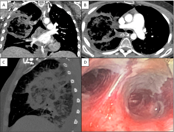
Due to ongoing fevers, empiric antimicrobial therapy was further broadened with meropenem, doxycycline, trimethoprim/sulfamethoxazole, and liposomal amphotericin B (L-AMB). Repeat cultures and imaging studies were ordered to evaluate for infection as a fever source. CT Angiography Chest (Image 1A-C) was performed and revealed an extensive non-enhancing area of ground glass opacity with peribronchovascular consolidation (“Reversed Halo” sign) in the right lung concerning for angioinvasive fungal infection. A tracheal aspirate was sent for bacterial and fungal culture. Subsequent bronchoscopy revealed extensive necrosis involving all visualized airways of the right tracheobronchial tree to the first subsegmental level (Image 1D). By contrast, the left lung appeared relatively normal and uninvolved. Bronchial washings of the right lung were also submitted for culture.

Image 1. Computed Tomography (CT) Angiography Chest images: Coronal (A), Cross-section (B) and Sagittal (C) sections reveal a large central ground glass opacity surrounded by a dense consolidation in the shape of a ring “Reversed Halo” sign. Bronchoscopy (D) revealed extensive necrosis and friable mucosa in the visible airways of the right lung.

Laboratory Identification

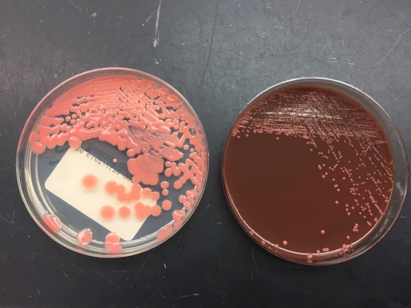
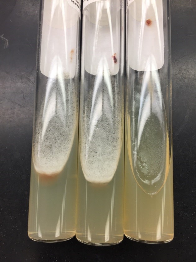
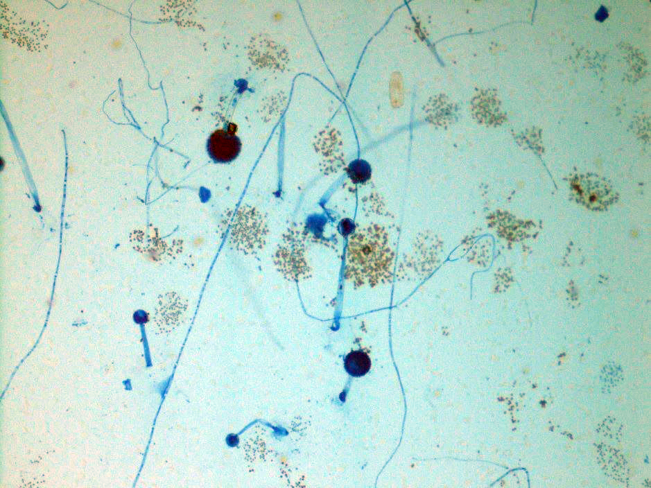
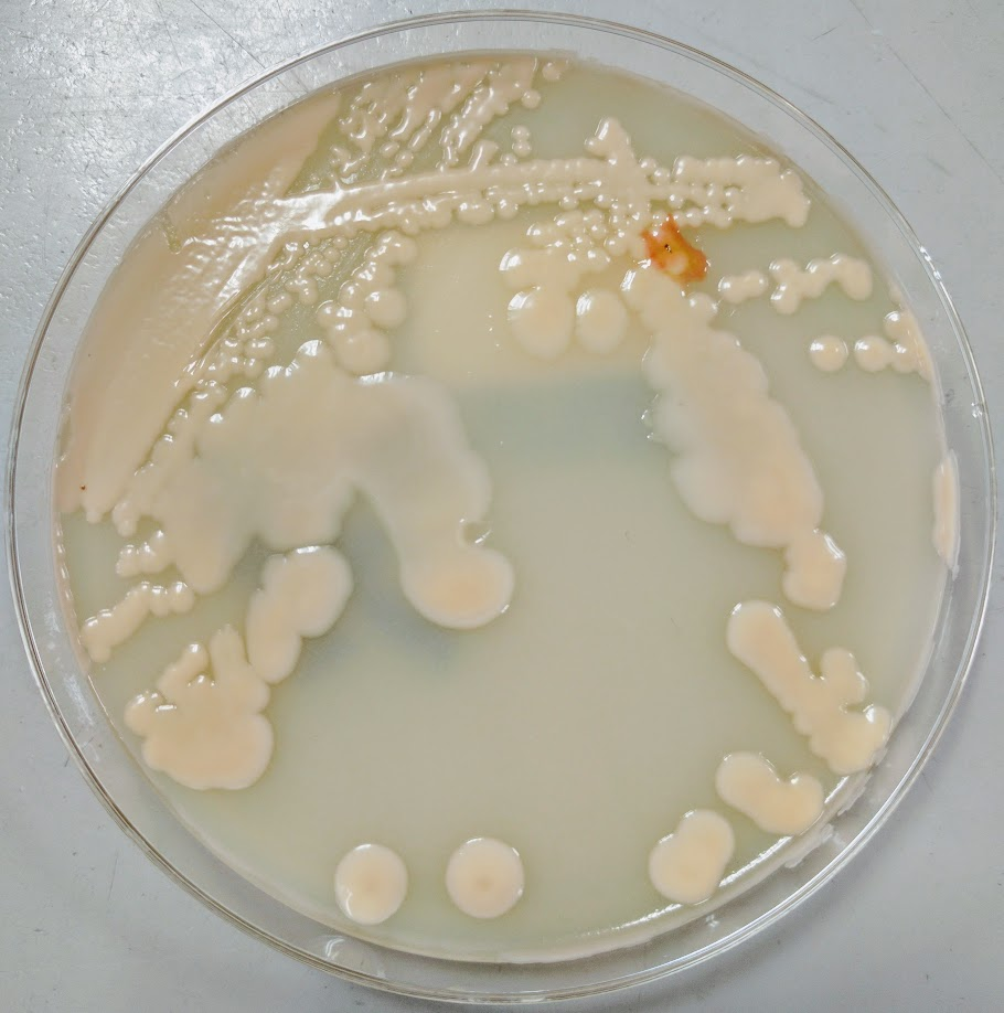
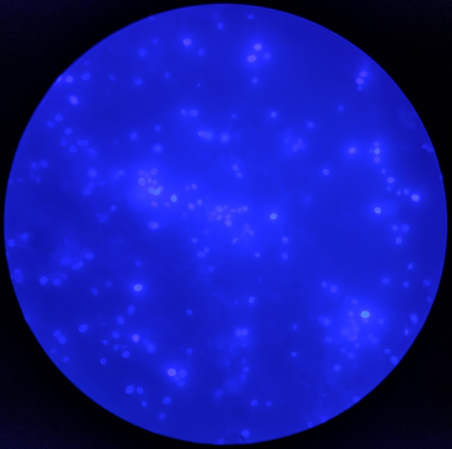
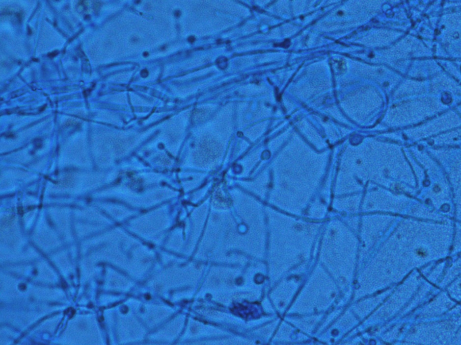
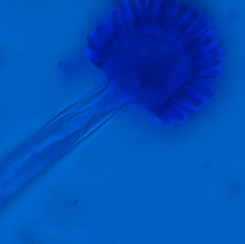
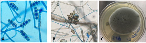
Respiratory specimens were sent to the microbiology laboratory for bacterial and fungal cultures. Hyphal elements were observed on the Gram stain of the submitted tracheal aspirate (Image 2A). Robust fungal growth was noted within 48 hours on Brain Heart Infusion and Inhibitor Mold agars from both the tracheal aspirate and bronchial wash specimens (Image 2B). A lactophenol cotton blue prep revealed broad hyphae with few septations, consistent with a member of the Mucorales (Image 3). Sporangiophores were noted to be long, dark and branched with round sporangia. Few rhizoids were observed and located between the sporangiophores. A definitive identification of Rhizomucor sp. was obtained through the use of matrix-assisted laser desorption ionization mass spectrometry (MALDI-TOF MS).

Image 2. A: Gram stain (400X) of direct sample from tracheal aspirate with broad, irregular, long, pauciseptate hyphae consistent with infection from a fungal species belonging to the order Mucorales. B: Fluffy “cotton candy” appearing light brown-grey colonies grew rapidly (<4 days) on brain heart infusion agar from both tracheal aspirate (pictured) and bronchial washing specimens.
Image 3. Lactophenol cotton blue stain obtained by tape preparation direct from fungal colony (A; 200X, B-D; 400X). Numerous branched dark brown non-apophysate sporangiophores with spherical columella were seen along with numerous broad, irregular pauciseptate hyphae with right angle branching (A, B). Rudimentary rhizoids developing off stolons (C, D) were rarely identified.

Discussion

Members of the order Mucorales can be identified in the laboratory by their rapid, robust growth and cotton candy-like appearance on conventional fungal media excluding those containing cycloheximide.2 Microscopically, these molds exhibit pauciseptate, broad (9-15 μm wide) hyphae with sporangia. Some species elaborate root-like structures called rhizoids (e.g Rhizopus, Rhizomucor) while others lack them (e.g. Mucor).2 Rhizomucor sp. can be differentiated microscopically from other related members of the Mucorales by its branched, dark brown sporangiophores with absent apophysis, round columella and the presence of few, short, rudimentary rhizoids (Image 3A-D). In practice, the rhizoids can be challenging to identify microscopically.2 Some species can also grow at elevated temperatures (~38-58°C) which can be utilized as a tool for use in identification.2 Newer methods including the use of MALDI-TOF MS and DNA probes allow for rapid, accurate identification of these fungi, but are not routinely available in all laboratories.

Infections caused by Mucorales (Mucormycosis) usually involve immunocompromised patients with defects in cell-mediated immunity. Rapid and often fatal, these infections can prove extraordinarily difficult to manage. They are known to be angioinvasive and can widely disseminate. Debridement of involved tissues and higher-level antifungal agents (e.g. posaconazole, amphotericin B) are mainstays of therapy. The most recognized group of patients where these infections are identified are uncontrolled diabetics, frequently in diabetic ketoacidosis, often with nasal/orbital sinus involvement. Patients with leukemia/lymphoma who are undergoing stem cell transplantation are another group often affected. In addition to the nasal/orbital sinuses, the gastrointestinal tract, skin and lungs also serve as important sites for these infections, especially if mucositis occurs.1

Rhizomucor sp. are an occasional cause of mucormycosis, but have a predilection for leukemic patients such as in this case.2 Given the significant bronchoscopy findings and the intraoperative presence of necrotic brain tissue, there was substantial clinical concern for invasive pulmonary mucormycosis with possible central nervous system involvement. Isavuconazole was discontinued, and posaconazole and micafungin were added to her antifungal therapy (L-AMB). Granulocyte infusions were used in an attempt to increase her cell-mediated immune response to the mold. Cardiothoracic surgery evaluated the patient but the lesion was deemed unresectable. Due to the presence of epistaxis, the otolaryngology service evaluated the patient for invasive fungal sinusitis; however, nasal endoscopy did not reveal any nasal/sinus involvement. The patient never regained significant neurological function and continued to medically decline during the hospitalization. She was placed on comfort care where she died shortly afterwards.

References

  1. Love GL, Ribes JA. 2018. Color Atlas of Mycology, An Illustrated Field Guide Based on Proficiency Testing. College of American Pathologists (CAP), p. 244-274
  2. Walsh TJ, Hayden RT, Larone DH. 2018. Larone’s Medically Important Fungi, A Guide to Identification. ASM Press, p. 185-190

-John Markantonis, DO is the current Medical Microbiology fellow at UT Southwestern and will be completing his Clinical Pathology residency in 2022. He is also interested in Transfusion Medicine and parasitic diseases.

Kim Stewart BS, MT(ASCP)SM holds a bachelor’s degree from Texas Tech University and is medical technologist in the microbiology section at UT Southwestern Medical Center with 35 years’ experience.

-Andrew Clark, PhD, D(ABMM) is an Assistant Professor at UT Southwestern Medical Center in the Department of Pathology, and Associate Director of the Clements University Hospital microbiology laboratory. He completed a CPEP-accredited postdoctoral fellowship in Medical and Public Health Microbiology at National Institutes of Health, and is interested in antimicrobial susceptibility and anaerobe pathophysiology.

Microbiology Case Study: A 40 Year Old Woman with Fever, Chills, and Leg Pain

Clinical History

A 40 year old African American female with a history of sickle cell disease presented to an outpatient clinic with fever, chills, and leg and back pain consistent with a sickle cell crisis. Her past medical history was also significant for asthma and seizures. She rated her pain as 10 out of 10, her vitals showed a temperature of 101.0°F, and she was also tachycardic and hypotensive. Her white blood cell count was 23.0 TH/cm2, hemoglobin 8.4 g/dL, hematocrit 26.0%, and platelets 619,000 TH/cm2. In clinic, she received pain medications and a fluid bolus, two sets of blood cultures were collected, and she was transferred to the emergency department for further work up.

Laboratory Identification

Image 1. Gram stain from a positive blood culture bottle showing small, gram positive budding yeast (1000x oil immersion).
Image 2. A mucoid, salmon-colored yeast grew on Sabouraud dextrose and chocolate agars.

Blood culture bottles were positive after approximately two days on the automated instrument. The Gram stain showed small, gram positive budding yeast (Image 1). The BioFire FilmArray for blood culture identification was negative for Candida albicans, C. glabrata, C. krusei, C. parapsilosis, and C. tropicalis. At this time, she was started on micafungin for antifungal therapy. A mucoid, salmon colored yeast grew on both Sabouraud dextrose and chocolate agars (Image 2) and was identified by Vitek 2 as Rhodotorula spp.

Discussion

Rhodotorula spp. are basidiomycetous yeasts that make up the normal microbiota on moist skin and can be found in bathtubs and on shower curtains. Rhodotorula spp. are usually considered contaminants, but can rarely cause fungemia in patients with central lines, endocarditis, peritonitis, and meningitis, especially in those that are immunocompromised. R. mucilaginosa, R. glutinis, and R. minuta are the species commonly associated with human disease. 

In the laboratory, Rhodotorula spp. grow as a mucoid, salmon colored yeast within 1-3 days of incubation. On Gram stain or lactophenol cotton blue prep, the yeast is small and round to oval with multilateral budding. Pseudohyphae are not usually present. Rhodotorula spp. produce urease and fail to ferment carbohydrates. R. mucilaginosa is negative for nitrate assimilation. Identification can also be confirmed by commercial kits, automated systems, and MALDI-TOF mass spectrometry. Rhodotorula spp. are intrinsically resistant to echinocandins and fluconazole.

In the case of our patient, she was switched to intravenous amphotericin B after the identification of Rhodotorula spp. was made. Reference laboratory testing identified the isolate as R. mucilaginosa with high minimum inhibitory concentrations (MIC) to fluconazole and echinocandins. Amphotericin had an MIC of 0.5 µg/ml. She successfully completed a 14 day course with close monitoring of creatinine, electrolytes, and platelet count. Repeat blood cultures were negative and no other focuses of infection were found on CT scans, transthoracic echocardiogram, and ophthalmology exam.

-Lisa Stempak, MD, is an Assistant Professor of Pathology at the University of Mississippi Medical Center in Jackson, MS. She is certified by the American Board of Pathology in Anatomic and Clinical Pathology as well as Medical Microbiology. She is the Director of Clinical Pathology as well as the Microbiology and Serology Laboratories. Her interests include infectious disease histology, process and quality improvement, and resident education.

Microbiology Case Study: A 24 Year Old Male with No Past Medical History Returning from Guatemala with Fevers, Myalgia, and Cough

Case History

A 24 year old male with no past medical history presented with fevers, myalgia, and cough following return from a 1-week trip to Guatemala where he spent significant time within caves. The patient described his cough as persistent, non-productive, and associated with mild shortness of breath at rest that significantly worsens with activity. In the emergency department, the patient was afebrile with a WBC of 10.2, Transaminitis, and chest X-ray showed diffuse reticular pattern. He underwent a bronchoscopy and BAL washout.

Laboratory Findings

Histoplasmosis Urine Antigen test came back positive.

Image 1. Fungal culture with white/tan, fluffy mold (growth at day 7).
Image 2. Scotch tape prep with tuberculate macroconidium. This mold was morphologically identified as Histoplasma capsulatum and sent to Mayo Laboratories for further confirmatory testing.

Discussion

Histoplasma capsulatum is an intracellular, thermally dimorphic fungus (grows as a yeast at body temperature/37°C in humans or culture media and as mold at 25°C in the environment/culture media). Histoplasma is found in soil, particularly in areas containing bird and bat droppings, such as caves. Within the United States Histoplasma in found in central and eastern states with a predominance in the Ohio and Mississippi River Valleys. This fungus is also found in parts of Central and South America, Africa, Asia, and Australia.

Infection with Histoplasma capsulatum causes significant morbidity and mortality worldwide. Upon inhalation of conidia, H. capsulatum transforms into the pathogenic yeast phase. This form replicates within macrophages that carry the yeast from the lungs to other organs. Histoplasmosis has three main forms:

  • Acute primary histoplasmosis which presents as a pneumonia with fever, cough, myalgia.
  • Chronic cavitary histoplasmosis which is characterized by pulmonary lesions that often resemble cavitary tuberculosis.
  • Progressive disseminated histoplamosis that spreads to infect many organs in immunocomprimised patients.

In the laboratory, culture of blood, tissue and respiratory specimens may be completed. In addition, a test for H. capsulatum antigen is sensitive and specific when simultaneous serum and urine specimens are tested. It is important to note that cross-reactivity with other fungi (Coccidioides immitis, Blastomyces dermatitidis, Paracoccidioides brasiliensis, Penicillium marneffei) has been identified.

Growth on fungal culture shows white/tan, fluffy mold that turns to brown to buff with age. The organism may also produce wrinkled, moist, or heaped yeast-like colonies that are soft and cream when grown at 37°C on certain media. Scotch tape preparation of the mold form shows tuberculate macroconidia, a diagnostic structure of Histoplasma capsulatum. The mycelia are septate and produce microconidia and macroconidia. Yeast forms of Histoplasma capsulatum are small (2 to 4 μm) and reproduce by budding. These budding forms may be seen on histology specimens. A commercially available DNA probe can be performed on culture material to confirm identification.

Patients with mild-moderate histoplasmosis can often have resolution of their symptoms without treatment. Those with more moderate-severe disease require antifungal agents including amphotericin B or itraconazole.

-Nicole Mendelson, MD is a 1st year Anatomic and Clinical Pathology resident at the University of Vermont Medical Center.

-Christi Wojewoda, MD, is the Director of Clinical Microbiology at the University of Vermont Medical Center and an Associate Professor at the University of Vermont.

Microbiology Case Study: A 49 Year Old with HIV and CNS Lymphoma

Case History 

A 49 year old African American female was transferred from an outside hospital due to orbital cellulitis. Her past medical history was significant for HIV, CNS lymphoma, for which she was taking methotrexate & rituximab, and type II diabetes. Her vitals were: blood pressure 181/145, heart rate 145, temperature 98.6°F and respiratory rate 20. On physical examination, her right eye was bulging, with conjunctiva & eyelid swelling, and her iris was non-reactive. Scant serous drainage was noted. Admission labs showed a normal white blood cell count (9.8 TH/cm2), glucose of 211 mg/dL (normal: 74-106 mg/dL), hemoglobin A1C of 7.7% (normal: 4.2-6.0%) and platelets were low at 41,000 TH/cm2. An infection was suspected and the patient was started on vancomycin and piperacillin-tazobactam. She had a head CT scan which showed right periorbital cellulitis and diffuse sinus disease but no abscess formation. Nasal endoscopy was performed and extensive adhesions & black colored, necrotic tissue of the right nasal cavity was noted in addition to whitish debris, consistent with fungal overgrowth extending into the nasopharynx. Biopsies were taken for frozen section and bacterial & fungal culture and Infectious Disease was consulted for management of a probable rhinocerebral fungal infection.

Laboratory Identification

rhiz1
Image 1. Biopsy of the right nasal wall showed tissue invasion and necrosis with broad, ribbon like hyphae that were pauciseptate and branched at right angles (H&E, 40x).

rhiz2
Image 2. Fluffy, white fungal growth on Sabouraud Dextrose and Sabouraud Dextrose with Chloramphenicol agars at 72 hours of incubation at 25°C. There was no growth on the Mycobiotic agar slant.

rhiz3
Image 3. Tape prep showed a round sporangium containing small sporgangiospores located directly below the rhizoids of the mold which is consistent with the diagnosis of Rhizopus spp. (lactophenol cotton blue, 40x).

Discussion 

Rhizopus spp. belong to the order Mucorales, are ubiquitous in the environment and are the most common etiologic agents of mucormycosis. Rhizopus spp. typically cause invasive infections in the nasal sinus, brain, eye and lung, particularly in patients that have uncontrolled diabetes, HIV or are immunosuppressed. Mucorales are angioinvasive, exhibit perineural invasion and there is usually thrombosis, infraction and necrosis of surrounding tissue. As the illness can progress quite rapidly, prompt diagnosis and treatment is necessary.

If a Mucorales is suspected, tissue specimens obtained during a surgical procedure should be sent for frozen section, direct examination with calcofluor white/KOH and fungal culture. On histologic exam or microscopic exam in the microbiology laboratory, the hyphae of Rhizopus spp. are wide & ribbion-like with few to no septations (pauci- or aseptate) and wide angle branching (90°) (Image 1). Further classification requires culture.

If a Mucorales is suspected, the tissue submitted for fungal culture should be minced into small pieces and directly applied to the appropriate fungal media. Grinding of tissue will kill the hyphae and result in no growth from culture. Mucorales will not grow on media containing cycloheximide. Rhizopus spp. grow rapidly within 1-4 days and start as white, fluffy colonies that become grey or brown in color as they mature (Image 2). The Mucorales are described as “lid lifters” due to their rapid growth and “cotton candy” like colonies that fill the plate. On lactophenol cotton blue prep, Rhizopus spp. have unbranching sporangiophores that terminate in a round sporangium and arise directly under well-developed rhizoids (Image 3). The sporangium ruptures when mature and releases many oval sporangiospores.

Treatment of patients with mucormycosis is usually a dual approach with wide surgical excision and amphotericin B, which has been shown to be an effective anti-fungal drug in the majority of Mucorales. In contrast, voriconazole has poor activity against these isolates. If susceptibility testing is needed, CLSI provides reference broth microdilution guidelines. In the case of our patient, due to the grave prognosis of her condition, in addition to her other comorbidities, the family elected for comfort care measures only and board spectrum anti-fungals were not started.


Stempak

-Lisa Stempak, MD, is an Assistant Professor of Pathology at the University of Mississippi Medical Center in Jackson, MS. She is certified by the American Board of Pathology in Anatomic and Clinical Pathology as well as Medical Microbiology. She is the Director of Clinical Pathology as well as the Microbiology and Serology Laboratories.  Her interests include infectious disease histology, process and quality improvement and resident education.

Microbiology Case Study: A 60 Year Old Female with Right Ear Pain

Case History

60 year old female presents to the emergency department with increased pain in her right ear and decreased hearing. She denies ear discharge. She endorses vertigo for 7 months that is precipitated by sudden changes in head position. On physical exam, the right ear canal is obscured by a foreign body. Ear swab is positive for growth on fungal culture.

Lab Identification

Image 1. Salt and pepper fungal colonies isolated from ear swab.

aspniger
Image 1. Salt and pepper fungal colonies isolated from ear swab.

aspniger2
Image 2. Septate hyphae with unbranched condidiophore connected to a swollen vesicle covered in phialides that produce chains of conidia.

The identification of Aspergillus niger is made based on macroscopic colony morphology and microscopic structures. On the potato flake agar, Aspergillus niger grows salt and pepper colonies. For microscopic examination, a slide is made by touching the colonies with a piece of clear tape, putting a drop of lactophenol analine blue on a glass slide, and placing the tape on the slide. Microscopically, Aspergillus niger appears as septate hyphae with long smooth unbranched conidiophores. Compared with other Aspergillus species, the phialides of niger cover the entire vesicle and form a “radiate” head, which splits into several loose columns.

Discussion

Aspergillus is a common mold that lives both indoors and outdoors. The Aspergillus genus is composed of 180 species, among which 34 are associated with human disease.1 A. fumigatus is the most common cause of aspergillosis syndromes. A. terreus is a species of particular concern due to its resistance to amphotericin. An invasive disease due to A. terreus has a poor prognosis.1

Healthy individuals inhale hundreds of conidia of Aspergillus per day without illness. However, people with a weakened immune system or lung disease are at higher risk of developing infections from inhaling the condidia. Presentations of aspergillosis range from allergy to fungal balls, to dissemination.1 Examples of aspergillosis include asthma, allergic bronchopulmonary aspergillosis, and allergic sinusitis.1

Invasive otitis externa due to Aspergillus is a rare, potentially life-threatening invasive fungal infection affecting immunocompromised patients.2 It spreads from the external auditory canal to adjacent anatomical structures such as soft tissues, cartilage, and bone.2 The condition can lead to osteomyelitis of the base of the skull with progressive cranial nerve palsies, irreversible hearing, and neurological impairment.2 The infection can be treated with antifungals.

References

  1. Barnes PD, Marr KA. Aspergillosis: spectrum of disease, diagnosis, and treatment. Infect Dis Clin North Am. 2006 Sep;20(3):545-61, vi.
  2. Parize, P. et al. Antifungal Therapy of Aspergillus Invasive Otitis Externa: Efficacy of Voriconazole and Review. Antimicrobial agents and chemotherapy. 2018 April; 62(4). http://aac.asm.org/content/53/3/1048.long


-Ting Chen, MD is a 1st year anatomic and clinical pathology resident at the University of Vermont Medical Center.

Wojewoda-small

-Christi Wojewoda, MD, is the Director of Clinical Microbiology at the University of Vermont Medical Center and an Associate Professor at the University of Vermont.

Microbiology Case Study: 42 Year Old Female with HPV

Case History

A 42 y/o female G2P2002 patient presented to her Ob/Gyn for Colposcopy for monitoring of persistent High-Risk HPV. She was originally found positive for HPV in 2015, but has had never had a Pap with a squamous intraepithelial lesion, abnormalities on colposcopy, or dysplasia seen on endocervical curettage. Additionally, she endorsed a complaint of vague diffuse pelvic/lower abdominal pain for approximately the last 2 months. She states that the pain is mild and comes and goes and is not associated with anything in particular. She has noticed some clear to gray-white discharge now and then since she first noticed the pain, but nothing that really worried her. Pt denies changes in bowel or bladder habits, denies nausea, fever, or chills. Pt has been in a monogamous relationship with her partner for the last 12 years. She had a Mirana IUD placed 4 years prior, without complication, and has not had menses since placement. Prior to that, the patient had normal, regular cycles. She has 2 children with the same father, both were delivered by spontaneous vaginal delivery without complications. She has mild anxiety and depression for which she is treated, but no other medical problems. There is no surgical history. She has 1-2 glasses of red wine every week, denies tobacco use, and denies illicit drug use.

Pelvic exam revealed a benign appearing cervix that was not painful to touch or motion. There was a clear to white mild discharge that was suspected to be normal vaginal secretions. IUD strings were noted. Colposcopy revealed an easily appreciated transformational zone without any obvious lesions. A routine endocervical curettage (ECC) was performed followed by observed increased clear discharge from the cervical os. ECC was sent for routine pathology:

actinomyces1
Actinomyces, H&E, 20x

actinomyces2
Actinomyces, H&E, 40x

Discussion

Actinomycosis is an infection by a species within the Actinomyces genus, generally seen in dental and other oropharyngeal abscess formations. However, rare occurrences of pelvic Actinomycosis can be seen in women with intrauterine devices in place. Pelvic infections can result in cervicitis and endometritis and progress into abscess formation within the fallopian tubes and the ovaries along with salphigitis. The more profound disease consisting of abscess formation generally presents with fever, specific lower abdominal tenderness, and elevated WBCs, thus can mimic acute appendicitis, ovarian torsion, or ectopic pregnancy (1). The first case reported in the literature was in 1967 (2).

Three main species of Actinomyces have been found to be associated with IUD-associated pelvic infection: A. naeslundii, A. odontolyticus (3), and A. hongkongensis (4). All of these species are obligate to facultative anaerobes, catalase negative, and nitrate reducing. A sub-species group of A. naeslundii, however, can be catalase positive and is CAMP test-positive. All members of A. naeslundii are urease positive while A. odontolyticus and A. hongkongensis are urease negative.

References

  1. Joshi et al. Pelvic Actinomycosis: a Rare Entity Presenting as Tubo-ovarian Abscess. Arch Gynecol Obstet. 2010, 281:305-306.
  2. Brenner et al. Pelvic Actinomycosis in the Presence of an Endocervical Contraceptive Device. Obstet Gynecol. 1967, 29: 71-73.
  3. Woo et al. Diagnosis of Pelvic Actinomycosis by 16S ribosomal RNA Gene Sequencing and its Clinical Significance. Diagnostic Microbiology and Infectious Disease. 2002; 43: 113-118.
  4. Flynn et al. Identification by 16S rRNA Gene Sequencing of an Actinomyces hogkongensis Isolate Recovered from a Patient with Pelvic Actinomycosis. J. Clin. Microbiol. 2013, 51(8):2721. DOI: 10.1128/JCM.00509-13.


-Jeff Covington, MD, PhD, is a 1st year anatomic and clinical pathology resident at the University of Vermont Medical Center.

Wojewoda-small

-Christi Wojewoda, MD, is the Director of Clinical Microbiology at the University of Vermont Medical Center and an Associate Professor at the University of Vermont.


Microbiology Case Study: A 52 Year Old Man with End Stage Renal Disease

Case History

A 52-year-old man with multiple medical issues including a history of end stage renal disease on hemodialysis, chronic pancreatitis status post distal pancreatectomy, intravenous drug use through dialysis catheter, and multiple types of bacteremia presented with chills and abdominal pain. Labs on admission included a white blood cell count of 28.64 k/cmm, hemoglobin 8.8 g/dL, and platelets 581 K. He was diagnosed with a pancreatitis flare and admitted for pain management, with further labs drawn. After one day, he felt much better and was discharged with a pending blood culture to follow up on. At 61 hours, one bottle flagged positive with yeast seen on gram stain.

Laboratory findings

cryptlaur1
Image 1: potato flake agar with creamy tan-white colonies.

cryptlaur2.png
Image 2: calcofluor white fluorescent stain showing round yeast forms.

The organism was identified as Cryptococcus laurentii via MALDI-ToF MS. A follow-up fungal culture was negative, however, repeat blood culture grew Stenotrophomonas maltophilia. His tunneled catheter was removed, and two days later the patient required urgent interventional radiology access for dialysis. He completed a two-week course of ceftazidime and was discharged. 

Discussion 

Cryptococcus laurentii is a very rare fungal pathogen. It is a psychrophilic organism, growing ideally at 15 °C, and is the most common yeast found in tundra.1 Major reservoirs include soil, food, and pigeon excrement.2 C. laurentii usually causes infection in immunocompromised hosts, although rare incidents of infection in immunocompetent patients have been reported. Reported manifestations have included fungemia, meningitis, peritonitis, pneumonia, pyelonephritis, keratitis, and skin infection.3

Cryptococcus laurentii is a urease-positive organism. Gram stain shows large budding yeasts without hyphae. The yeast grows on routine agar as whitish-yellow creamy colonies and on birdseed agar as whitish or greenish colonies. Staining with calcofluor highlights encapsulated yeast forms. Molecular diagnosis can be accomplished by ribosomal RNA sequencing of the internal transcribed spacer and D1/D2 regions. Treatment in most cases has been with fluconazole, although in one case of peritoneal dialysis catheter-related peritonitis, voriconazole was used due to low fluconazole susceptibility.4

References

  1. Molina-Leyva A, Ruiz-Carrascosa JC, Leyva-Garcia A, Husein-Elahmed H. Cutaneous Cryptococcus laurentii infection in an immunocompetent child. International Journal of Infectious Diseases. 2013;17(12). doi:10.1016/j.ijid.2013.04.017.
  2. Johnson, L. B., Bradley, S. F. and Kauffman, C. A. Fungaemia due to Cryptococcus laurentii and a review of non-neoformans cryptococcaemia. Mycoses. 1998;41: 277–280. doi:10.1111/j.1439-0507.1998.tb00338.x
  3. Furman-Kuklińska K, Naumnik B, Myśliwiec M. Fungaemia due to Cryptococcus laurentii as a complication of immunosuppressive therapy – a case report. Advances in Medical Sciences. 2009;54(1). doi:10.2478/v10039-009-0014-7.
  4. Asano M, Mizutani M, Nagahara Y, et al. Successful Treatment of Cryptococcus laurentii Peritonitis in a Patient on Peritoneal Dialysis. Internal Medicine. 2015;54(8):941-944. doi:10.2169/internalmedicine.54.3586.


-Prajesh Adhikari, MD is a 3rd year anatomic and clinical pathology resident at the University of Vermont Medical Center.

Wojewoda-small

-Christi Wojewoda, MD, is the Director of Clinical Microbiology at the University of Vermont Medical Center and an Associate Professor at the University of Vermont.

Microbiology Case Study: A 62 Year Old Man with a Lung Mass

A 62 year old male without a significant past medical or smoking history was referred to pulmonology for an abnormal chest CT.  Three months prior to presentation, the patient had developed a cough after doing some home remodelling that involved sanding drywall.  The cough became severe and blood-tinged, including some clots, so the patient sought medical attention.  The patient denied any other symptoms and reported feeling well overall.  Physical exam findings were all within normal limits.  A chest X-ray showed a round lesion in the left lower lobe.  Follow-up chest X-rays showed that the lesion had decreased in prominence but had not resolved.  Subsequently, a chest CT was performed that showed a 2.8cm mass-like focal area of consolidation in the left lower lobe without associated lymphadenopathy.  Because malignancy could not be excluded, the patient underwent bronchoscopy with biopsies obtained for cytopathologic evaluation as well as mycobacterial and fungal cultures.

blastoderm1
Image 1: Cytologic preparation (alcohol-fixed, Papanicolaou-stained) of lung, left lower lobe, 2.8cm mass, fine needle aspiration.

The cytologic preparation of fluid from a fine needle aspiration (Image 1) shows granulomatous inflammation with patchy necrosis.  Typically, a mixed inflammatory reaction is observed, with neutrophils, granulomas, epithelioid histiocytes, and foreign body giant cells.  Examination reveals several round-to-oval yeast cells, measuring 9-13μm in diameter.  Single broad-based (4-5 μm wide) buds and thick, double contoured, refractile cell walls are also characteristic of the yeast forms visualized here, leading to a rapid presumptive diagnosis.

blastoderm2
Image 2: Scotch Tape touch preparation of one white colony growing on potato flake agar (25°C) after 10 days of incubation.

Growth of the fungus on various culture media is more sensitive than direct examination and yields a definitive diagnosis.  On potato flake agar incubated at room temperature (25°C), one white colony that was tan on the reverse began growing at 8 days.  Typically, colonies appear in 1-4 weeks and range from white (initially) to brown (with age).  Microscopic examination of a Scotch Tape touch prepared at 10 days (Image 2) demonstrates the mold form of this dimorphic fungus has delicate, septate hyphae with right-angle conidiophores that bear single, terminal conidia (resembling lollipops).  A DNA probe is used to confirm the identification of Blastomyces dermatitidis.

Discussion

As described above, Blastomyces dermatitidis is a thermally dimorphic fungus.  In the environment, the mold form of B. dermatitidis is found in wet soil, particularly when enriched by animal droppings and decaying organic matter (1).  When a susceptible host (healthy or immunocompromised) disrupts wet earth that contains B. dermatitidis, infectious conidia are inhaled into the lungs.  Adult men are more likely to have blastomycosis, likely because they partake in outdoor activities (ex. hunting, fishing) that are associated with environmental exposure to airborne conidia.

Symptoms of blastomycosis are variable, ranging from asymptomatic or transient flu-like to severe pulmonary involvement.  Patients may present with symptoms of acute pneumonia (fevers, chills, cough, hemoptysis, and dyspnea) that can be indistinguishable from viral or bacterial causes.  Other patients, with chronic pneumonia, have systemic symptoms (weight loss, low-grade fevers, night sweats, productive cough, and chest pain) that overlap with pulmonary tuberculosis, histoplasmosis, or bronchogenic malignancy.  In addition to the primary pulmonary infection, approximately half of patients develop extrapulmonary symptoms from hematogenous dissemination to almost any organ; most commonly to skin, bones, male genitourinary, and the central nervous system.

Regardless of symptoms, a majority of patients with blastomycosis will have chest X-ray findings, alveolar infiltrates or a mass lesion involving any location that are non-specific and may mimic malignancy.  The mortality rate is 0% in healthy hosts and up to 30% in immunocompromised people, frequently due to disseminated disease.  There are no guidelines for susceptibility testing of dimorphic fungi.  The preferred treatment of mild to moderate pulmonary blastomycosis is itraconazole for 6-12 months.  Conversely, amphotericin B is used in moderately severe disease to treat chronic pulmonary symptoms, disseminated blastomycosis, CNS involvement, immunocompromised or pregnant patients.

Reference

  1. Saccente M, Woods GL. Clinical and laboratory update on blastomycosis. Clin Microbiol Rev. 2010;23(2):367-81.


-Adina Bodolan, MD is a 1st year anatomic and clinical pathology resident at the University of Vermont Medical Center.

Wojewoda-small

-Christi Wojewoda, MD, is the Director of Clinical Microbiology at the University of Vermont Medical Center and an Associate Professor at the University of Vermont.

Microbiology Case Study: A 55 Year Old Female with Respiratory Failure

Case History

A 55 year old female with a history of chronic obstructive pulmonary disease, alpha-1 antitrypsin deficiency, and current tobacco use was transferred to our hospital due to acute hypoxemic respiratory failure. She had a gradual six day onset of cough, fever, malaise, weakness, dizziness and wheezing. At the outside facility, she was hypoxic with an oxygen saturation of 67% at room air, hypotensive with a blood pressure of 80/50. She was intubated en route to our facility.

Labs were significant for a positive influenza B swab, leukopenia (WBC 1.2) with 59% bands, and acute kidney injury with a creatinine of 1.4 mg/dl and hyponatremia with a sodium level of 129 mEq/L. Blood cultures grew Streptococcus pneumoniae, sensitive to ceftriaxone. At our facility, she was started on ceftriaxone and azithromycin. She completed 14 days of ceftriaxone; however, she continued to have intermittent fevers above 38 degrees Celsius. Due to the patient’s continued fever, infectious work up was initiated and showed Candida in her urine and HSV lesions on her lips. She was started on a 14 day course of fluconazole and valacyclovir.

Tracheal aspirates on two occasions were also cultured and grew mixed gram positive and negative organisms as well as Syncephalastrum species. Four weeks after being admitted to our facility, she developed a right-sided hydropneumothorax in which 500 mL of exudative fluid was drawn and subsequently cultured. These cultures also grew Syncephalastrum species as well as Staphylococcus epidermis.

synrac1
Image 1: Syncephalastrum growing on a blood agar plate from the patient’s pleural fluid.

synrac2
Image 2: Lactophenol cotton blue stain of Syncephalastrum demonstrating the sporangiophore with tubular sporangia on the large round vesicle. The sporangia contain chains of round spores.

Discussion

Syncephalastrum racemosum is thought to be the only species out of the two Syncephalastrum species known to cause mucormycoses in humans (1). The only proven reported cases of infection have been due to percutaneous inoculation after trauma, however whether this is due to low pathogenicity, no case reports, or interpretation as a contaminant remains a mystery (1).

Syncephalastrum is a saprophytic fungus isolated throughout the world particularly in environments with decaying organic matter (1, 2). It is found in low levels in the air and has been reported to colonize both immunocompromised and healthy individuals after natural disasters (3).

Diagnosis of Syncephalastrum can be made by visualizing pauci-septate, ribbon-like mycelium and a merosporangial sack surrounding sporangiospores from the cultures using a lactophenol cotton blue mount preparation (1). Caution should be used in distinguishing Aspergillus niger from Syncephalastrum using a direct KOH mount due to the similarities in their fruiting bodies (1). On a petri plate, it begins as fast growing white fluff and then turns dark gray to almost black with the reverse side being white (4).


References

  1. Gomes MZ, Lewis RE, Kontoyiannis DP. Mucormycosis caused by unusual mucormycetes, non-Rhizopus, -Mucor, and -Lichtheimia species. Clin Microbiol Rev. 2011;24(2):411-45.
  2. Ribes JA, Vanover-sams CL, Baker DJ. Zygomycetes in human disease. Clin Microbiol Rev. 2000;13(2):236-301.
  3. Rao CY, Kurukularatne C, Garcia-diaz JB, et al. Implications of detecting the mold Syncephalastrum in clinical specimens of New Orleans residents after Hurricanes Katrina and Rita. J Occup Environ Med. 2007;49(4):411-6.
  4. Larone DH. Medically Important Fungi, A Guide to Identification. Amer Society for Microbiology; 2011.


-Angela Theiss is a pathology resident at the University of Vermont Medical Center.

Wojewoda-small

-Christi Wojewoda, MD, is the Director of Clinical Microbiology at the University of Vermont Medical Center and an Associate Professor at the University of Vermont.

Microbiology Case Study: A 16 Year Old with Rhinosinusitis

Case

A 16-year-old male presented with recurrent sinusitis and rhinitis. He had a history of left sinus surgery two years ago, and at that time pathologic examination of the tissue demonstrated eosinophilia and fungal culture grew Curvularia, consistent allergic fungal sinusitis. The patient was doing well without allergy or immunotherapy management until three months ago when he could not breathe out of the right nostril and began snoring loudly. He underwent bilateral endoscopic frontal sinusotomy with tissue removal of the ethmoid and sphenoid sinuses. Tissue was sent to the laboratory for fungal culture. After five days, fungal cultures grew mold on inhibitory mold agar with gentamicin. The surface was a gray speckled color (Image 1C). The reverse color of the mold colony was dark brown to black. The microscopic appearance can be seen in Image 1A-B.

Bipolaris figure
Image 1. A) Ellipsoid conidia with the common number of 3-5 septations stained with lactophenol cotton blue counterstain, B) a cluster of conidia surrounded by less lactophenol cotton blue stain better demonstrating brown melanin pigment in the cell wall, and C) a dark gray speckled fungal colony.


Discussion

These features are consistent with the identification of Bipolaris. Microscopic examination using lactophenol cotton blue tape prep demonstrated oblong conidia characteristic of Bipolaris (Image 1 A-B). The conidia are ellipsoidal with pale brown pseudoseptations that contain three to five septa. Four septa are the most common. Bipolaris is a dematiaceous fungus, meaning the cell walls contain dark brown melanin pigment. This can be seen by microscopic observation of the fungal cell wall which contains pigment (Image 1B) and is also demonstrated by the dark reverse color of the fungal colony.

To distinguish Bipolaris from Drechslera and Exserohilum, the Germ tube test can be utilized. When conidia are incubated with a drop of water on a glass slide for 8-24 hours, they will begin to form Germ tubes. Bipolaris species germinate from both poles of the oblong conidium at a 180 degree angles (hence the name “Bipolaris”), whereas Exserohilum germinate from just one pole at a 180 degree angle and Dreschslera species germinate at a 90 degree angle from the central cells of the conidium. Dreschslera can be confused for Bipolaris based on colony appearance and microscopic appearance, but unlike Bipolaris, Dreschslera is not associated with human disease.1

Pathogenic strains of Bipolaris include Bipolaris australiensis, Bipolaris hawaiiensis, Bipolaris maydis, Bipolaris melanidis, Bipolaris speicifera, and Bipolaris sorokiniana.2 The most common cause of infection is Bipolaris spifcifera. Bipolaris species are the most common cause of fungal sinusitis in immunocompetent individuals which often presents as allergic rhinitis. Allergic rhinitis could also be a risk factor for acquiring Bipolaris. Treatment often consists of prompt surgical excision to prevent expansion, superficial deformity and dissemination. If fungal chemotherapy is pursued, itraconazole and amphoterin B have been reported as effective agents.3

Bipolaris is one of the most common causes of allergic fungal sinusitis, typified by nasal polyps and mucus plugs consisting of eosinophils, fungal hyphae and Charcot-Leyden crystals. It is a type 1 and 3 hypersensitivity reaction mediated process due to high levels of mold-specific IgE.4 Skin prick testing is also positive in patients with allergic fungal rhinosinusitis (AFRS) which further indicates that the pathophysiology is an immunologic versus infectious process.4 While the exact process of fungal allergic sensitization has not been codified, chitin, a structural fungal protein has been shown to elicit a Th2 immune response.5 It will be interesting to see how this research evolves so that we might one day see why fungi can cause both erosive infections and allergies within human patients.


References

  1. Fothergill AW. Identification of Dematiaceous Fungi and Their Role in Human Disease. Clin Infect Dis. 1996; 22 (S2): S179-84.
  2. Shafili SM, Donate G, Mannari RJ, Payne WG, Robson MC. Diagnostic Dilemmas: Cutaneous Fungal Bipolaris Infection. Wounds. 2006; 18(1):19-24.
  3. Saenz RE, Brown WD, Sanders CV. Allergic Bronchopulmonary Disease Caused by Bipolaris hawaiiensisPresenting as a Necrotizing Pneumonia: Case Report and Review of Literature. The American Journal of Medical Sciences. 2001; 321(3):209-12.
  4. Manning SC, Holman M. Further evidence for allergic pathophysiology in allergic fungal sinusitis. Laryngoscope. 1998;108(10):1485–1496.
  5. Reese TA, Liang HETager AMLuster ADVan Rooijen NVoehringer DLocksley RM. Chitin induces accumulation in tissue of innate immune cells associated with allergy. Nature 2007; 3;447(7140):92-6.


 JS

-Jeffrey SoRelle, MD, is a 1st year Clinical Pathology Resident at UT Southwestern Medical Center.

Erin McElvania TeKippe, PhD, D(ABMM), is the Director of Clinical Microbiology at Children’s Medical Center in Dallas Texas and an Assistant Professor of Pathology and Pediatrics at University of Texas Southwestern Medical Center.