Microbiology Case Study: An Unconscious 72 Year Old Male

Case History:

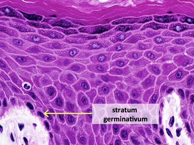
A 72 year old man had been complaining of many months of constipation followed by several months of severe diarrhea prior to admission. He did not receive medical care for these symptoms and had no medical history on file. He presented to an outside hospital unresponsive, and an emergent CT showed subcutaneous emphysema and stranding of the lower abdominal tissue extending to the left hemiscrotum. Also noted was irregular wall thickening in the distal rectum. Of note, his family reported that the patient had hit his left scrotum with a power cord recoil a few days prior to presentation. He was transferred to our hospital, where the surgical team reported severe cellulitis and necrosis of the scrotum, perineum, anterior abdominal wall, and upper thighs. Due to his poor prognosis, the family decided to transition his care to comfort measures only and he passed away several hours after presentation. An autopsy was performed, which revealed liquefaction of the subcutaneous tissue in the lower anterior abdominal wall, dusky gray connective tissue of the left testis as well as a small abscess at the superior pole, and a circumferential distal rectal mass.

Laboratory Investigation:

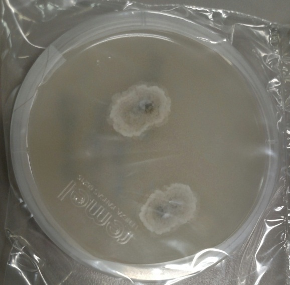
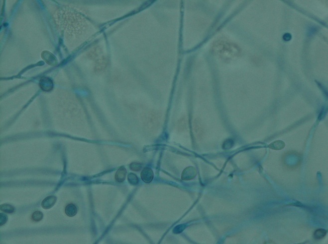
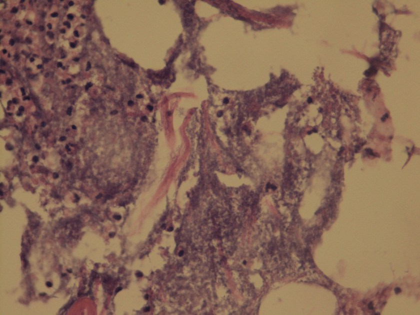
A gram stain of the scrotal abscess and tissue of the anterior abdominal wall both showed mixed gram positive and gram negative organisms. Cultures of these specimens both grew mixed gram positives including alpha hemolytic Streptococcus and coagulase negative Staphylococcus, as well as mixed anaerobes including Bacteroides fragilis group.

gang1
Gram stain of the scrotal abscess showing mixed gram negative and gram positive organisms.
gang2
Histologic section of anterior abdominal wall (400X) showing many polys and many bacterial organisms.
gang3
Chocolate agar plate showing mixed growth.

Discussion:

The case above represents an example of Fournier’s gangrene, a necrotizing infection of the external genitalia and/or perineum. These infections are most often polymicrobial and often include organisms that would not be particularly aggressive by themselves, suggesting a synergy between anaerobic and aerobic organisms. In a large literature review of over 4,000 cases of Fournier’s gangrene, the most common pathogens involved were Escherichia coli, Streptococcus, Bacteroides, Enterobacter, and Staphylococcus. Our case fits well with this profile, consisting of a mixture of gram positives and gram negatives including Streptococcus, Staphylococcus, and Bacteroides fragilis group. Antibiotic coverage in these cases must be broad spectrum and include coverage for aerobes, anaerobes, gram negative and gram positive organisms.

Risk factors for Fournier’s gangrene include diabetes mellitus, smoking, alcoholism, renal failure, hypertension, and coronary artery disease. It is unclear whether the patient in this case had any of these risk factors as he did not routinely seek medical care.

The rectal mass in this case proved to be adenocarcinoma. There have been multiple case reports of Fournier’s gangrene in association with rectal cancer. The theory behind the mechanism of this association is that perforation of the rectal cancer occurs with tumor infiltration of surrounding tissues spreading infectious organisms. It is possible that our case was caused by the patient’s rectal cancer, but it is also possible that the rectal cancer was simply coincidental. The recent trauma to the patient’s scrotum could also have acted as a vector for infection.

The patient in our case presented too late in the course of his disease to receive appropriate treatment, which would consist of broad spectrum antibiotic administration and aggressive debridement. Even with such treatment, the mortality rate of Fournier’s gangrene is quite high, ranging from 5% to 40% in different case series.

References:

Bjurlin MA et al. Causative pathogens, antibiotic sensitivity, resistance patterns, and severity in a contemporary series of Fournier’s gangrene. Urology 2013;81(4):752-8.

Bruketa T et al. Rectal cancer and Fournier’s gangrene – current knowledge and therapeutic options. World J Gastroenterol 2015;21(30):9002-9020.

-Laurie Griesinger is a Pathology Student Fellow at University of Vermont Medical Center.

Wojewoda-small

-Christi Wojewoda, MD, is the Director of Clinical Microbiology at the University of Vermont Medical Center and an Assistant Professor at the University of Vermont.

International Lung Cancer Experts Seek Public Comments on Revised Molecular Testing Guideline

From the press release:

BETHESDA, MD. June 28, 2016 — The College of American Pathologists (CAP), the International Association for the Study of Lung Cancer (IASLC), and the Association for Molecular Pathology (AMP) announced today the open comment period for the revised evidence-based guideline, “Molecular Testing Guideline for Selection of Lung Cancer Patients for EGFR and ALK Tyrosine Kinase Inhibitors.”

The open comment period begins today and will close on August 2, 2016. The online format provides an opportunity for public review of new draft recommendations for several key topics, as well as recommendation statements that have been reaffirmed since the initial guideline was jointly published online in April 2013 by Archives of Pathology & Laboratory Medicine, The Journal of Thoracic Oncology, and The Journal of Molecular Diagnostics.

The guideline revisions are designed to provide state-of-the-art molecular testing of lung cancer recommendations for pathologists, oncologists, and other cancer and molecular diagnostic laboratory professionals. The revisions are all based on evidence from an unbiased review of published experimental literature since 2013 and include the recommendations from an expert panel of renowned worldwide leaders in the field. The final recommendations will be approved and jointly published after consideration of the public comments, further panel discussion, and a complete evidence analysis. For more information and to provide comments, visit www.amp.org/LBGOCP.

Your Survival is in an Undisclosed Location

Over on NPR, Nell Greenfieldboyce writes about secret bunkers filled with healthcare supplies. You know, for the upcoming zombie apocalypse. (Okay, or maybe the next flu pandemic). It’s an interesting look at the logistics that go into managing stockpiles. Also, the author points out a sobering thought: while so much planning goes into inventory, dispersing that inventory in the event of an emergency could prove to be an issue.


Microbiology Case Study: A 44 Year Old Woman with Abdominal Tenderness

Case History

A 44 year old woman presented to an outside hospital with a chief complaint of abdominal tenderness and increased abdominal girth over the past few days. Her past medical history was significant for type II diabetes mellitus and associated complications including retinopathy and chronic kidney disease. As a result of her renal failure, she was currently undergoing peritoneal dialysis. Due to concern for infectious peritonitis, a paracentesis was performed and the resulting fluid obtained was sent to the Microbiology laboratory for Gram stain and bacterial culture. Because of difficulty in identifying the isolate, it was sent to our mycology section for further work up.

Laboratory Identification

prowick

Figure 1. Many sporangia of various sizes containing endospores arranged in a “soccerball” like pattern or morula configuration (Lactophenol cotton blue prep, 400x).


The isolate was received on potato dextrose agar (PDA) and appeared as discrete cream colored colonies, resembling a yeast. Upon transfer to cornmeal agar and incubation at 30°C, the isolate grew rapidly over the course of two days. The organism did not grow on media containing cycloheximide. A lactophenol cotton blue prep was performed (Figure 1) and showed many sporangia, ranging in size from 4-15 µm. In the larger forms, individual endospores were able to visualized and they were arranged in a “soccerball” like pattern. No budding or hyphae were present. Given these characteristics, the organism was identified as Prototheca wickerhammii.

Discussion

Prototheca wickerhammii is classified as an achlorophyllous algae and is known to cause human infections involving the skin and subcutaneous tissues, bursa of the elbow joint (olecranon bursitis) and rarely, systemic infections. P. wickerhammii is ubiquitous in nature and infection is usually the result from traumatic inoculation. Both immunocompetent and immunocompromised hosts can be affected, although more severe or systemic disease occurs in those who have defects in cell mediated immunity.

Prototheca spp. grow rapidly on PDA after incubation at 30°C for 2-3 days. Initially, it may be confused with a yeast based on plate morphology as they are cream colored and have a yeast-like consistency. When a lactophenol cotton blue prep is viewed under the microscope, sporangia of various sizes are identified (3-30 µm) that contain endospores arranged in a “soccerball” or symmetric daisy like pattern. Size of the sporangia, assimilation tests (such as the API 20C) and automated yeast identification systems (such as the Vitek yeast ID by bioMérieux) are helpful in identifying Prototheca to a species level. Both P. wickerhammii (4-11 µm in diameter) and P. zopfii (9-28 µm in diameter) can cause disease in humans, but P. wickerhammii is more common. While P. wickerhammii has symmetric internal divisions as seen in the above image, P. zopfii has random internal divisions.

The treatment of P. wickerhammii usually includes a combination of surgical management and anti-fungals depending on the site of involvement. For superficial skin infections, localized excision and the use of topical or systemic anti-fungals (azoles and amphotericin B) has been shown to have success. The treatment for olecranon bursitis focuses on bursectomy. In the case of systemic infections, amphotericin B has been the most successful treatment modality. Susceptibility testing is not routinely recommended to guide treatment of initial infections, as studies are few and results don’t always correlate with outcome.


Stempak

Lisa Stempak, MD, is an Assistant Professor of Pathology at the University of Mississippi Medical Center in Jackson, MS. She is certified by the American Board of Pathology in Anatomic and Clinical Pathology as well as Medical Microbiology. Currently, she oversees testing performed in both the Chemistry and Microbiology Laboratories.  Her interests include infectious disease histology, process and quality improvement and resident education. 

Collaboration is King

In the April issue of Transfusion journal, Joseph et al report their 1½ -year experience with the use of 4 Factor Prothrombin Complex Concentrate (4F-PCC) for urgent reversal of Vitamin K antagonists (Transfus 2016;l 56: 799-807).

As the authors mention, their “…study supports the safety of 4F-PCC for urgent vitamin K antagonist reversal even in unselected patients.”

I highlight this article for several reasons. It is incumbent upon those of us in the clinical laboratory, and especially the Blood Bank/Transfusion Service, to be aware of these new pharmaceutical agents that help provide rapid reversal of anticoagulants and allow for the potential elimination of unnecessary transfusions. I have found that often our clinical colleagues are unfamiliar with these strategies and we must take the lead in helping to establish protocols for their appropriate use. This article speaks, as well, to the need for ongoing evaluation of these drugs in, as they state in their title, the “real-world” of medical practice. Knowing how specific drugs affect outcomes outside of select studies with exclusions of particular patient populations (in this case, those with TE risk) is so valuable to our everyday work.

Another reason that this article is important is it underscores the importance of collaboration. The authors are representatives of departments of Pathology, the School of Medicine and Pharmacy. It is vital that we, as laboratory professionals, push to participate alongside our clinical colleagues in the assessment and implementation of new therapies and adjuvant treatments.

It is obvious from the Transfusion Medicine perspective, that our Pharmacy “friends” play a huge role in patient care, often spearheading and specializing in areas such as anticoagulant reversal strategies, release of factor concentrates, antifibrinolytics, IVIg and albumin. All of these pharmaceuticals can ultimately affect our laboratory testing and our potential interventions. Be certain you have representative from Pharmacy as a member of your Transfusion Committee.

It always pleases me to see, not only excellent literature, but also ongoing collaboration with laboratory professional often at the helm!


Burns

-Dr. Burns was a private practice pathologist, and Medical Director for the Jewish Hospital Healthcare System in Louisville, KY. for 20 years. She has practiced both surgical and clinical pathology and has been an Assistant Clinical Professor at the University of Louisville. She is currently available for consulting in Patient Blood Management and Transfusion Medicine. You can reach her at cburnspbm@gmail.com.

Microbiology Case Study: A 30 Year Old Male with Painful Right Eye

Case History:

A previously healthy 30 year old man presents with a painful right eye. Three weeks before he had been cutting concrete with an electric saw when a piece of hard concrete hit him in the eye. The eye became painful the following day. He was treated with empiric antibiotics but the eye pain failed to improve. He was eventually seen by ophthalmology, where ophthalmologic exam demonstrated findings suspicious for fungal keratitis. Corneal scraping was performed and sent for bacterial and fungal culture.

Laboratory Identification:

Bacterial culture showed no growth. Fungal culture demonstrated rapid growth of multiple white, cottony molds on potato flake agar. Over time these white colonies turned dark grey/brown. The reverse surface of the agar was white at first but also turned dark grey/brown over time.

Microscopically (scotch tape preparation), there were thin hyphae with single conidia arising directly off the tips of tapered conidiophores of variable length. Occasional conidia were also observed arising directly off of the sides of hyphae. The conidia were small and oval in shape with a truncated base, and somewhat darker as compared to the surrounding hyphae. Sexual forms were not observed.

scedo1
Potato flake agar showing a white, cottony mold with a central dark gray/brown coloration.


scedo2
Scotch tape preparation demonstrating thin hyphae with single oval-shaped conidia with truncated base. The conidia are small and have a “sperm-like” or “tadpole-like” morphology.

Discussion:

The clinical history and laboratory findings are characteristic of Pseudallescheria boydii/Scedosporium apiospermum.

Pseudallescheria boydii/Scedosporium apiospermum is an environmental mold which can be isolated from rural soils, polluted waters, manure and compost. Infection occurs secondary to local trauma. In immunocompetent individuals, infection is limited to the site of trauma, with some of the more common presentations being fungal keratitis, endophthalmitis, eumycotic mycetoma, sinusitis and pneumonia in the setting of near drowning. In immunocompromised individuals, infection can disseminate and involve any organ.

Pseudallescheria boydii/Scedosporium apiospermum is the most common cause of eumycotic mycetoma. Mycetoma is a chronic granulomatous infection of the subcutaneous tissue, usually involving the distal lower extremities, which can be caused by either a fungus (eumycotic mycetoma) or an actinomyces species bacteria (actinic mycetoma). On clinical exam there are multiple draining sinus tracts. The causative microorganisms aggregate into macroscopically visible groups (“granules”) which can be white, yellow or brown in color. Mycetoma may progress over time to involve underlying soft tissue, muscle, fascia and bone. Other less common causes of eumycotic mycetoma include Madurella spp., Acremonium spp., Fusarium spp., and Curvularia spp.

This organism has two names because historically different names were assigned to the sexual state (Pseudallescheria boydii) and to the asexual state (Scedosporium apiospermum). Morphologically, Pseudallescheria and Scedosporium are identical, the only difference being the presence of the sexual form, cleistothecia, in Pseudallescheria. Cleistothecia, when present, can be recognized as very large, dark-brown asci containing numerous ascospores. Technically, in the absence of cleistothecia the correct diagnosis would be Scedosporium boydii, however in clinical practice both names are usually listed regardless of whether cleistothecia are seen.

Pseudallescheria boydii/Scedosporium apiospermum is resistant to amphotericin B, but should be susceptible to azole therapy. The patient is being treated with topical voriconazole and oral fluconazole and currently (two weeks of antifungal therapy) has near complete resolution of symptomatology.

-Javier De Luca-Johnson, MD is a 3rd year anatomic and clinical pathology resident at the University of Vermont Medical Center.

Wojewoda-small

-Christi Wojewoda, MD, is the Director of Clinical Microbiology at the University of Vermont Medical Center and an Assistant Professor at the University of Vermont.


Microbiology Case Study: 47 Year Old Woman with History of Systemic Lupus Erythematosus

Case history

A 47-year old woman with a past medical history of Systemic Lupus Erythematosus (SLE) and liver cirrhosis of unknown etiology was admitted to the hospital for back pain and new onset neurological symptoms. She soon developed pancytopenia and study of her peripheral blood smear showed evidence of thrombotic microangiopathy. ADAMTS-13 inhibitor was negative ruling out thrombotic thrombocytopenic purpura (TTP). She then developed multiple thrombi, including a nonocclusive thrombus in the superior mesenteric vein with extension to the splenic vein as well as a femoral deep vein thrombosis. Her hospital course then became complicated by lupus cerebritis, a small ischemic focus in the left corona radiata and the left medial midbrain, and decompensated liver failure with hepatic encephalopathy. Despite intensive medical treatment, she became hypoxic and hypotensive requiring pressors, and expired in the ICU after several months of hospitalization. The autopsy was performed based on the relative’s request to better understand pathological processes that lead to patient’s demise. The flowing images were obtained from the brain at autopsy (Image 1).

toxo1
Light microscopy of H&E-stained sections of the hippocampus reveal encysted Toxoplasma bradyzoites as well as extracellular Toxoplasma tachyzoites in the CA1 region, suggestive of a subacute focal infection.

Discussion

Toxoplasmosis is considered to be a leading cause of death attributed to foodborne illness in the United States. More than 60 million men, women, and children in the U.S. carry the Toxoplasma parasite, but very few have symptoms because the immune system usually keeps the parasite from causing illness.

People typically become infected with Toxoplasma by contaminated food or animal-to human routes of transmission. Toxoplasmosis is not passed from person-to-person, except in instances of mother-to-child (congenital) transmission and blood transfusion or organ transplantation.

Persons with compromised immune systems may experience severe symptoms if they are infected with Toxoplasma while immune suppressed. Persons who acquire HIV infection and were not infected previously with Toxoplasma are more likely to develop a severe primary infection. The diagnosis of toxoplasmosis is typically made by serologic testing. A test that measures immunoglobulin G (IgG) is used to determine if a person has been infected. If it is necessary to try to estimate the time of infection, which is of particular importance for pregnant women, a test which measures immunoglobulin M (IgM) is also used along with other tests such as an avidity test. Due to the high rate of falsely positive Toxoplasma IgM testing, the FDA advises physicians testing pregnant women not to rely on the results of any one positive IgM test as the sole determinant for diagnosis of acute Toxoplasma infection.

Diagnosis can be made by direct observation of the parasite in stained tissue sections, cerebrospinal fluid (CSF), or other biopsy material. These techniques are used less frequently because of the difficulty of obtaining these specimens. Molecular techniques that can detect the parasite’s DNA in the amniotic fluid can be useful in cases of possible congenital transmission.

Clinical correlation

The case patient was at risk for developing toxoplasmosis due to SLE disease, and chronic immunosuppressive therapy that she was receiving for the aggressive course of her illness. However, most likely Toxoplasma gondii organisms seen in the brain parenchyma were in a dormant state due to lack of associated inflammation or architectural distortion. Her neurological decline is most likely related to thrombotic microangiopathy. Opportunistic infection is common in patients with SLE. In some patients, it is difficult to distinguish between the effect of infection and exacerbation of SLE because both can produce similar symptoms. There have been many reports of toxoplasmosis in SLE patients, with conditions such as cerebritis and pericarditis mimicking SLE manifestations.

References
1) http://www.cdc.gov/parasites/toxoplasmosis/

2) Seta N, Shimizu T, Nawata M et al. A possible novel mechanism of opportunistic infection in systemic lupus erythematosus, based on a case of toxoplasmic encephalopathy. Rheumatology (Oxford). 2002;41(9):1072-3.

3) Zamir D, Amar M et al. Toxoplasma infection in systemic lupus erythematosus mimicking lupus cerebritis. Mayo Clin Proc. 1999; 74(6):575-8.


Contributors

Written by Anastasia Drobysheva, MD, 2nd year Anatomic and Clinical Pathology resident, UT Southwestern Medical Center

Image provided by Bret Evers, MD, PhD, Neuropathology fellow, UT Southwestern Medical Center


-Erin McElvania TeKippe, Ph.D., D(ABMM), is the Director of Clinical Microbiology at Children’s Medical Center in Dallas Texas and an Assistant Professor of Pathology and Pediatrics at University of Texas Southwestern Medical Center.

Special Safety Considerations in the Anatomic Pathology Laboratory

Margie had worked in the histology department for years. She never used the chemical fume hood when pouring formaldehyde, but lately she had been coughing quite a bit, so she decided it was time to use the hoods. Soon she would be diagnosed with lung cancer.

Steve was a cytotechnologist working in the radiology department for a fine needle aspirate procedure. The radiologist was in a hurry, and when he handed the uncapped sample needle to Steve, the needle pierced the skin in Steve’s hand. The patient had Hepatitis C.

Jane had finished her long day of assisting with frozen sections and had to clean the cryostat. It was the end of the day, and she wanted to go home. She reached into the cryostat to change the blade with her gloved hands. She received a nasty cut on her finger, and since the blade had been used for multiple patients, Jane had to be treated for an exposure from an unknown source.

Anatomic Pathology laboratories present unique safety concerns that can differ from clinical lab concerns. Formaldehyde and other hazardous chemicals are used to preserve tissue specimens, workers can be exposed to large amounts of blood and body fluid during autopsies, and the sharp blades in cryostats and microtomes create additional cause for concern. Histology and Cytology employees need to be ever-vigilant to protect themselves from these exceptional hazards.

Formaldehyde and xylene are two chemicals typically handled in the AP lab. Xylene is a strong-smelling flammable liquid, and formaldehyde is listed by OSHA as a known carcinogen. If using these chemicals in the lab, you should have specific safety procedures for them, and vapor concentration monitoring should be performed to determine employee exposure levels. It is important to review the monitoring results as soon as possible, and never more than 10 ten days after receiving them. Discuss the results with each monitored employee individually or post the results in the department. Because monitoring is performed for tasks as well as locations, it is considered representative monitoring. That means one result may represent several employees who perform the same tasks or work in the same area.

If vapor concentration levels are elevated, make sure to take measures (such as using engineering controls) to reduce or eliminate staff exposure. OSHA’s Formaldehyde Standard requires annual monitoring if results are above the STEL (short term exposure limit). To discontinue sampling, the lab needs to obtain results from two consecutive sampling periods taken at least 7 days apart show that employee exposure is below the action level and the STEL.

OSHA requires a specific safety formaldehyde training for all employees who are exposed to formaldehyde concentrations of 0.1 parts per million (ppm) or greater. This formaldehyde training must include specific elements including education of the contents of the Formaldehyde Standard, the contents of the formaldehyde Safety Data Sheet (SDS), the health hazards of formaldehyde exposure, the proper use of necessary PPE, and spill response and clean-up procedures. This training is required by OSHA annually, and it may need to be provided to some employees who work outside the laboratory (operating room staff, labor and delivery staff, etc.). Even though the laboratory may not be responsible to provide this training, it is a good idea to communicate with other department leaders to ensure they get the required training for their staff.

Blades and other sharps pose great risks in AP procedure areas. Most microtomes and cryostats are equipped with knife guards- insist that they be used. Remove used blades with magnetic-tipped implements and insert new blades using rubber-tipped tweezers. Place disposable knives in sharps containers after use.

Odd as it may seem, there are Cytology procedures where the hand-off of uncapped needles containing samples sometimes occurs. Unprotected needles should never be passed from one person’s hand to another. These needles should be placed on a counter or the technologist should only take them from the collector with an implement such as tweezers. Be sure there is ample space to safely perform the cytology tasks in the procedure area away from the lab.

Laboratory Safety is not a “one-size fits all” umbrella for all areas where laboratorians work. Risk assessments and task assessments are critical to determine the particular hazards in a specific lab. In the Anatomic Pathology laboratory, these assessments should guide safety leaders to those specific safety measures which can help your staff prevent injuries and exposures which can be career and life-changing.


Scungio 1

-Dan Scungio, MT(ASCP), SLS, CQA (ASQ) has over 25 years experience as a certified medical technologist. Today he is the Laboratory Safety Officer for Sentara Healthcare, a system of seven hospitals and over 20 laboratories and draw sites in the Tidewater area of Virginia. He is also known as Dan the Lab Safety Man, a lab safety consultant, educator, and trainer.

Hybridization Conditions and Melting Temperature

Stringency is a term that many molecular technologists are all very familiar with. It is a term that describes the combination of conditions under which a target is exposed to the probe. Typically, conditions that exhibit high stringency are more demanding of probe to target complementarity and length. Low stringency conditions are much more forgiving.

  • If conditions of stringency are too HIGH → Probe doesn’t bind to the target
  • If conditions of stringency are too LOW → Probe binds to unrelated targets


Important Factors That Affect Stringency and Hybridization

  • Temperature of hybridization and salt concentration
    • Increasing the hybridization temperature or decreasing the amount of salt in the buffer increases probe specificity and decreases hybridization of the probe to sequences that are not 100% the same.
  • Concentration of the denaturant in the buffer
    • For example: Deionized Formamide and SDS can be used to reduce non-specific binding of the probe
  • Length and nature of the probe sequence
STRINGENCY AND BINDING
– Long Probe


– Probe has increased number of G and C bases


Binding occurs under more stringent conditions
– Short Probe


– Probe has increased number of A and T bases


Binding occurs under less stringent conditions

Melting Temperature (Tm) Long Probes

  • The ideal hybridization conditions are estimated from the calculation of the Tm.
  • The Tm of the probe sequence is a way to express the amount of energy required to separate the hybridized strands of a given sequence.
  • At the Tm: Half of the sequence is double stranded and half of the sequence is single stranded.
  • Tm = 81.5°C + 16.6logM + 0.41(%G+C) – 0.61(%formamide) – (600/n)

Where M = Sodium concentration in mol/L

n = number of base pairs in smallest duplex

  • If we keep in mind that RNA is single stranded (ss) and DNA is double stranded (ds), then the following must be true:


RNA : DNA Hybrids   More stable

DNA : RNA Hybrids        ↓

DNA : DNA Hybrids    Less stable


  • Tm of RNA probes is higher, therefore RNA : DNA hybrids increase the Tm by 20 – 25°C


Calculating the Tm for Short Probes (14 – 20 base pairs)

  • Tm = 4°C x number of G/C pairs + 2°C x number of A/T pairs
  • The hybridization temperature (annealing temp) of oligonucleotide probes is approximately 5°C below the melting temperature.

melt-temp

Sequence Complexity (Cot)

  • Sequence complexity refers to the length of unique, non-repetitive nucleotide sequences.
  • Cot = Initial DNA Concentration (Co) x time required to reanneal it (t)
  • Cot1/2 = Time required for half of the double-stranded sequence to anneal under a given set of conditions.
  • Short probes can hybridize in 1 – 2 hours, where long probes require more time.


Test Your Knowledge

  1. Calculate the melting temperature of the DNA sequence below:

ATCTGCGAAATCAGTCCCGG
TAGACGCTTTAGTCAGGGCC


Answer
If the number of G/C pairs = 11, and the number of A/T pairs = 9. The calculation is as follows:
4(11) + 2(9) = X
X = 62°C

L Noll Image_small

-LeAnne Noll, BS, MB(ASCP)CM is a molecular technologist in Wisconsin and was recognized as one of ASCP’s Top Five from the 40 Under Forty Program in 2015.


Show Us Some Skin

Which epidermal layer is indicated by the arrow?

  • A. Stratum corneum
  • B. Stratum germinativum
  • C. Stratum granulosum
  • D. Stratum lucidum
  • E. Stratum spinosum

Skin1


The answer is E, stratum spinosum. The stratum spinosum, also known as the prickle cell layer of the skin, is often several cell layers deep, and is located immediately above the stratum germinativum. Cells in this layer are polygonal, and are connected to each other by numerous desmosomes. During fixation, the cell membrane retracts around desmosomal contact points, giving the cells a prickly appearance. The cells contain many bundles of intermediate filaments as well as keratinosomes, which are membrane-bound granules thought to deposit a “toughening” layer on the surface of the cell membrane.

Skin2


The stratum germinativum (also known as the basal layer) is composed of a single layer of cells adjacent of the basal lamina. The cells are tall cuboidal or columnar, and are connected to the basement membrane by hemidesmosomes, and to other cells by desmosomes. Cells in the basal layer are mitotically active, and contain numerous polyribosomes and intermediate filaments.

Skin3


The stratum granulosum is 3 to 5 cell layers thick, and is composed of flattened, polygonal cells arranged with the long axis parallel to the basement membrane. The cytoplasm of these cells contains numerous basophilic granules, called keratohyalin granules, which are thought to be keratin precursors.

The stratum lucidum is not truly a distinct layer of skin, but rather a staining artifact. It is visible in some sections of thick skin as a glassy-appearing, eosinophilic artifact at the bottom of the stratum corneum. It is not present in this particular image of epidermis.

Skin4


The stratum corneum is the outermost layer of skin. The thickness of this layer varies considerably from region to region in the body. The cells of this layer are dead, flattened, and fused together, with completely keratinized cytoplasm.

Skin5


Krafts

-Kristine Krafts, MD, is an Assistant Professor of Pathology at the University of Minnesota School of Medicine and School of Dentistry and the founder of the educational website Pathology Student.