Global Health Narratives – An Interview Series Summary

2019 marked a very special year for me as I had the incredible opportunity to interview some of the most remarkable laboratory medicine specialists in the field of Pathology about their involvement in global health. Although their roles ranged from everything between medical technician, to PA, to medical student, to practicing pathologist, to the CEO of a major pathology organization, they all had one thing in common – they actively take the time to better their global community and contribute to improving pathology services in resource limited settings.

Now that the year is winding to a close, I’d like to take the opportunity to highlight all of these wonderful efforts and hopefully inspire you to take similar initiatives where applicable to your abilities and interest. Read on for a summary of each interview.

Dr. Kumarasen Cooper not only volunteers bi-annually in Botswana’s only academic pathology department as a way to give back to his native Africa, he has also worked to create an opportunity for residents at UPenn pathology to be involved too. Because of his efforts, the UPenn residents can accompany him and work together on the departments’ shared initiatives using official institutional elective time. This is a rare opportunity in pathology training, and is a model of how academic institutions can engage their trainees in global health initiatives.

Julie Papango, a medical technologist, has worked with Doctors without Borders/ Médecins Sans Frontières (MSF) to bring laboratory medicine to the world’s most remote places. She was one of MSF’s very few volunteers with laboratory experience and therefore has played a crucial role in projects ranging from addressing the infectious disease outbreaks in a Sudanese refugee camp, to helping the Cambodian Ministry of Health to improve their national tuberculosis detection program.

Dr. Ann Nelson is an expert in infectious disease pathology and has worked in many parts of Africa for more than 30 years. The focus of her work has been in HIV/AIDS pathology in the US and in sub-Saharan Africa. Currently she works on educational projects and capacity building in anatomic pathology, and linking anatomic pathology to ongoing clinical and epidemiologic research. She finds ways to be helpful in any new setting by just showing an open and willing attitude. “I went and built partnerships with everyone I could. You have to just go and talk to people, and ask them “What can we do?” With this approach, she’s been able to find countless ways to contribute her expertise to the world. She’s also spent innumerable hours in studying and publishing the issues affecting pathology services in Africa. Notably, she worked to conduct a landmark survey of African pathologists to determine the status of pathology resources in Sub-Saharan Africa.

Dr. Blair Holladay and Dr. Dan Milner have worked in global health most of their professional careers and now lead the American Society for Clinical Pathology’s efforts in improving laboratories worldwide. They are working with governments and local agencies to make sustainable changes in the neglected pathology and laboratory medicine landscape in low and middle income countries (LMICs). They are responding to the urgent need to improve pathology services to address the rapid increase in global non-communicable disease (NCD) incidence. As Dr. Holladay points out “Compared to the scale of the HIV crisis, NCDs are the health threat that gone unchecked, will go far beyond in affecting huge proportions of the global population.” In response to addressing this problem, Dr. Milner points out that the lab is the cornerstone to the solution: “In the field of cancer, which is a major problem in LMICs, you cannot treat the patient without a diagnosis – and the diagnosis must come from the laboratory.”

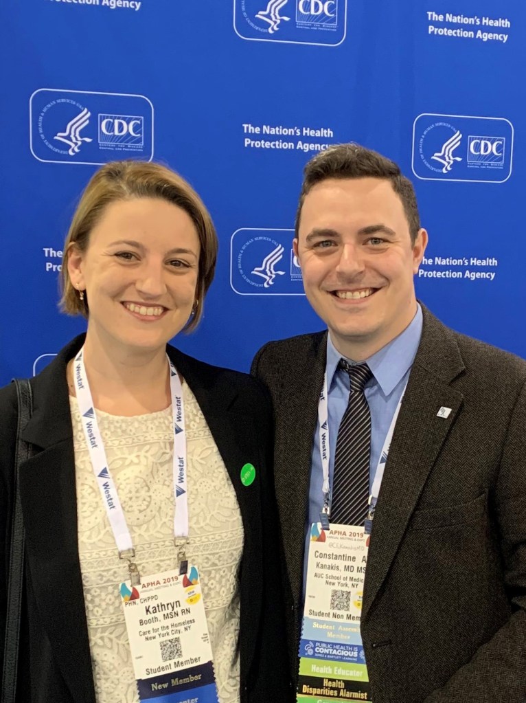
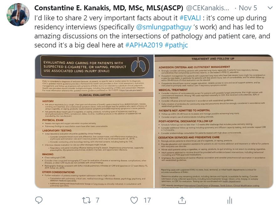
Dr. Constantine Kanakis is a medical student who decided to be an active part of the community of Sint Maarten while living there attending medical school. The community was facing multiple mosquito-borne infectious disease epidemics that includes Zika virus. In response, Dr. Kanakis took a service-learning elective course in medical school that focused on community outreach. He led the way to create an outreach program that has now been incorporated into the nation’s Ministry of Health Collective Prevention Services program. Dr. Kanakis encourages everyone to “Start by looking around at your immediate surroundings and take an assessment of the issues affecting the community. Anyone can do this, whether you are a physician, scientist, or a community member.”

Dr. Adebowale Adeniran, a cytopathologist, frequently works with the USCAP group “Friends of Africa” in which he speaks at the annual meetings, is involved in the group planning activities, and participates in educational initiatives and conferences in Africa. He encourages all academic institutions to engage in global health, stating “Academic institutions in the US can offer ways of enhancing training opportunities for African pathologists and trainees by offering short- or long-term exchange programs. This helps to bridge the gap between practiced based learning in resource limited vs. US institutions.”

Nichole Baker is a pathologist’s assistant that heard of a lab in Uganda that needed outside pathology help due to being severely understaffed. So Nichole decided to go visit the lab and see where she could help. One of the main issues was that the lab lacked an electronic medical record (EMR) system and keeping track of cases and patient reports was a real challenge. With no background in computer science, Nichole resourcefully reached out to her personal network to find someone that could help her build a free EMR and now the laboratory can track specimens, issue electronic reports, and has reduced their turnaround time as a result.

Dr. Drucilla Roberts is one of the world experts in perinatal pathology and has been working in Africa for over ten years with a focus on capacity building. Besides offering her surgical subspecialty expertise, she is also partnering with local pathologists to participate in ground breaking research on topics specific to low resource settings. She’s written widely on the need for pathology services in Africa. She says that one of the biggest problems in improving pathology services in Africa is that “there are not enough pathologists. You can help improve things in individual labs to a point, but for long term there has to be more pathologists working in Africa.” Dr. Roberts actively engages in solving this problem by helping train African pathology residents and by recruiting other pathologists to do the same.

Dr. Von Samedi, a cytopathologist, has worked with ASCP’s Center for Global Health at their partner sites all around the world. Dr. Samedi started working with ASCP as a resident, using his unique ability to speak French and Creole to assist ASCP in Haiti following the devastating 2010 earthquake. He has since worked on improving laboratory services in a vast array of ways, with everything from mentoring and local laboratorian training to running workshops on HIV related testing services. Volunteering gives Dr. Samedi a sense of purpose and he states that he “also benefits from interacting with my global colleagues and learning from them.”

There are so many more laboratory medicine specialists working in global health that I would have loved to feature on Lablogatory – but there are so many that I cannot capture all of their stories to share here. I hope that you have gained a snapshot of the potential ways that you can get involved, the possibilities are truly endless!

If you’ve been following this series, know that I am extremely grateful for your time and attention to this important matter. This will be my last post with Lablogatory for the time being, as I will be taking a break from writing to welcome my first child into the world! Wish me luck! J

If you want to find out more about volunteering in global pathology efforts, please visit my webpage that I have written in collaboration with Dr. Jerad Gardner: https://pathinfo.fandom.com/wiki/Global_Health_Opportunities_for_Pathologists

Please also take a moment to fill out this survey (https://www.surveymonkey.com/r/K7YK8LW) so that we can learn more about your interest and experience in global health and you can enter to win a global pathology prize pack!

-Dana Razzano, MD is a former Chief Resident in her fourth year in anatomic and clinical pathology at New York Medical College at Westchester Medical Center and will be starting her fellowship in Cytopathology at Yale University in 2020. She is passionate about global health and bringing pathology and laboratory medicine services to low and middle income countries. She was a top 5 honoree in ASCP’s Forty Under 40 in 2018 and was named to The Pathologist’s Power List of 2018 and 2019. Follow Dr. Razzano on twitter @Dr_DR_Cells.

Microbiology Case Study: A Child with Acute Abdominal Pain

Clinical presentation

An elementary school age child presented to the pediatric emergency department with an acute onset of abdominal pain. According to the parents, the patient recently had an ear infection and completed a course of amoxicillin. They noted the patient was more tired than usual, but did not have a fever. They reported no recent sick contacts or travel. Past medical history was significant for constipation, but normal bowel movements were noted over the past few days. On physical exam, the abdomen was soft and non-distended with diffuse mild tenderness noted on the right side. No masses were noted.  Laboratory testing was unremarkable and the WBC count, liver & pancreas enzymes, and alpha fetal protein were within normal limits. An abdominal CT scan revealed a mass with central necrosis in the liver concerning for an abscess. The patient was started on ceftriaxone & metronidazole and underwent a surgical procedure to drain the lesion.   

Laboratory Identification

Image 1. Gram stain of the direct liver aspirate showed many gram positive cocci in pairs & chains and numerous white blood cells (oil immersion).
Image 2. Rare small, white non-hemolytic colonies grew on CDC agar after 42 hours of incubation at 35°C under anaerobic conditions.

No bacterial growth was observed on blood or chocolate agars incubated at 35°C in CO2. MALDI-TOF mass spectrometry identified the isolate as a viridans groups streptococci, Streptococcus intermedius. The organism was susceptible to penicillin, ceftriaxone, and vancomycin by broth microdilution. Blood cultures were not collected for this patient.

Discussion

Streptococcus intermedius is a viridans group streptococci that belongs to the S. anginosus group. The S. anginosus group also includes S. anginosus and S. constellatus. This group of viridans streptococci composes the normal flora of the oropharynx, urogenital, and gastrointestinal tracts. These organisms are known for causing peritonitis and abscesses, particularly in the brain, breast, liver, and oral cavity.

Similar to other streptococci, S. intermedius is a gram positive cocci that grows in chains and is catalase negative. The anginosus group are facultative anaerobes and grow as pinpoint colonies (<0.5 mm) on blood agar. This is in contrast to pyogenic, beta-hemolytic streptococci which are greater than 0.5 mm in size after the same incubation period. The anginosus group streptococci can exhibit a variety of hemolysis patterns, including alpha, beta, or gamma hemolysis. A distinct butterscotch or caramel odor is noted on examination. The anginosus group can possess Lancefield antigens A, C, F, G, or be non-groupable, so it is important not to misidentify them as other streptococci that also have these antigens.

Historically, further identification of viridans group streptococci was challenging; however, the advent of automated systems and MALDI-TOF mass spectrometry has been useful in providing species level identifications for more common isolates. Molecular sequencing methods using sodA gene can be helpful as well for the most reliable results. While penicillin resistance is becoming more frequent in viridians group streptococci, it is still rare in the S. anginosus group.  

In the case of our patient, an echocardiogram was performed and found to be negative for endocarditis. The patient’s symptoms improved and they were discharged home on ceftriaxone and metronidazole. A follow up CT scan to confirm resolution of the abscess was scheduled.  

-Lisa Stempak, MD is the System Director of Clinical Pathology at University Hospitals Cleveland Medical Center in Cleveland, Ohio. She is certified by the American Board of Pathology in Anatomic and Clinical Pathology as well as Medical Microbiology. Her interests include infectious disease histology, process and quality improvement, and resident education.

Is it Christmas? Hematology Case Study: Coagulopathy

A 2 year old male was brought into the pediatrician’s office by his mother after tripping over a toy truck 2 days earlier. The mother stated that the child cut the inside of his lip in the fall, and the lip had been oozing blood for the past 2 days. The child had also experienced a bloody nose several times since the fall. Upon examination, the child appeared in general good health with no other bruising or bleeding. Examination of the joints revealed swelling in the right knee. The physician took a family history, and the mother reported that her younger brother has ‘some sort of bleeding problem’ and experienced prolonged bleeding after a tonsillectomy as a child, and after several surgeries as a young adult. The physician ordered blood work on the child.

  • Hgb 9.5 g/dl
  • Hct 30%
  • Platelet  185 x 103/ uL
  • INR  1.1
  • aPTT 57 sec
  • Mixing Test: corrected
  • Thrombin Time: normal

Based on these results, the prolonged aPTT warranted further investigation. A differential diagnosis involved ruling out other causes for the prolonged aPTT. The physician ordered mixing studies, factor VIII and factor IX assays and vWF. Mixing studies are used to determine if etiology of prolonged PT or PTT is due to a factor deficiency or an inhibitor. If the aPTT remains prolonged after mixing with normal plasma, this indicates an inhibitor. If the prolonged PTT becomes normal after the mixing studies, this would indicate a factor deficiency. The factor VIII and vWF were normal, but factor IX activity was 25%. Diagnosis: Factor IX deficiency. (It was also confirmed, after speaking with the child’s uncle, that he also had a factor IX deficiency)

So, you may ask, what does this have to do with Christmas? In the spirit of the season, I chose to present a Case Study on Factor IX deficiency, aka Christmas Disease. But, alas, this really has nothing to do with the holiday. Maybe it has something to do with the fact that the first article about this disorder was published in the British Medical Journal on Dec 27, 1954 (just 2 days after Christmas)? But, not so. Actually, Factor IX deficiency is also called Christmas Disease because it is named after Stephen Christmas, the first patient described to have Factor IX deficiency. Stephen Christmas was diagnosed with hemophilia in Toronto in 1949, at the age of 2. The family was visiting relatives in London in 1952 and it was there that doctors discovered that he was not deficient in Factor VIII, the cause of Classic Hemophilia as it was known at the time. It was discovered that he was deficient in another coagulation protein. This new protein was named Christmas protein and later became known as Factor IX.

A little bit more about the history of Factor IX deficiency. Before the discovery of the Christmas protein, it was thought that Hemophilia was a single disorder, caused by a deficiency of Factor VIII. With the discovery of this new protein, Classic Hemophilia (Factor VIII deficiency), was given the name Hemophilia A, and this new Factor IX deficiency became known as Hemophilia B. Yet another nickname for this disorder is the Royal Disease. Hemophilia was prominent in the European royal families in the 19rth and 20th centuries. Queen Victoria of Britain was a carrier of hemophilia and passed the gene on to three of her children. Her children and descendants married into the royal families of Germany, Russia and Spain, giving her the nickname the Grandmother of Europe. But, these marriages also served to spread the disease to these other royal houses, giving hemophilia the nickname Queen Victoria’s curse. The last known member of the royal families of Europe to carry the gene passed away in 1945, 9 years before that article in the British Medical Journal (December 27, 1954). So, how do we know that Hemophilia B is the hemophilia responsible for the Royal Disease? In 2009, DNA testing on bones identified as  Anastasia and Alexei Romanov, the last Russian royal family descendants of Queen Victoria, determined that the Royal Disease was Hemophilia B.

I remember teaching Hematology and Genetics before 2009 using a pedigree chart of Queen Victoria’s family to teach students about Hemophilia as an X linked recessive disorder. We created Punnett squares that showed the inheritance from Queen Victoria to her family members and descendants across Europe. I always enjoyed this lecture, because it was a fun piece of historical trivia paired with a good science lesson. After 2009, the science of the inheritance did not change, but we now knew that this Royal Disease was Hemophilia B. Hemophilia B is caused by mutations in the F9 gene which is responsible for making the factor IX protein.  The F9 gene is on the X chromosome. Hemophilia B, like Hemophilia A, is X linked, carried by the mother. 50% of males born to a carrier mother will have the disease and 50% of daughters will be carriers. All daughters of affected males will be carriers, but their sons will not be affected. Hemophilia A is more common than Hemophilia B, affecting about one in 5,000 males. Hemophilia B affects about one in 25,000 males. It has been though that up to about 30% of Hemophilia B cases occur as a spontaneous mutation and are not inherited. This has been thought to be the case with Queen Victoria. She has been believed to be ‘case zero’, the first hemophilia case in her family. However, some newer articles that have researched her family history suggest that she may have had a half-brother who had the disease.1 There are also other related disorders including a rare autoimmune acquired hemophilia B and another rare form of Hemophilia B called Hemophilia B Leyden.

The coagulation process involves many chemical reactions, from the initial event that triggers bleeding, to the formation of a clot. The sequence of events are generally depicted as a coagulation cascade to illustrate and simplify understanding of the process. The coagulation cascade is divided into 2 pathways, the intrinsic and extrinsic system, and a common pathway. This segregation of sections is not physiological, but allows for the grouping of factor defects and the interpretation of laboratory testing. Most problems with coagulation factors fall into one of three categories: a factor is not produced, there is a decreased production, or the factor is produced but not functioning properly. Hemophilia B is a factor IX deficiency. It is classified as mild, moderate or severe based upon the activity level of factor IX. In mild cases, bleeding symptoms may occur only after surgery or trauma and may not be diagnosed until later in life. In moderate and severe cases, bleeding symptoms may occur after a minor injury or even spontaneously. These moderate to severe cases are usually diagnosed at a younger age.

This child was diagnosed with Hemophilia B, based on coagulation studies, Factor IX assay results and family history. Treatment involves replacement of Factor IX to promote adequate blood clotting and prevent bleeding episodes.

References

  1. Turgeon, Mary Louise, Clinical Hematology: Theory & Procedures, 6th ed.  Lippincott Williams and Wilkins, Philadelphia, 2017.
  2. https://www.hog.org/publications/detail/the-royal-disease-a-family-history-update-on-queen-victoria
  3. https://rarediseases.org/rare-diseases/hemophilia-b/
  4. https://pediatriceducation.org/2015/12/14/what-is-it-called-christmas-disease/
  5. https://www.stago-us.com/hemostasis/tests-clinical-applications/hemophilia-b/

-Becky Socha, MS, MLS(ASCP)CM BB CM graduated from Merrimack College in N. Andover, Massachusetts with a BS in Medical Technology and completed her MS in Clinical Laboratory Sciences at the University of Massachusetts, Lowell. She has worked as a Medical Technologist for over 30 years. She’s worked in all areas of the clinical laboratory, but has a special interest in Hematology and Blood Banking. When she’s not busy being a mad scientist, she can be found outside riding her bicycle.

Recognizing Disruptive Innovation in Global Health

One of the challenges of providing healthcare to patients of any type is “staying current” or “keeping up with the literature.” This can be especially challenging in the diagnostics laboratory where novel or unique approaches to a given test or test method or disease may show early promise but have no clinical utility, be too expensive, or not actually significantly change work-flow and/or patient value to justify implementation. On the other hand, sometimes a technology or test which is in development or approval can be so anticipated that clinicians and laboratorians are frustrated that it is not yet available.

In global health, there is a different problem that is encountered every day. There are technologies and tests that are approved, have documented clinical utility, and add great value to patients but they are simply not available because of supply chain, cost, administration, or geography. In such situations, the practitioners in these settings face extreme frustration—especially with stock-outs—and can become jaded and non-dependent on laboratory testing as part of care. This latter issue is a major challenge in cancer care where cancer diagnoses are required before treatment can begin; yet, in a large number of countries, access to cancer diagnostics routinely is not available. It is to that end that ASCP along with a whole host of NGO, industry, academic, and government partners are making great efforts to improve cancer care in each part of the continuum.

In this environment, however, disruptive innovations are, in fact, much easier to recognize as forthcoming. In the early 2000’s when I was working and traveling in Malawi, our project had a landline in the hospital to call the landline at the doctor’s house for issues overnight with patients. This required 24-hour nurses to be physically in the ward, tied to the phone and the patients. Landlines were expensive to install, had a very long waiting list to be installed, and, for the most part, the majority of the population in the country had never had a phone line in their dwelling. By the mid-2000’s, our project had one or more cellphones (as did the nurses) and communications through texting were nearly constant (especially since it was less expensive than making a phone call). By 2010, cell phones were ubiquitous in Malawi (and almost everywhere else in Africa) and there was no demand for landlines. Although this is a commonly used example, consider the adoption of cellular telephones and now smartphones in the US compared with Africa. There was push back, denial, avoidance, and even refusal to use them because there was an existing, well established system of landline communication.  If you want to install cable television and internet in your home as late as 2016, you were often required to bundle with a landline. The point is that the adoption pattern was significantly different because there was a pre-existing competitor with the new technology although—clearly—the new technology was superior.

Now consider a woman of 35 years who has a breast mass on mammogram in downtown Boston today. She will likely have an imaging study with immediate ultrasound and fine needle aspiration and/or core biopsy subsequent. A pathological diagnosis will be issued within 3 to 4 business days (or sooner) which includes a histological diagnosis along with hormone receptor status and Her2 staining. She will see a clinician likely within a week for a positive cancer diagnosis and a treatment plan will be decided upon and executed. If we consider a similar woman in downtown Nairobi, Kampala, or Lagos, they may, in fact, have a similar experience because of the recent efforts globally to improve cancer awareness, diagnosis, and treatment. There may be some delays (reports may take several weeks), potential stock-outs, etc. but, in these major cities, the services might exist. They are likely, however, provided in private clinics, will cost a premium, and may or may not have any guarantees about quality.

The reality, however, is that the vast majority of women in the US or Europe who present with breast cancer do so at a very early stage because of active screening programs which include mammography. The vast majority of women in low- and middle-income countries (LMICs) present with later staged disease because of lack of screening. The latter group of women, however, often live in rural conditions and/or poverty conditions such that seeking care for a breast mass (of any size) will require them to spend time and money to travel to one of the major cities and attempt to access services. With this situation, many of these cancers are detected by the health system at a late stage where curative therapy windows have been missed.

Onto these observations let’s now overlay access to a test for a breast mass that can be performed on a fine needle aspiration biopsy and resulted in ~4 hours which will provide a diagnosis of cancer (or benign) along with prognostic features directing treatment. If we consider the woman in Boston, we may see such a test providing an incremental improvement in care because billing systems, litigation fears, compliance requirements, or accreditation standards still include routine histology and immunohistochemistry to be performed on a tissue biopsy. To some degree, the test may be rejected because it is adding a cost over the standard costs without adding value (other than speed) to the results. However, for the woman in the rural village who likely has access to a community health worker, access to such a test could mean that she starts oral therapy the same day she has the health visit without ever having to leave her village. We have now removed the journey to a clinic that can performed a biopsy, the costs associated with that travel, the time lost while traveling and waiting for a result, and removed the risk that this is not breast cancer—which would mean all the time and money were wasted. For this woman, enormous value is created for her with a test that is performed same day with immediate results.

This concept of point-of-care (POC) cancer diagnostics would arguable meet resistance in the US or European system because of competition with existing systems and other issues as mentioned previously. In an LMIC setting, as there may be no competition, such an innovation would sweep the system and become standard of care—almost regardless of cost. This last bit is very important because traditional systems for performing histology and IHC are complex, costly, and require multiple highly trained individuals to get a quality result. If that process costs $75 to $100 US dollars (to the health system) to provide and, for the individual patient, $10s to $100s of dollar for the travel, lodging, and lost wages, the cost of such a test could, in a stable, high-income country (HIC) market, fetch a hefty price. However, if such a test is priced at $25 to $50 USD (half the cost of the current system excluding the travel), the immediate replacement of the old system with this new system for the given indication must and will occur. This uptake is amplified in an LMIC when the POC test moves to the patient in a geographically distributed process. Breast cancer is an obvious target for such an approach because the tumors are easily accessible, the disease is quite common globally, and the primary therapies are very inexpensive. Could such a test have an impact in an LMICs for bone marrow-based, lung, bladder, colon, prostate, liver, kidney, or soft tissue tumors? The answer to that question lies in the availability of therapy, incidence of disease, and access to radiological equipment rather than availability of the actual POC device. That is, once you have a POC test for one cancer, creating a subsequent POC test for another cancer is a surmountable technical hurdle. But will such a test be able to have an impact because of the alignment of the other factors? It is likely that as you are reading this sentence, you have thought of a few yourself but there are certain cancers where you are likely thinking, “not possible”.

For breast cancer, two such POC approaches are coming down the pipeline. The first is the Cepheid GeneXpert Breast STRAT4 assay which measures quantitative RNA (qRNA) for ESR1, PGR, ERBB2, and MKi67. These four assays are surrogates for standard immunohistochemical staining for ER, PR, Her2, and Ki-67, respectively. In a series of published and in press feasibility and validation studies, the qRNA assay is essentially equivalent to IHC. There are nearly a dozen studies of this new testing cartridge using formalin-fixed, paraffin embedded (FFPE) tissue throughout Africa where the test is being compared to standard IHC. However, in at least one site, the test is being performed directly on FNA material. The second test is from the laboratory of Dr. Sara Sukumar at Johns Hopkins which uses a set of DNA methylation markers that can separate benign from malignant disease on FNA using only 10 markers. By combining these two approaches (benign vs. malignant followed by STRAT4 for positive tumors), a diagnosis of malignant breast disease with prognostic factors for treatment could be obtained in less than 4 hours.

Let’s jump forward to the point in time when both of these POCs are available (or, in fact, any POC for cancer is available). How would they change the approach to breast or other cancer in an LMIC? Because both tests require only an FNA of a mass and because tumors of the breast and other organs today are often late staged, community health workers could be trained to evaluate patients with masses, perform the sampling, and run the test in a remote village. Regardless of stage, starting a breast cancer patient on estrogen receptor antagonists can provide palliative relief or pre-surgical treatment. As a population down stages—which occurs as community health workers begin routine screening—the testing can triage benign and malignant disease at a fraction of the cost for both the system and the patient. Based on population epidemiology, nearly exact costs for these services can be predicted for a population and stock outs can be avoided. Corollary note: Only for those cancers for which you HAVE a POC.

How would these tests change the approach to breast cancer in an HIC? There would likely be resistance at many levels but, eventually, the relatively low cost and the increased patient value would allow the tests to replace or displace standard diagnostics. Without complete replacement, there could, at a minimum, be multimodality redundancy which increases quality. However, the tests would find purchase within the system because in some settings their cost and added value would make any other choice impossible.

For both settings, we can now add other market entrants, other tests for other cancers, and a generalize increased in cancer awareness in the community, all of which would increase demand, improve morbidity and mortality, but decrease costs. Such a situation would be highly valued by the patients and, therefore, is the most important eventuality as this disruption ensues. Recognizing forthcoming change is sometimes hard and sometimes easy; however, accepting and embracing forthcoming change in healthcare can lead to best outcomes for our patients—the central mission of ASCP.

Dr. Milner has no financial disclosures regarding this blog post and has received no fiscal or in-kind support from any entity, named or otherwise, that involves this blog post.

References

  1. Wu NC, Wong W, Ho KE, Chu VC, Rizo A, Davenport S, Kelly D, Makar R, Jassem J, Duchnowska R, Biernat W, Radecka B, Fujita T, Klein JL, Stonecypher M, Ohta S, Juhl H, Weidler JM, Bates M, Press MF. Comparison of central laboratory assessments of ER, PR, HER2, and Ki67 by IHC/FISH and the corresponding mRNAs (ESR1, PGR, ERBB2, and MKi67) by RT-qPCR on an automated, broadly deployed diagnostic platform. Breast Cancer Res Treat. 2018 Nov;172(2):327-338.
  2. Wasserman BE, Carvajal-Hausdorf DE, Ho K, Wong W, Wu N, Chu VC, Lai EW, Weidler JM, Bates M, Neumeister V, Rimm DL. High concordance of a closed-system, RT-qPCR breast cancer assay for HER2 mRNA, compared to clinically determined immunohistochemistry, fluorescence in situ hybridization, and quantitative immunofluorescence. Lab Invest. 2017 Dec;97(12):1521-1526.
  3. Downs BM, Mercado-Rodriguez C, Cimino-Mathews A, Chen C, Yuan JP, Van Den Berg E, Cope LM, Schmitt F, Tse GM, Ali SZ, Meir-Levi D, Sood R, Li J, Richardson AL,  Mosunjac MB, Rizzo M, Tulac S, Kocmond KJ, de Guzman T, Lai EW, Rhees B, Bates M, Wolff AC, Gabrielson E, Harvey SC, Umbricht CB, Visvanathan K, Fackler MJ, Sukumar S. DNA Methylation Markers for Breast Cancer Detection in the Developing  World. Clin Cancer Res. 2019 Nov 1;25(21):6357-6367.

milner-small

-Dan Milner, MD, MSc, spent 10 years at Harvard where he taught pathology, microbiology, and infectious disease. He began working in Africa in 1997 as a medical student and has built an international reputation as an expert in cerebral malaria. In his current role as Chief Medical officer of ASCP, he leads all PEPFAR activities as well as the Partners for Cancer Diagnosis and Treatment in Africa Initiative.

Microbiology Case Study: A 73 Year Old with Bacteremia Caused by an Unusual Pathogen

Case History

A 73 year old patient with a medical history significant for diabetes and diabetic foot ulcers presented to an outpatient orthopedic clinic complaining of right foot pain and fevers. Physical exam findings were significant for a right metatarsal ulceration that extended to the bone which prompted admission to a local hospital. Tissue from debridement of this ulcer was sent for routine bacterial culture and blood cultures were also collected. The patient was started on empiric antibiotics.

Laboratory Findings

The tissue culture gram stain showed mixed gram negative and gram positive bacteria. Two days after admission, an anaerobic blood culture bottle flagged positive with gram negative rods which could not be identified by Verigene nucleic acid detection test. It was plated on routine anaerobic and aerobic culture plates for further identification. Four days after admission, another blood culture set flagged positive with staphylococci which was identified on the Verigene as methicillin susceptible Staphylococcus aureus in the aerobic bottle. Seven days after admission, the gram negative organism grew and was identified by MALDI-TOF mass spectrometry as Campylobacter ureolyticus. The tissue culture grew mixed gram positive and negative bacteria including Staphylococcus aureus, Bacterodies fragilis group, and Trueperella bernardiae. The patient’s antibiotic therapy was tailored to cover the MSSA and Campylobacter and they were successfully discharged.

Image 1. Colony gram stain of Campylobacter ureolyticus isolated from the patient’s blood culture. The bacteria appear as thin S-shaped gram negative rods.

Discussion

The Campylobacter genus has 24 species of bacteria including C. jejuni and C. coli which are the most frequent cases of campylobacteriosis, a diarrheal illness which is described below. Other less frequent pathogenic species include C. fetus, C. upsaliensis, C. lari, and C. ureolyticus. Campylobacter species appear as a curved S-shaped spiral rods and are gram negative on gram stain, are nonspore forming, and motile, with the exception of C. ureolyticus, which is aflagellate. Because Campylobacter is difficult to culture, rapid detection tests have been developed including antigen detection tests, however, these lack specificity. Several FDA approved nucleic acid  amplification tests for Campylobacter exist, such as the BD MAX enteric bacterial panel that can detect C jejuni/C coli (speciation requires a reference lab).

Campylobacter grows best under microaerophilic conditions and at 42o C (closer to the body temperature of chickens). C. ureolyticus is unique as it grow anaerobically. Media that is selective and differential for Campylobacter, including charcoal cefoperazone deoxycholate agar (CCD) and charcoal based selective medium (CSM), is often used for stool specimens. Campylobacter appears as flat grey colonies that tend to spread along streak lines. Identification of Campylobacter includes a characteristic gram morphology, growth microaerophilically (expect for C. ureolyticus), and oxidase positivity.  C. jejuni are hippurate hydrolysis positive. C. coli are hippurate hydrolysis negative, however, there are C. jejuni that are hippurate hydrolysis negative, making this test non-specific.

Clinical and Laboratory Standards Institute (CLSI) recommends antibiotic susceptibility testing for C. jejuni and C. coli and includes testing for ciprofloxacin, erythromycin, and tetracycline resistance which requires microaerophilic conditions.

Campylobacter is mainly a zoonotic disease acquired from poultry, cattle, sheep, pigs, and domestic pets. C. ureolyticus, is thought to be transmitted most frequently form cattle, however, more research is needed in this area. A common cause of Campylobacter is consumption of undercooked meat, especially poultry due to the high prevalence of Campylobacter in US retail poultry. In 2015, 5,000 US retail poultry samples were tested for Campylobacter with 12% of samples testing positive; 24% of chicken breast samples tested positive and 0.2 % of ground turkey samples tested positive. The majority of isolates were C. jejuni and C. coli (65% and 34% respectively). In 2004, 60% of chicken samples tested positive in the US (1). Campylobacter most frequently infects young children ages 1-5 as well as adolescents and young adults and is most frequently seen between the months on June and August (1).

C. ureolyticus is a less studied species of Campylobacter, however there is evidence that this species can cause diarrheal disease and extra –intestinal infections. Some studies of fecal specimens from patients presenting with diarrhea illness in Ireland revealed 24% of Campylobacter positive stools were C. ureolyticus species (4). C. ureolyticus has also been isolated skin and soft tissue abscesses, however, C. ureolyticus is rarely the sole bacteria isolated, raising the questions of whether it a true soft tissue pathogen. The most frequent soft tissue site of infection is the perianal region (4).   

Campylobacter usually presents as a diarrheal illness, causing fever, diarrhea (can be bloody or non-bloody), and abdominal cramping with symptoms lasting days to weeks. The disease is usually self- limited, but in 10-15% of cases patients are admitted to hospitals (1). Generally, patients will clear campylobacter enteritis without the need for antibiotics. Indications for antibiotics include severe bloody diarrhea, relapsed cases, high fever, greater than 1 week course, and extraintestinal infections or immunocompromised status. Interestingly, presentations of C. jejuni/C. coli can mimic appendicitis and lead to unnecessary appendectomies. Extra-intestinal infections include bacteremia, septic arthritis, abscess formation, meningitis, peritonitis, prostatitis, urinary tract infections, and neonatal sepsis. Guilian-barre syndrome can be seen after C. jejuni infections, especially the heat stable serotypes HS19 and HS41, which is medicated by antibodies that develop against ganglioside-like epitopes in the bacterial cell wall LPS region which cross react with peripheral nerve gangliosides. C. jejuni/C. coli can also induce reactive arthritis and rarely have been implicated in inciting inflammatory bowel disease exacerbations and celiac disease (1-2).

In severe infections or extraintestinal infections, azithromycin is the preferred antibiotic as fluoroquinolone resistance is rising in the US. In 2014, 27% of C. jejuni and 36% of C. coli isolates were resistant to ciprofloxacin, and 2% of C. jejuni and 10% of C. coli isolates were resistant to azithromycin (1-2). In an Italian cohort of patients, greater than 60% of Campylobacter strains were ciprofloxacin or tetracycline resistant, while 29% of C. coli isolates were resistant to tetracycline, fluoroquinolones, and macrolides (3). Interestingly, use of these antibiotics in animal feed has been directly associated with the occurrence of antibiotic resistant Campylobacter stains (1-3). Antibiotic resistance and guidelines for the management of C. ureolyticus infections is largely unknown.

Overall, Campylobacter usually presents as a self-limiting diarrheal illness, however, less frequently extra-intestinal infections can occur such as in this patient’s case. The most common pathogenic species include C. jejuni and C. coli, while other Campylobacter species are seen less frequently. In this patient’s case, C. ureolyticus was isolated from the blood after the patient developed a right metatarsal ulcer. While we were unable to culture Campylobacter from the patient’s wound culture, this is the most likely source of their blood stream infection. 

References

  1. Whitehouse CA, Young S, Li C, Hsu CH, Martin G, Zhao S. Use of whole-genome sequencing for Campylobacter surveillance from NARMS retail poultry in the United States in 2015. Food Microbiol. 2018;73:122-128.
  2. Tack DM, Marder EP, Griffin PM, et al. Preliminary incidence and trends of infections with pathogens transmitted commonly through food – Foodborne Diseases Active Surveillance Network, 10 U.S. sites, 2015-2018. Am J Transplant. 2019;19(6):1859-1863.
  3. Garcia-Fernandez A, Dilonisi AM, Arena S, Iglesias-Torrens Y, et al. Human Campylobacteriosis in Italy: Emergence of Multi-Drug Resistance to Ciprofloxacin, Tetracycline, and Erythromycin. Front Microbiol. 2018 Aug 22;9:1906. doi: 10.3389/fmicb.2018.01906. eCollection 2018.
  4. O’donovan D, Corcoran GD, Lucey B, Sleator RD. Campylobacter ureolyticus: a portrait of the pathogen. Virulence. 2014;5(4):498-506.

-Liam Donnelly, MD is a 2nd year anatomic and clinical pathology resident at the University of Vermont Medical Center.

-Christi Wojewoda, MD, is the Director of Clinical Microbiology at the University of Vermont Medical Center and an Associate Professor at the University of Vermont.

Biomarker Testing for Cancer Patients: Barriers and Solutions, Part One

We are seeing an unprecedented amount of new targeted therapies for cancer treatment that are tied to diagnostic tests. Drug companies are heavily invested in ensuring the right patients get the right therapy. This is because it actually benefits pharma companies and patients. Patients get a very specific therapy that will likely improve their survival rate and improve their quality of life. By being selective and targeting only patient populations that are likely to respond based on the biology of their tumor, pharma companies show improvements over existing therapies which supports their request for FDA-approval.

With every pharma company tying their drug to specific rare biomarkers, broad molecular profiling such as NGS becomes more important than ever. We will never find the needle in the haystack if we don’t examine the entire stack. However, most cancer patient care occurs in the community where NGS testing is not usually offered locally. There are specific barriers to biomarker identification in the community setting. I will take the next few months to discuss specific barriers and how a lab might overcome these obstacles in order to increase patient access to precision medicine. Just as no barrier is identical between institutions, no solution will be one-size fits all. Feel free to cherry pick and modify solutions that you feel would address your local issues. Remember don’t let perfect be the enemy of the good. Small incremental improvements are impactful and generally require fewer resources than trying to revamp your entire process.

Here are the top 10 barriers that I’ve seen to biomarker testing in the community:

  1. High cost of testing.
  2. Long turnaround time for results.
  3. Limited tissue quantity.
  4. Preanalytical issues with tissue.
  5. Low biomarker testing rates.
  6. Lack of standardization in biomarker testing.
  7. Siloed disciplines.
  8. Low reimbursement.
  9. Lengthy complex reports.
  10. Lack of education on guidelines.

This month I will address the first two barriers that I commonly see with respect to biomarker testing. Molecular testing is expensive and turnaround time is often long. This was especially true for technology such as NGS. There are a few solutions to the high cost and long turnaround time for molecular testing that I’ve seen work well.

Solutions to costly molecular testing such as NGS:

  1. Insource NGS testing.
  2. Continue to send-out but renegotiate your contracts with reference laboratories to ensure pricing is as low as possible.

Let’s dig into the decision to insource NGS versus continuing to outsource testing. It’s easy for me to say insource the test and describe the benefits of doing so, but if your volume is low and you don’t have the facility or expertise, this solution is not likely to work for you. There is a new platform coming to market that claims to make it easier to insource NGS without extensive molecular expertise, however the company will need to provide data to support that claim. If they do show they can provide NGS testing with less expertise, then this could be a game changer for community labs looking to insource NGS testing.  

The benefits of insourcing testing include decrease cost of providing biomarker testing, decreased turnaround time on testing, and local provider input into the test menu. Some of the things that we considered when deciding to insource NGS was the cost to perform NGS testing versus sending it out, volume of specimens to be tested, expertise required, facility requirements, ease of workflow, did available panels meet our clinician and guideline needs, and if there was a comprehensive pipeline available from the vendor. We found a solution that fit our needs in all of these buckets.

After determining that insourcing NGS was the right thing to do for our health system we had to secure funding for the project. We prepared a business case using reference laboratory cost avoidance. This is an example business case for a NGS project:

  • Imagine that you currently send out 200 NGS tests per year for the same panel.
  • This reference lab NGS panel costs $3500 per sample.
  • You calculate that by insourcing the testing you can perform the test for $600 per sample (fully loaded with tech time, repeat rate, control cost, validation cost, QA cost, overhead).
  • This would save the health system $580,000 per year [($3500-$600)X(200 tests)].
  • Pretend the instrumentation required to perform the test in house cost $300,000.

Even the first year, the project could save the health system $280,000 ($580,000-$300,000). Subsequent years would be even more favorable. Showing a favorable return on investment (usually within a 5 year time period) would make it easy for the C-suite to approve insourcing this project.

Obviously money is not the only deciding factor when insourcing testing. I have to be able to perform a test cheaper, faster, and at least as well as the reference laboratory if not better or I will not insource a test.

There are a variety of reasons that you may not want to insource NGS testing. You may not have the expertise, facility, or volume for it to make sense to insource the testing. Are you stuck paying whatever your reference lab is charging you because you can’t in source the test? No.

If you have not negotiated the pricing and billing structure of your molecular pathology reference lab recently, it may be time to take a look around. Here are a few things to consider getting better pricing on send out testing:

  • Renegotiate. You can try to renegotiate with your current reference lab to decrease your contracted price.
  • Shop around. The molecular pathology lab market is growing. With competition comes better pricing.
  • Increase volume. You could try to standardize which lab your physicians are using to increase the volume to your reference lab. Most reference lab contracts are negotiated based on volume. So if you can increase the volume, it is likely that you can decrease the price you’re paying.
  • Direct billing. It is worth addressing who is billing the patient (and who has the highest risk of being stuck with the bill if the testing is not covered). Many molecular pathology labs now directly bill the patient (as long as the patient was not an inpatient within the last 14 days). You may want to explore this option when negotiating contracts.
  • Insurance coverage. You should also consider whether the test offered by the lab is approved for coverage by your most common payers.
  • Out of pocket costs. Many labs now have maximum out of pocket costs to patients that are reasonable. This ensures your patients are stuck with large bills.  

Whether you decide to insource or continue to outsource NGS testing, there are options that could decrease the cost and turnaround time for biomarker testing.

-Tabetha Sundin, PhD, HCLD (ABB), MB (ASCP)CM,  has over 10 years of laboratory experience in clinical molecular diagnostics including oncology, genetics, and infectious diseases. She is the Scientific Director of Molecular Diagnostics and Serology at Sentara Healthcare. Dr. Sundin holds appointments as Adjunct Associate Professor at Old Dominion University and Assistant Professor at Eastern Virginia Medical School and is involved with numerous efforts to support the molecular diagnostics field. 

From Safety Eyes to X-Ray Vision

In the Immunohistochemical stain lab, Rory made up his special stains under the chemical fume hood. One of the reagents he used was hydrochloric acid. At the end of each month there was usually a little bit of acid that needed to be disposed of as waste. He poured the waste acid into a glass jar and labeled the jar as “waste HCl.” He then carried the jar through the door to the room next door where there was an acid storage cabinet. That was where the contracted chemical waste vendor picked up other wastes from the lab.

Lydia was working the night shift in blood bank when she was changing the waste container on the automated type and screen analyzer. She splashed some waste into her eye when pulling the container out of the analyzer. She rubbed some water from the restroom sink in her eyes and decided not to report the incident as she was already in trouble with the supervisor for her continued absences.

I often talk to Lab Safety Professionals about using their “Safety Eyes” while performing their duties. It’s a latent ability we all have and can develop with some practice. With it, one can walk into a laboratory and quickly see safety issues and even make a swift assessment of the overall safety culture. Much of what can be seen using that super-power belongs to the lab’s physical environment- that which lies on the surface and should be visible to all. But sometimes there are deeper issues, those that may be more hidden. With practice, one might easily spot incorrect use of PPE, unlabeled chemicals or trip hazards. But how do you spot those other safety issues that can be just as dangerous- or even more so? How can your Safety Eyes ability be honed into something more powerful….like X-ray vision?

In the first scenario above, you may see nothing wrong, especially if you’ve performed that process yourself for years. One week later the EPA inspector came in for a laboratory waste audit, and they cited the lab for moving waste from the point of its generation to another area which was not designated as a Central Accumulation Area (CAA). Hazardous (chemical) waste cannot be moved to another location outside the line of sight of its generation point unless that other area is treated a CAA.

In the second scenario Lydia woke up the next day because her eye began to burn. She went to the emergency room and told her story. Because she missed the window of opportunity for proper treatment of an unknown source exposure to biohazards, she had to undergo long-term treatments which involved strong medications which have unpleasant side effects. She also had to be tested regularly for Hepatitis and HIV.

Some people you may know in the lab have been performing unsafe acts for years with little or no known consequences. Have they been doing the right thing or have they been lucky? What will it take to correct those unsafe actions? A fine? An exposure or injury? Hopefully not. Sometimes the reason unsafe acts occur is that staff is unaware of the regulations or the potential consequences. Influencing others’ safety behaviors is another more subtle super-power of the Lab Safety Professional, but it can be both important and useful.

As a safety professional, make sure you develop your basic super powers- your Influence and your Safety Eyes- but also be sure to augment what you already know how to use. Learn to use some X-ray Vision. Look more deeply for those processes and actions that may have been in place for years. It is not too late to make a change and prevent an incident that was years in the making.

Dan Scungio, MT(ASCP), SLS, CQA (ASQ) has over 25 years experience as a certified medical technologist. Today he is the Laboratory Safety Officer for Sentara Healthcare, a system of seven hospitals and over 20 laboratories and draw sites in the Tidewater area of Virginia. He is also known as Dan the Lab Safety Man, a lab safety consultant, educator, and trainer.

Pieces of PCR Products

Molecular diagnostics tests come in many forms, but one of the simplest assays is a fragment based assay. The principle of such an assay is to perform polymerase chain reaction (PCR) on a segment of DNA. If there is a mutation, the PCR fragments will be different in size. Notably, this method is good for detecting mutations that cause the insertion or deletion of multiple nucleotides. This type of assay is not suitable for single base pair changes or small insertion/ deletions.

The fragment size is analyzed by labeling the PCR products with a fluorescent dye and then running them through a Sanger capillary sequencer. The fragments will be separated based on size and ideally give clean peaks with low background (Figure 1).

Figure 1. Sample fragment analysis plot (x-axis is time, y-axis is fluorescence intensity) with smaller fragments coming off earlier (more to the left on x-axis). Red peaks represent the molecular size ladder for calibration. Other colors represent fragments labeled with other fluorophores. The ladder also helps you ensure that fragments of different lengths are coming off of the analyzer at similar levels.

One common application of this assay type is to detect FLT3 internal tandem duplications (ITD). FLT3, Fms Related Tyrosine Kinase 3, is a tyrosine kinase growth factor receptor for FTL3-ligand, and regulates hematopoiesis. Mutations in FLT3 are found in 1/3 of Acute Myeloid Leukemia cases and confer a worse prognosis. FLT3 mutations lead to ligand-independent activation by either disrupting the auto-ihibitory loop of the juxtamembraneous domain through an ITD mutation or by an activating point mutation in the tyrosine kinase domain (TKD) (Figure 2).  

Figure 2. Mechanisms of FLT3 activating mutations through internal tandem duplication (ITD) in the juxtamembraneous domain or activating point mutations in the tyrosine kinase domain (TKD).

The type of FLT3 mutation is also important as there are tyrosine kinase inhibitors (TKI’s) that are being investigated for use in FTL3+ cases. Type I inhibitors bind FLT3 in the active conformation either in the ATP binding pocket or at the activation loop; these inhibitors are useful for both ITD and TKD mutations. However, Type II inhibitors bind inactive FLT3 near the ATP binding domain, so they affect ITD but not TKD mutations.As the site of ITDs is consistently in exons 14 and 15 of FLT3, primers flanking this region were designed to detect any mutations in this area (Figure 3). As some artifacts can arise from the PCR process and create false positive peaks, a green primer labels PCR products from one direction and a blue primer labels PCR fragments from the other direction, therefore enhancing specificity (Figure 4). A wild type (WT) sequence will thus be 327bp in either direction.

Figure 3. Depicted is a representation of the FLT3 JM region and the activating loop of the kinase domain. Green and blue dots with black arrows represent the relative positions of primers that target the JM region for ITD and yellow dots with black arrows represent the relative positions of the primers that target TKD mutations in the activating loop of the kinase domain. The yellow box has vertical black lines that represent the position of the wild-type EcoRV restriction digest sites. Image adapted from InVivoScribe.
Figure 4. A FLT3-ITD positive case is shown on the top with a longer segment present with both green and blue peaks present confirming a larger PCR product size. This mutation is present in only minority of cells that represent the aberrant AML population. Image adapted from InVivoScribe.

As mentioned previously, fragment analysis is not suited to detecting point mutations as would be found for TKDs. However, the FLT3 assay has overcome this issue. Investigators determined that the TKD point mutation at codon D835 disrupts the endonuclease recognition site of the enzyme ecoRV (Figure 3). Customized primers again produce a unique PCR fragment (149bp long), which when digested with ecoRV will produce a 79bp fragment in wild type FLT3. If a FLT3-TKD mutation is present the ecoRV will not cleave the fragment at this location, but another ecoRV cleavage site (right side of yellow box) will create a 127bp fragment (Figure 5). Without this second cleavage site, an enzyme failure could be interpreted as a mutation. Thus, the enzyme, ecoRV, must be active and only functional at a single site to produce a TKD mutation.

Figure 5. Panels representing PCR fragments that are undigested by ecoRV (top), digested and have a TKD mutation present (middle) and no TKD mutation detected (bottom). Image adapted from InVivoScribe.

References

  1. Daver Naval, Schlenk RF, Russell NH, and Levis MJ. Targeting FLT3 mutations in AML: review of current knowledge and evidence. Leukemia 2019; 33:299-312.
  2. https://invivoscribe.com/products/companion-diagnostics-cdx/. Last accessed December 8th, 2019.
  3. Pawar R, Bali OPS, Malhotra BK, Lamba G. Recent advances and novel agents for FLT3 mutated acute myeloid leukemia. Stem Cell Invest. 2014; 1(3). doi: 10.3978/j.issn.2306-9759.2014.03.03

-Jeff SoRelle, MD is a Chief Resident of Pathology at the University of Texas Southwestern Medical Center in Dallas, TX. His clinical research interests include understanding how the lab intersects with transgender healthcare and improving genetic variant interpretation.

A 54 Year Old with Vomiting and Diarrhea Followed by Pneumonia

A 54 year old male former smoker and alcohol user presented to the Emergency Department with a five day history of nausea, vomiting, diarrhea, weakness, fever with chills, breaking out in sweats, and abdominal discomfort. He denied recent sick contacts, travel or exposure to potentially contaminated foods. He had a past medical history that was significant for Chronic Obstructive Pulmonary Disease (COPD), mitral valve regurgitation and ST elevation myocardial infarction (STEMI). Some of his medications are inhaled Fluticasone, Advair Diskus, Furosemide and Spironolactone. He has also had a mitral valve replacement.

His initial laboratory tests revealed leukocytosis with neutrophilia, non-specific electrolyte derangements and negative stool tests for enteric bacterial pathogens. His symptoms progressed within the first 24 hours of admission, with a decrease in oxygen saturation (SPO2) and dyspnea so further investigations were carried out. Subsequently, a chest X-Ray was done, which showed pneumonia. The patient had a bronchoscopy and bronchoalveolar lavage (BAL) fluid was sent to the laboratory for aerobic, fungal, and acid fast bacilli culture, as well as Legionella spp. and Pneumocystis jiroveci direct fluorescent antigen testing.

Image 1. Results of Bronchoalveolar Lavage. A. Direct Fluorescence Antibody to Legionella antigens; B. Legionella pneumophilia colonies on Buffered Yeast Charcoal Extract agar plate (BCYE), showing convex, round colonies with entire edges.

Discussion

The presence of pneumonia and diarrhea in the patient raised suspicion for Legionnaires’ Disease, so the patient’s specimens including BAL fluid and bronchial washings were tested by direct fluorescent antigen (Image 1A) which confirmed Legionnaires’ Disease as the diagnosis.

Legionnaires’ Disease (LD), is a form of pneumonia caused by Legionella species, most commonly Legionella pneumophilia. Legionella spp.are motile, obligate aerobic, facultative intracellular and weakly gram negative rods. They are also nutritionally fastidious, requiring specific nutrients such as L-cysteine, and iron. They live in amoebas or in biofilms all over the world and are seen in high concentrations in warm waters plumbing systems, water heaters, warm water spas and cooling towers, and in very low concentrations in freely flowing cold water and biocide-treated waters.1 They are disseminated by devices that aerosolize water such as cooling units, hot tubs, water fountains and showers and cause disease when this contaminated aerosolized water is inhaled. The inhaled bacteria then enter the bacteria-killing macrophages in the lungs. Once in, they hijack the intracellular mechanism of the macrophages, feed off them, multiply within them, and then kill the macrophages, releasing more bacteria into the surrounding tissues.1

The incubation period of Legionella infections is 2 to 14 days, with a median of 4 days. In humans, Legionella spp. causes Legionellosis which comprises two separate diseases. These are Pontiac fever, a mild, self-limited flu-like illness, and LD, an atypical form of pneumonia which affects multiple organs. LD can range from mild to fatal in severity and about 12% of patients die from the disease.1 In most cases, LD begins with fever and symptoms of gastrointestinal infection including diarrhea and vomiting before patients develop respiratory symptoms such as cough and difficulty in breathing. However, LD also involves other organs/systems, causing renal failure and cardiogenic shock. LD occurs world-wide and all-year round but most cases occur between late fall and early spring.1

Although LD is relatively rare in the US, it is believed to be underdiagnosed due to failure to test for Legionella infection, poor sensitivity of test methods used to detect the disease, and failure to report all diagnosed cases.1 However, the rate of reported cases in the US has increased by about 5.5 times in the past 20 years to 7,500 reported cases in 20172, which may be partially attributed to increased and improved testing. 

LD is more likely to occur in people with a suppressed immune system – particularly those on high-dose corticosteroids like Fluticasone, people with chronic lung, heart or kidney diseases, people who smoke or smoked in the past, people who travel, especially overnight travel, people who have received solid organ transplants, and people who use certain medications such as anti-tumor necrosis factor drugs.1 LD is often fatal and survival depends on how severe the pneumonia when treatment starts, the presence or absence of other serious comorbidities, and how early specific treatment for the disease is commenced.1 Therefore, prompt diagnosis is very important to survive LD.

Unfortunately, LD patients often present with nonspecific symptoms as well as chest X-ray, biochemical and hematological laboratory tests results. Therefore microbiological investigations which identify Legionella spp. are crucial in the management of these cases. The most commonly used test method is the Urinary Antigen test which detects the most common cause of Legionnaires’ disease, L. pneumophila serogroup 1. But, it does not detect other potentially pathogenic Legionella species and serogroups. Legionella spp. can also be cultured and identified, but this requires the use of Buffered Charcoal Yeast Extract [BCYE] agar which provides the specific growth requirements of Legionella spp.Common specimens for culture include lower respiratory secretions such as BAL and bronchial washings, lung tissue and pleural fluid. Other methods used to diagnose LD include polymerase chain reaction (PCR), direct fluorescence antibody (DFA), and paired serology. However, the Centers for Disease Prevention and Control, CDC recommends testing with culture and urinary antigen test in combination.2

LD is treated with Azithromycin or Levofloxacin. 95 to 99% of cases can be cured if they are otherwise healthy but treatment is started early.1

References

  1. Edelstein, Paul H. and Lück, Christian. “Legionella.” In Manual of Clinical Microbiology, Eleventh Edition, pp. 887-904. American Society of Microbiology, 2015.
  2. Centers for Disease Control and Prevention. Legionella (Legionnaires’ Disease and Pontiac Fever). https://www.cdc.gov/legionella/clinicians.html. April 30, 2018

Adesola Akinyemi, M.D., MPH, is a first year anatomic and clinical pathology resident at University of Chicago (NorthShore). He is interested in most areas of pathology including surgical pathology, cytopathology and neuropathology–and is enjoying it all. He is also passionate about health outcomes improvement through systems thinking and design, and other aspects of healthcare management. Find him on Twitter: @AkinyemiDesola

-Erin McElvania, PhD, D(ABMM), is the Director of Clinical Microbiology NorthShore University Health System in Evanston, Illinois. Follow Dr. McElvania on twitter @E-McElvania. 

Up in Smoke

Hello again everybody, and welcome back! Last month, I was flattered by a double feature with my post about giving a TEDx talk and Dr. Razzano interviewing me for her global health series. This month, I’d like to address a topic that’s been literally everywhere lately and is just as hard to ignore as…well, second-hand smoke. So, fasten your seatbelts, ensure your seats and tray tables are in the upright position, make sure your biases are stowed in the seat before you, and (of course) please note the no smoking sign as we take off on the topic of vaping!

Image 1. What? I’ve been traveling a lot. There’s inspiration everywhere!

The Smoking Gun

You may have noted that in the past few weeks or months the topic of vaping has been a mainstay of nighttime news stories and front-page print articles. That’s because there’s a lot happening, and from a lot of different angles. It can be messy and confusing, especially because there’s a scientific and non-scientific debate: availability, marketing, health risk, research, and more—all happening at once. I’m going to talk a little bit about all of this, but mostly we’ll look at the medical aspect of vaping as some fantastic publications are making their way into medical journals, including our very own American Journal of Clinical Pathology (AJCP). Recently, friend, colleague, and fellow member of the ASCP Social Media Team and pulmonary pathologist at the Cleveland Clinic, Dr. Sanjay Mukhopadhyay (@smlungpathguy on Twitter) published a noteworthy article with AJCP demonstrating the histopathologic findings of vaping associated lung injury. In essence, vaping causes acute lung injury which is recognized in tissue, supporting the case that both further studies are mandated for health and safety and that vaping should currently be considered a potential critical health risk.

Image 2. About half of the official ASCP Social Media Team (#ASCPSoMeTeam!) from left to right Lab scientist and educator Aaron Odegard (@odie0222), myself (@CEKanakisMD), Dr. Sanjay Mukhopadhyay (@smlungpathguy), famous resident Dr. Adam Booth (@ALBoothMD), and Dr. Kamran Mirza (@Kmirza). If you want updates with great pathology and lab medicine stories and content—follow ALL OF THESE twitter handles!

In this paper, Dr. Mukhopadhyay, et al, tried to capture the direct tissue-related effects of vaping. EVALI, Electronic-Cigarette or Vaping use Associated Lung Injury, has received quite a bit of spotlight in the media as I mentioned. Case series featured in the New England Journal of Medicine (NEJM) highlighted patients in the Midwest with EVALI-type pulmonary disease, but the number of publications on the topic is currently scarce—let alone ones that demonstrate the actual pathophysiology in-process in those affected patients. In the AJCP paper, lung biopsies from a small number of male patients who havdrespiratory illness and concurrent histories of vaping were examined. With all other pulmonary pathology worked up and negative, their biopsies showed various patterns of acute lung injury. The NEJM cases were also worked up and found to be negative for the differentials of pulmonary disease whether infectious, inflammatory, or otherwise; adding credence to a developing body of research supporting the connection between vaping and EVALI.

Image 3. Here’s the mainstay paper I keep referencing. It’s part of a growing number of published works on the topic and part of our expanding understanding of EVALI and its health implications from both public health and pathologic/diagnostic viewpoints.

Where There’s Smoke, There’s…a Lot of Stuff, Actually

There are a ton of stories in the lay-press about vaping-related illnesses. The surveillance data from those NEJM case series and the CDC show a median age of 19 with an overwhelming 94% being hospitalized and roughly two-thirds of those requiring ICU intervention and one-third having to be placed on mechanical ventilation. Of note, 11% of these patients claimed that they vaped pure nicotine product, while 89% smoked cannabinoids/THC in their vape products. Most of them presented to medical care with oxygen saturations <89% on room air (normal O2 sats are variable by patient, but they should be above 95% in ideally healthy individuals). This is neither an endorsement or comment on the medical uses of cannabinoids or a statement on their health effects. Instead, it should be worth mentioning that not only are electronic-cigarette products a new way of smoking higher concentrations of tobacco-obtained or synthetic nicotine but also other products, which have very little data with regard to their associated health risks.

Image 4a. If you haven’t been able to read Dr. Mukhopadhyay’s paper yet, don’t worry I got you. Here are a few cases’ computed tomography (CT) scans that show clinically diagnostic evidence of pulmonary disease visible as (A) ground-glass opacity, GGO, with a pattern that mimics a peripheral eosinophilic pneumonia, (B) more GGOs with areas of consolidation, or solid-looking lung tissue, (C) lower lung GGOs and consolidation with some thickened tissue, and (D) patchy GGOs. All of these cases and more demonstrated some kind of pneumonia and lung tissue pathology but had been worked up and found negative for other causes of disease aside from their shared history of vaping.
Image 4b. Okay, this is a blog for medical laboratory professionals, right? So here’s some slides for the glass pushers! PLEASE NOTE: this is just a sample of a number of histopathologic findings published in the paper, so to see the rest go to the primary source. I’ve highlighted these images as they demonstrate the two major lung injury patterns seen in the EVALI entity: organizing pneumonia seen in (A) & (B) and diffuse alveolar damage (DAD) seen in figures (C) & (D).

Put This in Your Pipe and (please don’t) Smoke It

Okay, I mentioned cannabinoids. Now that I have your attention, I want to walk you through a unique piece of the EVALI discussion you may have seen in the media: the implication of Vitamin-E substances as a potential culprit for these lung-related injuries. The New York Times recently published a piece that cites the CDC’s consideration of Vit-E Acetate as a “a very strong culprit.” Think about it this way: the aerosol generated by vaping devices can reach very high temperatures (higher than traditional cigarettes), if a substance is inhaled at this temperature, and contains lipid-soluble-contents like Vitamin-E acetate, you’re breathing in a grease fire! Here’s an oversimplification: some studies of vaping came up with a theory that a grease fire would cause injury in the lungs similar to a pattern caused by inadvertent inhalation of mineral oil into the lungs known as “exogenous lipoid pneumonia”. However, when expert lung pathologists including Dr. Mukhopadhyay looked at lung biopsies from EVALI patients, they didn’t find even a single case of exogenous lipoid pneumonia. What does this mean? Not much at this point. It’s certainly possible that vitamin E acetate causes lung damage but not in the way mineral oil does. As the CDC materials state, this is early days if it is indeed a health epidemic (it probably is though, please stop vaping). More research is needed, as always, but you can read the NYT article and CDC primer article here.

Image 5. Not all that glitters is…Vitamin-E Acetate. The paper includes images of exogenous lipoid pneumonia (not from the cases studied) and endogenous lipoid pneumonia (from an EVALI case) as a comparison. Note that from a tissue standpoint, the lipid- filled macrophages on the right from an EVALI patient do not resemble the lipid-filled macrophages on the left (caused by mineral oil). Sure, there’s lipid in macrophages in the EVALI lung, but is that because a lipid is causing the damage, or because lipid from the membranes of injured cells is being cleaned up by macrophages? Lung pathologists think that the latter is more likely.

Fired Up, Ready to Go and Sending Smoke Signals

So, imagine you’re a vaper. Imagine you started because it helped you quit traditional cigarettes. That’s fantastic, good for you. You’re on the road to smoking cessation and better health! But perhaps the vaping-associated lung injury cases has made you a little defensive. Trust me I learned the hard way as I joined in the discussion earlier this month on a live-tweet pathology journal club on the AJCP article featured here. They happen under the hashtag #PathJC and lots of folks jump into the discussion from different places, institutions, time zones, and across disciplines—but its not just a bunch of pathologists analyzing an article in an academic bubble. Twitter is a public forum and that brings with it public scrutiny and commentary. As such, there were lots of lay people participating in the discussion and many individuals who held a positive opinion of electronic cigarettes. So not only did we have a very comprehensive discussion in the merits and shortcomings of published literature on the topic of EVALI, we also had to field questions and engage in non-jargon conversations with concerned (and sometimes passionate) members of the non-scientific community. Suffice it to say, it’s a tricky tightrope to walk when you’re trying to balance your anti-smoking public health crusade with some good old-fashioned medical education challenged with a sprinkle of vitriol on the most open of forums, the internet. But that’s okay! I strongly think, that in the future of medical practice, those of us in any discipline (but especially pathology and lab medicine) should lead the charge as champions of truth to connect our revered medical data to people in real terms—basically translate translational medicine.

Image 6. Why am I showing you my twitter profile picture? Easy: one of those “incendiary” comments in discussing smoking and vaping in a public forum actually included someone screen capturing my profile and accosting my “smug” pose and taste for esophageal damage in drinking hot coffee, citing poor data references for caffeine related deaths versus that of smoking. How do you deal with this? Calmly, with open honest information and, most importantly, with humility to address the barriers in communication between opposing points of view. Champions of truth, remember? But once you notice you’re talking to folks online who represent companies in the tobacco industry, ABORT MISSION, you went too far, haha! (True story, yikes!)

Once the Smoke Settles

Basically, everything’s going to be okay. There’s always a crisis or an epidemic happening that we have to address with limited data, developing knowledge, and some cohort of representative push back. That’s the nature of public health. But I’ll pull straight from the authors’ conclusion in the AJCP paper and remind you that not only is this just one, single study with very small number of cases to measure clinical outcomes, but further study is needed to support what is just beginning to be a correlation between vaping and lung injury.

TL;DR – it might seem obvious to some that hot smoke burns your lungs, but we’ve got to prove it and take steps to protect our patients everywhere.

And the good news is there are lots of us working on this. Scientists, public health officials, researchers, reporters, medical professionals, and especially pathologists are here collecting data and adding knowledge to that growing body of evidence to address this …hot topic.

Image 7. Here’s at least two of those people. Spoiler: it’s my wife and me. Here we are the recent American Public Health Association (APHA) conference in Philadelphia where the topic of vaping, smoking, and lung injury were very much in the forefront of public health research as it fits into the context of social determinants of health, medical literacy campaigns, and other concurrently related health issues like asthma and COPD.
Image 8. I actually joined that live tweet #PathJC journal club discussion from the APHA 2019 conference and was lucky enough to have, in-hand, the official EVALI clinical information release from the CDC booth in the expo floor. Check it out on my Twitter feed.

Breathe Easy

What’s past this smokescreen challenge? The same thing as always: hard work, collaboration, innovation, and paradigm shifting. If you’ve read my previous posts, you know I like to wax a bit about the future of medicine and the humanity behind our profession. Taking everything into consideration with this newest and hottest of public health concerns, our role as diagnosticians and translational representatives is as important as ever. And, if we want to ensure the recognized contributions of pathology in the wider field of medicine (and health-at-large) we should work with our colleagues in and out of the medical profession to demystify this kind of research, cleanly communicate health data to the public, and push the boundaries of personalized health and improved patient outcomes. But beware: when you address big topics like smoking, vaping, EVALI, and THC use, it can be easy to get too hot, and even burn out.

Thanks again, see you next time, and hope you had a Happy Thanksgiving!

(This is absolutely stolen from @iHeartHisto on Twitter, but enjoy a slice of pump-skin pie!)

Constantine E. Kanakis MD, MSc, MLS (ASCP)CM completed his BS at Loyola University Chicago and his MS at Rush University. He writes about experiences through medical school through the lens of a medical lab scientist with interests in hematopathology, molecular, bioethics, transfusion medicine, and graphic medicine. He is currently a 2020 AP/CP Residency Applicant and actively involved in public health and education, advocating for visibility and advancement of pathology and lab medicine. Follow him on Twitter @CEKanakisMD