Microbiology Case Study: A 60 Year Old Woman with Endometrial Cancer and Uterine Bleeding

Case History

A female in her 60s with no significant past medical history presented to the emergency department with continued abnormal uterine bleeding for the past two months. She reported never entering menopause. Patient complained of recent unintentional weight loss, shortness of breath, urinary frequency, and new-onset night-time fever and severe fatigue. Physical exam was notable for large uterus and left lower quadrant abdominal tenderness on palpation. Sterile speculum exam revealed a friable tissue extruding from the uterus and malodorous mixture of thin white purulent discharge and blood. Endometrial biopsy, tissue and blood cultures were collected. Labs were notable for normocytic anemia and leukocytosis. Urine analysis was suggestive of urinary tract infection. Pelvic ultrasound showed an enlarged uterus, thickened endometrium with mixed echogenicity. The patient was admitted to the hospital for work up of potential gynecological malignancy and was started on empiric antibiotic therapy for possible urinary tract infection.

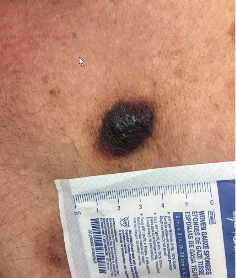
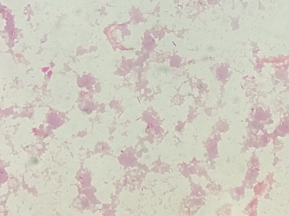
The patient’s tissue Gram stain was notable for 2+ WBCs (PMNs) and 4+ gram negative rods. The predominant organism grew on 5% sheep blood and chocolate agars but there was no growth on the MacConkey agar (Image 1).

Image 1. Pasteurella multocida grows on blood (A) and chocolate (B) agar plate.
Image 2. Gross cross section of the friable fungating endometrial lesion. (A) Microscopic pathological findings consistent with undifferentiated carcinoma. (B)

Discussion

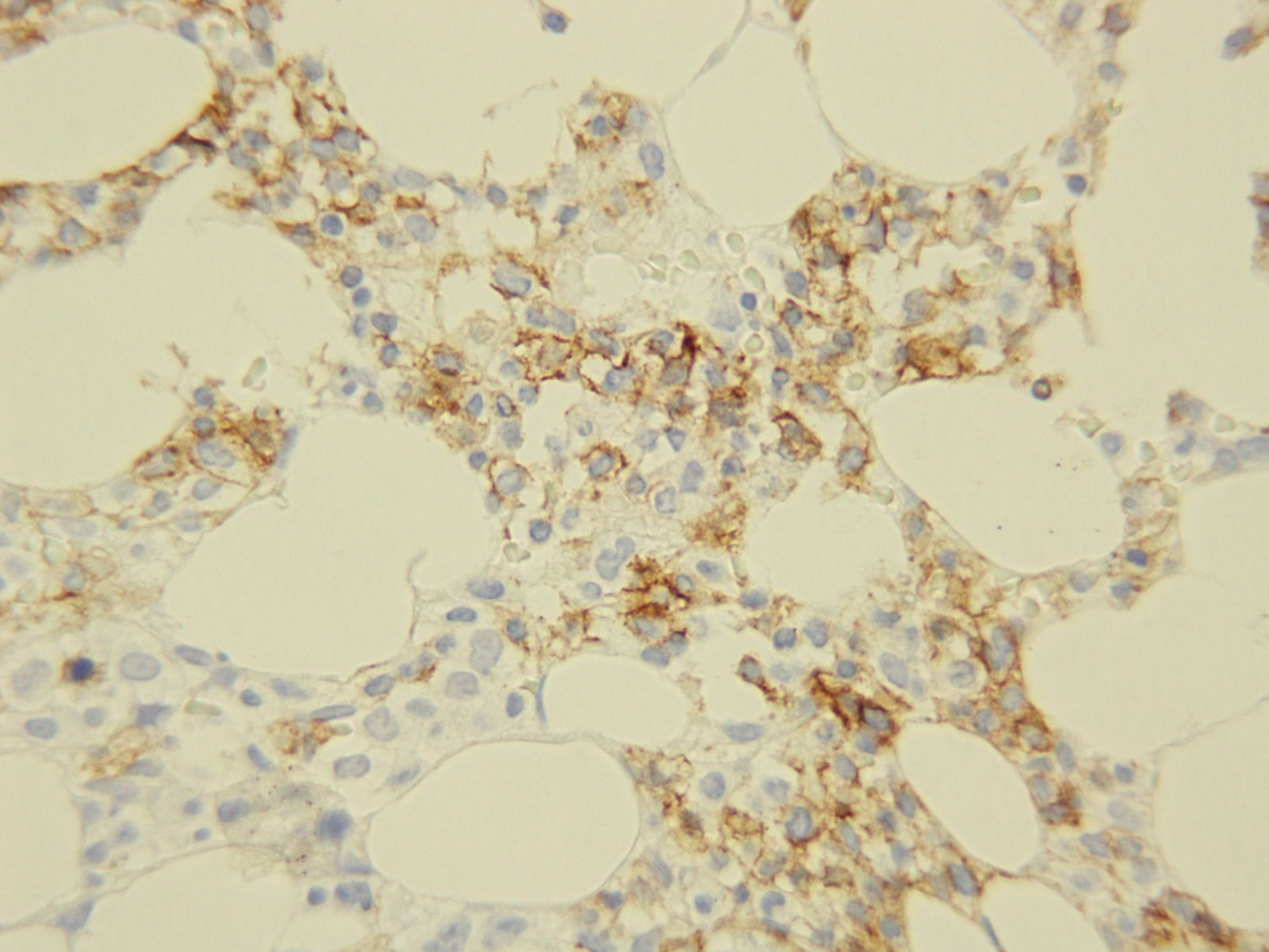
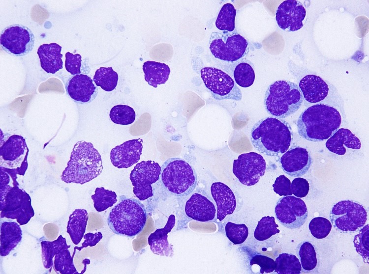
The predominant organism was identified by MALDI-TOF MS as Pasteurella multocida. Given the microbiological findings, patient was further questioned but she denied having pet at home or contact with animals. The patient underwent a surgery to remove her uterus, both ovaries and Fallopian tubes. The gross pathological examination of her uterus revealed an exophytic endometrial lesion with extensive area of necrosis surrounding the lesion (Image 2A). Final microscopic pathological diagnosis was significant for a highly malignant and invasive tumor of the uterus (Image 2B) and active infection of her left Fallopian tube.

Here we present a rare case of a P. multocida intrauterine infection lacking history of animal exposure in the context of gynecologic malignancy. P. multocida is a small, non-motile, non-spore forming, aerobic and facultative anaerobic gram negative coccobacillus that is associated with animal exposure. [1] This organism is found in the digestive and respiratory tract of domesticated animals (cats, dogs, cattle, sheep etc.) as well as wild animals (lions, buffaloes, panthers etc.). The suggested hypothesis for transmission of Pasteurella to human involves close contact with the infected animal’s oral cavity. Thus, the most likely route of infection in humans is through direct animal bites or scratch, depositing the bacteria on compromised skin or if an infected animal lick on an exposed human mucosal surface [1, 2]. In the general population, P. multocida causes local infection that may progress to abscess formation, but bacteremia is uncommon. However, elderly patients and those with associated immunosuppressed states from chronic disease or cancer are at higher risk of more complicated severe infections [2].

The presented case highlights the importance of clinical microbiology in the setting of obtaining detailed history and conducting physical examination.  Uncommon cases of P. multocida infection secondary to pet scratches or bite inducing genital infection and causing tubo-ovarian abscess have been reported. Similarly, rare cases of immune-compromised P. multocida bacteremia without history of animal contact have been described. The most likely explanation is the re-activation of previously asymptomatic colonization of the respiratory and gastrointestinal tract in the context of immunosuppression. As such, patients presenting with systemic Pasteurella infection without a documented animal exposure should undergo extensive evaluation in order to exclude the presence of an underlying immunodeficiency [1, 3-4].

References

  1. Weber DJ., Wolfson JS., Swartz MN., Hooper DC. Pasteurella multocida infections – report of 34 cases and review of literature. Medicine. 1984;63(3):133-54.
  2. Raffi F, Barrier J, Baron D, Drugeon HB, Nicolas F, Courtieu AL. Pasteurella multocida bacteremia- report of 13 cases over 12 years and review of the literature. Scand J Infecti Dis. 1987; 19:385-93.
  3. Kimura K., Hagiya H., Yamamoto N., Yoshida H., Akeda Y., Nishi I., Tomono K. Pasteurella multocida multiple intrapelvic abscesses in a young woman with uterine cervical cancer. J Infect Chemother (2018).
  4. Lukban JC., Baker MS. Pasteurella multocida isolation from a tuboovarian abscess. A case report. J Reprod Med 1995;40:603-5.

– Pouya Jamshidi, MD is a 1st year anatomic pathology resident at University of Chicago (NorthShore). Academically, Pouya has a particular interest in neuropathology and cellular basis of neuroplasticity. In his spare time, Pouya enjoys listening to classical music and opera.

-Erin McElvania, PhD, D(ABMM), is the Director of Clinical Microbiology NorthShore University Health System in Evanston, Illinois. Follow Dr. McElvania on twitter @E-McElvania. 

Over the River and Through the Woods…

Specimen handling and transport is a vital training topic in the realm of Laboratory Safety. There is much to consider here that affects specimen quality and integrity, and ultimately affects patient results. There are also considerations involving employee safety at every step of these processes. One group of employees that can easily be overlooked when it comes to proper safety training is lab couriers. They perform the important role of properly and safely transporting specimens for testing, but without the proper education and tools, these team members can quickly fall into situations of harm.

The courier was running late, and she had one last stop on her route at a medical office building with multiple physician offices and drop boxes. It was cold, and she decided to leave the vehicle running while she went inside to pick up more specimens and deliver lab reports. The car was also left unlocked. When she came back outside, the car was gone. It was found hours later in am empty field, but it had been set on fire. There were lab specimens and reports strewn all over the field and into the nearby woods.

Couriers need to be trained about the importance of their role, and that training should include information about security and protected health information (PHI). Be sure to include HIPAA training for all courier staff. When discussing security, enforce specific processes such as always turning off vehicles before exiting and properly securing all patient specimens and any paperwork being transported. Whether couriers use company vehicles or their own personal transportation for the job, making sure harm does not come to the vehicle nor any contents being transported is key.

The courier knew he had a long drive ahead of him because of the toll bridge, and he had several specimens that needed to be delivered as frozen. He went to the lab’s cooler and scooped a large pile of the dry ice into a big box using his hands. It was cold, but it helped to wake him up a bit. He placed the specimens in the box and placed it in the back seta of his vehicle. It wasn’t very warm out, so as he began his drive, the courier made sure the heat was on high and that all windows were closed. After a few miles, the courier began to fell very tired. He struggled to stay awake, and he couldn’t figure out why. After sitting in traffic on the bridge for a time, he pulled off the road and called the dispatcher to let them know he could not continue. When he got out of his vehicle, he began to feel better.

CAP regulations require that laboratory staff have dry ice safety training, but that requirement extends to anyone who may acquire the dangerous substance in the lab. Make sure staff are aware of the need for proper PPE use when handling dry ice. Insulated gloves, the use of a scoop or tongs, and face protection are necessary when scooping ice into a container. Couriers should carry no more than three pounds of dry ice in a vehicle, and there should always be adequate ventilation, including open windows in the vehicle when transporting dry ice. Dry ice converts rapidly from a solid state to a gas, and that gas rapidly displaces oxygen in the air making it difficult to breathe or stay conscious. High volumes of dry ice in a car can create a very deadly road situation in a short amount of time.

The courier was transporting pathology specimens in a cooler, but was unaware that the lid had popped off of one of the specimens and formaldehyde was splashing all over the inside of the cooler. As time went by, the courier began to feel queasy. After realizing that something did not smell right in the vehicle, she eventually stopped the van and pulled to the side of the road to investigate. She opened the cooler and quickly pulled out dripping specimens and set them on the carpet before feeling too sick to continue cleaning up the mess. She had to be taken to the Emergency Room for formaldehyde exposure symptoms while the Lab safety Officer had to bring spill clean-up supplies to the van to neutralize the formaldehyde. The carpet had to be removed and disposed of properly.

Courier vehicles need to be equipped with spill clean up supplies that can handle whatever types of spills could occur during transport. If formaldehyde is transported, couriers need training in the proper transport and clean up of that chemical. Biological spill kits should be available as well, and spill training should be a regular part of overall courier safety training.

I wish I could say that these were imaginary stories, but sadly, that is not the case. The stories, though, illustrate clearly what can happen when proper safety management and training are lacking. Every part of the laboratory pre-analytical process is important, and every lab team member involved in the process needs to be considered. Employ complete safety training, and assess safety competency on a regular basis. By providing the proper tools and safety training to couriers, you can ensure the quality of lab results, and you can prevent incidents like these with your employees.

Dan Scungio, MT(ASCP), SLS, CQA (ASQ) has over 25 years experience as a certified medical technologist. Today he is the Laboratory Safety Officer for Sentara Healthcare, a system of seven hospitals and over 20 laboratories and draw sites in the Tidewater area of Virginia. He is also known as Dan the Lab Safety Man, a lab safety consultant, educator, and trainer.

Hematopathology Case Study: A 65 Year Old Male with a Skin Lesion on the Right Shoulder

Case History

A 65 year old Caucasian male presents with a skin lesion on his right shoulder. Physical examination reveals a 3 .0 cm  ×  1.5  cm hyperpigmented plaque with mild hyperkeratosis on his right shoulder and multiple scattered erythematous macules and plaques on the trunk and back Skin biopsy reveals involvement by Blastic Plasmacytoid Dendritic Cell Neoplasm (BPDCN). PET scan reveals no extracutaneous involvement.

The patient undergoes CHOP chemotherapy followed by autologous hematopoietic stem cell transplantation. He is subsequently treated systemically with lanolidomide, venetoclax, and idelalisib due to relapses of disease.

The patient returns to clinic three years later for follow-up. While his original cutaneous lesions are completely resolved, new lesions are noted on his back (representative lesion, Image 1). Hematologic evaluation is remarkable for pancytopenia with hemoglobin 8.7gm/dL, white blood cells 1.4 K/uL, and platelets 39 K/uL. A biopsy of the bone marrow is performed.

Image 1. Skin lesion on back.

Biopsy Findings

Core biopsy
CD56
Aspirate

H&E stained sections demonstrate a normocellular bone marrow with diminished trilineage hematopoiesis and sheets of amphophilic, blastoid cells with irregular borders occupying most of the marrow cells. Immunohistochemistry demonstrates a cellular population with CD56. The aspirate smears show similar findings with numerous clustered blastoid cells (92%) with a monocytoid appearance, often with basophilic vacuolated cytoplasm. There is also a decrease in myeloid and erythroid precursors.

Flow cytometric analysis performed on the bone marrow aspirate reveals a dim CD45 population with expression of CD4, CD56, partial CD7, dim and partial CD5, and CD38. The same population lacks expression of immaturity markers such as CD34, MPO, and TdT. The morphologic and phenotypic findings found in the marrow specimen are diagnostic of extensive involvement of the marrow by BPDCN.

Discussion

BPDCN is a rare and highly aggressive malignancy derived from precursors of plasmacytoid dendritic cells. Its nomenclature has constantly changed over years as the understanding of this entity has been improved. It has been variously known as blastic natural killer cell lymphoma/leukemia, agranular CD4+ natural killer cell leukemia, and CD4+CD56+haematodermic neoplasm. It is currently classified under acute myeloid leukemia and related precursor neoplasms in the most recent WHO classification of tumours of haematopoietic and lymphoid organs.

Limited data exist regarding the incidence of BPDCN; however, it is estimated to account for 0.7% of primary cutaneous skin lymphomas and 0.44% of all hematological malignancies. This hematodermic malignancy predominantly affects elderly male patients with mean age ranging from 60 to 70; however, a few cases have also been reported in childhood and infancy. As demonstrated in our case, the patients typically present with multiple violaceous skin lesions, which may be associate with erythema, hyperpigmentation, purpura, or ulceration. Extracutaneous involvement is reported to occur in the bone marrow, peripheral blood, and lymph nodes.

Diagnosis of BPDCN relies on histological and immunophenotypic findings. Histologically, BPDCN may show a monomorphic infiltrate of medium-sized immature blastoid cells with round nuclei, finely dispersed chromatin, and cytoplasmic vacuoles. They typically display immunophenotypic expression of markers CD4, CD56, CD123, and T-cell leukemia/lymphoma 1 (TCL1) without any lineage-specific markers of T cells or B cells. Chromosomal abnormalities involving 5q, 12p,13q, 6q, 15q, and 9p have been reported. The differential diagnosis entails, but is not limited to, mature T-cell lymphoma, nasal-type NK/T-cell lymphoma, myeloid sarcoma/acute myeloid leukemia and T-cell lymphoblastic lymphoma/leukemia

The clinical course of BPDCN is aggressive, with a median survival of 9 to 16 months. The patients with disease limited to the skin may have a better prognosis, while advanced age and advanced clinical stage are indicators of poor prognosis.  There is currently no consensus on optimal management and treatment because of low incidence of BPDCN; however, most patients are treated with regimens used for other hematopoietic malignancies (i.e. CHOP and hyperCVAD) followed by allogeneic stem cell transplantation for eligible patients. They often respond well to chemotherapy with complete resolution of skin lesions; however, relapse of disease can occur due to resistance to chemotherapeutic agents, which may have happened in our case.

References

  1. Lim MS, Lemmert K, Enjeti A. Blastic plasmacytoid dendritic cell neoplasm (BPDCN): a rare entity. BMJ Case Rep. 2016;2016:bcr2015214093.
  2. Grushchak S, Joy C, Gray A, Opel D, Speiser J, Reserva, Tung R, Smith SE. Novel treatment of blastic plasmactoid dendritic cell neoplasm: a case report. Medicine (Baltimore). 2017 Dec;96(51):e9452.
  3. Dhariwal S, Gupta M. A case of blastic plasmacytoid dendritic cell neoplasm with unusual presentation. Turk J Haematol. 2018 Jul 24. doi: 10.4274/th.2018.0181.
  4. Shi Y, Wang E. Blastic plasmacytoid dendritic cell neoplasm: a clinicopathologic review. Arch Pathol Lab Med. 2014 Apr;138(4):564-9.
  5. Bulbul H, Ozsan N, Hekimgil M, Saydam G, Tobu M. Report on three patients with blastic plasmactoid dendritic cell neoplasm. Turk J Haematol. 2018 Sep;35(3):211-212.
  6. Kerr D 2nd, Sokol L. The advances in therapy of blastic plasmacytoid dendritic cell neoplasm. Expert Opin Investig Drugs. 2018 Sep;27(9):733-739.
  7. Pagano L, Valentini CG, Pulsoi A, Fisogni S, Carluccio P, Mannelli F, et al. Blastic plasmactoid dendritic cell neoplasm with leukemic presentation: an Italian multicenter study. Haematologic. 2013 Feb;98(2):239-246.

-Jasmine Saleh, MD MPH is a pathology resident at Loyola University Medical Center with an interest in dermatopathology and hematopathology. Follow Dr. Saleh on Twitter @JasmineSaleh.

-Kamran M. Mirza, MD PhD is an Assistant Professor of Pathology and Medical Director of Molecular Pathology at Loyola University Medical Center. He was a top 5 honoree in ASCP’s Forty Under 40 2017. Follow Dr. Mirza on twitter @kmirza.

Solutions, Not Resolutions

The turn of the year is a quasi-inspiring time for many people who attempt to change something about themselves or their situation with “New Year’s Resolutions.” When my friends and I were heading to brunch on New Year’s Day in southern California, there were many people running (alone or in groups) which I hadn’t seen before and my one thought was, “How long will that last?” When I returned to Chicago after the holidays, I dusted off my gym membership card and logged some treadmill time—my one thought was, “I hope this lasts!” But we are all too familiar with the breaking of these resolutions by most of us, and the ultimate regret we feel in the latter part of the year when our hopes and dreams of thinness/money/power/rare pokemon/fame have been dashed by the collision with our actual lives and the limited time we have to get done what needs to be done. Fortunately, we are human beings and we are allowed to be disappointed with ourselves over this (or these) tiny failings as long as our life trajectory is heading the way we want*. Then there are those handful of people that each of us will ultimately know who stick to their resolution and shed the weight, get a new job, or (hopefully with increasing numbers!) quit smoking! And we are more inspired by their actual doing of these things than by our lack of resolve.

As individuals, this trivial annual mindset is acceptable and even entertaining. But as a society, empty promises and feigned changes are simply unacceptable. To certain things, we must as a society—and as individuals in that society—commit. Recycling, for example, is a strategy that is both an economic and environmental boon. But as of January 2018, our major “solution” for plastics (especially from the West) that was China has now ended. Other nations willing to take these recyclables stopped their acceptance as well towards the end of 2018. What can we do to solve (not resolve) this situation permanently? Each country should consider first the role of plastics in their society and perhaps, like many African nations, simply ban the product(s). Secondly, encouraging personal recycling and reuse of plastics, for example through water filters to refill plastic bottles or fees on reusable grocery bags, can minimize impact. But, ultimately, each nation needs a sustainable recycling plan that represents a balance of production and utilization, creating a negative plastic total impact (i.e., no new plastic created).

In pathology, the theme of recycling is very important for any laboratory but can have major benefits for laboratories in developing nations. Formalin, xylene, alcohol, and paraffin (the four principle reagents for pathology processing), can be recycled using devices or process plans that can have minimal capital costs to set up. Consider that a given country may have shipping challenges such that an order placed today for 10 gallons of neutral buffered formalin may take 6 weeks to 6 months to arrive and cost 3 to 5 times the price in another country. In that setting, recycling formalin is clearly a superior approach and extrapolates to xylene and alcohol. Process approaches to paraffin (e.g., collecting waste paraffin from trimming and lids, using minimized mold sizes, lateral flow to minimize contamination) can optimize the use of the wax and reduce costs.  As these four reagents represent core elements to the process, efficient utilization, reuse, and management can keep costs low and processes running. But the laboratory must commit to this process and adhere to it every moment of every day to change patient’s outcomes for the better.

Similarly, core histology equipment (unlike many clinical laboratory machines) is almost indestructible when properly managed and maintained. Laboratories in developed nations may replace this equipment when it is several years to a decade old when the equipment may have another decade (or sometimes two) left of life. Decisions to replace functional equipment are left to the individual laboratory; however, once this process occurs, functional equipment should not be left to collect dust and should be moved to a new location where it can be of value. Every laboratory considering the replacement of older equipment must ask the question, “What is the remaining functional life of this device?” If that number is many years or the often stated 70%, a plan for donation of the equipment is highly suggested.  It is this philosophy that inspired the ASCP Center for Global Health program along with many other groups to actively seek out donated, functional equipment and transfer it to nations and colleagues who desperately need it to maintain their pathology services (Do you have equipment for donation? Email us!). This is especially important because the perceived demand for histology equipment in many low-income countries is so low that manufacturers and distributors refuse to become involved with the equipment (especially with trade and tariff barriers standing in the way). But, in truth, the demand is the same per population as in any other country with at least one high volume, functional pathology laboratory needed for every 1 to 3 million people (depending on population age structure and clinical utilization).

As we begin a new year together, reflecting on what we did (and didn’t) do in 2018 and what we can (and should) do in 2019 is an iterative process that can guide us through many decisions. I hope that everyone reading this blog takes a few moments (or even an hour if you can spare it!) to delve into 2018 and really plan for 2019 with true solutions in mind for any challenges you identify. And, lastly, always take some time every day to think outside yourself and even your laboratory to others in your local community or in foreign lands. Consider what little (and big) things you can do that may improve the life of just one person other than yourself and commit to those things.

*If your life trajectory is not going the way you want, consider performing a personal SWOT (strengths, weaknesses, opportunities, threats) analysis and think outside the box about where you are and where you want to be. Don’t be afraid to make life changes or new life choices that give you a better piece of mind and stronger sense of self and self-awareness. A room full of happy people who are self-aware and emotionally intelligent can solve problems at light speed because their personal issues (good or bad) don’t get in the way. So, for 2019, I strongly encourage everyone to consider really solving (not resolving) the problems you perceive in your life so that we can all work together to solve (not resolve) the challenges we face as a society moving into the next decade.

milner-small

-Dan Milner, MD, MSc, spent 10 years at Harvard where he taught pathology, microbiology, and infectious disease. He began working in Africa in 1997 as a medical student and has built an international reputation as an expert in cerebral malaria. In his current role as Chief Medical officer of ASCP, he leads all PEPFAR activities as well as the Partners for Cancer Diagnosis and Treatment in Africa Initiative.

Evaluating and Analyzing Next Generation Sequencing Specimen Results

Welcome back – in my previous blog we discussed how a run is evaluated on the Ion Torrent instrument. This quarter’s blog will review the individual specimen results from that run.

First off, we take a look at how many reads per specimen have been sequenced and how those reads performed over the areas that are targeted. For the AmpliSeq Cancer Hotspot Panel v2 that we run, there are a total of 207 amplicons that are created and sequenced. To assess the depth of coverage over these amplicons, we need to think about the biology of the tumor cells and the limit of detection of the assay. We feel confident that we can detect 5% variant allele frequency for single nucleotide changes, and 10% variant allele frequency for insertions or deletions. In order to be confident that we are not missing variants, we require the specimen has a tumor percentage greater than 20%. This is because, for a given tumor, it can be assumed that if it is mutated, it will be only heterozygous – only one of the two alleles will have the variant. This automatically halves the possible allele frequencies from any given tissue. If a colon specimen that we are given to test has a tumor percentage of 40%, it can be assumed that any variant will have a variant allele frequency of no more than 20%. Because of this then, we also require the amplicons that are sequenced to have at least 500x coverage – they need to be sequenced at least 500 times so that if we have a 5% mutation, we will see it in 25 of the reads and we can feel confident this is an actual change, as opposed to background noise.

Next, we look at the On Target percentage and Uniformity percentage (over 95% for each is expected). The On Target value tells us what fraction of the amplicons actually cover the 207 amplicons that are in the panel. Uniformity informs us of how even the number of reads is over all the 207 amplicons – were they all equally represented or were there a subset of these that had more coverage than the others? This information can actually lead us to further testing – if there is a subset of amplicons that have more coverage than the rest, and it they are all from one gene, this may indicate gene amplification. In these cases, the clinician is alerted and additional testing can confirm the amplification.

All of this coverage information is provided by one of the “plugins” we run after the basecalling and alignment are finished:

The most useful (and interesting!) information is gathered from the variant calling plugin. This plugin compares the specimen sequences with the reference sequences and reports the differences – the “variants”. Many of the variants that are detected are single nucleotide polymorphisms (variants that are detected in greater than 1% of the population). They could also be known artifacts of the sequencing itself. These are all analyzed and categorized in the validation of the assay and then can be filtered out when analyzing clinical data. After filtering out the known SNPs and artifacts, the somatic changes can then be evaluated. Generally, the panel will detect 15-20 variants, but after filtering only 1-4 variants will be somatic changes. Each change that is detected is reviewed using a program called IGV, shown below. We compare the sequence to confirm that what the plugin is reporting looks correct in the actual reads from the sequencer. See screenshots below of a subset of variants called, then filtered, and analyzed in IGV. While the plugin is exceptionally good at variant calling, no program is perfect and visualizing the data is still necessary to confirm there is not anything else going on in the area that is sequenced. The fastq file from the run is also run through a secondary software to compare results. The variants for each specimen are assessed for variant allele frequency, coverage and quality in both software.

VariantCaller Output

Filtered Calls: White cells means SNP, Blue cells mean possible somatic call

IGV Output for KRAS and STK11 calls:


Lastly, the results are brought into yet another software to be reported. This software will allow the pathologists to assign significance to the variants. It will also pull in any treatment information linked to the variants and then allow the pathologist to pick any applicable clinical trials in order to assist the clinician as much as possible. In future blogs we will take a look at cases like this to see interesting findings of oncology cases.

rapp_small

-Sharleen Rapp, BS, MB (ASCP)CM is a Molecular Diagnostics Coordinator in the Molecular Diagnostics Laboratory at Nebraska Medicine. 

Microbiology Case Study: A 54 Year Old Woman with Fever

Patient History

A 54 year old woman is seen by her oncologist with complaints of rigors and fevers for the past two months. Her past medical history is significant for Stage IV Follicular Lymphoma. Previous treatment included rituximab and bendamustine that was completed a year prior. Currently her therapeutic plan is to receive rituximab maintenance therapy every two months (her most recent dose was 2 days prior to this visit) via a port. She has two dogs, both of which had a diarrheal illness three months prior. She has chickens and spent the summer RV camping around New England. She has not had diarrhea or noticed any rashes. Her travel history includes a trip to Europe  4 years prior. A blood culture was collected due to her fevers and rigors and was found to be positive 29 hours post incubation.

Gram stain of the blood culture bottle showed small, faintly staining gram negative, curved rods (Image 1). The patient was started on ciprofloxacin and referred to the infectious disease clinic. After 3 days of incubation, small slightly mucoid yellow gray colonies grew on 5% sheep blood and chocolate agar (Image 2). There was no growth on the MacConkey agar. Initial Gram stain revealed similar gram negative rods to the Gram stain performed on the blood culture media. MALDI-TOF identified the organism as Campylobacter jejuni. The patient was asked to stop taking ciprofloxacin and start azithromycin. She also had her port removed on this day. Four days after her oncology visit, the patient’s blood was negative for organisms. Her fever and rigors resolved as well. Susceptibility testing showed the organism to be resistant to ciprofloxacin and susceptible to azithromycin.

Image 1. Gram stain from the blood shows faintly staining curved gram negative rods.
Image 2. Growth on chocolate agar after 3 days (Not in a microaerophilic environment).

Discussion

Campylobacter jejuni is a small, gram negative, curved rod. It is the most common cause of bacteria-mediated diarrheal disease globally (1). In immunocompromised patients, it can cause a variety of extraintestinal diseases: septicemia, meningitis, septic arthritis, and endocarditis (2). Infection can also lead to Guillain-Barre syndrome. This is an autoimmune disease of the peripheral nerves that is thought to be due to antigenic cross reactivity between the surface lipopolysaccharides of the bacteria and the patient’s peripheral nerve gangliosides (3). Of patients diagnosed with Guillain-Barre syndrome, 20-40% will have had a history of Campylobacter jejuni infection (2).

Campylobacter jejuni grows best at 42°C in a microaerophilic environment (5% O2,10% CO2, and 80% N2). It displays a darting motility in broth and will not grow in 3.5% NaCl. Almost all are oxidase and catalase positive. On Campy-BA, a selective blood agar plate for Campylobacter jejuni, colonies will be peach colored. This media is made up of a Brucella agar base, sheep red blood cells, and various antibiotics that suppress the growth of normal fecal flora(2).

Macrolides (erythromycin, azithromycin) are considered the treatment of choice, however it has been reported that resistance approaches 1.7% (1 and 2). Fluoroquinolones like ciprofloxacin can be used, however resistance is higher due to widespread use of this drug in medical and veterinarian practices and agricultural businesses.

References

  1. Yang W, Zhang M, Zhou J, Pang L, Wang G, Hou F. The Molecular Mechanisms of Ciprofloxacin Resistance in Clinical Campylobacter jejuni and Their Genotyping Characteristics in Beijing, China. Foodborne Pathog Dis. 2017;14(7):386-392.
  2. Tille P. Bailey & Scott’s Diagnostic Microbiology. Fourteenth Edition. Elsevier;2017.
  3. Murray P. Medical Microbiology. Seventh Edition. Elsevier; 2013.

-Angela Theiss, MD is a 3rd year anatomic and clinical pathology resident at the University of Vermont Medical Center.

-Christi Wojewoda, MD, is the Director of Clinical Microbiology at the University of Vermont Medical Center and an Associate Professor at the University of Vermont.