Investing in the Best Testing

“Damn that q-tip goes in deep!

But it lit up negative so d/c to street

But it was flu, cuz he bounced back again

And now my Press Gayney’s a minus ten…”

Video 1. Another classic excerpt from a favorite: ZDoggMD, singing about this year’s flu season and available testing options on the horizon—because, let’s face it—rapid flu tests aren’t quite cutting it anymore.

Hello again everyone! Back again to talk about a new set of recommendations from last month’s post. This time it’s about influenza. Recommendation: get vaccinated. Thank you. See you next time…

Seriously, as the 2018-2019 flu season dawns upon us, it’s time to talk about vaccines, tests, prevention, and health literacy. I’m sure many of your social media pages are filled with various debates, articles, and fake news stories on one side or another pitting science, pseudo-science, and non-science all against each other for public spectacle. In the lens of laboratory science and medicine at large, I think most if not all of us agree that preventable diseases should be prevented, and if not, at the very least detected accurately, sensitively, and early. Influenza A/B is a prime example of a consistent threat to our health and safety that has wavered responses in various socio-medical circles.

Official communication and guidance from the Centers for Disease Control and Prevention (CDC) clearly tells those of us in health-care to embrace a multi-tiered approach to protecting public health regarding the flu. That approach includes vaccination, testing, infection control, anti-viral treatment, and anti-viral prophylaxis. And why such a fuss over the flu? It’s a big deal! Last year, the CDC reported approximately 80,000 deaths associated with influenza as a primary cause. 80,000 deaths! That’s almost 7 times as many that died from H1N1/Swine Flu complications back in 2009, where only 12,000 patients were killed by the virus. And even more so, in the terrifying Ebola epidemic of 2016—in which there was a staggering 1 recorded death in the US—nearly 29,000 people were infected globally and only 11,300 died (despite under-reporting). I’m being dramatic, I know. But it’s important for us to recognize true epidemics when they happen, and even more important for societies like ours to be at the forefront of preventing them from developing any further.

flu1.jpg
Image 1. The CDC recommends you get your flu shot every year, because obviously.

flu2.jpg

Image 2. I’m not here to talk about the anti-vax elephant in the room. That’s not fair to elephants. But imagine if the CDC reported 44% of flu vaccine misconceptions were addressed!

As an aside, I’ll probably recommend that you get your annual flu shot a hundred times in this post alone. But just to have a clear reference, please look at the following table. It’s critical to be able to both distinguish common cold versus influenza symptoms for yourself, as well as educate your patients and peers about the differences between the two. This information can change the way people perceive treatments (i.e. why the doctor only recommended rest/Tylenol and didn’t give out antibiotics for their symptoms) and why it’s absolutely crucial to protect vulnerable populations from an otherwise fatal virus. So, micro-rant aside, it should be clear that by now we should be working on a way to both improve our prophylaxis with vaccines and medications as they always leave room for improvement—I’m looking at you Tamiflu and Relenza! Notwithstanding any analysis of efficacy for the flu vaccine, the CDC reports a variable and transparent success rate of vaccines. It can be difficult to predict and assess epidemiologic trends and mutations as the influenza virus continues to change annually.

flu3.JPG
Table 1. Distinguishing the common cold, and “flu-like symptoms” from a proper influenza viral infection.
flu4.jpg
Image 3. CDC Report on seasonal flu vaccine effectiveness since 2004.

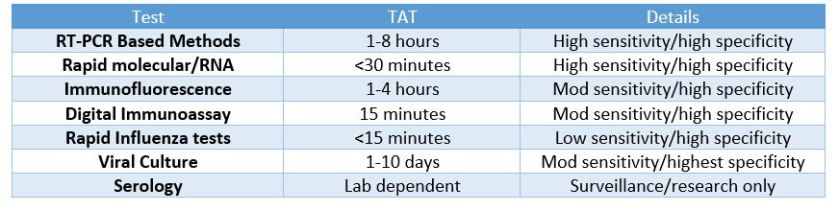
So, what was the deal with ZDoggMD plugging some PCR testing in the opening credits here? That’s a good question and one that inspired this article in the first place. Obviously, if you follow my posts you know I follow his, and at the end of this latest video he discusses new available options for influenza point-of-care testing (POCT) for clinics and emergency rooms. This was a partnership with the company Cepheid and linked with their promoting their POCT PCR-based FluA-B testing. Here’s a quick paraphrasing of the CDC recommendations on influenza testing: because of the numerous false negative tests every season, the bests tests in order of preference are RT-PCR, immunofluorescence, and rapid antigen testing. Did you catch that? Rapid Flu swabs are bottom of the barrel stuff here. UpToDate, the clinical resource for current practices and standards discusses rapid influenza tests as sacrificing turn-around-time (TAT) for accuracy: “commercially available rapid antigen tests for influenza virus yield results in approximately 15 minutes or less but have much lower sensitivity than RT-PCR, rapid molecular assays, and viral culture.” (I didn’t bold those words, they did). Most of the places I’ve worked run through boxes of rapid flu swab kits ALL DAY LONG. But what are we missing? Clinically, this is supposed to be an important “no miss” diagnosis—it’s dangerous, it’s contagious, it’s mutatable…

Who remembers learning biostatistics in school? Remember SPIN and SNOUT? “Specificity is used to rule IN, Sensitivity is used to rule OUT” So why are we relying on the LOWEST sensitivity available to us for ruling out influenza? Probably because of technological/practical limitations up to this point in time, and of course the most glaring limiting reagent of all: funding, also known as “administrative buy-in.” Have I hit enough lab management buzz words in this post? Not yet.

flu5.JPG
Table 2. Per UpToDate, these are the quick and dirty details on our favorite available flu tests currently on lab benches across the country. I’d say there’s got to be a better way, but there already is.

Sweet. So, it’s a little expensive but ultimately better for our patients, right? Done and done, whip up a cost-benefit-ratio report for the suits upstairs and let’s start a validation project! Well, yes and no. I’m a big proponent of utilizing MALDI-TOF—the mass spectrometry based system to replace traditional bacterial identifications. A 2015 study published in the Journal of Clinical Microbiology stated, “The use of MALDI-TOF MS equated to a net savings of 87.8%, in reagent costs annually compared to traditional methods. …The initial cost of the instrument at our usage level would be offset in about 3 years. MALDI-TOF MS not only represents an innovative technology for the rapid and accurate identification of bacterial and fungal isolates, it also provides a significant cost savings for the laboratory.” What promise! Cepheid’s ED POCT PCR Flu test promises 18% fewer tests needed, 17% fewer antibiotics prescribed, and overall savings per patient visit of up to $700. But this sounds like another, too familiar, recent promise from another voice in our profession. Something about quick, easy, and accurate testing on chips with micro-laboratories available commercially and only using microliters of whole blood for analysis. “Unfortunately, none of those leads has materialized into a transaction. We are now out of time,” read the goodbye letter to the company’s stockholders—Theranos, that’s the one. The moral of the story here: it’s good to remain fiscally prudent when deciding what your clinic or hospital should invest in with regard to testing. However, when something has been a proven and successful replacement which ultimately is recommended by multiple societies within the field then something’s got to give.

flu6
Image 4. MALDI-TOF saves money! You spend a little upfront, but then your hospital can write articles about how your bacteriology department has a swab-to-sensitivity TAT of a few hours. Less errors, less antibiotics, more likes on social media!

What do you see in your practice or laboratory as far as influenza testing? Are there issues I missed? What is your experience with rapid tests, or PCR testing? Is anyone else as big a fan of MALDI-TOF as I am? Did you get your flu shots yet? Leave your comments and questions below! Share with a colleague today!

See you next time!

I have absolutely no affiliation with Cepheid, financial or otherwise, but as an educational/professional resource read more information about Cepheid’s molecular rapid flu tests, read their literature at www.GetTheRightTest.com

flu7

References

  1. Carreyrou J. (2018) Blood Testing Firm Theranos to Dissolve. Wall Street Journal. Health: Theranos Co. Letter to Shareholders. Accessed at: http://online.wsj.com/public/resources/documents/Theranos_Stockholders_Letter_2018.pdf?mod=article_inline
  2. Cephid (2018) Is it really flu? Cutting emergency department costs with bedside rapid molecular tests. Accessed at: http://www.cepheid.com/images/Cepheid-WP-ED-Cost-FINAL.pdf
  3. CDC (2017) Interim guidance for influenza outbreak—management in long-term care faciltites. e Recommendations of the Advisory Committee on Immunization Practices – United States, 2016-17 Season. Accessed at: http://cepheid.com/images/CDC-interim-guidance-outbreak-management.pdf
  4. CDC (2018) Seasonal Influenza Vaccine Effectiveness, 2004-2008. Accessed at: https://www.cdc.gov/flu/professionals/vaccination/effectiveness-studies.htm
  5. CDC (2018) Flu Symptoms and Complications. https://www.cdc.gov/flu/consumer/symptoms.htm
  6. Dayhoff-Brannigan, M (2018) To Tamiflu or Not to Tamilflu? National Center for Health Research. Accessed at: http://www.center4research.org/tamiflu-not-tamiflu/
  7. Dolin, R. (2018) Diagnosis of Seasonal Influenza in Adults. UpToDate. https://www.uptodate.com/contents/diagnosis-of-seasonal-influenza-in-adults?search=influenza&source=search_result&selectedTitle=6~150&usage_type=default&display_rank=6#H1289544319
  8. McNeil, D. (2015). “Over 80,000 Americans Died of Flu Last Winter, Highest Toll in Years” The New York Times.
  9. McNeil, D. (2015). “Fewer Ebola cases go unreported than thought, study finds”. The New York Times
  10. ZDoggMD (2018) This Flu Test https://www.youtube.com/watch?v=YKTYw-7ikJQ#action=share
  11. Tran A, et al. (2015) Cost Savings Realized by Implementation of Routine Microbiological Identification by Matrix-Assisted Laser Desorption Ionization–Time of Flight Mass Spectrometry. Journal of Clinical Microbiology. DOI:1128/JCM.00833-15

ckanakisheadshot_small

–Constantine E. Kanakis MSc, MLS (ASCP)CM graduated from Loyola University Chicago with a BS in Molecular Biology and Bioethics and then Rush University with an MS in Medical Laboratory Science. He is currently a medical student actively involved in public health and laboratory medicine, conducting clinicals at Bronx-Care Hospital Center in New York City.

Hematopathology Case Study: A 63 Year Old Man with Fatigue

Case history

A 63 year old male presented with extreme fatigue and weakness of unknown duration. Physical examination revealed scattered petechiae and mildly decreased muscle strength. His past medical history included a one year history of cough that had recently improved. Laboratory investigation demonstrated severe anemia and thrombocytopenia with a mild leukopenia.

Review of the peripheral blood smear showed smudge cells, circulating neutrophils with Döhle bodies and toxic granulation. CT scan of the chest showed upper/anterior mediastinal lymphadenopathy without hilar lymphadenopathy.

A biopsy of the bone marrow was performed.

Microscopic Findings

hod1.jpg


hod 2.jpg

The bone core biopsy revealed a hypercellular marrow for the patient’s age with a pronounced lymphohistiocytic infiltrate involving 30-40% of the biopsied marrow space. Interspersed along the infiltrate were large, atypical lymphoid cells with pleomorphic nuclei and prominent nucleoli. The marrow aspirate smear reveals progressive trilineage hematopoiesis with scattered hemophagocytic histiocytes.

Immunophenotype

hod3.jpg

The large atypical lymphoid cells were positive for CD30 and EBER, while being dimly positive for PAX5 and negative for CD20.

Diagnosis

The detection of mononuclear Hodgkin cells staining for CD30 along with a characteristic reactive infiltrate, together with dim PAX-5 staining, positive EBER, and negative CD20 is sufficient to diagnose involvement of a secondary site by Hodgkin lymphoma. The lymphoma was associated with a secondary hemophagocytic lymphohistiocytosis.

Discussion

Hodgkin lymphoma (HL) is a B-cell derived monoclonal lymphoid neoplasm. HL has a bimodal age distribution, with teenagers or patients in their early 20s and patients older than 55 years having the highest incidence. Although the typical presentation is with peripheral lymph node involvement, extranodal sites may be involved by either direct invasion or hematogenous dissemination. These sites include the spleen, liver, lung and bone marrow. About one third of patients have constitutional symptoms such as high fevers, night sweats, and weight loss.

Two broader forms of Hodgkin lymphoma exist: Classic Hodgkin lymphoma (CHL) and the less common nodular lymphocyte-predominant Hodgkin lymphoma (NLPHL). NLPHL tends to preserve the entire B-cell transcriptional phenotype, while the neoplastic cells in CHL fail to do so.

CHL is composed of mononuclear Hodgkin cells and multinucleated Reed-Sternberg cells surrounded by an infiltrate of non-neoplastic reactive cells that might encompass small lymphocytes, plasma cells, eosinophils, neutrophils, and histiocytes. Fibrosis may also be present in the form of bands or may be more diffusely spread. The four histological subtypes: nodular sclerosis CHL, lymphocyte-rich CHL, mixed cellularity CHL, and lymphocyte-depleted CHL are based on the composition and characteristic of the reactive infiltrate, and the cytological features of the neoplastic cells.

The classic Reed-Sternberg cell is binucleated, with prominent eosinophilic nucleoli, often referred to as having an “owl’s eye” appearance. However many neoplastic cells are not of the typical Reed-Sternberg variant, and can be mononuclear, termed Hodgkin cells, or cells with more condensed cytoplasm and pyknotic reddish nuclei known as mummified cells.

Hodgkin/Reed-Sternberg cells (HRS) in Classic Hodgkin Lymphoma fail to preserve their B-cell traits, and this is reflected by their immunophenotype. The majority of cases are negative for CD45, and although CD20 may be expressed, it is usually present only on a minority of the neoplastic cells and stain with varied intensity. The HRS cells stain with PAX5 with a lower intensity than the surrounding reactive cells, making them easily detectable. The HRS cell stains positive for CD30 and CD15 in nearly all cases. Both of them stain the membrane with accentuation around the Golgi apparatus. EBV associated Hodgkin Lymphoma will stain positive with EBER, detecting EBV-encoding small RNA.

Bone marrow involvement is rare, ~5-10% of cases, and suggest vascular dissemination of the disease. Bone marrow trephine biopsies are commonly performed in the staging of patients with newly diagnosed CHL which guides the further treatment and gives us information about prognosis. Involvement of the bone marrow represents stage IV disease (advanced stage) in the Ann Arbor staging classification and patients with advanced stage disease typically receive a more prolonged course of chemotherapy. The 5-year survival rate of stage IV Hodgkin lymphoma is ~65%,  a much worse prognosis when compared with stage I, stage II, and stage III with ~90%, ~90%, and ~80% 5-year survival rates respectively.

References

  1. Stein H, Pileri SA, Weiss LM, et al. Hodgkin Lymphomas. In Swerdlow SH, Campo E, Harris NL, Jaffe ES, Pileri SA, Stein H, Thiele J, editors: WHO classification of tumours of haematopoietic and lymphoid tissues, revised ed 4, Lyon, France, 2017, IARC Press, pp 423-464
  2. Ansell SM. Hodgkin Lymphoma: Diagnosis and Treatment. Mayo Clin Proc. 2015 Nov;90(11):1574-83.
  3. Howell SJ, Grey M, Chang J, Morgenstern GR, Cowan RA, Deakin DP, Radford JA. The value of bone marrow examination in the staging of Hodgkin’s lymphoma: a review of 955 cases seen in a regional cancer centre. Br J Haematol. 2002 Nov;119(2):408-11.
  4. Clarke C, O’Malley C, Glaser S. Hodgkin lymphoma. In: Ries LAG, Young JL, Keel GE, Eisner MP, Lin YD, Horner M-J, eds. SEER Survival Monograph: Cancer Survival Among Adults: U.S. SEER Program, 1988-2001, Patient and Tumor Characteristics. National Cancer Institute, SEER Program, NIH Pub. No. 07-6215, Bethesda, MD, 2007.


Hans.jpg

-Hans Magne is a 6th- year medical student at Poznan University of Medical Sciences. Follow Hans on Twitter @HHamnvag

Mirza-small

-Kamran M. Mirza, MD PhD is an Assistant Professor of Pathology and Medical Director of Molecular Pathology at Loyola University Medical Center. He was a top 5 honoree in ASCP’s Forty Under 40 2017. Follow Dr. Mirza on twitter @kmirza.

Regulatory Inspections: Are You Ready?

Part Two: The inspectors are here!

In last month’s post we reviewed the importance of being prepared for your regulatory inspections, as well as some tips for how to accomplish this task. This month we’ll focus on the inspection process itself – areas the inspectors may focus on, and how your preparation work last month will make the process go smoothly.

Once your formal inspection is underway, don’t panic. Based on all of your preparation work in the months prior, you should be organized and ready to answer any question the inspectors may have regarding your current procedures and policies. The focus of each inspection will vary based on the regulatory agency it is being performed by, but the following areas have a high likelihood of being reviewed.

Utilize a Patient Tracer.  One of the easiest ways to evaluate your laboratory processes from pre-analytical, through analytical phase, and finally the post-analytical phase is to utilize a patient tracer. Inspectors may pick a specific date or date range, and ask to see all associated documents for a particular test.

Pre-Analytic. Gather a copy of the test requisition indicating the patient information, ordering physician, and specific tests requested. Ensure your phlebotomy team knows where your policies related to patient identification and specimen collection are located, and ensure they are following the requirements within these procedures. Inspectors may ask to observe the phlebotomy collection process, so prepare your staff ahead of time to reduce the potential for nervousness.

Analytic. All records related to the testing of the sample will need to be produced. This includes the actual instrument print-out for the sample in question and/or worksheets used to document results, quality control records for the day of testing, instrument maintenance and calibration records, as well as the training and competency records for the technologist who performed the test that day. Training and competency assessment are two different tasks – ensure you have documentation to support both of these activities at the required intervals for the staff member in question. Depending upon the actual circumstances surrounding that particular sample, there may be additional documentation requested such as corrective action logs, critical call notification records, or confirmation testing records.

Post-Analytic. Inspectors will also need to review how your results are being documented and displayed on the patient charts. They will be focusing on accurate transcription of results including units of measure and reference ranges; the correct timing of sample collection, receipt in the lab, and result reporting times; in addition to all of the patient demographics properly being displayed. They may also ask to see a corrected report to see how clinicians are notified about any changes, so patient treatment can be adjusted accordingly in a timely manner.

Proficiency Testing (PT) Results. One of the common requirements of a laboratory accreditation program is the participation in a proficiency testing program for all regulated analytes. Since the intent of this program is to ensure accuracy in your patient testing results, inspectors will be focused on any unsuccessful PT surveys, and the root cause analysis you performed to investigate the occurrence. Was this an isolated and random error, or is there a systemic quality issue which caused the inaccurate result? Did you perform a look-back to confirm accuracy of patient results being reported between the time of PT analysis and when the laboratory was notified of the unsuccessful event? Were your preventive actions implemented and sustained, or are you still continuing to experience accuracy problems with your testing? Be sure to document all steps of your investigation, and have that documentation available to inspectors for review.

Quality Metrics. Laboratory directors have a responsibility to provide oversight of their laboratory’s quality program, and to ensure that medically reliable data is being generated. There are many ways to monitor the quality of your laboratory program, and you should be prepared to speak on your methods in use to the inspectors. Although labs are not expected to be perfect, there is a responsibility to monitor for issues and initiate appropriate corrective and preventive actions when they are identified. Ensure that your monthly performance improvement metrics are reviewed and signed by your laboratory director, and any metrics not meeting performance goals have documented corrective action initiatives. Metrics should be meaningful and demonstrate continuous monitoring and improvements within the laboratory.

Be Honest and Transparent. If an inspector asks for specific documentation which you do not have, be upfront and let them know. Trying to hide a problem or misdirect an inspector away from a problem area can result in even more citations as it creates an environment of mistrust. Inspections are an opportunity to identify and improve upon the weak points in your laboratory program, and the inspectors themselves can offer ideas and suggestions on better ways to meet certain requirements that you may be struggling with. Some regulations can be interpreted differently by different individuals – ensure that your staff can speak to your practices in use and explain to the inspectors how you are satisfying the requirements.

Coming up in part 3, we’ll discuss what to do next – how to address any issues identified during the inspection process, and how to keep the overall experience positive and beneficial to your staff.


Nevins-small

-Kyle Nevins, MS, MLS(ASCP)CM is one of ASCP’s 2018 Top 5 in the 40 Under Forty recognition program. She has worked in the medical laboratory profession for over 18 years. In her current position, she transitions between performing laboratory audits across the entire Northwell Health System on Long Island, NY, consulting for at-risk laboratories outside of Northwell Health, bringing laboratories up to regulatory standards, and acting as supervisor and mentor in labs with management gaps.

The Best Laid Plans: A “Trial by Fire”

From around 2009 to 2016, I worked very closely with a USA-trained surgeon, Dr. Brian Camazine of Earthwide Surgical Foundation, who visits Nigerian Christian Hospital in Aba, Nigeria for one month every quarter. He performs between 200 to 300 surgeries, which produce 40 to 60 surgical pathology specimens each visit.  Dr. Camazine has invested time, energy, and money into training local Nigerians in surgical skills, acquiring surgical and medical supplies to support his patient population, and following up all of his patients with Skype clinics after he returns.

My role in Dr. Camazine’s activities was to receive the surgical pathology samples, process them, and return results for him as quickly as possible. When Dr. Camazine contacted me, there was no pathology laboratory at NCH. Dr. Camazine uses a heavily subsidized model for all of the services provided at NCH such that a patient may pay ~$200 for a surgery (complete care including pathology) that would have cost them $2,000 to $4000 elsewhere in Nigeria. My hospital at the time had an ongoing project of a similar fashion with several sites in Africa but the costs of that program were growing. Dr. Camazine agreed to pay a fee of $25 per sample to my hospital to offset the technical costs of our laboratory processing the samples, and I provided all diagnostic results pro bono. Dr. Camazine was only charging patients $20 per case for pathology; thus, he subsidized the service further.

I had many long and difficult discussions with Dr. Camazine about this program and how we needed to focus on a sustainable solution that did not involve transport to the US for processing for many reasons including (but not limited to): a) danger and difficulty with sending tissue, b) long turnaround time because of shipping delays, c) chain of custody and requisition challenges, and d) capacity building in pathology. We kept at it with this long-term plan in mind but, as I departed my hospital to join ASCP in 2016, a drastic decision had to be made because I would no longer be able to shepherd this service. Dr. Camazine reached out locally to Nigerian laboratories and was fortunate to meet Dr. Chidi Onwuka from the Department of Histopathology at the University of Uyo Teaching Hospital. Brian and Chidi came to a feasible financial arrangement and, with the closeness of the laboratory, Chidi can return results to Brian in about 1 week (Meet Chidi and read Brian’s Blog here). This was a great success for Brian and Chidi because it represented moving from a non-sustainable, bridging program (i.e., what I had set up with Brian) to a permanent solution with the local laboratory. For over two years, Chidi has provided high quality service with quick turnaround time and massively improved the patient care journey for NCH patients.

On June 27th, 2018, however, that complete pathology solution came to a screeching halt when a fire swept through the laboratory and destroyed all of the equipment and reagents. The laboratory in question had just been completely updated with 40 Million Naira (~$115,000 USD) worth of equipment and upgrades, but it was all lost. Dr. Chidi reached out to Brian, myself, and many others with an urgent request to help him get a replacement laboratory up and running. After so much success, it was heartbreaking to hear such a loss had occurred.

IMG_20180627_143654 (1).jpg

The ASCP Partners for Cancer Diagnosis and Treatment in Africa Initiative was launched in 2015 with a goal of bringing 100% access to cancer diagnostics services to all patients. Although the population of patients Brian cares for and Chidi diagnoses are within Africa and within the scope of the Partners Initiative, at the time of the fire, there were at least 10 laboratory projects (including equipment, training, IHC, telepathology, etc.) in process through the Partners project. We were seemingly “at capacity” to help. What could we do? Although we have ASCP member volunteers that donate equipment, we have a waiting list of labs wanting to receive the equipment. Although Brian and Chidi are my colleagues and friends, the distribution of global health resources, assistance, and capacity should always be done with equity. As part of the Partners Initiative, ASCP Center for Global Health acquires equipment (typically through donation which means donor requirements of the local countries) and covers shipping costs to move the equipment to the recipient sites but we had not yet formalized this process. But, for Chidi, I simply didn’t have the equipment available to send.

Then, I received a WhatsApp message from Chidi on August 3rd with a small bit of good news. He had located a microtome in the USA that he could purchase; however, he did not have sufficient funds to ship the equipment. Now, finally, ASCP could help him! But it was not quite that easy!

ASCP staff member Dr. Debby Basu got the microtome in the USA to Chidi in Nigeria. This was not an easy task. Debby faced two major challenges for organizing Chidi’s shipment. First, she had to establish key templates and tools necessary to facilitate donation. Although we have several sets of donated equipment that are to be shipped from ASCP to other sites, Chidi’s microtome was the first actual piece of equipment that would go with our new shipping agent. As this was our first shipment with Bollore, she first had to work with Bollore to determine what documentation ASCP was responsible for providing. She then developed the in-house documents, templates and tools needed to facilitate shipment using Bollore’s services (e.g. commercial invoice, packing list, Shipper’s Letter of Instructions (SLI) Form (customs information), donor letters, etc.). She served as the liaison between the original vendor, recipient and shipper to make sure that donation and shipping documentation was consistent, and that information was clear and available to all parties. The second challenge was understanding the complex international shipping guidelines for exporting scientific instruments and goods on US side and importing donation on receiving end. To address this on the domestic side, she worked closely with the shipper directly to clarify domestic customs guidelines specific to the context of the items being shipped and ensure customs documentation was completed appropriately. On the Nigerian side, she connected Chidi to Bollore’s Nigeria-based shipping team to establish a local point of contact for him. She then coordinated with both the US-based and Nigeria-based shipping teams to clarify country-specific importation requirements and provide Chidi with necessary documentation to ensure smooth receipt of instrument. It had been ASCP’s intention to use Bollore for the donation program but Chidi’s emergency pushed our agenda forward and Debby was able to race into action to make the process go. Now, Chidi has his microtome (and is replacing his other equipment) and ASCP’s shipping donation program has its process finalized for the next series of donations.

SPC L4 (1).jpg

ASCP is so grateful to all of our members and member volunteers who have made the Partners Initiative a functional and impactful global health program. We are careful in our assessments, planning, and development of implementation plans with each of our sites and their leadership. However, terrible things happen unexpectantly. We hope that ASCP can always be a light in the dark when all others have gone out.


milner-small

-Dan Milner, MD, MSc, spent 10 years at Harvard where he taught pathology, microbiology, and infectious disease. He began working in Africa in 1997 as a medical student and has built an international reputation as an expert in cerebral malaria. In his current role as Chief Medical officer of ASCP, he leads all PEPFAR activities as well as the Partners for Cancer Diagnosis and Treatment in Africa Initiative.

Vending Laboratory Safety

When you put your money into a vending machine, there is always a gamble. There is a risk of the machine not working- it will take your money but not dispense any products, or the item might just get stuck inside the machine and no amount of banging or tipping will help. As humans, though, we take that risk, and the “danger” is only the loss of some money.

The potential danger for a patient in the hospital can be higher. For years, healthcare organizations have been working with other agencies to improve patient safety. Two professions that often serve as the gold standards of safety culture are the airline and nuclear industries. I have seen many speakers over the years from those agencies give amazing speeches on attaining such high safety ratings. On my more cynical days, I often think that hospital caregivers will probably never reach the same level of safety that is seen in the nuclear and airline industries, and I feel there is a “logical” reason for that. If a pilot or an employee at a nuclear plant makes an error, it potentially places his or her own life at risk, so more attention is paid and fewer errors are made. If an employee makes a mistake when treating patients, the error affects the patient and not the employee, so paying constant attention may not seem as urgent to the worker (I told you these were cynical thoughts).

Now let’s go back to the vending machine. There is some risk to take when putting money into the machine, but once the money is accepted, we feel free to make our selection. Now, if you’ve ever watched someone make such a selection, you may notice that they will not risk making a mistake- they will check, double-check, and even triple-check to make sure they press the right button combination so they get the correct item. The outcome of any mistake made here directly affects the person craving that specific soda or candy bar, so the caution taken to ensure a proper selection is greater. Is that just human behavior? Do we make safer choices if the risk directly affects us?

If that theory is true, then laboratory employees should always work safely. They should always wear proper PPE, they should never eat or drink in the labs, and they would never use their cell phones in the department. Yet many lab safety professionals know that these unsafe behaviors still exist, even in today’s world where we handle highly infectious organisms and deal with bloodborne pathogens daily. If unsafe behaviors lead to exposure- to harm that directly affects the employee- why do these behaviors remain? What’s missing from the picture? I believe the answer lies somewhere between complacency and education, but I also believe both can be handled with increased safety awareness.

Staff who have been in the lab for many years can lose their respect for the chemicals and samples they handle every day. They know that they have worked with them for many years with no negative outcomes, and older lab employees remember the days when all of those unsafe behaviors ran rampantly. Ask a mature lab tech about smoking in the lab, placing party casseroles in the microbiology incubator to keep it warm for the party, and even mouth pipetting. Many laboratory employees worked in environments like that and came out unscathed. But not everyone did.

The reason OSHA and other lab accrediting agencies put forth more stringent safety regulations over the years is because so many lab employees were infected, injured, or killed as a direct result of those unsafe actions. Even in the span of my ten years in lab safety, I can tell a different horror story to each person who says they are fine not paying attention to safety rules. It’s important to do that. Injuries and exposures occur every day in labs, and if they happen in your lab, it is vital the story is told to other staff. Transparency and discussing methods of prevention with staff makes an impact because it makes the danger real and more personal. If you’re in a lab where accidents are rare, that’s great- but make sure you continually raise awareness of the inherent dangers in the lab work place by finding stories of events in other labs and talking about them. Tell stories of near miss events as well. It is good to discuss events that were averted through solid safety practices as well.

Lab safety education, both initial and on-going, are key to helping staff understand the environments in which they work. Safety competencies, drills, and tests are good tools to keep awareness of the lab’s safety issues on the minds of employees every day. Telling safety stories and sharing incidents are other actions that can also reduce safety complacency. Every day our employees come to work, and the potential dangerous possibilities are always there in the lab “vending machine.” Help them to be careful to make the correct selection so they can remain healthy and happy with the career choice they have made.


Scungio 1

Dan Scungio, MT(ASCP), SLS, CQA (ASQ) has over 25 years experience as a certified medical technologist. Today he is the Laboratory Safety Officer for Sentara Healthcare, a system of seven hospitals and over 20 laboratories and draw sites in the Tidewater area of Virginia. He is also known as Dan the Lab Safety Man, a lab safety consultant, educator, and trainer.

Laboratory Medicine for Transgender Patients: An Introduction

Welcome to a new series where I’ll explore the role of lab medicine in the care of transgender patients! Many of you may be asking yourself, “Why should I care? I’m in the lab far separated from these dicey patient care issues.” However, the lab plays important roles as the patient moves through the healthcare setting. Everywhere from name confirmation by phlebotomists and before blood transfusion to sex-specific reference intervals, the lab interacts with the healthcare of transgender patients in important ways. With more transgender patients presenting for clinical management, and more clinicians armed with hormone therapy guidelines created and endorsed by the Endocrine Society, it will be expected for laboratory professionals to know how to manage these patients too.

For me, my first encounter with transgender healthcare through the laboratory was during my clinical chemistry rotation when the lab paged me about a very high estradiol value 10 times higher than the upper limit of normal. I found that the patient was a transgender woman taking excessive hormone doses. Their doctor counseled them and persuaded them to stick with their prescribed dose, because the risks of supraphysiologic estrogen is not known. While we were glad the patient didn’t have an estrogen secreting tumor, I wondered how this hormone therapy may affect other aspects of their health and physiology as reflected by lab values.

After a literature review, I found there were few studies that addressed changes in lab values with hormone therapy. Those papers I found had limited numbers of patients, so I decided to find the answers for myself. Subsequently, I (along with two medical students) studied a large number of patients attending transgender specific clinics.  I’ll discuss our findings as a part of this series.

For now, I’ll go over terminology so everyone can be on the same page. Many of us are likely unfamiliar with the experiences of transgender individuals and don’t realize how what appears to be a verbal misstep can be offensive. The first distinction to make is the difference between sex assigned at birth and gender. Sex is assigned at birth to a child, often based on external anatomy. Gender is the set of behaviors and roles that society or culture assigns to a person that ranges from masculine to feminine. However, gender identity is a deeply held internal sense of whether you consider yourself male, female, both or neither. This is distinct from sexual orientation, which one colleague explains: “orientation is who you go to bed with, gender expression is what you go to bed wearing, and gender is who you go to bed as.” When one’s gender identity is concordant with their sex assigned at birth, they are called cisgender; whereas, discordance between sex assigned at birth and gender identity is termed transgender (I think of cis and trans stereochemistry in organic chemistry). The process of using medical or surgical interventions to transition is referred to as gender-affirming hormone therapy or gender-affirming surgery.

The easiest way to address someone whose preferred name doesn’t match their sex in their record is to address them as they appear: use female pronouns if they are dressed as a woman and male pronouns if they are dressed as a man. And if you’re not comfortable with that, a simple “How would you like to be addressed?” is appreciated. I will go into the importance and challenges of legal sex/name and pronouns in the electronic health record in a later discussion.

To round out the topic of terminology, I’d also like to mention a few terms that should be avoided. “Transgendered” adds an unnecessary “-ed” as transgender is already an adjective. It is further confusing, because it makes the word sound past tense (we wouldn’t say “lesbianed,” for example). Rather, a person undergoes gender transition as they accept and express their gender identity through a set of social, physical, medical or legal changes (sometimes call gender affirmation process). Using terms like pre-op/ post-op/ sex change overly emphasizes the role of surgery in the process, and thus gender transition is more inclusive. Similarly, asking for someone’s “real name” overly emphasizes their legal name and there are limited situations where that would be necessary to use. Derogatory terms include tranny, hermaphrodite, or transvestite and shouldn’t be used even when referring to people who are intersex or wear clothes of the opposite sex.

Thanks for making it all the way through this first post, I look forward to hearing any questions you have and exploring this topic together further!

References

  1. Goldstein Z, Corneil TA, Greene DN. When Gender Identity Doesn’t Equal Sex Recorded at Birth: The Role of the Laboratory in Providing Effective Healthcare to the Transgender Community. Clinical Chemistry 2017; 63(8):1342-1352.
  2. Rosendale N, Goldman S, Ortiz GM et al. Acute Clinical Care of Transgender Patients. JAMA Intern Med. Published online August 27, 2018.
  3. Roberts TK, Kraft CS, French D et al. Interpreting laboratory results in transgender patients on hormone therapy. Am J Med. 2014;127(2):159-62.

SoRelle Picture

-Jeff SoRelle, MD is a Molecular Genetic Pathology fellow at the University of Texas Southwestern Medical Center in Dallas, TX. His clinical research interests include understanding how the lab intersects with transgender healthcare and advancing quality in molecular diagnostics.

In Favor of Co-Testing

Recently, Lablogatory interviewed R. Marshall Austin, MD, PhD, in regards to the benefits of using both liquid-based cytology and HPV testing to screen for cervical cancer. The interview below has been lightly edited for brevity and clarity.


Hi, Dr. Austin. Thanks for joining us today. Can you tells us a bit about your background?

I consider myself a gynecological pathologist, which includes surgical pathology and cytology. I’ve been involved with cervical screening issues for quite some time. Going back to the 90s, and even before that with CLIA ’88. My PhD is in virology, which is relevant now with the all the HPV issues. I did my subspecialty training in GYN and breast pathology and cytology at the armed forces institute of pathology.

Over your career you’ve authored or co-authored over 80 papers relating to cervical cancer screening. What made you so interested in this field of study?

It was an area that became a hot topic with CLIA ’88. CLIA ’88 was precipitated by a Wall Street Journal expose on Pap smear screening in the United States, which was ironic because the Pap smear has been the most effective cancer screening test in the history of medicine. This drew me in, since it was my subspecialty area of interest. There had been technological advances in the field even though the Pap smear itself hadn’t changed that much since it was introduced during World War II. Computer-assisted screening, liquid-based cytology, HPV testing all really have dramatically changed the field.

What was your initial reaction when the US preventative services task force released their draft document on cervical cancer screening recommendation in September of 2017?

I thought it was a mistake. I wrote a letter why I thought so, and apparently a lot of other people did, too.

How integral was the pathology and medical community’s reaction to this draft document in changing the USPSTF’s recommendations to include co-testing?

I’m sure that the feedback had a cumulative impact. I’ve heard different views on what components were most instrumental.

What made you decide to perform the recent study that appears in AJCP?

I had read online at the end of last year a pre-publication paper published by the Journal of the National Cancer Institute. I had seen their figures presented by Walter Kinney as early as October at a meeting in Amsterdam. I asked him where these figures available, and he said they were going to be published. I thought their results were probably different than what we would have seen in our own lab. So I thought we really need to look at our own data in the exact same format as the data presented in the JNCI. We were able to do that by about March.

Wow! That seems really fast, considering how large your data set is.

We have kind of an unusual set up here because I work with two information scientists here at the University of Pittsburg. We automatically have all of our data being taken from our LIS into a cervical screening model which we call the Pittsburgh Cervical Cancer Screening Model and we have over 13 years of data. So we’re in kind of a unique position to very quickly put our data into different types of formats. Agnieszka Onisko, the information scientist on the publication, was able to quickly look at our data and get it into the same format as the paper from Kaiser. Once I saw how different our results actually were, my goal was to get the paper out before the USPSTF report came out. We had our tables and figures by March and I submitted the manuscript to AJCP in early May.

Let’s talk a little bit about the benefits of cotesting, and some of the downsides.

Well, the reason I always tell people, the reason that women get screened is because they don’t want to get cancer and they don’t want to die of cancer. Getting screened isn’t a pleasant experience, necessarily, but women don’t get screened because they’re worried about dysplasia or some other condition. They’re worried about cancer. The other thing that’s always been misunderstood is the limitations of screening. Screening was effectively sold to the American public by the American Cancer Society and the National Cancer Institute, and while it was an effective campaign, it basically left women with the impression that if they get screened, they won’t get cancer. Although cervical cancer screening has been the most effective cancer screening program ever, it’s never been perfect. A paper out of England in 2016 had a sophisticated analysis about the effects of cytology screening on cancer rates in England. It estimated that about 70% of cancer mortality was being eliminated with screening, and could potentially be as high as 83%, which still isn’t 100%. So when women get cancer, they get upset. My general philosophy has always been that we should do as much as we can to minimize cancer in the screened population because that’s what the public wants and expects.

The disadvantages of cotesting is one, it adds costs. Two tests cost more than one, after all. And also, cotesting adds the potential for more red flags that require potential investigation that can increase the number of procedures. Having said that, and having been involved in a number of years especially in cases where litigation is involved the public wants and expects the most protection possible. So, to me, the extra cost is still in line with what the public wants: the maximal possible protection.

Up to Date or Up for Debate?

Hello again everyone! Welcome back.

This month I think it’s important to take a step back from clinical pearls, developing our interpersonal skills, and interdisciplinary dynamics and go back to what I started writing about here on Lablogatory: public health and shaping policy. (Sorry, no Transformers, Simpsons, or Star Trek this time.)

Now, you may or may not have heard in recent news that the United States Preventive Services Task Force (USPSTF) updated their long-standing guidelines for screening women for cervical cancer. Normally I wouldn’t file this away under “exciting must-read,” but I was piqued when I also read that ASCP along with the College of American Pathology (CAP), American Society of Cytopathology (ASC), American Society for Colposcopy and Cervical Pathology (ASCCP), the Society of Gynecologic Oncology, the American College of Physicians (ACP), the American Society for Cytotechnology (ASCT), the American Cancer Society (ACS) the Papanicolaou Society of Cytopathology, as well as the American College of Gynecologists (ACOG) and other professional institutions and individuals voiced concerns over the changes to the USPSTF standard.

This is a topic that can be discussed for days, but I’ll do my best to give you the readers’ digest and present the main contentions regarding this standard of patient care and laboratory methodology.

Woah. What’s going on?

Basically, because of some new research and recommendations, the USPSTF—a body which publishes the standard of care for nearly every conceivable aspect of preventive care in the US—rolled back on the algorithms for screening women for HPV and cervical cancer. It all comes down to the utilization of co-testing (doing both Pap smear cytology and HPV testing for certain age demographics) as a point of contention. Under a banner of addressing keywords like “cost” and “harm,” these new recommendations have left clinicians both in and out of the lab in stirrups—sorry, couldn’t resist that one. But don’t worry, I wouldn’t be able to track these changes or even understand them without some sort of visualization. When it comes to recommendations, standards, and guidelines I’m about as proficient as a broken manual diff counter…

cotesting1
Figure 1. These are the guidelines as they stand from each of the major professional organizations concerned with cervical cancer screenings. Dissenting/recommended opinions are highlighted. To the untrained eye this is very unexciting. The bottom line is that the USPSTF no longer recommends co-testing for screening. Source: adapted from UpToDate

Slow down. Explain co-testing and primary testing? What exactly do the old and new recommendations mean?

Okay. When women undergo routine cervical cancer screening they receive Pap smears (cytologic examinations) every three years. This testing has been the standard for a number of years and is adequately sensitive for women up to the age of 30. Often times, these younger women may have slight intraepithelial changes (LGIL) which are considered low grade and remiss on their own. After that age it has become standard practice to add the additional test (while collecting the Pap specimen) of HPV DNA testing. This adds an increased level of sensitivity/specificity and is called co-testing. The new recommendations depart from this co-testing model, citing that there are increased harms (in the form of false positives) which ultimately lead to waste and unnecessary testing for women after the age of 30. Primary testing would mean only screening with HPV DNA assays after 30. According to the National Cancer Institute, all available literature on the subject of HPV and cervical cancer testing adequately demonstrates that co-testing is the best option. A number of studies were compiled to address the harm vs. benefit of Pap and HPV testing. Together, however, these tests decrease the incidence of cervical cancer. New guidelines were made based off mathematical projections and cost-benefit analyses which try and minimize losses for screening. Dr. J. Kim, a public health researcher at Harvard, was integral in contributing models which projected the cost/benefit of changing HPV guidelines. Essentially, the study projected that, when considering “harm” (i.e. colposcopy/false negative) abandoning co-testing changed the mortality rate from 0.3-0.76 per 1000 women with co-testing, to 0.23-0.29 per 1000 women with primary HPV testing. An impressive and significant statistical advantage. However, the total number of unscreened women with mortality rates was between 1-2%. This study was a microsimulation done from historical data within rates of cytologic detection and retrospective testing data on women, projected for a future hypothetical 5 year interval. Fascinated by this study, I tried to reach out to Dr. Kim to discuss limitations in using models and simulations and public health evidence to change practices, but I’m sure she is busy and could not respond in time.

So, to co-test or not to co-test, that’s the question. Right?

In its simplest sense, yes. The major medical professional societies also publish their most current recommendations for practice standards—and the issue is that the USPSTF took a departure from what most of the professional societies recommend regarding co-testing. Late last year, the CAP, ASC, ASCT, ASCP, and the PSC issued a statement under their independent collaboration called the Cytopathology Education and Technology Consortium (or CETC). In this response to the USPSTF guideline changes, they discussed their concerns. Specifically, their objections center around the fact that without co-testing for screening, cancer prevention might be impacted negatively. The CETC claimed that sensitivity is already maximized with previously recommended co-testing guidelines. They also cite that there is only one FDA approved HPV primary screening test available—and not all labs have it! More so, CETC discussed the need to keep morphological testing continuous for women who have histories of Pap smears, the potential to overwhelm colposcopy services for screening all positive HPV patients, and the honest reality that not all clinicians would be compliant with the way the USPSTF recommends testing. The bottom line from this consortium:

  1. Cytology and high-risk HPV co-testing should be kept as the standard screening for women aged 30-65
  2. Primary HPV screening should only be done with validated, FDA approved testing methodologies
  3. HPV screening methods should continue their current schedule until longitudinal data can offer new evidence for changes

So, what’s the current technological climate for how we test for HPV?

Currently, most clinicians do co-testing. The standard for Pap smears utilizes a physical tool to collect epithelial cells from the cervix at vaginal, ectocervical, and endocervical sites. The swabs are prepped on 1-2 slides, fixed with alcohol or other spray cell-preservatives and sent to labs for cytologic examination. The basic Papanicolaou staining procedure uses hematoxylin for nuclear staining, and two cytoplasmic counterstains.  This is essentially a modified H&E stain to clearly visualize morphology.  Staining is rarely done manually and some instruments offer stain/prep combination capability. I couldn’t find too much information on this, but I remember there not being too many official FDA approved prep machines for Pap specimens. Cytotechs and pathologists read the slides and issue sign outs on morphology according to the Bethesda system—very heavy read, don’t bother; essentially it has three main categories of normal, benign changes, and abnormal. According to ASC “for squamous lesions, TBS terminology includes atypical squamous cells of undetermined significance (ASCUS), low grade intraepithelial lesion (LGSIL or LSIL), high grade intraepithelial lesion (HGSIL or HSIL) and squamous cell carcinoma.  Some laboratories also incorporate other terminologies of dysplasia and/or cervical intraepithelial neoplasia (CIN) into their reports.  For glandular lesions, TBS terminology includes atypical glandular cells of undetermined significance (AGUS) and adenocarcinoma.”

As of now FDA approval for HPV primary testing for high-risk strains is limited to the Roche Cobas hrHPV test. I could link you to their website, but you’ll be sold right away. They tout the future of HPV screening is HPV primary testing and to do away with the Pap! Their graphs and figures are impressive (just like their price tag!) and there’s no doubt that sensitivity is something that real-time PCR provides more than cytologic examination. But, as always, more assays will be approved, and advancements will tweak the sensitivity and specificity higher and higher.

Got it. So, technology and lab tests are always advancing, why can’t we make this change?

It’s not so easy to change the method or assay we use to screen or diagnose patients in the lab. If you recall, I talked about how the hospital I’m currently rotating in is leading the region in advancing the new high-sensitivity troponin assays. It’s still a hard sell to many even though the data and projections seem to all point to a green light. But that’s a paradigm shift that involves side-stepping from one immunochemical assay to a more sensitive immunochemical assay. It’s the same stuff, just sharper and with more clinical data to interpret with regards to acute coronary symptoms and clinical risk stratification. Swapping an old car for a new car. This conversation is a bit more complex. The recommendations for cervical cancer screening suggest that we should move away from mostly morphologically-driven, human-based cytology interpretation and move toward PCR-based assays for detection. Literally apples to oranges. We might think we know which one is better right now, but longitudinal studies are the only way to really tease out if this change in practice to improve patient outcomes in the long run.

Where do we go from here?

Ultimately, I think a few things need to happen for this recommendation to become standard practice. First, professional societies in every discipline from gynecology to cytology need to come to an agreement. It remains to be seen whether certain agencies will adopt and recommend the USPSTF guidelines, and statements from groups like CETC reveal a vote of no confidence in this current climate. Ultimately, because of numerous objections (including the ones from ASCP and the CETC) the USPSTF does say that co-testing is still optional between patient and provider, so we’re not really in crisis mode. But what happens when the advancements and the recommendations catch up to our ability to abandon the cytologic contributions of a future useless Dr. Papanicolou? We could probably deal with that when it comes to fruition, but until then we have a real discussion about what “harm” really is. Is colposcopy flat out harm? Or are the false positives that reflex to further testing? Is the current practice a safety-net for populations across socio-economic tiers with varying access to screening in the United States? When compared to other countries, HPV prevention, vaccination, and screening is much more easily facilitated. Is this a contributing factor for our messy guidelines? Will there be more options for FDA approved methodology in the near future? There remain a number of good questions which require examining cross-sections of data and patient outcomes. And, I believe, we may see change soon—but not quite yet.

What are your thoughts? What have you experienced in your lab or clinic? Leave your comments below!

Thank you and see you next month!

References

  1. ASCP One Lab. 2018. ASCP Declares Victory for Patients with Revised USPSTF Cervical Cancer Recommendation. Aug 21, 2018. Accessed Sep 2018: http://labculture.ascp.org/community/news/2018/08/21/ascp-declares-victory-for-patients-with-revised-uspstf-cervical-cancer-recommendation
  2. 2018. Vaccines and Preventable Diseases. HPV Vaccine Recommendations. Centers for Disease Control and Prevention. Atlanta, GA. Accessed Sep 2018. https://www.cdc.gov/vaccines/vpd/hpv/hcp/recommendations.html
  3. 2017. Response to New USPSTF Draft Guidelines for Cervical Cancer Screening. Cytopathology Education and Technology Consortium. Accessed Sep 2018: https://s3.amazonaws.com/ascpcdn/static/ONELab/pdf/2017/CETC+-USPSTF+Letter+10-2-17.PDF
  4. Basu, S. 2013. Complexity in Mathematical Models of Public health: A Guide for Consumers of Models. PLOS, Medicine. Oct 29, 2013. https://doi.org/10.1371/journal.pmed.1001540
  5. Felscher, K. 2018. The science behind new screening guidelines for cervical cancer. An Interview with Dr. J. Kim. Harvard T.H. Chan, School of Public Health. Accessed Sep 2018: https://www.hsph.harvard.edu/news/features/science-behind-new-screening-guidelines-cervical-cancer/
  6. Kim, J. 2017. Screening for Cervical Cancer in Primary Care. Journal of the American Medical Association (JAMA). 2018;320(7):706-714. Doi:10.1001/jama.2017.19872
  7. Lerman, L. 2018. Screening for Cervical Cancer – New Tools and Opportunities. Journal of the American Medical Association (JAMA) – Opinion, Editorial. Vol. 320(7):647-649
  8. Nayar, R. 2017. Primary HPV Cervical Cancer Screening in the United States: Are We Ready? Journal of the American Society of Cytopathology (2017) 7, 50e55
  9. Nelson, R. 2018. USPSTF Updated Recommendations for Cervical Cancer Screening. Medscape. Accessed Sep 2018: https://www.medscape.com/viewarticle/900985#vp_3
  10. 2018. Cervical Cancer Screening (PDQ) Health Professional Version. National Cancer Institute. Accessed Sep 2018: https://www.cancer.gov/types/cervical/hp/cervical-screening-pdq#link/_133_toc
  11. Sawaya, G. 2018. Cervical Cancer Screening—Moving from the Value of Evidence to the Evidence of Value. Journal of the American Medical Association (JAMA), Internal Medicine. doi:10.1001/jamainternmed.2018.4282
  12. Up To Date. 2018. Cervical cancer screening recommendations from United States professional organizations. Accessed Sep 2018: https://www.uptodate.com/contents/image?topicKey=7575&search=&source=outline_link&imageKey=PC%2F82951
  13. 2018. Cervical Cancer: Screening. Recommendation Summary. August 2018. Accessed Sep 2018: https://www.uspreventiveservicestaskforce.org/Page/Document/UpdateSummaryFinal/cervical-cancer-screening2
  14. USPSTF. 2018. Screening for Cervical Cancer, US Preventive Task Force Recommendation Statement. US Preventive Task Force. Journal of the American Medical Association (JAMA) 2018;320(7):674-686. Doi:10.1001/jama/2018.10897


ckanakisheadshot_small

–Constantine E. Kanakis MSc, MLS (ASCP)CM graduated from Loyola University Chicago with a BS in Molecular Biology and Bioethics and then Rush University with an MS in Medical Laboratory Science. He is currently a medical student actively involved in public health and laboratory medicine, conducting clinicals at Bronx-Care Hospital Center in New York City.

History of Generations: GenZ

The newest generation, Generation Z, is born in the 21st century. The oldest are now 18, which means that some have started entering the work force in entry-level positions. This generation is even more comfortable with technology than Millennials, as they grew up with computers, laptops, cellphones, internet and social media all around them.

The older Gen Zers are aware of the financial crisis that occurred, which created a strong focus on saving money. This generation was brought up with a sense of “Stranger Danger” so they are concerned with their own and public safety. They have a strong family orientation and consider themselves global citizens. They are characterized by an entrepreneurial spirit, the idea that anyone can be famous, are open-minded, and care deeply about the environment.

Because of the rising cost of education, many are worried about the price of college and about saving money for their parents. It is a little too early to tell because this generation is still young, but they could have feelings of unsettlement and insecurity due to the state of the economy, environment, and world. They are very loyal, compassionate and independent and have friends around the world, even if they have never traveled abroad themselves.

lotte-small

-Lotte Mulder earned her Master’s of Education from the Harvard Graduate School of Education in 2013, where she focused on Leadership and Group Development. She’s currently working toward a PhD in Organizational Leadership. At ASCP, Lotte designs and facilitates the ASCP Leadership Institute, an online leadership certificate program. She has also built ASCP’s first patient ambassador program, called Patient Champions, which leverages patient stories as they relate to the value of the lab.


I think we’re embarking on an incredible generation. I interviewed someone from each of our generations about how they observed, interacted with, learned from the Generation Zs. Here are their thoughts.

The Traditionalist: Ned the Grandpa

As the grandpa of two Gen Z grandsons, Ned found them to have an expanded knowledge base of the entire world. They are sophisticated in their analysis and critical thinking because of their exposure to information that their phones and computers provide them.

Lastly, they value human diversity far more than his own generation.

The Baby-boomer: Donna the Grandma

Donna is a “Grandma Boomer” and finds the Gen Z grandchildren’s vocabulary amazing. She says they are obsessed with the mechanical stuff and are used to doing 2-3 things at the same time. They still love sports, however, it’s like a class that they study. They attend practices but still play with their friends on their computers or phones. However, they “only” text. They don’t talk on the phone.

The Gen Z’s are far more sophisticated than the Boomers, yet they can’t write or spell as well as other generations. They don’t know cursive, and the first question they ask when going somewhere is, “do they have WIFI?” Oh, and “do you have a charger?”

Another Boomer: Susan the Grandma

Susan’s greatest concern was that many high-schoolers were being treated for levels of anxiety. Why? There’s no “turn off switch” with the world. They are almost required to stay tuned to respond or react to friends 24/7. Life is all about them from Instagram to Twitter, and Snapchat and tracking the number of followers.

The GenXer, Kim the Aunt

Her nephews are definitely focused on technology. They do not like talking on the phone and prefer to only text. They have incredible access to information, but they still like to play family games because they value tradition. Her nephews are great travelers and most comfortable with airports, planes and trains, Vs. just cars or bicycles. This is attributed to their expanded world. So what’s their greatest fear? A dead battery!

Maddie the Millennial

Maddie was shocked when she noticed that her sister, who is a Gen Z, was communicating via texting with her friend who was in the same room!


Stakenas-small

-Catherine Stakenas, MA, is the Senior Director of Organizational Leadership and Development and Performance Management at ASCP. She is certified in the use and interpretation of 28 self-assessment instruments and has designed and taught masters and doctoral level students.  

Hematopathology Case Study: A 43 Year Old Man with History of Latent TB

Case History

43 year old Vietnamese speaking man with a history of treated latent TB who presented with one month of fevers, night sweats, weight loss, and acute left facial swelling with associated pain, nasal congestion and 2 nose bleeds. The patient was found to have a polypoid mass within the left interior nasal cavity.

Biopsy Left Nasal Mass

nkt-he10x
H&E 10x
nkt-he40x
H&E 40x
nkt-cd3
CD3
nkt-cd5
CD5
nkt-cd56
CD56
nkt-perforin
Perforin
nkt-67
KI-67
nkt-eber.jpg
EBER

Diagnosis

The biopsy shows nasal mucosa with a dense submucosal lymphoid infiltrate and large areas of necrosis. The lymphocytes are somewhat pleomorphic, medium to large in size with irregular nuclear contours, vesicular chromatin and inconspicuous nucleoli. There are scattered mitoses and apoptotic cells.

By immunohistochemistry, CD3 highlights the lymphoma cells, which comprise the majority of the lymphoid infiltrate. The lymphoma cells co-express CD56 and CD7 (dim) and are negative for CD2 and CD5. The lymphoma cells also express cytotoxic markers perforin and granzyme (major subset). CD20 highlights only rare small clusters of B-cells. The lymphoma cells are also positive for EBER (Epstein-Barr Virus encoded RNA) in situ hybridization.  The Ki67 (MIB1) proliferation index is 60% with focal areas exhibiting up to 80%.

Taken together, the morphologic and immunophenotypic findings are consistent with an extranodal NK/T cell lymphoma, nasal type.

Discussion

Extranodal NK/T cell lymphoma, nasal type is an aggressive lymphoma that is more prevalent in Asian and South American populations. It occurs most often in adults and is more common in men than women. It is generally located in the upper aerodigstive tract, with the nasal cavity being the prototypical site. Patients tend to present in a manner similar to the patient described in this case, with rhinorrhea, pain, nasal obstruction and epistaxis due to a mass lesion. 1 The term “lethal midline granuloma” was once used to describe this entity because patients can present with locally destructive mid-facial necrotizing lesions. The early non-specific symptoms can pose a diagnostic challenge, and often result in treatment delays, which makes the aggressive disease more lethal. 2

The entity is described as NK/T cell lymphoma because although most cases are of NK-cell origin, some cases are comprised of cytotoxic T-cells. Natural killer (NK) cells are non-T and non-B lymphocytes that are part of the innate immune system. They respond immediately to antigenic challenge and are able to directly kill virally infected cells without the help of antigen presenting cells. They also secrete cytokines to increase the innate immune response. NK cells are classically positive for cytoplasmic CD3 and CD56, as well as cytotoxic molecules granzyme and perforin. Of note, NK-cells lack recombination activating gene enzymes and therefore have no clonal molecular marker for gene rearrangement such as the T-cell receptor or Immunoglobulin heavy chain.  3

Microscopically, the involved sites generally have widespread mucosal destruction. There is an angioentric and angiodestructive growth pattern that results in extensive necrosis. Another important diagnostic consideration is the very strong association with EBV. EBV is present in a clonal episomal form. This means that the infection occurs prior to and likely plays a pathogenic role in the development of NK/T cell lymphomas. 3

Following diagnosis, staging and management of the disease involves quantification of circulating EBV DNA. This can be used as a laboratory marker for disease status and progression or remission of disease. PET/CT is performed for accurate staging and patients are most commonly treated with a combination of radiotherapy and the SMILE regimen, which includes dexamethasone, methotrexate, ifosfamide, L-asparaginase and etoposide. NK/T cell lymphomas are aggressive and patients tend to have a short survival and poor overall response to therapy. 3

A recent study by Kwong, et al. showed the potential use of PD1 (programmed death ligand 1) blockade drug pembrolizumab in the treatment of relapsed or refractory NK/T-cell lymphoma. As mentioned above, the lymphocytes in this entity are invariably infected with EBV. PD1 is known to be up regulated in cells infected with EBV. In the study, seven patients who had failed treatment with the SMILE regimen were treated with pembrolizumab. After a medium follow-up of 5 months, 5 patients remained in complete remission and all patients had objective responses to treatment. 4 This shows promise as a potential new treatment for patients with this uncommon, but deadly disease.


References

  1. Swerdlow SH, Campo E, Harris NL, et al. WHO Classification of Tumours of Haematopoetic and Lymphoid Tissues (Revised 4th edition). IARC: Lyon 2017.
  2. Mallya V, Singh A, Pahwa M. Lethal midline granuloma. Indian Dermatology Online Journal. 2013;4(1):37-39. doi:10.4103/2229-5178.105469.
  3. Tse, E, Kwong, Yok-Lam. The diagnosis and management of NK/T-cell lymphomas. Journal of Hematology and Oncology. 2017:10:85. Doi: 10.1186/s13045-017-0452-9.
  4. Yok-Lam, K, Thomas, S.Y. Chan, Daryl Tan, et. al. PD1 blockade with pembrolizumab is highly effective in relapsed or refractory NK/T-cell lymphoma failing L-asparaignase. Blood. 2017:129:2437-2422. Doi: 10/1182/blood-2016-12-758641.


Marcus, Chelsea_099-Edit

Chelsea Marcus, MD is a third year resident in anatomic and clinical pathology at Beth Israel Deaconess Medical Center in Boston, MA and will be starting her fellowship in Hematopathology at BIDMC in July. She has a particular interest in High-grade B-Cell lymphomas and the genetic alterations of these lymphomas.