Tissue is the Issue: Splitting Specimens, Part 1

When we think about infectious disease (ID) and specific syndromes (culture-negative endocarditis, for example), it can be difficult to know the etiology.1 This is because different microorganisms can cause similar symptoms and depending on the specimen submitted to the laboratory for testing, you may need to split your specimen. It may be that the infection is localized to a small area (valve vegetation) and all you get for processing is a small volume of tissue. Another scenario is that you get a sufficient amount of specimen, but you must split the specimen for culture, multiple send out studies, pathology, etc. Or even worse–a small volume specimen that you need to split for multiple diagnostic tests.

We recently had a case of endocarditis. The patient’s blood cultures were negative, and she was going to have her mitral valve replaced. The ID team requested that we send the tissue for broad-range bacterial and fungal sequencing. We can do that- not a problem.

The Issue

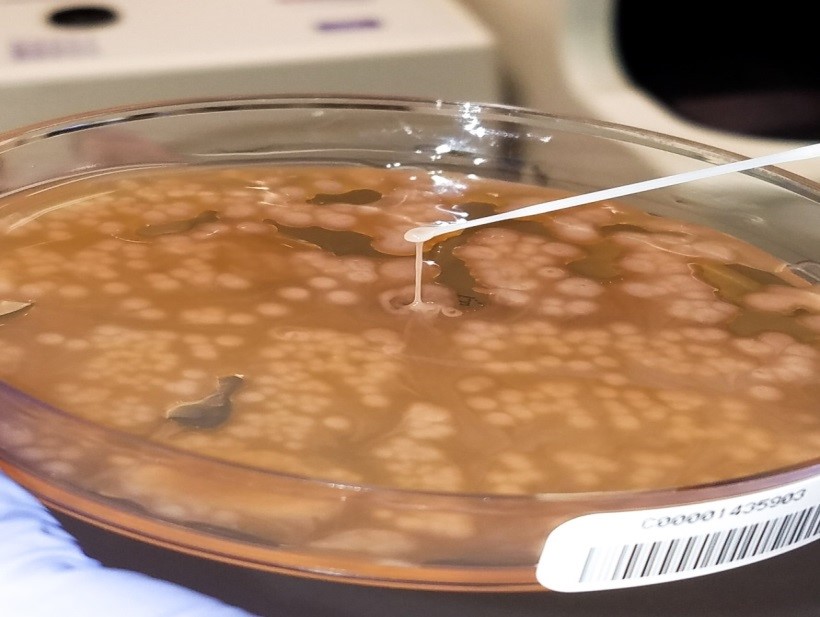
As requested, the specimen (mitral valve vegetation) was split once it was received in the laboratory. Half went into the freezer for sequencing requests which we send to a reference laboratory and the other half was processed for bacterial (including mycobacteria) and fungal cultures.

In our experience, if no organisms are observed, then no DNA is detected. Therefore, it is not beneficial to send tissue for sequencing if we do not observe an organism (or something that looks suspicious for an organism) to begin with. In parts 2 and 3 of this series we will go into greater detail of the workflow for examining tissue for infection, but for now we will focus on the processing piece.

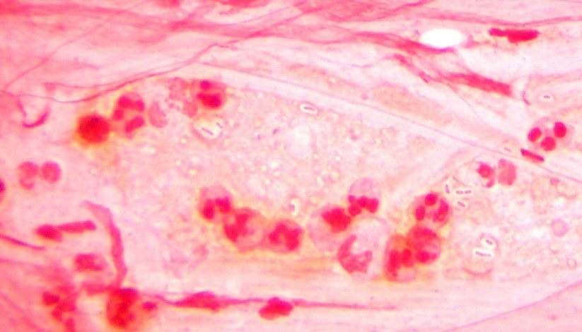
No organisms were observed in the direct smears (Gram, fungal, and acid-fast) and all cultures were negative. Because no organisms were observed, we did not send the tissue for sequencing. However, the patient was not improving and ID insisted that we send the tissue for sequencing anyway. As a last ditch effort we decided to homogenize the frozen tissue to see if by chance organism was present.

Long story short: the Gram stain did not reveal organism, but acridine orange2 did. We cultured the tissue and recovered an organism. Moral of the story: specimen processing can be tricky. It is an inherent issue that we must be aware of. How were we to know in which part of the tissue the organisms were? By definition, this is sampling error at its finest.

The Solution

Moving forward, rather than split the specimen prior to processing we have changed our protocol to homogenize the tissue first, then split the specimen. We believe this will eliminate similar scenarios from happening again in the future.

The Conclusion

All specimens are different in their composition. Unlike body fluids, which are easy to vortex and make homogeneous; tissue is more complex. Whatever the specimen, make sure your protocol(s) reduces sampling error.

References

  1. Subedi SJennings ZChen SC. 2017. Laboratory Approach to the Diagnosis of Culture-Negative Infective Endocarditis. Heart Lung Circ. 26(8):763-771.
  2. Lauer BAReller LBMirrett S. 1981. Comparison of acridine orange and Gram stains for detection of microorganisms in cerebrospinal fluid and other clinical specimens. J Clin Microbiol.14(2):201-5.


Martinez Headshot-small 2017

-Raquel Martinez, PhD, D(ABMM), was named an ASCP 40 Under Forty TOP FIVE honoree for 2017. She is one of two System Directors of Clinical and Molecular Microbiology at Geisinger Health System in Danville, Pennsylvania. Her research interests focus on infectious disease diagnostics, specifically rapid molecular technologies for the detection of bloodstream and respiratory virus infections, and antimicrobial resistance, with the overall goal to improve patient outcomes.

Chemistry Case Study: Conjugated Bilirubin in Neonatal Jaundice

Case History

Patient was a 1-week-old infant in the level 2 NICU born at 37 weeks. This infant was initially born with indirect hyperbilirubinemia but now also has increasingly elevated level of direct bilirubin (see measurements in table below). Neonatologist requested conjugated and unconjugated bilirubin test due to increasing elevated level of direct bilirubin. Conjugated bilirubin test is not routinely performed in our hospital laboratory and needs to be send out.

Question: What’s the difference between conjugated bilirubin and direct bilirubin? When does conjugated bilirubin need to be assessed?

Ref Range 3/6/18 3/7/18 3/9/18 3/10/18 3/12/18
Bilirubin total, neonatal 1.0-10.5 mg/dL 9.2 8.7 10.8 10.2 8.6
Bilirubin direct, neonatal 0.0 – 0.6 mg/dL 0.5 0.7 1.8 1.8 2.1

Discussion

Neonatal jaundice is commonly seen in newborns in the first few days of life, mainly due to increased bilirubin formation from break down of red blood cells and limited conjugation of bilirubin. Total bilirubin normally peaks at day 2-3 and should decline by day 4-5. Sample is collected via heelstick in green top tube and protected from light. Measurement of total bilirubin is interpreted based on the Bhutani Nomogram to assess risk of hyperbilirubinemia. Most often, unconjugated bilirubin is elevated in neonatal jaundice owing to hemolytic causes. In cases with prolonged jaundice, conjugated bilirubin needs to be determined to rule out cholestasis.

Conjugated bilirubin refers to bilirubin conjugated with one or two glucuronic acid, and this term “conjugated bilirubin” is often used interchangeably with direct bilirubin. Direct bilirubin refers to bilirubin fractions that can directly react with diazo reagent without the addition of accelerator, such as methanol or ethanol. This fraction usually includes conjugated bilirubin and delta bilirubin. Delta bilirubin is formed by covalent bonding between conjugated bilirubin and albumin, and has a similar half-life as albumin, 21 days. Therefore, direct bilirubin measurement overestimate conjugated bilirubin and in cases with persist or atypical jaundice, clear differentiation between conjugated and direct bilirubin is important. Clinician should know what the laboratory is measuring when interpreting the bilirubin fraction results.

In laboratories, conjugated bilirubin can be assessed by the VITROS BuBc dry slide, which simultaneously measures unconjugated (Bu) and conjugated (Bc) bilirubin by use of a mordant. In the presence of the mordant, the visible spectra of conjugated and unconjugated bilirubin are different, allowing measurement of both species from a single slide. Fractions of bilirubin can also be separated by HPLC, but this is not practical to use in a routine clinical laboratory. In this case, conjugated bilirubin was measured by VITROS BuBc slide test, and result came back elevated at 1.0 mg/dL (ref range: < 0.3 mg/dL).


Ketcham

-Megan Ketcham, MD is a 4th year anatomic and clinical pathology resident at Houston Methodist Hospital. She will be completing both hematopathology and dermatopathology fellowships. Her interests include pathology resident and medical student education and skin lymphomas.

Xin-small

-Xin Yi, PhD, DABCC, FACB, is a board-certified clinical chemist, currently serving as the Co-director of Clinical Chemistry at Houston Methodist Hospital in Houston, TX and an Assistant Professor of Clinical Pathology and Laboratory Medicine at Weill Cornell Medical College.

Blood Bank Case Study: Transfusion Transmitted Malaria

Case Study

A 26 year old African American female with sickle cell anemia presented to a New York emergency room with cough, chest pain, fever and shortness of breath. Laboratory results showed an increased white blood cell count, slightly decreased platelet count and a hemoglobin of 6.2 g/dl. Her reticulocyte count was 7%, considerably below her baseline of 13%. Consulting the patient’s medical records revealed history of stroke as a child and subsequent treatment with chronic blood transfusions. She was admitted to the hospital for acute chest syndrome and aplastic crisis and care was transferred to her hematologist. Two units of RBCs were ordered for transfusion.

The blood bank technologists checked the patient’s blood bank history and noted her blood type was A, Rh(D) positive, with a history of a warm autoantibody and anti-E. The current blood bank sample confirmed the patient was blood type A, RH(D) positive with a negative DAT but the antibody screen was positive. Anti-E was identified. Per request of the hematologist, phenotypically similar units were found and the patient was transfused with 2 units of A RH(negative), C/E/K negative, HgS negative, irradiated blood. The patient’s hemoglobin rose to 8g/dl and she was discharged from the hospital 3 days after transfusion.

Ten days after discharge the patient returned to the emergency room with symptoms including aching muscles, fever and chills. A delayed transfusion reaction was suspected. A type and screen was immediately sent to the blood bank. The post transfusion type and screen remained positive for anti-E, DAT was negative. No additional antibodies were identified. However, a CBC sent to the lab at the same time revealed malarial parasites on the peripheral smear. The patient was consulted for a more complete medical history and reported that she had never traveled outside of the country. A pathology review was ordered and the patient was started on treatment for Plasmodium falciparum.

plasfal1

Discussion

Red Blood cell transfusions can be life saving for patients with sickle cells anemia. These patients are frequently transfused by either simple transfusion of red cell units or by exchange transfusion. Because of this, alloimmunization is reported to occur in 20% to 40% of sickle cell patients.1 Blood bank technologists are very diligent in adhering to strict procedures and follow a standard of practice aimed to prevent transfusion reactions. While preventing immune transfusion reactions may be the most forefront in our minds when transfusing the alloimmunized patient, it is important to consider transfusion transmitted diseases as a potential complication of blood transfusions.

Malaria is caused by a red blood cell parasite of any of the Plasmodium species. Mosquito transmitted infection is transmitted to humans through the bite of an infected mosquito. Transfusion-transmitted malaria is an accidental Plasmodium infection caused by a blood transfusion from a malaria infected donor to a recipient.

Donors, especially those from malarial endemic countries who may have partial immunity, may have very low subclinical levels of Plasmodium in their blood for years. Even these very low levels of parasites are sufficient to transmit malaria to a recipient of a blood donation. Though very rare, transfusion-transmitted malaria remains a serious concern for transfusion recipients. These transfusion-transmitted malaria cases can cause high percent parisitemia because the transfused blood releases malarial parasites directly into the recipient’s blood stream.

Blood is considered a medication in the United States, and, as such, is closely regulated by the FDA. Blood banks test a sample of blood from each donation to identify any potential infectious agents. Blood donations in the US are carefully screened for 8 infectious diseases, but malaria remains one infectious disease for which there is no FDA-approved screening test available. For this reason, screening is accomplished solely by donor questioning.2 A donor is deferred from donating if they have had possible exposure to malaria or have had a malarial infection. Deferral is 12 months after travel to an endemic region, and 3 years after living in an endemic region. In addition, a donor is deferred from donating for 3 years after recovering from malaria. It is important, therefore, for careful screening to take place by questionnaire and in person, to make sure that the potential donor understands and responds appropriately to questions concerning travel and past infection.

Malaria was eliminated from the United States in the early 1950’s. Currently, about 1700 cases of malaria are reported in the US each year, almost all of them in recent travelers to endemic areas. From 1963-2015, there have been 97 cases of accidental transfusion-transmitted malaria reported in the United States. The estimated incidence of transfusion-transmitted malaria is less than 1 case in 1 million units.4 Approximately two thirds of these cases could have been prevented if the implicated donors had been deferred according to the above established guidelines.3 While the risk of catching a virus or any other blood-borne infection from a blood transfusion is very low, a blood supply with zero risk of transmitting infectious disease may be unattainable. With that being said, the blood supply in the United Sates today is the safest it has ever been and continues to become safer as screening tests are added and improved. Careful screening of donors according to the recommended exclusion guidelines remains the best way to prevent transfusion-transmitted malaria.

References

  1. LabQ, Clinical laboratory 2014 No.8, Transfusion Medicine. Jeanne E. Hendrickson, MD, Christopher Tormey, MD, Department of Laboratory Medicine, Yale University School of Medicine
  2. Technical Manual, editor Mark K. Fung-18th edition, AABB. 2014. P 201-202
  3. https://www.cdc.gov/malaria/about/facts.html. Accessed April 2018
  4. The New England Journal of Medicine. Transfusion-Transmitted Malaria in the United States from 1963 through 1999. Mary Mungai, MD, Gary Tegtmeier, Ph.D., Mary Chamberland, M.D., M.P.H., June 28, 2001. Accessed April 2018
  5. Malaria Journal. A systematic review of transfusion-transmitted malaria in non-endemic areas. 2018; 17: 36. Published online 2018 Jan 16. doi: 1186/s12936-018-2181-0. Accessed April 2018
  6. http://www.aabb.org/advocacy/regulatorygovernment/donoreligibility/malaria/Pages/default.aspx


Socha-small

-Becky Socha, MS, MLS(ASCP)CM BB CM graduated from Merrimack College in N. Andover, Massachusetts with a BS in Medical Technology and completed her MS in Clinical Laboratory Sciences at the University of Massachusetts, Lowell. She has worked as a Medical Technologist for over 30 years. She’s worked in all areas of the clinical laboratory, but has a special interest in Hematology and Blood Banking. When she’s not busy being a mad scientist, she can be found outside riding her bicycle.

To Be (MDS) or Not To Be? The Conundrum of Cytoplasmic Vacuolation in Hematopoietic Precursors

Every hematopathologist and pathology trainee knows to be wary of the myriad of causes that could mimic the dysplastic changes seen in marrows involved by MDS. Many times morphology alone, without genetic or cytogenetic evidence of clonality can be tricky. The list of things that can recapitulate changes seen in MDS seems to grow longer every day – and with it the length of our ‘canned comments’ on ruling out reactive causes of dysplasia. Within the recent past, two bone marrow biopsies crossed my microscope, both sent to ‘rule out’ MDS. Both had almost identical morphologic findings, but very different diagnoses. Here are some representative images from the marrow aspirates and iron stains:

mds
Figure 1. Representative Wright-Giemsa stained cells from Case 1 (A and B) with accompanying iron stain (C) showing numerous ring sideroblasts.  Representative Wright-Giemsa stained cells from Case 2 (D) with accompanying iron stain (E) showing some ring sideroblasts. 

Discussion

Images A through C come from case 1, a 67-year-old woman with a past medical history of non-alcoholic steatohepatitis (NASH) complicated by hepatic encephalopathy and recurrent ascites who underwent bone marrow biopsy for new onset pancytopenia with transfusion-dependent anemia. The marrow was slightly hypercellular for age and showed progressive trilineage maturation. Granulocytic and erythroid progenitors did not reveal quantitatively significant dysplasia. The one dysplastic megakaryocyte identified is pictured here (panel A). Interestingly many erythroid and granulocytic precursors showed cytoplasmic vacuolation (panel B showing granulocytic vacuolation). An iron stain (panel C) revealed 44% ring sideroblasts. Case 2 is represented in images D and E and was from a 64-year-old man with no significant past medical history who presented with lethargy and anemia. This marrow was also slightly hypercellular for his age and showed borderline-significant dysplasia in megakaryocytic maturation. Granulopoiesis and erythropoiesis were unremarkable except for cytoplasmic vacuolations in many cells (panel D). An iron stain showed 8% ring sideroblasts (panel E).

Both cases were signed out descriptively, urging the clinician that we needed to rule out reactive causes of dysplasia before a definitive diagnosis of MDS could be rendered. In both cases we suggested waiting for the cytogenetics results for a more comprehensive analysis. Additionally, we recommended testing for serum copper since copper deficiency can be the cause of dysplastic morphology, cytoplasmic vacuolation, and ring sideroblasts.

Case 1 revealed markedly diminished copper and normal cytogenetics. Copper replenishment was curative. Case 2 revealed normal copper levels and a complex karyotype that contained numerous MDS-associated abnormalities confirming the clonal, and therefore malignant nature of these changes. Despite being almost identical morphologically, these case were diagnostically and prognostically poles apart.

Copper is an element that serves as a micronutrient required for hematopoiesis. It’s presence in many readily available foods including meat, fish, nuts, and seeds renders diet-related copper deficiency a rare phenomenon. Zinc-supplementation is one of the causes of copper deficiency in published reports. Copper deficiency has been well documented to mimic dysplastic changes seen in MDS; but these morphologic findings and cytopenia are reversible. Characteristically, cytoplasmic vacuolation is an important morphologic clue that there could be an underlying paucity of serum copper.  Another aspect of copper deficiency is the presence of ring sideroblasts which also can mean MDS. It is very important to consider this differential diagnosis when dealing with marrow specimens sent to rule out MDS. This Lablogatory post highlights the significant overlap between presentation and morphologic findings between MDS and copper deficiency supporting the notion that a high index of suspicion, good communication, stat copper levels, and cytogenetics or MDS FISH studies are very helpful in delineating benign from malignant.

References

  1. Dalal N. et al. Copper deficiency mimicking myelodysplastic syndrome. Clin Case Rep. 2015 May; 3(5): 325–327.
  2. Willis M.S. Zinc-Induced Copper Deficiency: A Report of Three Cases Initially Recognized on Bone Marrow Examination. AJCP. 2005 Jan; 123(1): 125–131
  3. D’Angelo G. Copper deficiency mimicking myelodysplastic syndrome. Blood Res. 2016 Dec; 51(4): 217–219.
  4. Karris S and Doshi V. Hematological Abnormalities in Copper Deficiency. Blood 2007 110:2677


Mirza-small

-Kamran M. Mirza, MD PhD is an Assistant Professor of Pathology and Medical Director of Molecular Pathology at Loyola University Medical Center. He was a top 5 honoree in ASCP’s Forty Under 40 2017. Follow Dr. Mirza on twitter @kmirza.

Microbiology Case Study: A 79 Year Old Male with Rheumatic Heart Disease

Case History

The patient is a 79 y/o male with past medical history of rheumatic heart disease, permanent atrial fibrillation, mechanical aortic and mitral valves (2004), status post single chamber pace maker for bradycardia (2010), and prostate adenocarcinoma treated in 2000. He had new MRI compatible pace maker placed on Oct 19, 2017. During follow-up he was noted to have a hematoma over the incision site. He had a revision done on Nov 3, 2017. At that time, the blood from the incision site was sent for culture. 

Laboratory Identification

Gram stain showed moderate amount of polys with no bacteria seen. The isolate was a gram-negative rod that was identified on the MALDI-ToF as Burkholderia multivorans.


burkmult1
Image 1: Semi-mucoid, yellow-grey colonies on Chocolate agar and on Blood agar plates.

Discussion

The Burkholderia genus appears as gram-negative medium-sized straight rods, with the exception being B. mallei which is a coccobacillus. The will grow on blood, chocolate, and MacConkey agar. Oxidative-fermentative-base-polymyxin B-bacitracin-lactose (OFPBL) agar can be used to isolate B. cepacia and Ashdown medium can be used to isolate B. pseudomallei. They are non-lactose fermenters on MacConkey, but B. cepacia can turn into a dark pink to red due to oxidation of lactose after 4-7 days.

B. multivorans is a species within the Burkholderia genus, which are normal to plant, soil, and water, but not normally considered common human flora. Formerly of the Pseudomonas genus, B. cepacia, B. mallei, and B. pseudomallei are the most commonly seen as infections in humans. Further, B. cepacia and B. mallei are not typically human pathogens in a healthy human host. Because of the rarity of this genus to infect humans, their pathogenicity is not well known; but, importantly, they are intrinsically resistant to many antibiotics and can thus be associated with hospital acquired infections.

Of this genus, very little literature is present on B. multivorans specifically, and of the literature that does exist, most of it is in relation to cystic fibrosis patients. Taxonomic advances has shown that B. cepacia complex is a cluster of nine closesly related genomic species or genomorvars (1).  B. multivorans represents genomorvar II. Hospital acquired clinical infections from this complex (but perhaps not specifically from this particular genomorvar) has been seen following catheterization, cystoscopy, heart surgery, and with contaminated ventriculoatrial shunt (2). B. multivorans biochemically is oxidase positive, catalase positive, lipase positive, nitrate-reducing, urease positive, resistant to colistin, and can grow at 42C (3, 4).

A recent comparative genomic study showed that B. multivorans is a highly evolutionarily preserved genome with genomic characteristics from the environment and isolated from cystic fibrosis patients to be similar, and that isolates from different continents are also similar (5). Further, a murine model for pulmonary infections showed that B. multivorans could persist in the host by establishing an intracellular presence within macrophages, which could explain the persistence of this pathogen in cystic fibrosis patients (6). Importantly though, due to the conserved and common genomic structure, there rests a possibility for potential vaccination for cystic fibrosis patients against B. multivorans.

The patient was prescribed a single dose of oral Bactrim and then advised to come into the hospital for admission for IV antibiotics. IV ceftazidime was started with pending blood cultures, which are negative at the time of this documentation.

References:

  1. Coenye T. et al. Taxonomy and identification of the Burkholderia cepacia complex. J Clin Microbiol 2001;39:3427-3436.
  2. Pallent LJ. et al. Pseudomonas cepacia as contaminant and infective age. J Hosp Infect 1983;4:9-13.
  3. Henry DA. et al. Phenotypic methods for determining genomovar status of Burkholderia cepacia complex. J Clin Microbiol 2001;39:1073-1078.
  4. Vandamme P. et al. Occurrence of multiple genomovars of Burkholderia cepacia in patients with cystic fibrosis and proposal of Burkholderia multivorans sp. nov. Int J Syst Bacteriol 1997;47:1188-1200.
  5. Peeters C. et al. Comparative genomics of Burkholderia multivorans, a ubiquitous pathogen with a highly conserved genomic structure. PLoS One. 2017, 21; 12 (4): e0176191.
  6. Chu KK. et al. Persistence of Burkholderia multivorans with the Pulmonary Macrophage in the Murine Lung. Infect Immun 2004; 72 (10): 6142-6147.


-Jeff Covington, MD, PhD, is a 1st year anatomic and clinical pathology resident at the University of Vermont Medical Center.

Wojewoda-small

-Christi Wojewoda, MD, is the Director of Clinical Microbiology at the University of Vermont Medical Center and an Associate Professor at the University of Vermont.

Faculty Insights: Teaching Medical Laboratory Science in a Blended Learning Format

Thus far, we’ve talked about the structure of our blended model of curriculum delivery and the learner experience, but what about the instructor experience teaching in this format?

I surveyed a few of our faculty members about their experience teaching in our Medical Laboratory Science (MLS) Program, and their comments about our blended model of curriculum delivery fell into a few themes:

Benefits of the “flipped classroom” model

“Having students complete the lecture content as homework and then meeting with them for the hands-on ‘face-to-face’ learning adds another layer and more reinforcement of key concepts. If learners can’t understand what they’ve read or interacted with in the online component, they’ve got another chance to hear a short review of the material and actually perform a hands-on, real-world lab activity to reinforce again what they have learned. This is what makes blended learning so effective—read, learn, see, do—it hits the learning from every angle.”

“I believe the blended learning style with the online component is very effective, and I would bet that students retain more information and retain the information longer than in traditional formats. It’s all about reinforcement.”

Role of the instructor from “Sage on Stage” to a facilitator of the student’s knowledge acquisition and enhanced student learning

“As an instructor, I am no longer required to be the “Sage on the Stage,” and the questions from our students tend to be more specific, in that they come into the classroom prepared, having some base knowledge of the content.”

“I love teaching with a blended format. Having the online component allows the student to review the learning content prior to coming to lab where we meet face to face. The online blackboard format allows for several different ways to attack the learning, which is nice for the variety of students that we have (age, gender, and background), as well as a variety of learning styles.”

“Teaching with the online component allows for embedded written lessons, recorded lectures, PowerPoint handouts, images, YouTube videos, interactive activities such as a discussion board and wikis, and online worksheets—all at which the student can work through as fast or as slow as needed. Online learning allows for multiple levels of reinforcement to help make the information stick. It also helps learners be in control of their own learning. Access to information is not just a one-time shot in a live lecture. It’s there to use and review as much as needed.”

More time for instructor-led hands-on activities

“Time is another factor. We are only given so much time with the MLS students, and if we had to present all the lecture material in the classroom, we would not have time for all the laboratory activities that we have developed.”

“I like the fact that it puts the onus on the learner to engage with the material ahead of time, which allows for more hands-on learning in the classroom. The blended format makes it extremely conducive for a laboratory-based class.”

“When teaching morphology of cells, I like to use online question ‘banks’ with images of cells, crystals, casts, etc., for the students’ practice. They can review these question banks as much or as little as needed outside of the classroom. They can practice morphology identification at home, outside of class—all without the need of a microscope. Not only do they come into the classroom/lab knowing their cells, but they can work more efficiently and progress more quickly to advanced case studies.”

“I think the flipped model we implement is a great way to enhance our students’ reading skills and comprehension, while holding them accountable for completing the required assignments.”

“The blended approach allows us to address more difficult concepts. While the students may be able to grasp the concepts from their online reading, they also need talking points to confirm that they actually understand and can apply the concepts. We have found that giving the students the task of learning the online concepts can only really be successful if we follow up with them the next day, starting with a discussion about their online homework. We also give quizzes and have designed laboratory activities that apply the online concepts.”

Varied thoughts about course maintenance

“While the time to develop online content can be extensive, once it’s built in this format, it is easier to update and maintain on an annual basis.”

“It is not so easy to maintain the details in the online course. It takes a lot of time and effort to update all of the dates for assignments and other activities for each class section. Once the core components are built, one can easily add to the content. However, if one is building a new module or lesson, it can take a lot of time. It seems that the time to maintain an online course is similar to the time it takes to keep materials up-to-date in a traditional course.”

“At first, it took some getting used to grading assignments online, but I am used to it now and actually prefer it. It’s so easy for a student to do a “copy/paste” when filling out an online worksheet, so I do question typing (copy/paste) vs. writing things out on a worksheet and how well the information is sticking. With the intensity of our program, time is of the essence. I like that as soon as students submit their online assignments, I can grade it. Some students like to work ahead, and some turn things in at the last second. With the online submissions, I can grade as they come in, instead of getting hit with 24 assignments at once, which is a big time-saver for me.”

Repurposing

“One of the greatest positives with [the software] Blackboard Learn is that we can use the system with multiple learners. The learner has easy access to the course once he/she is added to system. One cannot always say that with traditional classroom teaching/learning. Unless the content/didactic is recorded, there is not easy access to the materials.”

In summary, I would stress the following key points as benefits of adopting a blended approach to curriculum delivery:

  • Increased classroom time for hands-on activities that are more closely aligned with what the students will actually be doing once they graduate, get jobs, and go to work.
  • Increased instructor satisfaction.
  • Students are more prepared for the classroom activities.
  • Increased ability to engage students with higher-learning concepts.
  • Course maintenance is more efficient, and learning tools are enhanced.
  • Time and cost savings are realized, related to repurposing of curriculum across different learners.


Lehman_small

-Susan M. Lehman, MA, MT(ASCP)SM graduated from the University of Wisconsin-Madison in 1983 with a BS in medical technology. She is program director for the Medical Laboratory Science Program and course director for Clinical Microbiology I and II; her areas of interest include distance education and education methodology.

Transparency in Injury Reporting

Susan was getting ready to work in the microbiology lab. She sat down after donning her lab coat, but before she put on gloves, she picked up some reports that were on the counter. As she picked them up, she noticed she got a small paper cut on her finger. Thinking nothing of it, she put her gloves on and went to work.

Chuck opened the door to walk into the back of the main lab. A cardboard box was in the walkway, and Chuck hit it with his toe and fell to his knee. He figured he wasn’t hurt, so he didn’t say anything since filling out paperwork was such a nuisance- and no one saw it happen.

Jean was walking into the hospital during the ice storm to get to work. Shortly after she closed the car door, she slipped and landed on her wrist. It hurt a little, but she figured it would be fine, so she didn’t say anything.

Accidents happen often in the laboratory setting, and many of them go unreported. The first thing that should occur after an injury is first aid. Then the incident needs to be reported. That may mean telling someone in charge in the department- a lead technologist or a manager. That can vary depending on the department and the time of day. Next, the incident should be reported to an institutional Occupational Health department or to a designated authority (such as the emergency Department) if the Occupational Health office is closed. This step is vitally important.

Make sure the details of the incident are recorded accurately, and that any witnesses are identified. Some facilities use an electronic reporting system, and others require a nurse to fill out the forms. Good communication is important here so that a thorough follow-up by the lab safety professional can occur later. The fewer details left out, the better.

We are human, and accidents happen, but the route to a better safety culture in the department is transparency. All injuries at work need to be reported. There is no shame in an injury, and there should be no reprisals, and reporting leads to prevention of injuries. The communication about the event is crucial- the reporting may prevent someone else from being injured in the same way. In some labs there have been serious injuries that occurred because no one reported a previous similar event. That can and should always be avoided. There are other reasons to report injuries as we – those stories at the beginning of the article did not have a happy end – because they were not reported.

After a week, Susan noticed that her little paper cut had become red and swollen. She made an appointment with her physician who prescribed an antibiotic. The antibiotic didn’t work, and after a serious bout of septicemia, Susan had to have part of her hand amputated to prevent the spread of the rare bacterial infection.

A day after Chuck tripped, Elaine walked into the lab and tripped on the same cardboard box. Elaine fell hard and broke her hip. She needed immediate surgery. She would have retired in another month.


Two weeks after her fall in the parking lot, Jean decided to go to the urgent care since her wrist was still hurting. An x-ray revealed a fracture that would need a surgical repair. Jean went to the Occupational Health office to report the event. Because there was such a delay in reporting, the compensation department decided they could honor the claim, and Jean’s medical follow-up was not covered.

There are many reasons to report an injury at work. The first one is about you- protect your own health and your future- that’s worth a few minutes of paperwork and a short visit to the Occupational Health office. The second reason to report is about everyone else. If something is unsafe in your environment and it has caused an injury, let someone know. That sort of communication and transparency is important to the entire team. Accidents happen, but even when they do, we can respond quickly and communicate so that safety improves after the event. As a lab safety professional, make sure you talk about accident transparency, and make sure it is something practiced by the entire team.


Scungio 1

Dan Scungio, MT(ASCP), SLS, CQA (ASQ) has over 25 years experience as a certified medical technologist. Today he is the Laboratory Safety Officer for Sentara Healthcare, a system of seven hospitals and over 20 laboratories and draw sites in the Tidewater area of Virginia. He is also known as Dan the Lab Safety Man, a lab safety consultant, educator, and trainer.

Microbiology Case Study: A Middle-Aged Man with Malaise, Shaking, and Chills

Case history

A middle-aged male presented to the hospital emergency room with the complaints of malaise, shaking and chills for the last two days. He denied any runny nose, cough, abdominal pain, nausea, vomiting, headache or known sick contacts. His past medical history was significant for alcohol use disorder. Imaging of the abdomen revealed an ill-defined region of decreased attenuation in the right lobe of the liver measuring 4.8 x 4.7 x 2.2 cm. The Gram stain of the abscess showed 4+ WBCs (PMNs) and 4+ gram negative rods with a very large capsule surrounding them (Image 1).  The organisms grew very mucoid colonies on 5% sheep blood, chocolate, and MacConkey agars (Image 2).  A string test performed on the mucoid bacterial colonies was >5 mm (Image 3).

kleb1
Image 1. Gram stain of abscess showing 4+ WBCs and 4+ GNR with large capsule.
kleb2
Image 2. Cultures showed mucoid colonies on the chocolate and MacConkey agars.
kleb3
Image 3. A string test was performed on the mucoid colonies and was positive (mucoid capsule “string” > 5mm).

Discussion

The organism was identified as Klebsiella pneumoniae by MALDI-TOF MS.  Based on the mucoid capsule and positive string test, this organism was further identified as hypermucoviscous K. pneumoniae.

Hypermucoviscous K. pneumoniae is a relatively newly recognized hypervirulent variant of K. pneumoniae. It was first described in the Asian Pacific rim and is now increasingly recognized in Western countries. Defining clinical features include serious, life-threatening community-acquired infection in younger healthy hosts, an unusual feature for enteric gram negative bacilli in the non-immunocompromised population. It can cause a variety of diseases including, but not limited to liver abscess, pneumonia, meningitis, osteomyelitis, necrotizing fasciitis and endophthalmitis.

Intestinal colonization, appears to be a critical step leading to infection. It is seen mostly in Asians, raising the issue of a genetic predisposition vs. geospecific strain acquisition.  The increased virulence might be due to the ability to more efficiently acquire iron and perhaps an increase in capsule production, which confers the hypermucoviscous phenotype to the organism. The vehicles for acquisition and subsequent colonization appear to be food and water, person-to-person transmission (e.g., close contacts such as family members or sexual partners) or animal-to-person transmission (e.g., between pets and their owners).

To date, most strains of hypermucoviscous K. pneumoniae have been very susceptible to antimicrobials except ampicillin.  However, in recent literature, propensity for hypermucoviscous Klebsiella pneumoniae to become multi-, extreme or pandrug-resistant, including the acquisition of extended-spectrum β-lactamases (ESBL) and carbapenemases has been reported. Since hypermucoviscous K. pneumoniae strains often cause abscesses, source control is a major aspect of the overall management plan and a need to drain abscesses and closed space infections is essential for optimal outcome.

References

  1. Alyssa S. Shon, Rajinder P.S. Bajwa and Thomas A. Russo; Hypervirulent (hypermucoviscous) Klebsiella pneumonia: A new and dangerous breed; Virulence 4:2, 107–118; February 15, 2013; 2013 Landes Bioscience
  2. Bonnie C Prokesch, Michael TeKippe, Jiwoong Kim, Prithvi Raj, Erin McElvania TeKippe, David E Greenberg; Primary osteomyelitis caused by hypervirulent Klebsiella pneumonia; The Lancet Infectious Diseases , Volume 16 , Issue 9 , e190 – e195


MA

-Muhammad Ahmad, MD is a 2nd year anatomic and clinical pathology resident at University of Chicago (NorthShore) program based at Evanston Hospital, Evanston, IL. His academic interests include breast pathology and cytopathology.

-Erin McElvania, PhD, D(ABMM), is the Director of Clinical Microbiology NorthShore University Health System in Evanston, Illinois.

CMS’s National Coverage Determination for Next Generation Sequencing (NGS) – What Does This Mean for the Future of NGS Testing for Molecular Oncology?

I thought I’d take a break from the next generation sequencing (NGS) wet bench description this month to review news occurring in the world of reimbursement of testing of cancer specimens with next generation sequencing.  As a tech, I don’t deal with the nitty gritty of insurance reimbursement of our tests on a day to day basis, but this one caught my eye as it would have had a real impact on the NGS testing in our lab.  On November 30th, 2017, a proposal was released by the Centers for Medicare & Medicaid Services (CMS) to review the national coverage analysis tracking sheet for NGS for Medicare beneficiaries with advanced cancer.  In the original wording of the proposal, one thing it stated was that CMS should only reimburse NGS testing for advanced cancers when the testing was done with an FDA approved assay.  This caught me, as well as many others in the molecular community, by surprise.  The reason?  Currently, there are only a few FDA approved assays on the market; much of the testing occurring right now for oncology assays by NGS are lab-developed tests (LDTs), including the ones that we run in our lab.  Under the proposal’s language, these types of assays would not be reimbursed for Medicare patients (and where CMS reimburses, the major insurance companies follow), making it very difficult for us to continue the testing that we perform.

The process for a proposal such as this one includes posting the proposal, then allowing a period for public comments about the proposal.  Six weeks were given for people to post their comments online, during which, 315 comments were left.  These comments included praise to CMS for recognizing that NGS testing is increasingly useful for precision medicine, but also stressed the limitations of only allowing FDS approved assays to be used.  Some comments pointed out how clinicians and pathologists work together in the institutions performing the NGS assays in a way that would be impossible if forced to use an assay from an outside institution.  They also indicated how difficult it would be for all NGS testing to be performed by the very small number of FDA approved assays and how it is almost impossible for small academic institution labs to get FDA approval for assays due to the amount of money and time the approval process takes.

On March 16, 2018, the final decision memo was released with altered wording compared to the original and can be found here https://www.cms.gov/medicare-coverage-database/details/nca-decision-memo.aspx?NCAId=290 and is also shown below:

“A.  Coverage

The Centers for Medicare & Medicaid Services (CMS) has determined that Next Generation Sequencing (NGS) as a diagnostic laboratory test is reasonable and necessary and covered nationally, when performed in a CLIA-certified laboratory, when ordered by a treating physician and when all of the following requirements are met:

  1. Patient has:
    1. either recurrent, relapsed, refractory, metastatic, or advanced stages III or IV cancer; and
    2. either not been previously tested using the same NGS test for the same primary diagnosis of cancer or repeat testing using the same NGS test only when a new primary cancer diagnosis is made by the treating physician; and
    3. decided to seek further cancer treatment (e.g., therapeutic chemotherapy).
  2. The diagnostic laboratory test using NGS must have:
    1. FDA approval or clearance as a companion in vitro diagnostic; and
    2. an FDA approved or cleared indication for use in that patient’s cancer; and
    3. results provided to the treating physician for management of the patient using a report template to specify treatment options.
  3. The diagnostic laboratory test using NGS must have:
    1. FDA approval or clearance as a companion in vitro diagnostic; and
    2. an FDA approved or cleared indication for use in that patient’s cancer; and
    3. results provided to the treating physician for management of the patient using a report template to specify treatment options.
    4. Other

Medicare Administrative Contractors (MACs) may determine coverage of other Next Generation Sequencing (NGS) as a diagnostic laboratory test for patients with cancer only when the test is performed in a CLIA-certified laboratory, ordered by a treating physician and the patient has:

  1. either recurrent, relapsed, refractory, metastatic, or advanced stages III or IV cancer; and
  2. either not been previously tested using the same NGS test for the same primary diagnosis of cancer or repeat testing using the same NGS test only when a new primary cancer diagnosis is made by the treating physician; and
  3. decided to seek further cancer treatment (e.g., therapeutic chemotherapy).

See Appendix D for the NCD manual language.”


In part B, it addresses those assays that are not FDA approved, but are run in a CLIA-certified laboratory.  This part was added in the final decision and makes it possible for non-FDA approved assays run in CLIA-certified laboratories to be reimbursed, dependent upon the local MACs.  While this is a huge improvement over the previous, there are still questions regarding some of the wording and we will have to see how this affects testing for our patients.  For example, in 1b, where it mentions repeat testing – some patients have multiple mutations that are followed over time for hematological malignancies – will this be considered repeat testing? It will remain to be seen.  Needless to say, I am happy to be able to continue doing my job.

rapp_small

-Sharleen Rapp, BS, MB (ASCP)CM is a Molecular Diagnostics Coordinator in the Molecular Diagnostics Laboratory at Nebraska Medicine.