Traditionalists are the oldest working generation in today’s professional environments. They bring a wealth of information, knowledge, and experience with them. Therefore, organizations that work with Traditionalists either on their staff or on their Boards are fortunate to have access to their input. In order learn as much as possible from this generation, while they are still present in the workplace, it is critical to know and understand their preferred way of communicating, leading, and working. It is also important to know how and when to adapt your own preferred communication, behavioral, and leadership styles to meet the needs and preferences of this
generation.
Typically, Traditionalists prefer face-to-face communication. They grew up with limited communication technology and they prefer to connect in person when possible. If you cannot communicate in person, pick up the phone and call them. Not only is this respectful to their own preferences, it will allow you to increase your verbal communications skills when there is no written form used. Having a personal touch is important, so try not to talk business right away but take time to get to know one another.
When meeting with Traditionalists, some formal protocol is appreciated. Have someone else introduce you, or if you are in charge of the meeting make sure to introduce everyone properly. You can add a personal touch if appropriate. For example, say “This is Betty Jones. She is the current President of our Board of Directors and has been a member of our organization for over forty years. She is here to provide us with strategic details about our new direction. Also, she is an avid fly-fisher!” Additionally, pay attention to meeting protocols such as offering something to drink and sending the agenda ahead of time so that they can prepare. This is, of course, good to do with everyone, but Traditionalists respond especially well to such protocol.
Their leadership style is based on a chain of command and creating contingency plans. They dislike indecisiveness, disrespect, profanity, and poor dress. They appreciate a sense of formality and high quality work. I always think about how Traditionalists dressed, and sometimes still dress, when going on a plane. They dressed very formal, especially compared to today’s travelers. Keep this in mind when meeting with them in person. Forego the jeans and sweaters and wear something more traditionally professional. Finally, use formal address, such as Sir, Doctor, and Madam. Again, the more professional protocol you use, especially in the beginning, will set you up for success when working with them.
Personally, I learned and witnessed that if you include this generation in inquiry-based conversations and discussions that you can learn about additional leadership approaches to increase your own adaptability. Learn from other generations as much as possible, especially the ones that are currently leaving the workforce. There is a lot to be gained from generational diversity and increasing your own ability to meet the needs of every generation in the workplace.
-Lotte Mulder earned her Master’s of Education from the Harvard Graduate School of Education in 2013, where she focused on Leadership and Group Development. She’s currently working toward a PhD in Organizational Leadership. At ASCP, Lotte designs and facilitates the ASCP Leadership Institute, an online leadership certificate program. She has also built ASCP’s first patient ambassador program, called Patient Champions, which leverages patient stories as they relate to the value of the lab.
At the ASCP Annual Meeting this October, I had the privilege of facilitating a Roundtable Discussion about diversity in the workplace. I anticipated that we might be talking about issues such as culture, religion, gender, ethnicity, educational level, ability/disability and possibly age and generational issues. I was anticipating a very rich and “diverse” list of topics for this discussion.
To my surprise, generational differences was the primary topic for this Roundtable Discussion. There were nine people at our table with representation from both sub-sets of the Baby Boomer group, as well as, the Gen Xers, and Millennials (Gen Y). There seemed to be a strong disconnect between the Millennials and Gen Xers and the older people in the lab, meaning the Boomers and Traditionalists.
The Traditionalist generation only represents about 5% of the workers in clinical labs, however, the Baby Boomers still represent about half of the work force in the clinical labs. The strongest point of dissention seemed to center on “work life balance.” There was clearly a lack of knowledge and understanding on both parts. Baby Boomers are known for their work ethic and learned well from their Traditionalist’s parents and role models. They identify with their job, profession, and career. This is why we still have Traditionalists and Boomers working in the laboratories. They possess the institutional knowledge, relationships, and a strong sense of loyalty.
The Gen X and Y “work life balance” issue collided with the strong sense of work ethic characterized by the Traditionalists and Boomers. However, once each generation were able to share what they valued, there was a light bulb that appeared at the table and the bridge of understanding began to be built.
So what’s the key to collaboration? It’s all about talking with each other and asking good questions. The Traditionalists can learn from our Gen Xers and Millennials and focus on work life balance. Just as it is important for the Gen Xers and Gen Ys to learn about the institutional knowledge and work practices that can be gleaned from the Traditionalists.
-Catherine Stakenas, MA, is the Senior Director of Organizational Leadership and Development and Performance Management at ASCP. She is certified in the use and interpretation of 28 self-assessment instruments and has designed and taught masters and doctoral level students.
This time, I’ve got something to talk about that’s a little more serious. I don’t like to deviate from fun lab-related memes and insights,but every now and then something really strikes a chord. Enough so to talk to all of you about it. Some of you reached out to me after my post discussing clinician burnout and suicide in healthcare and that felt great; connecting with people who had some powerful stories to share really validated that conversation. Today, I want to talk about guns. Specifically, the public health epidemic of gun violence, the current conversation about whose “lane” (read:responsibility) belongs to whom, and what role those of us in laboratory medicine play.
I was horrified to see the recent shooting and murder of three in my Chicago home at Mercy hospital in the Near South Side. I won’t rehash the details that are on the news. Emergency resident physician Dr.Tamara O’Neal, newly minted Chicago Police Officer Samuel Jimenez, and pharmacy resident Dayna Less were all shot and killed point-blank by a gunman in the Mercy Hospital emergency department. A place that is supposed to be for healing, safety, and hope. Senseless.
Image 1. Victims of the Mercy Hospital shooting: (L-R) emergency room Dr. Tamara O’Neal, Chicago police Officer Samuel Jimenez, and pharmacy resident Dayna Less. These were all new to their careers, whose lives were abruptly ended by senseless gun violence. Source: ABC7 Chicago.
This now presses the start button on America’s newest tradition: a very short-lived, ill-timed, and often tone-deaf debate about the firearm subculture in our nation. Okay, bias check: you should know that I am not a fan of guns of any kind. If it were up to me, they would either belong in museums or find more useful lives melted and repurposed as metal used to reinforce hurricane-prone buildings or safe hypodermic needles for patients in need. That said, this isn’t a gun debate article; nor is it an open forum to discuss gun control, the second amendment, the NRA, or anything political. I respect opinions and educated civil discourse, but this piece today is focused on health—public health.
The epidemic of gun violence in America is a problem. The American Public Health Association (APHA) posted on their website extensively on the topic of gun related deaths which “kill more than 38,000 people and cause nearly 85,000 injuries each year. As a longtime advocate for violence prevention policies, APHA recognizes a comprehensive public health approach to addressing this growing crisis is necessary.” (Read their fact sheet here)Furthermore, the American College of Physicians (ACP) published a position paper on the topic in the Annals of Internal Medicine journal (read it here)where they establish a comprehensive set of recommendation from a conglomerate of clinical medical specialty organizations. Increasingly now more than ever does this prevalence of gun related injury and death present itself as a major health concern: a public health epidemic. I could talk to you about the number of mass shootings in our country, or the epidemiologic incidence of gun-related deaths compared to other countries, even the policy discussion around gun ownership and regional policies regarding safety and gun control—it doesn’t matter. All the charts and graphs any recycled article on the subject will just fade into the mist of “yet another shooting.” That’s not okay. I don’t want to drown you in data. Better put, I can’t. See, the problem is you’ll see the same pieces of information regarding the gun debate as you scroll through the news on your social media. Something new I want to add to this conversation is the overwhelming emphasis on the simple truth that this is a public health issue.
Image 2. In the US, we fund approximately as much research for gun violence as we do for drowning and falls. Of the three, mortality related to gun violence is about the same as sepsis—and that’s heavily documented. Source: Journal of the American Medical Association.
This unfortunate new reality is no different from other public health programs that have addressed various issues over the past decades. What do deaths from motor vehicle accidents, fires, smoking-related ung cancer, obesity and type 2 diabetes, heart attacks, antibiotic resistant bacterial infections, and traumatic brain injuries have in common? Per the American Foundation for Firearm Injury Reduction in Medicine (AFFIRM), they were all public health crises that pushed medicine past a breaking point in clinical burden and forced us to invest in research which conclusively provided results to address related mortality and morbidity. AFFIRM is a non-profit organization which is building a coalition in medicine for the purpose of researching and addressing this newest public health issue. They argue that,without medical evidence we won’t be able to find solutions to the senseless loss of life from gun violence. Death from car accidents gave us the seatbelt and tickets for disobeying its required legal usage. Death in home fires got us the smoke detector and regulations surrounding their installation. Lung cancer deaths led to smoking cessation programs, increased taxation, and policy changes regarding access to cigarettes. Sugar-related morbidities created a conversation about healthy diets, public policies addressing food deserts, and taxation programs for drinks with added sugar. Heart attack deaths gave us longitudinal studies for best care practices and lifestyle recommendations.Resistant bugs established a new discussion on antimicrobial stewardship. Brain injuries gave us new guidelines for concussions. I could go on. That’s only the tip of the public health iceberg. The point is that if there is an epidemiological trend where people are literally dying, data married with health metric-oriented research create solutions!
But let’s add deaths from gun violence to that list. What then do they all have in common, besides the concern for improving public health? Save for the tragically evident lack of a solution, the similarity becomes clear: there is lobby, interest, power, and support. Cars didn’t always have seat belts, cigarettes used to be cheap and doctors used to smoke at work,no one talked about cheeseburgers giving you heart attacks and diabetes decades ago, and helmet-clashing football players didn’t always receive the treatment they needed. Why? Because some entity—corporate, societal, etc.—wasn’t keen on“buying in.” Much like it takes justification and convincing for administration to buy your fancy chemistry analyzer, so do the public and oppositional lobby groups which require swaying toward the intervention(s) being proposed.
Image 3. The Dickey Amendment passed in 1996 was a small rider in a bill that was part of a larger budgetary spending bill. Effectively, it completely disallows public health research into gun related deaths, gun violence, or any publication that would endorse gun control or limitations. Source: 104th Congress.
Often, the data stacks high enough to influence decisions on its own. But that isn’t the case with gun related mortality. I see gun related violence as sort of the opposite of the vaccine debate: with the flu shot there seems to be too much data and not enough stories to convince the anti-vax movement to realize the significant threat being addressed. On the other hand, gun related violence exhibits far too many stories without any significant amounts of data. Possibly, this might be related to the limitations placed upon the CDC since the mid-1990’s that forbid them from using funds “to advocate or promote gun control.” Yes, really. Just last month, I wrote about the newest advancements in influenza testing and the best practice of vaccinating annually.I cited thousands of deaths related to vaccine-preventable or epidemiologic illness; 80,000 dead from influenza last year, thousands from swine flu over a decade ago, etc. But when you try and cite proper, medical data regarding guns in public health, its … not so easy. No data, no research. No research, no change.
Many of you have undoubtedly read about the current social media “discussion” regarding whose “lane” gun violence is to navigate: The National Rifle Association (NRA) asserted in a tweet that doctors, discussing the issue only within their field should leave it to more “qualified” groups like them. That’s been a tinder box of vitriol the medical community, for lack of a better term, is up in arms about. I followed and read tons of comments about this as it unfolded, hearing from endless doctors, nurses, and laboratorians posting with blood spattered scrubs, decimated trauma bays, and emptied blood bank refrigerators that this growing epidemic is enraging clinicians about. Earlier, I highlighted similarities between public health problems and their respective solutions citing that they all shared oppositional lobby groups. What better profession to handle the topic in question than medicine—whose associated lobby power from professional societies like ours to Big Pharma amass one of the largest voices in policy making in America. And another thing, as gun violence is a public health concern, whose literal job is it to address health, mortality, and morbidity? All of ours. Nurse educators lead patients through lifestyle modifications they can employ to curtail some effects of diabetes, physicians manage patient treatment regimens balancing input from pharmaceutical tools to professional guidelines,clinicians like us strive to provide the best resources available by advancing hemoglobin A1c levels or point of care testing. We all play roles in every single healthcare matter that translates to life or death, so why not this one?
So, I touched on it a little here, but what role does the medical laboratory professional play? Besides bullets in tissue section, how does the public health epidemic of gun violence reach the lab? I wasn’t so sure, until I read a story about Dr. Julie Melinek, a forensic pathologist with UC Davis and the Alameda County Sheriff’s Department. In response to the NRA’s“stay in your lane” tweet regarding gun deaths, Dr. Melinek tweeted “Do you have any idea how many bullets I pull out of corpses weekly? This isn’t my lane. It’s my [expletive] highway.” She proceeded to turn her phone off and work for a few hours. When she returned, things were viral. In an interview with Medscape, she discussed this story and the topic at large with editor-in-chief Dr. Eric Topol. She talked about the epidemiologic role clinicians of all specialties play in risk assessment and harm reduction,saying “…if we see something that’s dangerous for the pediatric population,like a toy that breaks apart or is a choking hazard, we report it to the Consumer Product Safety Commission and it gets recalled because it’s a hazard.”She and Dr. Topol explored the ways clinicians can advocate for patients and public health at large, concluding with some poignant words, encouraging those of us in medicine to reach out to elected officials. The internet facilitates such an easy way to communicate, she says that it becomes paramount to voice the opinions held within the medical community to those in policy-making; especially clinicians who may own guns or be active NRA members! Because, ultimately, this isn’t about gun ownership or second amendment rights—its about the health, well-being, and safety of our patients.
Dr. Melinek represents a single voice within the pathology community. You’ve read my posts about lab management values, interdisciplinary team work, attainable goals, and utilization of data to make clinical decisions. Those of us in lab medicine find ourselves at the forefront of translating data into decisions. When quality control measures on instrumentation fail to correct after countless interventions, do we continue running assays? No! We work-up and investigate what root cause is the problem and fix that if possible; thinking outside the box, looking at lesser-than-obvious causes, investigating all possible solutions, etc. In pathology we’re the first to implement new, highly advanced tests and corroborate with other specialties about what the new changes mean for patient care and management of diseases (i.e. 5thgeneration high-sensitivity troponins and evolving to a new standard of care for acute coronary syndromes). We’re also the first to notice trends that impact patient outcomes and the first to provide solutions: think back to the last time you spent a few minutes reading your labs metrics and goals posted somewhere at work. Dr. Melinek collecting bullets from her autopsy patients is no different than forensic pathologists historically noting trends in mortality statistics, iatrogenic, environmental, and other causes of death.And, when those trends get published and presented, they call for further research and investment into public health interventions that may prevent those deaths in the first place. Pathology, public health, epidemiology, and laboratory medicine are built for this. We’re the tangible bridge between what gets discovered and what gets researched. We’re also in a privileged position to have a bird’s eye view of a larger clinical, epidemiologic picture as pathologists see populations of patients.
In a recent Lablogatory post, ASCP’s Lotte Mulder (ASCP Leadership Institute and Patient Champions programs) wrote about Moral Capacity, Courage, and Resiliency. Specifically, she said “It is not enough to understand and recognize a moral dilemma, it is important to act on it… it is critical for leaders to understand that culture influences moral and ethical behavior.” If America’s gun violence problem is one that desperately needs data, then why shouldn’t we, then, be professional and cultural leaders and advocate through data collection, analysis, and translation like we always do? Let’s use our tools and our talent for lab medicine, in partnership with the growing coalition of clinical professional specialties, and cultural humility for the populations we protect, and address this once and for all.
Thank you.
–Constantine E. Kanakis MSc, MLS (ASCP)CM graduated from Loyola University Chicago with a BS in Molecular Biology and Bioethics and then Rush University with an MS in Medical Laboratory Science. He is currently a medical student actively involved in public health and laboratory medicine, conducting clinicals at Bronx-Care Hospital Center in New York City.
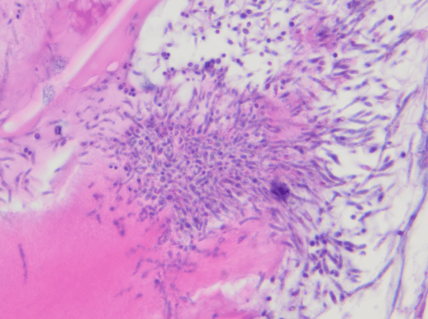
The patient is a 54 year old woman who presented to the hospital after a fall, which revealed a pathologic fracture of T1 and a spinal lesion from C6/C7 to T2. CT of the chest/abdomen and pelvis at the time showed a large mass in the anterior mediastinum with extensive lymphadenopathy and lytic lesions in the spine and ribs.
C7-T1 Soft Tissue Excision
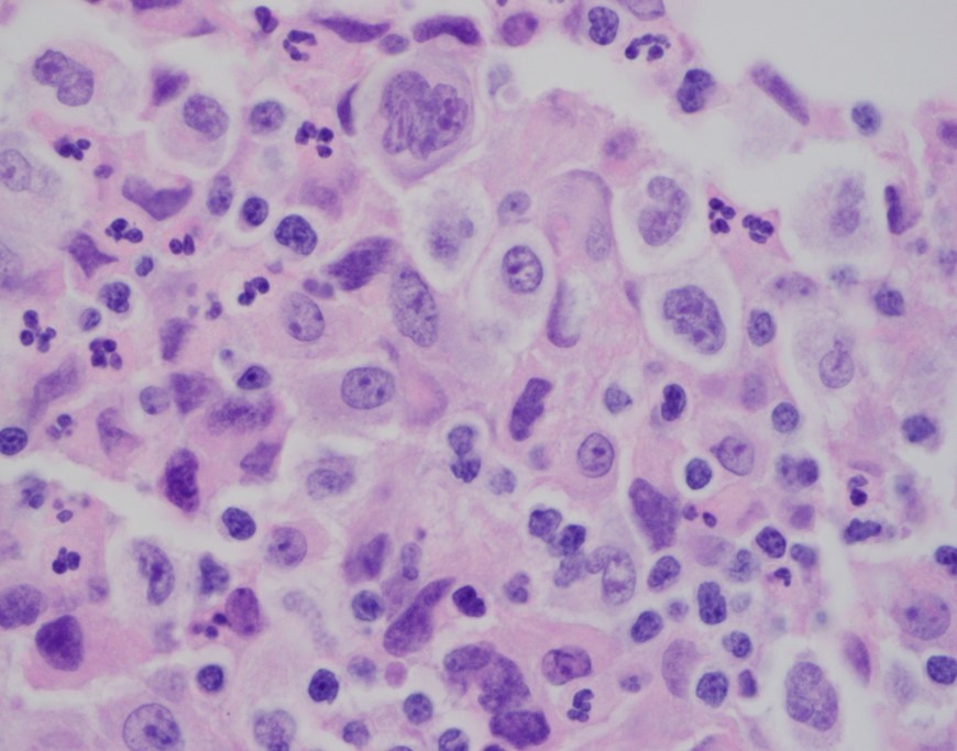
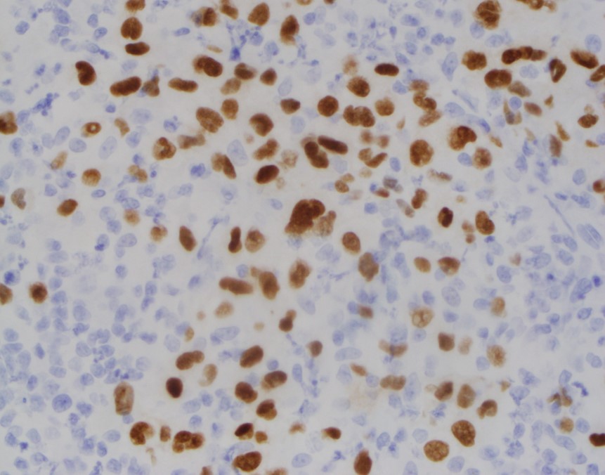
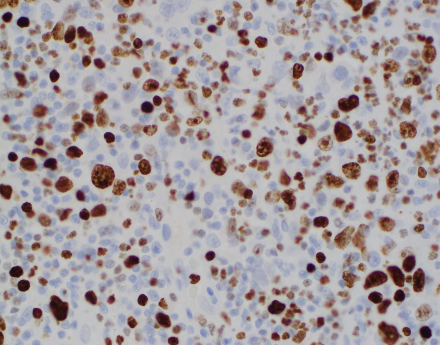
H&E 20X H&E 50X H&E 100X CD30BSAP/PAX5KI-67
Diagnosis
Sections show sheets of large epithelioid-like cells with segmented nuclei with variably prominent nucleoli and ample amounts of eosinophilic cytoplasm.A majority of these larger cells have abundant cytoplasm and lobulated nucle iwith multiple nucleoli and a surrounding halo. They are consistent with Lacunar cells. These cells form large aggregates and are admixed with numerous neutrophils, histiocytes and scattered lymphocytes.
Occasional Hodgkin cells and multi-nucleated Reed-Sternberg cells are seen, as well as scattered medium size hyper chromatic cells with irregular nuclear contours and scant cytoplasm consistent with mummy cells.
Immunohistochemical staining revealed that the largea typical cells are immunoreactive for CD30, CD15 and PAX5/BSAP. CD45 highlighted background lymphocytes but showed infrequent dim staining in the large atypical cells. By Ki-67, the proliferation index is 50-60% in the large atypical cells. Taken together, the findings are consistent with Classic Hodgkin Lymphoma, nodular sclerosis, syncytial variant.
Discussion
Classic Hodgkin lymphoma (CHL) has four distinct subtypes including nodular sclerosis, lymphocyte-rich, mixed cellularity and lymphocyte-depleted. These subtypes differ based on characteristics of the background non-neopalastic reactive cells and the histomorphology of the Hodgkin/Reed-Sternberg cells (HRS). Nodular sclerosis Classic Hodgkin lymphoma accounts (NSCHL) for approximately 70% of all CHLs. The mediastinum is the most commonly involved site and it generally occurs in people between the ages of 15-34 years old. Generally, the histology shows nodules with surrounding fibrosis. There are a variable number of Hodgkin/Reed-Sternberg (HRS) cells mixed with other inflammatory cells. The characteristic HRS cell is called a lacunar cell. This is a type of HRS cell with more cytoplasm, less prominent nucleoli and can show retraction of the cytoplasm in formalin-fixed tissue that gives the cell a halo or “lacunae.”1
The syncytial variant (SV) of CHL, nodular sclerosis was first described in the 1980s. It presents in 5-15% of cases of NS CHL. It is characterized by sheets and clusters of “lacunar cells” typical of the type of HRS cell most commonly seen in NS CHL. Previous studies had determined the SV of CHL to have a worse prognosis and more aggressive course than other subgroups. In a more recent study by Sethi, et. al. the clinical features and response to treatment of patients with SV were compared to patients with typical NS CHL. Within the cohort, 43 patients with SV were compared to 124 patients with typical NS CHL. The study found that there was no significant difference in age, sex, performance status, stage, bulky disease, number of nodal sites and chemotherapy regimens used between the two groups.2
As far as treatment outcomes, the rate of complete response in the SV group was 74% vs. 87% in the NS group. This result approached statistical significance with a p=0.05. The medium progression-free survival in the SV group was significantly shorter compared with the NS group. The overall survival, however was not statistically different, indicating that salvage chemotherapy was ultimately able to match the clinical outcomes for patients with SV type to patients with NS type. 2
Currently, all CHLs are treated with adriamycin, bleomycin,vinblastine, decarbazine (ABVD) chemotherapy regimen plus or minus radiation therapy regardless of subtype. Patients with relapsed or refractory disease are treated with a “salvage” chemotherapy regimen followed by an autologous stem cell transplant. Emerging therapies including PD-1 inhibitor nivolumab have shown great effect in patients with CHL. PD-1 or programmed death ligand 1 is overexpressed on HRS cells. This ligand binds with receptorson T-cells to prevent the T-cell immune response and reduce cytokine activation and targeted response against the proliferating HRS cells. By using an antibody against the PD-1 ligand in CHL,the ability of the tumor to suppress the immune response is decreased and patients have been shown to have better clinical response rates.3
Patients with SV do need to be recognized as a distinct subgroup that may have a higher risk of disease progression with first line chemotherapy agents. Due to the high numbers of HRS cells seen in patients with SV and the increased failure rate of initial chemotherapy agents, antibody therapies such as PD-1 inhibitors may be even more successful in those patients.
References
Swerdlow SH, Campo E, Harris NL, et al. WHO Classification of Tumours of Haematopoetic and Lymphoid Tissues (Revised 4thedition). IARC: Lyon 2017.
Sethi, T.K., et al. Differences in Outcome of Patients with Syncytial Variant Hodgkin Lymphoma (HL) Compared with Typical Nodular Sclerosis HL. Blood. 2015;126(23),1441. Retrieved from http://www.bloodjournal.org/content/126/23/1441.
Bond DA, Alinari L. Emerging treatment options for the management of Hodgkin’s lymphoma:clinical utility of nivolumab. J Blood Med. 2017;8:41-54. Published 2017 May 11. doi:10.2147/JBM.S117452.
–Chelsea Marcus, MD is a third year resident in anatomic and clinical pathology at Beth Israel Deaconess Medical Center in Boston, MA and will be starting her fellowship in Hematopathology at BIDMC in July. She has a particular interest in High-grade B-Cell lymphomas and the genetic alterations of these lymphomas.
A 44 year old male presented to the emergency department with severe, throbbing back pain in his mid-thoracic spine. He states the pain began a couple weeks ago and noted no recent fevers or night sweats, but does admit to chills. His past medical history is significant for end stage renal disease requiring dialysis, insulin dependent diabetes mellitus, and multiple amputations. On physical examination, there was tenderness to palpation along spine in mid-thoracic region. Lab work showed a normal white blood cell count, C reactive protein of 0.90 mg/dL (0.00 – 0.50 mg/dL), and an erythrocyte sedimentation rate of 60.0 mm/hr (0.0 -10.0 mm/hr). MRI of the spine was consistent with discitis and osteomyelitis at T7-8 with compression fractures causing spinal stenosis and cord compression. Given the concern for an infection process, blood cultures were collected and interventional radiology performed a bone biopsy. The specimen was sent for bacterial, fungal, and AFB cultures as well as for histology.
Laboratory Identification
Image 1. Discrete, creamy colonies growing on blood agar (left) and Sabouraud dextrose agar (right) after 48 hours of incubation at 35°C. Image 2. Fragments of bone with focal necrosis, foci of acute and chronic inflammation with clusters of yeast forms and pseudohyphae consistent with a Candida spp. infection.
The organism grew as discrete, creamy colonies growing on blood agar and Sabouraud dextrose agar after 48 hours of incubation at 35°C and resembled a yeast. MALDI-TOF mass spectrometry identified the isolate as Candida parapsilosis. Similarly, the surgical pathology specimen showed necrotic bone with inflammation and yeast forms and pseudohyphae consistent with a Candida spp. infection. Blood cultures were negative. On chart review from an outside hospital, it was discovered the patient had an episode of candidemia ten months ago which was thought to be related to his dialysis port.
Discussion
Yeasts are ubiquitous in the environment and make up the normal microbiota of human skin as well as the oral cavity, gastrointestinal tract and genitourinary tract. In general, when Candida spp. cause infections it is thought to an opportunistic infection acquired endogenously and due to exposure to prolonged antibiotics, suppressed immune system, or as a result of intravascular catheters. Those with diabetes mellitus, mucositis, bowel perforations, and intravenous drug users are most susceptible. Infections with Candida parapsilosis are becoming more common, and have the potential to cause invasive disease, such as fungal endocarditis and severe infections in the neonatal population.
In the microbiology laboratory, C. parapsilosis grows rapidly as discrete, creamy colonies on a variety of agars. On cornmeal-Tween 80 agar, C. parapsilosis grows as short, curved pseudohyphae with blastoconidia arranged singly or in small clusters at points of constriction. The arrangement is sometimes described as resembling a sage bush. C. parapsilosis is germ tube negative and is negative for urease. In many laboratories currently, identification is achieved by automated methods, such as Vitek 2, or mass spectrometry, allowing for more rapid and accurate identification.
Anti-fungals, such as echinocandins, azoles, and amphotericin B, are all potential therapeutic options to treat C. parapsilosis infections. CLSI C.parapsilosis specific breakpoints exist for fluconazole, voriconazole,micafungin, caspofungin, and anidulafungin in the M27-S4. Susceptibility testing should be performed on significant isolates from normally sterile sites.
In the case of our patient, infectious disease was consulted and he was started on IV micafungin and then transitioned to oral fluconazole. He had a transesophgeal echo and eye exam performed to ensure he didn’t have endocarditis or an invasive eye infection due to hematogenous spread of the yeast. He was discharged home on long term oral fluconazole.
-Rim Alkawas, MD, is a second year Anatomic and Clinical Pathology resident at the University of Mississippi Medical Center.
-Lisa Stempak, MD, is an Assistant Professor of Pathology at the University of Mississippi Medical Center in Jackson, MS. She is certified by the American Board of Pathology in Anatomic and Clinical Pathology as well as Medical Microbiology. She is the Director of Clinical Pathology as well as the Microbiology and Serology Laboratories. Her interests include infectious disease histology, process and quality improvement, and resident education.
In the previous 2 blog posts we discussed how to prepare for your inspection, and what to expect during the inspection itself. In the last of our 3 part series on regulatory inspection preparedness, today we’ll be covering what to do after the inspection ends.
Throughout the inspection itself, the inspectors should be communicating any issues or citations they uncover; ensure that your management staff is taking notes on any of these potential findings. Based on these notes, you should start working to address and correct any issues right away. Formal documentation regarding the nature of any official citations can take several weeks to receive back, depending upon the regulatory agency performing the inspection. Waiting for the formal report to begin making corrections will reduce the time you have to form a plan of correction, and can further impact patient care depending upon the citation received.
Have a Plan. Draft a spreadsheet to record: 1) each issue identified, 2) laboratory department(s) it was found in, 3) associated risk factor (patient care or safety issues = 1, regulatory requirements = 2, recommendations = 3), 4) staff member assigned to investigate and correct the issue, 5) due date for investigation response, and 6) status of the investigation (in progress, on hold, completed). Share this spreadsheet with your management team, and review at weekly/monthly staff meetings for updates on progress completion.
Risk 1 Issues. The safety of your patients and staff, along with ensuring accuracy in testing results is the number one priority of a laboratory. If the inspectors identified any weaknesses in these areas, they should be addressed first. This would include items such as staff not adhering to required safety precautions, not following manufacturer requirements for quality control testing or instrument maintenance/calibration, lack of follow-up for QC or proficiency testing failures, along with any other finding which questions the integrity and accuracy of the testing being performed.
Risk 2 Issues. Double check the regulatory standard to ensure you fully understand the requirements, and that you have appropriate evidence of compliance. As the testing activity menu and complexity of testing increases, the amount of documentation requirements can increase as well. Even with a paperless system, it is easy to overlook a signature of review or checkmark on a log. “If it’s not documented, it wasn’t done.” For simple administrative oversights, review your current processes to identify any gaps or areas that can be improved upon to ensure all documentation is properly filled out each month. If the inspectors noted a discrepancy between your current policy and how staff are actually performing a test, review the testing process to see where the true discrepancy is – is the policy outdated and needs to be revised, or do staff need to be retrained on the current policy with competency assessed for compliance?
Risk 3 Issues. Inspections are a great opportunity for further education for all those involved, both the inspector and staff being inspected as well. For some regulations, there is no one set way that must be followed in order to demonstrate compliance with a requirement. Hearing how someone else is meeting the requirements may spark an innovative idea from your own staff on how your current processes can be improved. Be open to hearing new ideas, and find ways to implement those which you feel would be successful at your institution.
Evaluate All Sections of the Lab. When investigating a finding in one laboratory department, ensure that any process improvements are shared across all areas of the lab. Just because microbiology didn’t get caught with expired reagents like hematology did this inspection, doesn’t mean that they aren’t at risk for future inspections.
Focus on the Positives. Congratulate and recognize your staff on their successes in the areas you performed exceptionally well in. It’s a joint effort to ensure the lab is inspection ready; be sure to pass along any compliments received throughout the inspection process to all levels of staffing. Focus on what you’re doing well and how you can continue to maintain those processes and implement them in additional areas.
A little bit of preparation ahead of time will make the inspection process smoother and less stressful for all involved. When viewed as a learning experience and opportunity for improvement rather than a visit from the “lab police”, laboratory inspections can be a useful tool to confirm the quality of your overall laboratory program.
-Kyle Nevins, MS, MLS(ASCP)CM is one of ASCP’s 2018 Top 5 in the 40 Under Forty recognition program. She has worked in the medical laboratory profession for over 18 years. In her current position, she transitions between performing laboratory audits across the entire Northwell Health System on Long Island, NY, consulting for at-risk laboratories outside of Northwell Health, bringing laboratories up to regulatory standards, and acting as supervisor and mentor in labs with management gaps.
Leaders’ decisions and actions have moral ramifications, both on an individual and an organizational level. There are three factors of moral development, namely moral capacity, moral courage, and moral resiliency.
Being able to recognize a dilemma as a moral issue is one of the critical aspects of leadership. Such awareness is referred to as moral capacity, which influences the characteristics and recognition of a moral issue. There are multiple aspects that influence a leaders’ moral capacity. The first aspect is their previous experiences with moral dilemmas and how much they learned from them. The second is to what extent a leader is able to see and understand the multiple perspectives of an issue. The third is how leaders view their role and whether or not that incorporates a moral view.
It is not enough to understand and recognize a moral dilemma, it is important to act on it. Such moral courage is especially important when under pressure to act immorally. Such pressure can come from peers, supervisors, or the entire organization. Therefore, the more moral courage someone has, the more likely it is that they take a moral action or make a moral decision. One important aspect of moral courage is the notion of willpower. Willpower is a muscle that people can practice with small tasks, such as drinking a glass of water before breakfast. The more people practice it on small tasks, the more likely they are to use it during challenging situation, such as making a moral decision when pressured to do otherwise.
Moral resiliency is an extension of moral courage. While moral courage focuses on the strength to make moral decisions in the short-term, moral resiliency is a process through which leaders continuously adapt their moral compass and actions. Moral resiliency is this what creates sustainable moral decision-making.
Depending on how leadership effectiveness is defined, moral behavior can either make leaders more or less effective. When looking at effectiveness in the short term, it is possible that moral behavior can impede effectiveness if measured in terms of money or short-term success. However, when looking at effectiveness in the long term, moral behavior increases leaders’ effectiveness. The more honest, and thus morally, people behave, the more effective they are. In a world that is becoming more globalized, it is critical for leaders to understand that culture influences moral and ethical behavior. In other words, what is moral in one culture might be immoral in another. To increase leaders’ effectives it is therefore important to understand the cultural implications of behavior and to be aware of the differences in appropriate and effective behavior.
-Lotte Mulder earned her Master’s of Education from the Harvard Graduate School of Education in 2013, where she focused on Leadership and Group Development. She’s currently working toward a PhD in Organizational Leadership. At ASCP, Lotte designs and facilitates the ASCP Leadership Institute, an online leadership certificate program. She has also built ASCP’s first patient ambassador program, called Patient Champions, which leverages patient stories as they relate to the value of the lab.
#PathTweetAward is a crowdfunded award created
on Twitter by pathologists to recognize pathologists and pathology trainees who
post exemplary educational tweets in the field of anatomic or clinical pathology.
How and when
was #PathTweetAward started?
The idea for an “educational pathology Tweet ofthe year” award was proposed on Twitter on April 6, 2018.
The tweet that launched the concept of #PathTweetAward.
The hashtag#PathTweetAward was created the next day based on a suggestion by Dr. Amy Deeken (@AmyHDeekenMD), a pathologist from Ohio who is active on social media. A Twitter handle (@PathTweetAward) and a promotional video to create awareness about the award were created shortly thereafter by Dr. Muhammad Ahsan (@ahsanuitis), a pathologist from Pakistan. The award was supported online by several pathologists with a prominent social media presence, including Dr. Jerad Gardner, Dr. Kamran Mirza, Dr. Christina Arnold, Dr. Julie Teruya-Feldstein and Dr. Kalyani Bambal.
What is
crowdfunding? How was #PathTweetAward crowdfunded?
Crowdfunding is a fundraising method in which small amounts of money are contributed by a large number of people, typically via the internet. Crowdfunding for #pathtweetaward was made possible by Dr. Amy Deeken, who started up a GoFundMe account for the award on April 7, 2018 (gofundme.com/5dakxso) and created a promotional video. Incredibly, the fundraising goal of $1000 was reached within a single day of setting up the GoFundMe account. At the time of this writing, the account has raised $1550, generously donated by 26 individuals (predominantly pathologists) over a period of 6 months.
The GoFundMe account that funds #PathTweetAward.
Who is
eligible for the award?
Pathologists in practice, pathology residents,
and pathology fellows anywhere in the world are eligible for the award. To be
considered, a tweet with educational value should be posted on Twitter and
brought to the attention of the screening judges by using the hashtag
#PathTweetAward.
Who is
allowed to use the hashtag #PathTweetAward?
Anyone can use the hashtag. Any pathologist anywhere in the world can tag an educational post on Twitter in a reply or retweet. An example is shown in the image below. Pathologists can also self-tag their own tweets if they wish.
To nominate a tweet for the award, simply reply to the original tweet and type #PathTweetAward. You can also “retweet with comment” and type #PathTweetAward in your comment.
Are the
awards monetary?
Yes, the current plan is to award four
monetary prizes each year ($500, $300, $200 and $100), including two awards in
the open group and two awards for trainees. The
Pathologist magazine and the American Society for Clinical Pathology (ASCP)
have generously offered additional non-monetary support, mainly in the form of promotion
of award-winning tweets and awardees.
How are
winners selected?
Judges for #PathTweetAward are pathologists who are active on Twitter. There are three panels of judges, selected with diversity and inclusion in mind; judges include women and men, trainees and faculty, community pathologists and academics. A different panel of judges will be selected each year. The bulk of the work is shouldered by two panels of five screening judges each. One, led by Chicago hematopathologist Kamran Mirza, MD, PhD,screens tweets from trainees only. The other, led by Canadian head and neck pathologist Bin Xu, MD, screens tweets from an “open” group that includes trainees and practicing pathologists.
Screening
judges (for trainee tweets only)
Screening
judges (for “open” category, including all pathologists, including trainees)
Final
judges
Kamran
Mirza, MD (group leader)
Bin Xu, MD (group leader)
Jerad
Gardner, MD (group leader)
Elvira
Gonzalez-Obeso, MD, PhD
Eman
Aljufairi, MD
Silvija
Gottesman, MD
John
Gross, MD
Daniel
Skipper, MD
Valerie
Fitzhugh, MD
Adam L. Booth,
MD
Yiang Hui,
MD
Laura G.
Pastrian, MD
Pallavi A.
Patil, MD
Chen Yang,
MD
Geronimo
Junior, MD
Table 1. Judges for #PathTweetAward (2018)
Each week, tweets tagged with the hashtag #PathTweetAward are collated by a screening judge and posted publicly on Twitter using the “Moments” feature. These collections serve as a repository of the best tweets, from which each panel of screening judges selects a “tweet of the month” to be sent at the end of the year to the final panel of judges.The latter are tasked with selecting four final prize-winners, two from the open group and two from the trainee-only group. The final four shortlist will be posted publicly in a Twitter poll, at which time the general public can vote to determine the final ranking. The process, summarized by screening judge Dr.Elvira Gonzalez-Obeso, is shown in this image:
Summary of the process by which #PathTweetAward winners are selected.
-Sanjay Mukhopadhyay, MD is a Staff Pathologist at the Cleveland Clinic in Cleveland, OH. He is an expert thoracic pathologist,author of a lung pathology textbook, and a pioneer in the use of social media for teaching lung pathology globally, free of charge. Follow Dr. Mukhopadhyay on Twitter @smlungpathguy, and check out his freely accessible educational videos on YouTube.
Platelets are our first line of defense in controlling bleeding. Abnormally low numbers of platelets can lead to easy bruising, tiny leaks from capillaries into the skin and mucous membranes, causing petechiae, and bleeding. The platelet count is a significant parameter in the CBC and it is therefore vital to be able to report accurate and precise platelet counts. Furthermore, physicians must be able to use this information to diagnose the cause of the thrombocytopenia in order to recommend treatment.
What a platelet count alone cannot tell us is the reason for thrombocytopenia. Just as there can be many reasons for a low hemoglobin, and many causes for an increased or decreased WBC, there are numerous causes for a decreased platelet count. After ordering a CBC, the next steps in determining etiology of thrombocytopenia have historically been a thorough physical with attention to any bleeding symptoms and organ enlargements, and a medical history. The medical history should include family history, and notation of recent viruses or drug therapies. After these tests, a bone marrow aspirate and biopsy may also be necessary to clarify etiology. While modern, automated hematology analyzers produce reliable platelet counts, measuring only the circulating platelet count does not give us any information as to the etiology, so there is a need for further testing. With thrombocytopenia, platelet counts can be less reliable than with normal counts.Platelet counts were originally performed by impedance methods, then better accuracy and precision was obtained with optical platelet counts. Physicians rely on precision with very low platelet counts to make informed decisions about when to transfuse patients. The problem with the impedance counts at the low end, is that RBC fragments, schistocytes and microcytic RBCs can be counted as platelets, giving a falsely high count. On the other hand, measuring platelets by size can miss large platelets leading to a falsely low count.
Historically, the mean platelet volume (MPV) has been used along with the platelet count to aid in making a differential diagnosis. The MPV is analogous to the red cell distribution width (RDW) for red cells, and can be used to as an indicator of the maturity of platelets. Young platelets are the largest, and as they age, the size decreases. The normal ranges for MPV are generally about 9-12 femtoliters (fl).The MPV will be higher if more platelets are being released from the bone marrow, and lower if fewer are being newly released and we are counting mature platelets. Thus, the MPV can be used as an indirect marker for platelet production. However, an inherent problem with the MPV is that, similarly to the impedance platelet count, this count can be unreliable because any RBC fragments or particles may interfere with the measurement.
So, what is a physician to do?And how can the lab provide information to help them make the best differential diagnosis and transfusion decisions? In an effort to provide a parameter that could help differentiate causes of thrombocytopenia, the concept of reticulated platelet counts (retPLT) was first introduced in research in the late 1960s. The term is used to describe immature, functional platelets in the peripheral blood.Reticulated platelets are to mature platelets as reticulocytes are to mature red blood cells. These are the youngest platelets, within 24 hours of being released from the bone marrow. Reticulated platelets are large, with increased amounts of RNA, and the number in the circulation can be used to provide an estimate of the rate of thrombopoiesis. Originally, these were stained with new methylene blue and manual counts were done, much like a manual reticulocyte count; tedious,and imprecise. It wasn’t until about 30 years later that a flow cytometry method was described for measuring retPLT. Using traditional flow cytometry, reticulated platelets can be stained with a Thiazole Orange dye and passed through a flow cytometer. This method, however, has been shown to have wide normal ranges from 1-15% because of the lack of analytical standardization. Variations in the concentration of the thiazole dye used, the timing, and the gate settings all make it difficult to compare results obtained from one laboratory to another. In addition, flow cytometry is time consuming, labor intensive and costly.
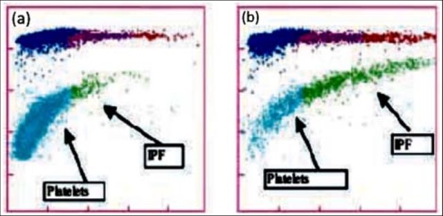
Newer flow cytometry methods are now available on select hematology analyzers. There are currently 2 companies that have analyzers that can report retPLT using routine CBC reagents and controls. Reticulated platelets can be measured with the same K2 EDTA tube used for the CBC. The test is automated, simple to perform, fast, and gives standardized results with tighter normal ranges. The Abbott CELL-DYN Sapphire measures the retPLT using a fluorescent dye and flow cytometry with 2 dimensional gating. Sysmex XE and XN analyzers offer several Advanced Clinical Parameters including measures of reticulated platelets, expressedas the Immature Platelet Fraction (IPF%) and the Absolute Immature Platelet Fraction Count (IPF#). Sysmex offers a fluorescent platelet count (PLT-F) as an addition to impedance counting (PLT-I) and optical counting (PLT-O). PLT-F is more reliable because it uses a platelet specific dye which eliminates noninterference seen with other methods. The fluorescent dye labels the RNA and forward scatter is used to determine size while fluorescence is used to measure RNA content. With gating set based on cell volume and RNA content, the PLT-Fcan be measured. When there is an abnormal scattergram or a low platelet count,the PLT-F is reflexed and the IPF% and IPF# are also reported.
What’s the clinical utility of the IPF? Thrombocytopenia can have many causes.Immature platelets are functioning platelets, and an increased IPF means that we have more newly formed immature platelets circulating. The IPF helps physicians to differentiate thrombocytopenia caused by platelet destruction or consumption versus thrombocytopenia caused by deficient platelet production in bone marrow failure. It is vital to know the pathogenesis of thrombocytopenia in making decisions about treatment. With these advanced parameters, these decisions can often be made without costly, time consuming flow cytometry,without an invasive bone marrow biopsy and without waiting for the results of such biopsy. This can often save a patient an unnecessary platelet transfusion.
The reference range for IPF% in healthy individuals is1.0-7.0%. Together with a low platelet count, an increased IPF indicates an increase in platelet production. This is seen in patients with excessive destruction of platelets. An example of the clinical utility of the IPF can be seen in the diagnosis of immune thrombocytopenic purpura (ITP). ITP is an autoimmune bleeding disorder in which the immune system makes anti-platelet antibodies which destroy platelets. Acute forms occur more often in children while adults can have chronic ITP. ITP can be diagnosed on clinical findings but laboratory confirmation is often necessary. This can be expensive with long turnaround times using traditional flow cytometry and/or bone marrow aspirates.An IPF reported with a CBC is fast, inexpensive, and be extremely beneficial in aiding a timely diagnosis. Patients with ITP have been shown to have the consistently highest IPF values with ranges from 7-28%.1 As their platelet counts recover, the IPF% returns to the normal range, without the need for transfusions. Thus, the IPF can be used not only to help diagnose but also as an indicator of remission.
Figure1. Platelet scattergrams from a healthy individual with a normal IPF (a) and a patient with a high IPF (b). Mature platelets appear as blue dots, green dots represent the IPF with increased cell volume and higher fluorescence intensity compared to mature platelets1
In contrast to what we see with ITP, thrombocytopenia with alow normal or decreased IPF indicates decreased bone marrow production of platelets. Patients with bone marrow failure are more likely to have bleeding episodes with low platelet counts and may need transfusion. Rapid differential diagnosis using the IPF can help physicians help these patients get early treatment.
IPF may also be a reliable indicator of bone marrow recovery. Traditionally, neutrophil counts have been used as an indicator of recovery after a bone marrow transplant. IPF can be used as an indicator of imminent platelet recovery. It has been shown that,post-transplant, the IPF% increases before the platelet count. In a study done with stem cell transplant patients, it was shown that the absolute neutrophil count took an average of 13 days to recover, compared to 9 days for the IPF. The IPF was shown to recover before the Immature reticulocyte count, platelet count and absolute neutrophil count, giving physicians earlier indication that the transplant was successful.2 This is significant because it can eliminate the need for bone marrow biopsies and platelet transfusions.
Thrombocytopenia is not an uncommon finding in neonates, particularly in the neonatal intensive care unit (NICU). There are various causes for this, including sepsis, placental insufficiency and immune thrombocytopenia. The IPF% and IPF# can be used to diagnose and distinguish the cause of thrombocytopenia in neonates, and direct the treatment. When platelet count platelet count drops below 50 x 103/Lin an otherwise healthy appearing infant in the first 72 hours of life, neonatal alloimmune thrombocytopenia (NAIT) can be suspected. This condition is similar in pathogenesis to hemolytic disease of the fetus and newborn (HDFN), and is caused by an incompatibility in human platelet antigens between mother and baby. This occurs most often when the mother is HPA-1b and the father and baby are HPA-1a. The mother forms anti-HPA-1a which crosses the placenta and destroys the fetus’ platelets.This is a thrombocytopenia caused by platelet destruction, and the IPF% is high. The condition is self-limiting and resolves in 1-4 weeks. Neonatal sepsis can also present with a high IPF, but typically is found in very sick or premature babies and the degree of thrombocytopenia is not as severe as with NAIT. In contrast, neonatal thrombocytopenia due to placental insufficiency would exhibit a decreased IPF due to a deficiency in platelet production. Using the IPF% and IPF# to help differentiate the causes of neonatal thrombocytopenia can help steer the treatment and save infants from unnecessary invasive procedures and transfusions.
TheIPF has proved to be very valuable in the clinical setting. It has been used in the investigation of etiology in secondary thrombocytopenias due to chronichepatitis C, liver disease and HIV. It has been used to guide treatment in thrombocytopenias such as thrombotic thrombocytopenic purpura (TTP). IPF can also be useful in evaluation of hereditary platelet thrombocytopenias. The IPF% and IPF# can be compared after transfusion to support the theory that, after platelet transfusion, theIPF% will decrease due to the newly increased platelet count, but the IPF#remains the same. This validates that the IPF is a reflection of continual platelet production by the bone marrow.4
IPF%and IPF# are expanded CBC parameters that physicians can use to aid in differentiation of various thrombocytopenic states. Treatment for the different classes of thrombocytopenia can differ drastically, and knowing the class of thrombocytopenia helps direct the management. The IPF parameters are automated,easy to perform at the same time as the CBC, and provide standardized results that are inexpensive and available 24 hours a day in the hospital setting. Using the IPF can also reduce diagnostic costs for the patient. Many studies have been conducted on the varied applications of the IPF and research continues investigating possible further uses of this advanced clinical parameter. This is the new hematology, constantly providing the clinician with better tools for making diagnoses and treating patients. Platelet counts alone and MPVs are out. Make room for the new kid on the block; the IPF is in.
References
Arshi Naz et al. Importance of Immatureplatelet Fraction as a predictor of immune thrombocytopenic purpura. Pak J MedSci 2016 Vol 32 No 3:575-579
Zucker ML et al. Immature Platelet fraction asa predictor of platelet recovery following hematopoietic progenitor celltransplanatation. Lab Hematol 2006 12(3):125-30
Briggs,C. Assessment of an immature plateletfraction (IPF) in peripheral thrombocytopenia. Br J Haematol 2004Jul;126(1):93-9
Sysmex White Paper. The role of the ImmaturePlatelet Fraction(IPF) in the differential diagnosis of thrombocytopenia. www.sysmex.com/us
Fujii,T et al.. A new approach to detectreticulated platelets stained with thiazole orange in thrombocytopenicpatients. Thromb Res. 2000 Mar 15;97(6):431-40
Cremer Malte The immature platelet fraction(IPF) in neonates. Diagnostic Perspectives 2011 Vol1:36-42
Cremer M. et al. Immature platelet values indicateimpaired megakaryopoietic activity in neonatal early-onset thrombocytopenia.Thrombosis and Haemostasis 2010; May;103(5):1016-21
-Becky Socha, MS, MLS(ASCP)CM BB CM graduated from Merrimack College in N. Andover, Massachusetts with a BS in Medical Technology and completed her MS in Clinical Laboratory Sciences at the University of Massachusetts, Lowell. She has worked as a Medical Technologist for over 30 years. She’s worked in all areas of the clinical laboratory, but has a special interest in Hematology and Blood Banking. When she’s not busy being a mad scientist, she can be found outside riding her bicycle.
A young adult female presents to an urgent care clinic with the chief complaint of a “bump and surrounding redness” on her right medial thigh. The patient reports the bump had been present without change for 1 year. Approximately 2 days prior to presenting at the urgent care clinic the patient states she nicked the bump while shaving, and subsequently the bump became tender with surrounding erythema and produced purulent drainage. The patient denies any similar prior lesions and denies any significant past medical history. On physical exam, the lesion measured 1 cm with the surrounding erythema measuring 5cm. The urgent care physician performed an incision and drainage and noted a scant amount of white purulent material within the lesion. A cyst wall was identified and was removed as much as possible. A swab of the purulent material was collected and submitted to microbiology for culture.
Laboratory Identification
The primary gram smear of the swab specimen was interpreted as no bacteria or polys seen. Routine culture media including blood, chocolate, MacConkey, and CNA agar were inoculated and incubated aerobically. Following incubation, the blood agar showed few gram positive cocci consistent with usual skin flora and few single morphology of medium to large sized gray colonies without hemolysis. On the MacConkey agar, few single morphology non-lactose fermenting colonies were identified. The gray colonies identified on the blood agar gram stained as gram negative bacilli with unremarkable morphology. An oxidase test was performed and the bacteria was found to be oxidase positive. The key biochemical and physiologic characteristics of the isolate included: good growth on thiosulfate citrate bile salts and sucrose (TCBS) agar with yellow colonies, good growth in 6% NaCl nutrient broth, and no growth in 0% NaCl nutrient broth. The organism was identified by matrix-assisted laser desorption/ionization time of flight (MALDI-TOF) as Vibrio alginolyticus.
Image 1. Blood agar isolate of medium sized gray colonies.
Image 2. MacConkey agar with non-lactose fermenting colonies.
Discussion
Vibrio spp. are water organisms commonly found in marine or brackish water environments. These organisms are gram negative bacilli which classically have “comma” shaped morphology on gram smear, though this is not an absolute. On sheep blood agar, these organisms are variably beta hemolytic medium to large gray colonies and on MacConkey agar are non-lactose fermenting (with the exception of Vibrio vulnificus). Vibrio spp. are oxidase positive, ferment glucose, and readily grow on most isolation media with growth being enhanced with the addition of 1% NaCl to the media. The growth characteristics on media containing different concentrations of NaCl can be used in differentiating the different Vibrio spp. Thiosulfate Citrate Bile Salts and Sucrose (TCBS) agar is both selective and differential for Vibrio spp. with sucrose fermentation being detected as yellow colony formation.
Vibrio angiolyticus typically causes extraintestinal infections, with wound infections and otitis externa being the most frequent. Transmission frequently occurs via traumatic aquatic injuries in contaminated water. Vibrio angiolyticus rarely causes intestinal disease and is isolated in less than 5% of stool cultures in patients with Vibrio associated diarrhea. Growth characteristics of Vibrio alginolyticus include yellow colonies on TCBS due to its ability to ferment sucrose and good growth on 6% NaCl and no growth on 0% NaCl. Additional key biochemical characteristics of Vibrio alginolyticus include oxidase positivity, nitrite positivity, negative for myo-Inositol fermentation, negative for arginine dihydrolase, positive for lysine decarboxylase, and variable positivity for ornithine decarboxylase. Most wound infections due to Vibrio alginolyticus are non-severe, and most mild infections will clear without antibiotic therapy.
References
Procop GW, Koneman EW. Koneman’s Color Atlas and Textbook of Diagnostic Microbiology, North American Edition. LWW; 2016.
Morris, J., Calderwodd, S., and Bloom, A. Minor Vibrio and Vibrio-like species associated with human disease. In: UpToDate, Post, TW (Ed), UpToDate, Waltham, MA, 2017.
-Justin Rueckert, DO is a 3nd year anatomic and clinical pathology resident at the University of Vermont Medical Center.
-Christi Wojewoda, MD, is the Director of Clinical Microbiology at the University of Vermont Medical Center and an Associate Professor at the University of Vermont.
While Maria was working in Microbiology, she cut her finger while pulling reports off of the printer. It was a minor paper cut, so she ignored it, put her gloves on and continued her work back on the bench. A week later, the tiny cut was swollen and red. She decided to report the incident to her manager since it wasn’t healing. The manager asked Maria to report to the Occupational Health department, but was unsure if any treatment would be covered since the incident was not reported while she was at work.
Steve and Josh were bored during the night shift and they created a ball made from rubber bands to toss around. When Josh didn’t catch the ball, it hit the open tray of formaldehyde on the gross bench, and it splashed into Josh’s eye. He rinsed his eyes in the eyewash station for a couple of minutes, but both men were afraid to report the incident for fear of getting in trouble. Josh’s eye irritation continued to worsen, and he had to go to the eye doctor for treatment.
There are obvious consequences for injuries that occur in the laboratory, and reporting them is important for many reasons. Staff may be motivated in some instances to not report, but that creates problems for the individual, the department, for the facility, and even for other labs across the country! That may seem like a stretch, but it will become clearer with exploration.
The value in injury and accident reporting starts with medical follow-up. Those incidents which require treatment or abatement of infection can and should be dealt with quickly, and appropriate monitoring can be done if necessary. Some injuries may require immediate first aid, and a trip to the emergency department may even be necessary. Not reporting those types of injuries can be very dangerous for staff. Other incidents may require physician office visits or other monitoring, and employees who need it should be encouraged to comply.
In many work places the injury follow-up visits and treatment are covered financially by the institution, either via a structured occupational health program or through reimbursement. Some organizations may not offer financial coverage, however, if the incident that occurred at work is not reported as soon as possible. That reporting delay can raise suspicion as to whether or not the injury actually did occur while on the job, and since the written reporting protocol was not followed, there may also be no obligation for employer medical coverage.
Departmental issues will arise when incident reporting in not part of the overall lab safety culture. Sometimes there can be reprisals for unsafe behaviors which lead to accidents, but if the safety culture is good and if managers and employees coach against such practices, then there should be fewer overall incidents to report. That said, a culture of secrecy regarding injuries or exposures can also be dangerous. There is value in talking to all staff about an incident that occurred within the department. Staff can learn from the event and have a healthy discussion about how to keep it from reoccurring. A discussion of events can bring important safety issues to light, particularly if similar incidents happen with multiple people. This sharing of information can also promote awareness of good safety practices that can aid in the prevention of further incidents for all who work in the department.
OSHA requires the reporting of certain work place injuries, those that may have led to time away from work or that need medical follow up, for example. This injury data is compiled and reported nation-wide. It becomes a good source for benchmark data, a way to be able to compare your lab injury rates with others across the country. The U.S. Bureau of Labor Statistics provides this data as information labs can use. One way to utilize the information is to see if the number of reportable injuries you are seeing in your lab is comparable to a national average. That assessment can give you a starting point in determining whether or not your lab’s safety incidents are at typical levels. Of course, lab safety professionals want to see zero injuries, but if you see your lab injury numbers are very high compared to benchmark data, you can begin to see where to focus in on fixes for the lab physical environment or on creating specific safety training.
There is great value in talking about safety incidents that may result in injury or exposure in the lab setting. These “safety stories” raise awareness of safety issues, and they can act as a deterrent for repeat incidents. Create a culture where staff feel free and comfortable to report incidents, and be sure to discuss them with all staff, and record reportable injuries as well. Having reliable national data also provides helpful information to other labs, and better information can help to improve safety in laboratories everywhere!
–Dan Scungio, MT(ASCP), SLS, CQA (ASQ) has over 25 years experience as a certified medical technologist. Today he is the Laboratory Safety Officer for Sentara Healthcare, a system of seven hospitals and over 20 laboratories and draw sites in the Tidewater area of Virginia. He is also known as Dan the Lab Safety Man, a lab safety consultant, educator, and trainer.