WATS3D Esophageal Screening for Barrett’s Esophagus

Recently, a new technology has become available for screening the esophagus for early detection of at-risk patients for cancer. Esophageal cancer has one of the highest mortality rates. Watch this interview with Rob Odze, a pathologist who works with the technology, and learn more about how this could impact the practice of clinicians, pathologists, and laboratory professionals involved in upper endoscopy services.


milner-small

-Dan Milner, MD, MSc, spent 10 years at Harvard where he taught pathology, microbiology, and infectious disease. He began working in Africa in 1997 as a medical student and has built an international reputation as an expert in cerebral malaria. In his current role as Chief Medical officer of ASCP, he leads all PEPFAR activities as well as the Partners for Cancer Diagnosis and Treatment in Africa Initiative.

Lab Value Changes in Transgender Males

For patients with gender dysphoria, the Endocrine Society has endorsed the use of hormone therapy to promote secondary sexual characteristics of the desired gender. These guidelines were first established in 2007 and revised last year, and gave the first evidence guided recommendations for clinicians treating transgender patients.

For transgender males, testosterone by itself is prescribed as an injectable oil-based solution. These doses are given as intramuscular injections- usually into the thigh. If that’s too painful, subcutaneous injections have been shown to have similar efficacy. The doses given to transgender males is much higher (50-100mg/ injection) than that given to men with testosterone deficiency (30-50 mg/ injection). Primarily because the men have more testosterone to start with. Also, whereas topical testosterone gel may be sufficient for men with “low T,” it doesn’t seem to provide enough testosterone to transgender males and is quite expensive, so it is generally not used.

Nov1.png
Image 1. Picture of testosterone cypionate vial from mcguffmedical.com. This is used for intramuscular injections.

Upon starting testosterone injections, the frequency of injections is every one to two weeks. However, the onset of physical secondary sexual characteristics takes 3-6 months to begin. After about 3 years, most of the changes to occur will have manifested. These physical changes are outlined in the table below. You’ll notice how certain traits like cessation of menses and fat redistribution start within the first 6 months whereas muscle growth and voice change take effect after 6 months. Also, the time certain effects take maximal effect varies; the voice doesn’t deepen further after 2 years, but hair growth continues to increase through 5 years.

Physical Effect Begins Maximal Effect
Facial/body hair growth 6-12 mo 4-5y
Skin oiliness/acne 1-6mo 1-2y
Scalp hair loss 6-12 mo
Increased muscle mass 6-12 mo 2-5y
Fat redistribution 1-6mo 2-5y
Cessation of menses 1-6mo
Deepening of voice 6-12 mo 1-2y

Table 1. Timeframe of physical traits that manifest in transgender males while taking testosterone hormone therapy. Based on Hembree et al. 2017 (1).


Just as hormone therapy induces physical manifestations of secondary sexual characteristics for transgender men, we would suspect that internal aspects of physiology are affected too.  Values measured by the laboratory provide meaningful insight into how our body and its different organ systems are functioning. Accordingly, the Endocrine Society also recommended laboratory monitoring of transgender patients starting hormone therapy.

  1. Measure Testosterone and hemogoblin/ hematocrit every 3 months for the 1st year, then 1-2x/ year afterwards.
  2. Monitor Lipids at regular intervals

Previous studies have monitoring these lab values found consistent increases in hemoglobin and hematocrit (2,3). This is due to the stimulation of erythropoiesis by testosterone (4).  While excessive testosterone could lead to polycythemia (excessive RBCs in the blood), it is not a commonly described complication in transgender patients. Some summary results from our study for hemoglobin and hematocrit are shown in Figure 1A, which shows a clear shift in levels.

However, reports on lipids have been varied LDL and triglyceride changes (2,3). The only consistent finding was that HDL decreased in transgender males taking testosterone (2,3). In our study, we found triglycerides were increased with decreased HDL (Figure 1B). The take-away is that because cardiovascular cut-offs are based on risk and not a reference range, patients and clinicians will have to be aware of these possible metabolic changes.

Creatinine, when it was checked, increases for transgender males (5). We found creatinine was strongly increased in our study to become similar to baseline creatinine in transgender women before taking hormone therapy (Figure 1C). This topic as it relates to glomerular filtration rate is very complex and will be discussed further in a future post.

To illustrate lab value changes in transgender men, I’ll direct you to data that I found in a large study of over 300 transgender patients including about 80 transgender men. The completed manuscript is not currently available but will be printed soon:

Nov 2

However, this does not mean Cisgender male reference intervals are adequate for transgender men. This topic needs further exploration and ideally a prospective trial to be performed in a controlled manner. A double-blind study would not be possible as it would be unethical to perform.

References

  1. Hembree WC, Cohen-Kettenis PT, Gooren L, Hannema SE, Meyer WJ, Murad MH, et al. Endocrine Treatment of Gender-Dysphoric/Gender-Incongruent Persons: An Endocrine Society* Clinical Practice Guideline. J Clin Endocrinol Metab. 2017
  2. Wierkx K, et al. Cross-Sex Hormone Therapy in Trans Persons is Safe and Effective at Short-Time Follow-Up: Results from the European Network for the Investigation of Gender Incongruence. J Sex Med, 2014. 11(8):1999-2011.
  3. Mueller A, Kiesswetter F, Binder H, Beckmann MW, Dittrich R. Longer-term administration of testosterone undecanoate every 3 months for testosterone supplementation in female-to-male transsexuals. J Clin Endocrinol Metab. 2007
  4. Paller CJ, Shiels MS, Rohrmann S, Menke A, Rifai N, Nelson WG, et al. Association Between Sex Steroid Hormones and Hematocrit in a Nationally Representative Sample of Men. J Androl. 2012 33(6): 1332-1341.
  5. Fernandez JD, Tannock LR. Metabolic Effects of Hormone Therapy in Transgender Patients. Endocr Pract. 2016;22:383–8.


SoRelle Picture

-Jeff SoRelle, MD is a Molecular Genetic Pathology fellow at the University of Texas Southwestern Medical Center in Dallas, TX. His clinical research interests include understanding how the lab intersects with transgender healthcare and advancing quality in molecular diagnostics.

Gram Stain Examination – Beyond Infectious Organisms

Case History

A 72 year old female with past medical history of stage IV ovarian adenocarcinoma treated with chemotherapy and interval debulking surgery, presented to emergency room with a one week history of confusion and worsening balance.

CT scan of the head showed new communicating hydrocephalus.  A magnetic resonance imaging couldn’t be performed initially because of patient’s uncontrolled agitation.  Lumbar puncture (LP) was performed.  Following this procedure the patient’s mental status showed some improvement and therefore neurosurgery team decided to insert ventriculoperitoneal (VP) shunt to treat her hydrocephalus and prevent recurrence of seizures.

It was Friday afternoon when a microbiology technologist brought the patient’s cerebrospinal fluid (CSF) gram stain to be reviewed.  It was confirmed that no inflammatory cells and organisms were present.  However, cells in the background looked very atypical (Image 1a, b).

CSF1
Image 1:  Gram stain of CSF showing atypical epithelial cells at (a) 40x and (b) 100x with oil.
CSF2
Image 1b.

Discussion

The gram stain is used to provide preliminary information about the microorganism present in the specimen.  Gram stain differentiates bacteria into two fundamental varieties of cells.  Bacteria that retain the initial crystal violet stain (purple) are said to be “Gram-positive,” whereas those that are decolorized and stain red with carbol fuchsin (or safranin) are said to be “Gram-negative” (1).  An adequate examination of a gram-stained smear includes observing numerous representative fields and the fields containing neutrophils yield the most information (2).  Gram stain provides information about number of bacteria present, gram reaction and shape of the bacteria.  In background we can also see epithelial cells and inflammatory cells.  However, it’s a good practice to also appreciate and investigate any odd looking findings.

To investigate further, we visited the hematology laboratory to view their CSF slide to determine if these cells were a processing artifact.  After it was confirmed that hematopathology saw the same atypical cells, a cytopathologist was requested to review the gram stain since patient’s CSF cytology specimen was to be processed after the weekend.  Cytopathologist favored our suspicion and decided to process the cytology specimen late in the day on Friday and it was confirmed that those atypical cells were consistent with the metastatic adenocarcinoma.

Neurosurgery team was immediately contacted to reconsider insertion of the VP shunt as the shunt would drain fluid from the CSF into the peritoneal cavity and thus there was concern for transferring of malignant cells from central nervous system into abdomen/pelvis. However, after consulting oncology team it was later decided to proceed with the procedure since patient’s primary tumor originated in abdomen/pelvis and current intraabdominal tumor burden was not significant as compared to the symptoms driven by CNS involvement. Proceeding with this procedure was considered to be palliative and the best course of action to improve the patient’s quality of life.

References

  1. Beveridge TJ. Use of the gram stain in microbiology. Biotech Histochem.2001 May;76(3):111-8.
  2. Barenfanger J, Drake C. Interpretation of gram stains for the nonmicrobiologist. 2001 July;32(7):368–375.

KM

-Kiran Manjee, MD, is a 1st year anatomic and clinical pathology resident at University of Chicago (NorthShore).

-Erin McElvania, PhD, D(ABMM), is the Director of Clinical Microbiology NorthShore University Health System in Evanston, Illinois. Follow Dr. McElvania on twitter @E-McElvania. 


Hematopathology Case Study: A 56 Year Old Man with Sinus Congestion and Axillary Adenopathy

Case History

A 56 year old male presented to his PCP complaining of sinus congestion, rhinorrhea, night sweats, decreased appetite and fevers of up to 101-102 every evening. Hematologic evaluation revealed a neutropenia and a lymphopenia. An infectious disease work up was negative. His LDH was elevated. Physical examination reveals an enlarged left axillary lymph node. An excisional biopsy was performed.

Biopsy Findings

Figure 1.jpg

Figure 2.jpg

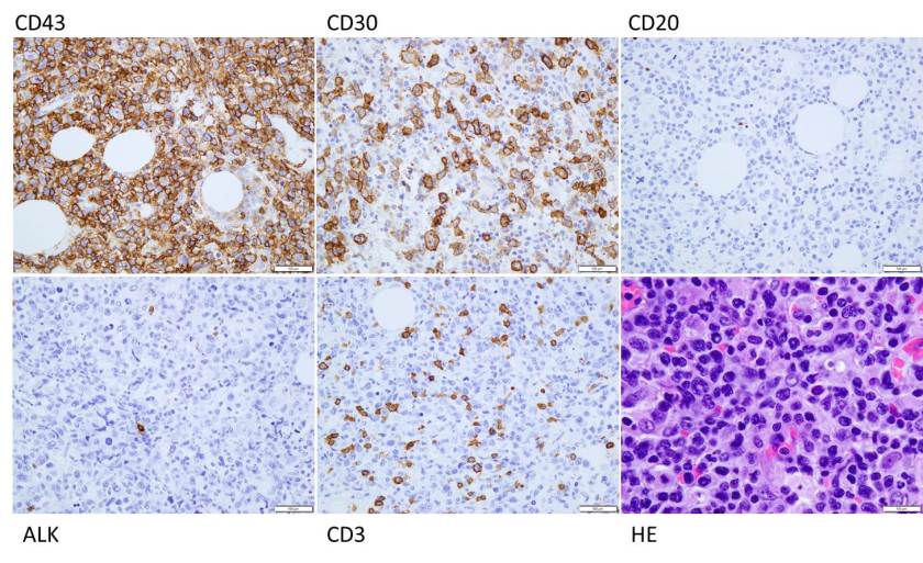
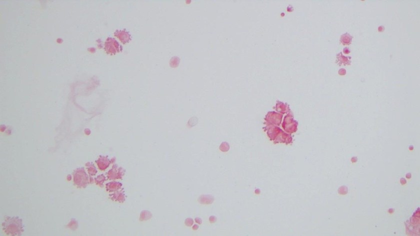
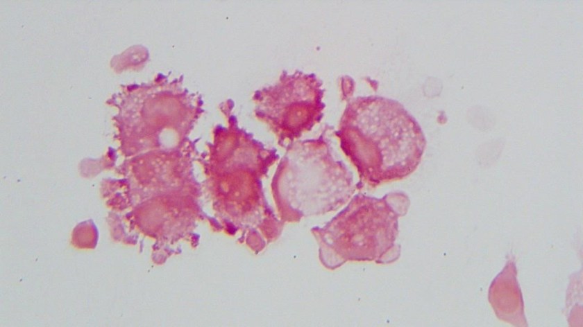
H&E stained sections demonstrate an enlarged node with effaced architecture and scattered residual follicles with small, mature cells. There is a proliferation of intermediate to large, to very large, atypical and highly pleomorphic cells many of which demonstrate bizarre forms, irregular nuclear morphology and acidophilic nucleoli. The lymphoma cells are noted to focally traverse through adipose tissue. Occasional hallmark cells are appreciated.

To further characterize the infiltrate, immunohistochemical stains were performed and interpreted with appropriate controls. The lymphoma cells were diffusely positive for CD45 (LCA), CD43, and CD30 (membranous and Golgi) with a Ki-67 of 80-90%. These cells were negative for CD20, PAX-5, CD3, CD4, CD8 (mostly), CD5, D10, BCl-2, BCl-6 and ALK1.

The morphologic features and immunophenotype of the cells was diagnostic of anaplastic large cell lymphoma, ALK negative.

Discussion

Anaplastic Large Cell Lymphoma (ALCL), ALK-negative (ALK-) is defined as a CD30+ T-cell neoplasm that morphologically resembles ALK-positive ALCL, but lacks ALK protein expression. It most commonly affects adults (aged 40-65 years), and has a slight male preponderance with a male-to-female ratio of 1.5:1. T. Most patients present with advanced disease (stage III-IV), lymphadenopathy and B symptoms. The most common differential diagnosis is ALK-positive ALCL.

The molecular deciphering of ALCL began in the 1990s with the discovery of a recurrent t(2;5) (p23;q35) translocation fusing the ALK gene and the nucleophosmin gene generating a NPM-ALK fusion protein, as well as other ALK translocations resulting in a high ALK kinase activity. This triggers the major oncogenic pathway in ALK-positive ALCL. Pharmacologic therapy has been developed to target ALK, and has shown efficacy. Thus, compared with ALK-negative cases, ALK-positive occurs in younger patients and has a better prognosis. ALK-negative ALCL also tends to involve both lymph nodes and extranodal tissues, although extranodal sites are less commonly involved than in ALK+ ALCL.

The other differential diagnoses of ALK- ALCL includes, primary cutaneous ALCL (C-ALCL), other subtypes of CD30+ T-cell or B-cell lymphoma with anaplastic features and classic Hodgkin Lymphoma. If a single lymph node or cutaneous cases are suggestive of ALK- ALCL, C-ALCL needs to be considered. Any cases that involve the gastrointestinal tract need to be distinguished from CD30+ enteropathy-associated and other intestinal T-cell lymphomas.

Molecular analysis of ALK- ALCL shows characteristic strong expression of CD30, in equal intensity in all the cells. Loss of T-cell markers is frequently seen, however, more than half of all cases express one or more T-cell markers. CD2 and CD3 are more commonly expressed than CD5, and CD43 is almost always expressed. CD4+ is frequently positive, while CD8+ is rare. Many cases also express cytotoxic markers TIA1, granzyme B, and/or perforin.

The genetic profile in ALK-negative ALCL has been found to be pretty heterogenous. Most notably, activating mutations of JAK1 and/or STAT3 have been shown to lead to activation of the JAK/STAT3 pathway. Chromosomal rearrangements of DUSP22 (i.e. chromosomal rearrangements in or near the DUSP22-IRF4 locus on 6p25.3) occur in 30% of the cases, and rearrangements of TP63 occur in about 8% of cases. Neither of the rearrangements have been reported in ALK+ ALCL.

From a prognostic standpoint, studies have shown that the rearrangements have effects on the survival rate. TP63-rearranged cases were shown to have an unfavorable prognosis worse than ALK- ALCL with neither rearrangement, while DUSP22-rearranged cases were shown to have favorable outcomes similar to ALK-positive ALCLs.

References

  1. Gaulard P, de Leval L. ALK-negative anaplastic large-cell lymphoma. 2016 Jan 14;127(2):175-7.
  1. Edgardo R. Parrilla Castellar et al., ALK-negative anaplastic large cell lymphoma is a genetically heterogeneous disease with widely disparate clinical outcomes Blood. 2014 Aug 28; 124(9): 1473–1480.


Bradon Zelman

-Brandon Zelman is 4th year medical student at the Philadelphia College of Osteopathic Medicine and an aspiring pathologist. You can follow Brandon on Twitter @ZelmanBrandon.

Mirza-small

-Kamran M. Mirza, MD PhD is an Assistant Professor of Pathology and Medical Director of Molecular Pathology at Loyola University Medical Center. He was a top 5 honoree in ASCP’s Forty Under 40 2017. Follow Dr. Mirza on twitter @kmirza.

Microbiology Case Study: A 64 Year Old Man with Metastatic Colon Cancer

Case History

A 64 year old man with metastatic colon cancer and a history of recent motor vehicle collision with polytrauma presented from a rehabilitation facility with fever up to 105 degrees Fahrenheit. Two months prior to admission he was hospitalized for the motor vehicle collision in which he sustained orthopedic injuries to multiple extremities. In addition to external fixation of several injuries, he returned to the operating room on multiple occasions for additional incision and drainage of a wrist wound which demonstrated gross purulence with cultures growing Enterococcus, Prevotella, and an extended spectrum beta-lactamase-producing Morganella morganii. Antimicrobial regimen initially consisted of surgically placed antibiotic beads and broad-spectrum therapy with vancomycin, piperacillin-tazobactam, and then meropenem. The patient was eventually transitioned to an oral antibiotic regimen consisting of linezolid and ciprofloxacin for an anticipated course of six weeks and he was discharged to a rehabilitation facility.

After several weeks at the rehabilitation facility, the patient became febrile and was admitted for workup of his fever. Initially the fever was of uncertain origin with malignancy (rectal cancer with metastasis to the liver), drug fever (linezolid), and wound site infection on the differential. Linezolid was discontinued, daptomycin initiated, and ciprofloxacin maintained. Fever persisted and ciprofloxacin was discontinued as another possible source of drug fever. Ertapenem was initiated. Initially, prior wounds and surgical sites appeared well-healing. Blood cultures all yielded no growth. However, on day five of this hospitalization, purulent drainage was noted from the site of a left leg surgical wound. Arthrocentesis yielded 0.5 mL of bloody fluid which was sent for cell count, differential, and culture.

Laboratory Findings

Initial Gram stain showed few polymorphonuclear leukocytes with no bacteria seen. Cell count was unable to be performed due to viscosity of the specimen but differential showed 80% neutrophils. There was no growth on aerobic blood, chocolate, or MacConkey agars. Anaerobic Schaedler (non-selective) agar grew 1-2 mm brownish colonies (Image 1). Gram stain of this isolate revealed gram variable bacilli forming long filaments (Image 2). The isolate was identified using MALDI-TOF MS (Vitek) as Clostridium ramosum.

cloran1
Image 1. 1-2 mm brownish colonies on anaerobic Schaedler agar
cloran2
Image 2. Gram stain showing gram variable bacilli in filamentous chains

The patient was taken to the operating room for incision and drainage of the left knee with two additional samples sent for culture which grew Clostridium ramosum.

Discussion

Clostridium species are anaerobic, spore-forming, gram positive bacilli. C. ramosum is non-motile and is normally found in the human colon and the environment. One study identified C. ramosum in the feces of 83% of sampled adults. Former names include Eubacterium filamentosum, Ramibacterium ramosum, Actinomyces ramosus, and Eubacterium ramosum. Figure 2 demonstrates a notable characteristic of C. ramosum, i.e. its variable appearance on Gram stain. The morphology here may be described as gram negative or “over-decolorized”, though gram positive bacilli are clearly seen forming many of the filaments. Its terminal endospores are often difficult to identify on Gram stain and this is true of Figure 2. These characteristics on Gram stain have historically made identification difficult, though use of MALDI-TOF MS facilitated identification in our case. Biochemically, C. ramosum ferments glucose and hydrolyzes esculin; it is negative for lecithinase and lipase.

C. ramosum possesses an IgA protease though it is not commonly pathogenic. When it is pathogenic, the spectrum of disease overlaps with that of other anaerobes and includes deep-seated abscesses, e.g. intra-abdominal abscess secondary to trauma. Osteomyelitis and primary bacteremia are also possible, particularly in immunocompromised patients. Otitis media in children is another possible clinical scenario.

Septic arthritis due to Clostridium ramosum

A 2016 case report described two cases of septic arthritis due to C. ramosum. In one case, a patient with rheumatoid arthritis on methotrexate and prednisone and history of revision knee arthroplasty eight years prior presented with knee swelling. Synovial fluid aspirate was consistent with an infectious process; the prosthesis was removed but synovial and intraoperative cultures were negative. Antimicrobial therapy with linezolid and ciprofloxacin was administered for six weeks with clinical improvement. Two weeks after discontinuation of antibiotics the patient became febrile. Blood cultures were negative but culture of synovial fluid grew C. ramosum. The patient required multiple operations due to joint destruction and was ultimately managed with intravenous penicillin and clindamycin with transition to oral metronidazole for three months of therapy. The second case of C. ramosum septic arthritis presented in this report was ultimately managed with surgical debridement and amoxicillin-clavulanate. Both cases presented in patients with immunocompromising comorbidities and the course of their septic arthritis was chronic, recurring, and destructive but non-fatal with both patients dying from other causes.

These clinical and laboratory characteristics are consistent with the case of C. ramosum septic arthritis identified at our institution. The case of septic arthritis presented here involved an immunocompromised host (malignancy) with history of trauma, foreign body placement (external fixator), and long-term antibiotic therapy. This patient’s wound required debridement in the operating room on three occasions. Once clinically stable, the patient was discharged to a subacute rehabilitation facility and continued on ertapenem with amoxicillin for an expected duration of six weeks with the plan to switch to amoxicillin-clavulanate and ciprofloxacin for suppressive therapy.

References

  1. Forrester JD, Spain DA. Clostridium ramosum bacteremia: case report and literature review. Surg Infect (Larchmt). 2014 Jun;15(3):343-6. doi: 10.1089/sur.2012.240. Epub 2013 Nov 27. Review. PubMed PMID: 24283763.
  2. García-Jiménez A, Prim N, Crusi X, Benito N. Septic arthritis due to Clostridium ramosum. Semin Arthritis Rheum. 2016 Apr;45(5):617-20. doi: 10.1016/j.semarthrit.2015.09.009. Epub 2015 Oct 1. PubMed PMID: 26546506.
  3. Procop, Gary W et al. Koneman’s Color Atlas and Textbook of Diagnostic Microbiology. Seventh ed., 2017.


-Benjamin F. Smith is a Pathology Student Fellow at University of Vermont Medical Center.

Wojewoda-small

-Christi Wojewoda, MD, is the Director of Clinical Microbiology at the University of Vermont Medical Center and an Associate Professor at the University of Vermont.