Pathologist on Call: There Is No Perfect Lab Test for Smoking Assessment

Cigarette smoking can affect both innate and adaptive immunity, and introduces concerns when evaluating a patient’s eligibility for surgery. It has been shown to hinder time required for healing and long-term survival of patients. It can promote vascular complications, increase the rates of hepatocellular carcinoma and reduce lung function.1 For lung transplantation, one of the common requirements of eligibility is smoking abstinence for at least 6 months. Smoking post-surgery is associated with worse outcomes for the patients including complications and higher rates of mortality.2 Relapse to smoking post lung transplantation has been reported to range from 11% to 23% in various patient populations.3 As a result, clinical testing for cigarette smoking abstinence is an important part of initial workup and follow-up of transplant patients.

In some situations, the burden of lung allocation weighs heavily on a single clinical laboratory result that is perceived to definitively confirm or exclude active cigarette smoking. This subsequently factors into the decision by the physicians to deem the patient eligible to receive a lung transplant. The perception of nicotine testing as definitive proof of smoking is misleading and does not reflect the complexity of situations that can lead to a positive test result.

How can we assess smoking?

Ideally, many factors should weigh into the final smoking status determination including self-reporting (used historically), witnesses to behavior, odor, and past history including cessation attempts. Clinical laboratory testing is important and thought to be more reliable means for smoking assessment. It can involve testing for nicotine (originating from tobacco or nicotine replacement therapy, NRT) and its metabolites: cotinine, 3-hydroxycotinine (3-OH-cotinine), and nornicotine. Moreover, nicotine contains a number of alkaloids that are not usually present in nicotine-replacement therapies (NRTs) including anatabine and anabasine.4 Nicotine testing can involve a combination of metabolites such as cotinine as well as alkaloids like anabasine. Various sample types have been used including saliva, blood and urine.5 In addition, measurements of the exhaled carbon monoxide (CO) have been used to assess recent smoking status (within the last 8 hours).6

Clinical case: patient with detectable nicotine metabolites

A case involving a patient being considered for lung transplantation was received by our department. The patient had been tested for anabasine, nicotine, and its metabolites in urine. Testing of random urine specimens was performed by liquid chromatography tandem mass spectrometry (LC-MS/MS) at different time points from samples collected during hospital visits (days 0, 38, and 62). The urine contained variable concentrations of nicotine and its metabolites, with anabasine concentrations below the detection limit in 2 out of the 3 testing instances. Testing at day 0 showed an interfering substance that prevented the determination of accurate anabasine concentration. The nicotine and its metabolite concentrations in the random urine specimens were lower from day 0 to day 38, but a noticeable increase of 3-OH-cotinine and cotinine concentrations was observed in the specimen collected on day 62. The physician was seeking information about the current smoking status of the patient and was planning to use this information to determine the patient’s lung transplant eligibility.
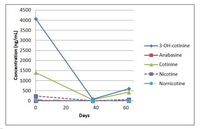
smoking-1


Days 0 38 62
Analyte concentration (ng/mL)
3-OH-cotinine 4074 89 603
Anabasine interf. subst. < 3 < 3
Cotinine 1404 47 425
Nicotine 241 < 2 72
Nornicotine 58 < 2 6


Figure and table 1. Nicotine, metabolite and anabasine concentrations (ng/mL) at different time points for a patient evaluated for lung transplantation eligibility. Anabasine was not detected on days 38 and 62, with an interfering substance preventing quantitation on day 0.

How definitive are these results?

No information was available regarding self-reported smoking or NRT use history for this patient. The physician had high suspicion that the patient was an active smoker and was attempting to use higher concentrations of nicotine and metabolites observed on day 62 as evidence of recent tobacco use.

For cotinine, values can range from 20-550 ng/mL for daily tobacco use.5 Nicotine concentrations in urine can approach over 5000 ng/mL with daily use. Together, high nicotine and cotinine can support tobacco or high-dose nicotine patch use. Furthermore, presence of nornicotine above 30 ng/mL along with anabasine greater than 10 ng/mL would be consistent with current tobacco use rather than NRT.7

Given that these were random urine specimen and the urinary creatinine values are not routinely measured, it’s important to consider the possible contributions of the variable urine concentration to the analyte concentrations. It has previously been reported that individuals abstaining from smoking for at least two weeks should present with nicotine of <30 ng/mL, cotinine of < 23 ng/mL, 3-OH-cotinine of <120 ng/mL, nornicotine < 3 ng/mL, and anabasine of < 2 ng/mL in urine.7 Based on these cut-offs, all analytes except anabasine would suggest new nicotine intake within the last two weeks.

In general, a positive anabasine result, in combination with the presence of nicotine metabolites, is consistent with active use of a tobacco product, whereas anabasine values of < 2ng/mL may suggest that NRT is the likely source.8 This can imply that the patient is abstinent from smoked or chewed tobacco if anabasine is not detected. However, anabasine is not a sensitive marker of smoked tobacco. It has been reported that the compound may not be detectable in 60% of self-reported smokers (N=51; 3 ng/mL cut-off in urine)9  and its urinary concentrations do not correlate well with self-reported tobacco use.8

As a result, anabasine has low sensitivity for determining eligibility for UNOS (United network for organ sharing) listing. There are some recommendations that this marker should not be used alone. Given that other alkaloids can originate from tobacco plant, it has been proposed that anatabine should be added to analysis due to higher expected concentration.9 However, this alkaloid is not completely specific to tobacco as it has been proposed to also arise from other plant sources 10,11  leading to possible implications for the patient that may be misclassified. In addition, anatabine sensitivity in detecting smoked tobacco use varies depending on the tobacco source and the clinical cut-off used. Clinical tests that include anatabine are not routinely available.

Can we improve this process?

Unfortunately, there is no definitive marker distinguishing smoking from NRT.

The determination of smoking status has advanced from reliance on self-reporting to quantitative and specific measurements of metabolites of nicotine and minor components of tobacco. Additional analyte incorporation into a test panel leads to additional complexities and considerations in interpretation of the results. Therefore, it is important to educate the physicians about various nicotine sources causing a positive nicotine and/or metabolite test result including NRT or e-cigarettes. It is also important to convey the limitations of tobacco alkaloid testing in such scenarios. Both the lab and the physician need to be cautious about implying active smoking in the absence of indirect supporting evidence and/or positive clinical test results.

At the same time, there is a need to improve the utility and availability of other tobacco alkaloid testing in distinguishing cigarette smoking from NRT in specific transplant populations and consider the value of testing alternative specimens. This may lead to a more effective implementation of secondary markers of tobacco use.

References

  1. Qiu, F.; Fan, P.; Nie, G. D.; Liu, H.; Liang, C.-L.; Yu, W.; Dai, Z., Effects of Cigarette Smoking on Transplant Survival: Extending or Shortening It? Frontiers in Immunology 2017, 8, 127.
  2. Zmeskal, M.; Kralikova, E.; Kurcova, I.; Pafko, P.; Lischke, R.; Fila, L.; Valentova Bartakova, L.; Fraser, K., Continued Smoking in Lung Transplant Patients: A Cross Sectional Survey. Zdravstveno varstvo 2016, 55 (1), 29-35.
  3. Vos, R.; De Vusser, K.; Schaevers, V.; Schoonis, A.; Lemaigre, V.; Dobbels, F.; Desmet, K.; Vanaudenaerde, B. M.; Van Raemdonck, D. E.; Dupont, L. J.; Verleden, G. M., Smoking resumption after lung transplantation: a sobering truth. The European respiratory journal 2010, 35 (6), 1411-3.
  4. Hukkanen, J.; Jacob, P., 3rd; Benowitz, N. L., Metabolism and disposition kinetics of nicotine. Pharmacological reviews 2005, 57 (1), 79-115.
  5. Raja, M.; Garg, A.; Yadav, P.; Jha, K.; Handa, S., Diagnostic Methods for Detection of Cotinine Level in Tobacco Users: A Review. Journal of clinical and diagnostic research : JCDR 2016, 10 (3), Ze04-6.
  6. Sandberg, A.; Skold, C. M.; Grunewald, J.; Eklund, A.; Wheelock, A. M., Assessing recent smoking status by measuring exhaled carbon monoxide levels. PloS one 2011, 6 (12), e28864.
  7. Moyer, T. P.; Charlson, J. R.; Enger, R. J.; Dale, L. C.; Ebbert, J. O.; Schroeder, D. R.; Hurt, R. D., Simultaneous analysis of nicotine, nicotine metabolites, and tobacco alkaloids in serum or urine by tandem mass spectrometry, with clinically relevant metabolic profiles. Clinical chemistry 2002, 48 (9), 1460-71.
  8. Jacob, P., 3rd; Hatsukami, D.; Severson, H.; Hall, S.; Yu, L.; Benowitz, N. L., Anabasine and anatabine as biomarkers for tobacco use during nicotine replacement therapy. Cancer epidemiology, biomarkers & prevention : a publication of the American Association for Cancer Research, cosponsored by the American Society of Preventive Oncology 2002, 11 (12), 1668-73.
  9. Feldhammer, M.; Ritchie, J. C., Anabasine Is a Poor Marker for Determining Smoking Status of Transplant Patients. Clinical chemistry 2017, 63 (2), 604-606.
  10. Lanier, R. K.; Gibson, K. D.; Cohen, A. E.; Varga, M., Effects of dietary supplementation with the solanaceae plant alkaloid anatabine on joint pain and stiffness: results from an internet-based survey study. Clinical medicine insights. Arthritis and musculoskeletal disorders 2013, 6, 73-84.
  11. von Weymarn, L. B.; Thomson, N. M.; Donny, E. C.; Hatsukami, D. K.; Murphy, S. E., Quantitation of the minor tobacco alkaloids nornicotine, anatabine, and anabasine in smokers’ urine by high throughput liquid chromatography mass spectrometry. Chemical research in toxicology 2016, 29 (3), 390-397.


VG

-Dr. Valentinas Gruzdys developed interest in clinical chemistry early in his academic training which led him to pursue and obtain a PhD in Clinical and Bioanalytical Chemistry at Cleveland State University. Valentinas is enthusiastic about teaching and helping improve the understanding of limitations and utility of clinical laboratory testing. He is currently enrolled in a clinical chemistry fellowship program at the University of Utah. He enjoys learning more about various aspects of clinical chemistry and cannot wait to make his own contributions to the field after his training.

Microbiology Case Study: A 10 Year Old Boy with Right Knee Pain

Case History  

A 10 year old Caucasian male presented to the pediatric emergency department due to significant pain in his right knee with exposed hardware. His past medical history was noteworthy for Perthe’s Disease, a condition leading to avascular necrosis of the femoral head. In addition, he is affected by an autosomal dominant congenital disorder and has had ischemic strokes in the past. Recently, he had surgery performed on multiple joints to correct abnormalities and they were complicated by dehiscence & infection with methicillin sensitive Staphylococcus aureus. On current admission, his mother reported he has been afebrile and was consistently taking cephalexin to treat the above infection. He was taken to the operating room for incision & drainage and hardware removal of the knee. Bacterial cultures were collected at the time of surgery and he was started on IV clindamycin.

Laboratory Identification

eikcor1
 Image 1. Clear, spreading colonies that showed “pitting” of the agar after 48 hours incubation at 37°C in 5% CO2.  
eikcor2
Image 2. Gram stain directly from the colony showed slender gram negative rods (100x oil immersion).

On direct Gram stain, there was no organisms seen and rare white blood cells. Initially, there was no growth at 24 hours, but two morphologies were observed on the second day. The first organism was identified as the S. aureus that was previously isolated from this site. The second organism was clear with spreading colonies that pitted the agar (Image 1). A distinct bleach like odor was observed. There was no growth on MacConkey agar even though the Gram stain showed gram negative rods, making this organism most likely to be classified as fastidious (Image 2). Benchtop biochemical tests were negative for catalase and positive for oxidase. MALDI-TOF mass spectrometry identified the isolate as Eikenella corrodens. 

Discussion

Eikenella corrodens is a fastidious Gram negative rod that is a member of the HACEK family. It is considered normal flora in the oral cavity and possibly the gastrointestinal tract of humans.  Infection results from these endogenous sources and can be the result of poor oral hygiene, mucositis or dental procedures. E. corrodens causes juvenile and adult periodontitis and is commonly implicated in bacteremia and infective endocarditis, particularly in IV drug users who lick needles prior to injection. Infections of the abdomen, bones/joints and brain are less common.

In the laboratory, E. corrodens is slow growing and is usually present as clear, spreading colonies after 48 hours incubation at 37°C in 5% CO2 on blood and chocolate agars. A unique feature of the organism is that it pits or corrodes the agar, lending to its species name. Also, E. corrodens produces a bleachy smell due to the production of hypochlorite. It does not grow on MacConkey agar despite the fact it is a Gram negative rod. Biochemical tests are negative for catalase, positive for oxidase and negative for indole. Automated instruments and MALDI-TOF mass spectrometry are both able to identify E. corrodens with confidence.

Susceptibility guidelines can be found in the 3rd edition of the CLSI M45 document. In general, E. corrodens is susceptible to penicillin, board spectrum cephalosporins, carbapenems, azithromycin and fluoroquinolones. Resistance to narrow spectrum cephalosporins, macrolides and clindamycin has been documented. In general, susceptibility testing should be performed on E. corrodens when it is isolated from a normally sterile site or is identified in pure culture. Beta lactamase testing is recommended routinely on E. corrodens, and if positive, the isolate is resistant to penicillin, ampicillin and amoxicillin. In the case of bite wounds caused by E. corrodens, susceptibility testing may not be necessary if it is treated with amoxicillin-clavulanate acid (Augmentin) due to a high probability of susceptibility to this antibiotic.

In the case of our patient, he responded to the antibiotic therapy used to treat his S. aureus and E. corrodens infections and healed well. He was placed on long term oral antibiotic therapy until additional hardware is able to be removed at a future date.
RA

-Rim Alkawas, MD, is a first year Anatomic and Clinical Pathology resident at the University of Mississippi Medical Center. 

Stempak

-Lisa Stempak, MD, is an Assistant Professor of Pathology at the University of Mississippi Medical Center in Jackson, MS. She is certified by the American Board of Pathology in Anatomic and Clinical Pathology as well as Medical Microbiology. She is the director of the Microbiology and Serology Laboratories. Her interests include infectious disease histology, process and quality improvement and resident education.

Boards and Wards

As a little detour before I start my medical school clerkship rotations as a 3rd year student, I’d like to take a moment to appreciate—yes appreciate—board exams. I just sat for the daunting and arduous United States Medical Licensing Exam (USMLE) called “Step 1.” It is roughly an eight-hour endeavor to prove that some of the tomes of information I was exposed to throughout my first two years of medical school made it somewhere into my hippocampus. That said, yes board exams are always daunting and yes, they can even be quite stressful. There’s a lot depending on your scores, in any field you find yourself testing in. Some are pass/fail and some provide you with a scaled score performance.

For what feels like forever ago to me now, I sat for a state licensure exam for the Illinois Department of Public Health as an Emergency Medical Technician Basic provider, or EMT-B. I absolutely failed it—missed it by a point or so. Scheduled a retake, studied hard, and passed round two. Lesson learned. That license opened many doors for me back in the day, and that’s precisely the point: professional certification, official licensures, and (often) professional society membership will bolster anyone looking to get ahead in their career.

Other times, these board exams are highly encouraged. After graduate school at Rush for my MLS degree I had to sit for the ASCP BOC Board Exam for the professional credentials of a Medical Laboratory Scientist, or MLS (ASCP). When I passed, I was able to advance in my career then and have excellent opportunities that would be unavailable otherwise. More so, certain jobs would have been completely unavailable to me without those clinical credentials! I would say that like ASCP cites 70% of patient results originate from the lab, 70% of my CV depends on those professional credentials.

aki-fig-1
Figure 1. A previously renewed ASCP BOC certificate, proudly displayed.

This brings up a somewhat related point. There is a professional debate that’s been going on for a few years: board certification vs. regulatory licensure. Organizations like ASCP and CAP have been on board with licensure for a while, citing the critical roles we play in patient care and the specialized education training required. An article from 2015 had circulated well explaining the advantages and regulatory compliance improvement offered by licensure as medical laboratory science evolved since the Clinical Laboratory Improvement Act of 1988 (known as CLIA ’88). Those authors established that virtually all laboratory professional organizations, as well as local state public health departments, favor licensure to guarantee regulatory oversight for the quality of personal and testing results (Rohde et al., 2015). With so many questions today about what qualifies laboratory personnel since the Center for Medicaid Services decision in 2016 that says a bachelor’s degree in nursing is sufficient to perform and manage laboratory moderate to complex testing, professional organizations like ASCP, CAP, and ASCLS continue to investigate what measures would maintain quality and regulations for positive patient outcomes.

aki-fig-2
Figure 2. States with licensure, and without. I was trained and practiced medical laboratory science in Chicago, Illinois, a state that does not require licensure. (Rohde et al., 2015)
aki-fig-3
Figure 3. These graphs show the number of sanctions under CLIA imposed on labs in the following states. This demonstrates the ineffectiveness of CLIA improving laboratory testing or personnel quality. (Rohde et al., 2015)

Like the EMS exam, the USMLE is absolutely mandatory if I in any capacity wish to continue my medical education, match into a residency program, and ultimately practice as a physician. So, as daunting as these tests might be, they provide a good benchmark standard for the quality of physicians from around the world who want to practice in the United States. USMLE actually has a series of four board exams I’ll be taking in the coming years—so bear with me as I try to stay positive. The Step exams check the depth and breadth of one’s understanding of medical concepts from anatomy to the minutiae of biochemistry. Like ASCP’s board exam, it was a mix of hematology, microbiology, immunology, with added clinical vignettes and patient outcomes. At the end of the test day, I didn’t have a single neuron left working at 100%, but I’ve since recovered. And now it’s onto the next chapter: clinicals. Hope to catch you all again soon, as I’ll try to write up some interesting lab-related cases I will most assuredly come across. Thanks!


aki-fig-4
Figure 4. One of many medical students’ bibles. (Stock photo from Amazon.com)

References

Rohde, R., Falleur, D. Ellis, J. (2015) “Almost anyone can perform your medical laboratory tests – wait, what?” Elsevier.com March 10th, 2015; retrieved from: https://www.elsevier.com/connect/almost-anyone-can-perform-your-medical-laboratory-tests-wait-what


Centers for Medicaid and Medicare Services (2016) Personnel Policies for Individuals Directing or Performing Non-waived Tests, Revised due to typographical error under citation of §493.1443(b)(3). Center for Clinical Standards and Quality/Survey & Certification Group. April 1, 2016; retrieved from: https://www.cms.gov/Medicare/Provider-Enrollment-and-Certification/SurveyCertificationGenInfo/Downloads/Survey-and-Cert-Letter-16-18.pdf


ckanakisheadshot_small

Constantine E. Kanakis MSc, MLS (ASCP)CM graduated from Loyola University Chicago with a BS in Molecular Biology and Bioethics and then Rush University with an MS in Medical Laboratory Science. He is currently a medical student at the American University of the Caribbean and actively involved with local public health.

Decoding Generations

When I started my professional life, nobody talked about generations. Experience was everything: none, little, some, significant or expert. Now, conversations about generational similarities and differences are integrated into professional and personal life.

There are currently five generations at work today: Traditionalists, Baby Boomers, Generation X, Generation Y/Millennials, and Generation Z.

generations

Understanding generations allows people to adapt their behavior according to certain preferences.Take, for example, communication styles. When communicating with someone from the Baby Boomers generation, picking up the phone might be appreciated, while sending an email to a Generation X is the best way to communicate with them. Always keep in mind, however, that generational preferences are generalizations, and knowledge about them does not substitute understanding each employee and colleague on an individual basis.

Another example of the differences between the generations is how they define their aspirations. Traditionalists value home ownership, Baby Boomers want job security, Generation X aspires to achieve work-life balance, Generation Y prefers flexibility and freedom, and Generation Z values security and stability. Understanding each generation’s aspirations allows leaders to tailor their communication style and job aspects to each individual.


lotte-small

-Lotte Mulder earned her Master’s of Education from the Harvard Graduate School of Education in 2013, where she focused on Leadership and Group Development. She’s currently working toward a PhD in Organizational Leadership. At ASCP, Lotte designs and facilitates the ASCP Leadership Institute, an online leadership certificate program. She has also built ASCP’s first patient ambassador program, called Patient Champions, which leverages patient stories as they relate to the value of the lab.


As a former member of the ASCP Resident Council, I volunteered for the opportunity to serve as a beta tester for the ASCP Leadership Institute. To obtain certification, I completed 10 modules which often included a pre-course reading or interactive video assignment along with a pre-recorded webinar, post-test, and post-course evaluation. Some of these courses are also available in-person at live meetings and can include personal coaching live, online, or by phone.

A cursory internet search will reveal a plethora of written and video resources available on the topic of leadership. Additionally, many of us have participated in evaluations or trainings that sought to not only define our personal leadership skills/style but also help us to gain essential self-awareness and skills to better lead or be part of a team. Despite such experiences and even though I’ve held many leadership positions over the years, I still find it difficult to reconcile what it means to be a leader, both in terms of expectations that I hold for myself and those that others hold of me and how to build these expectations to realize a shared objective.

I was intrigued by the title of the module “DeCoding Generations”. This module was especially salient for me since I was a non-traditional medical student after initially studying to be a neuroscientist. I’ve generally been older than my fellow trainees and younger than the majority of my teachers. This generational gap has also been similarly evident within the teams I’ve participated in since I matriculated into medical school.

This module explored the core values of the following “generations”: traditionalists, baby boomers, gen X, gen Y (also known as millennials), and gen Z to help the learner understand what drives members of each group. The course then further defined the aspirations, attitudes toward technology and their careers, and preferred communication media and preferences of members of each generation. This was all with the goal of facilitating interactions, especially as a leader, with members from each generation. For instance, different generations prefer and respond better to different types of communication: in-person, phone, email, video conferencing, text, or a combination of these modalities. That’s where the “decoding” part of the module comes in. As leaders, we need to recognize how best to interact with each team member to acknowledge their core values and foster the most harmonious working relationships while working toward a shared goal.

I’m a very visual learner and intuitive person but not the most eloquent or at ease with verbal communication despite friends remarking that I’m a “social butterfly”. This module helped me evaluate ways to adapt my communication style especially when interacting with others in the two most numerous generations in the workforce: millennials (42%) and baby boomers (29%). I fall in the middle as a gen X’er (23%) and have often found myself confounded by the attitudes and behaviors of millennials and this module helped me to understand their perspective and preferred modes of communication. But what I learned most was to look at not only the differences that impair our interactions but also the similarities we share that can be used to prevent or resolve conflicts and to encourage team creativity and solidarity.


Chung

-Betty Chung, DO, MPH, MA recieved a BA from The University of Chicago, MA from Boston University School of Medicine, DO from UMDNJ-School of Osteopathic Medicine (now Rowan-SOM), and MPH from Columbia University and a decade of experience in basic science research. She completed her AP/CP residency at the University of Illinois at Chicago (PGY1-2) and Rutgers Robert Wood Johnson Medical School (PGY3-4). Her current interests lie in graduate medical education, quality improvement, hematopathology, and molecular genetic pathology.

Components of an Online CLS Course

When interviewing prospective candidates for Mayo Clinic’s program in medical laboratory science (MLS), I provide an overview of one of our blended courses and compare and contrast it to one of our more traditionally taught (lecture-based) courses. This gives me the opportunity to emphasize expectations and recommended study habits based on some of the “best practices” we’ve learned from our students.

Our online MLS courses include the following components:

  • Syllabus
  • Weekly calendar
  • Online lessons
  • Homework assignments
  • Discussion boards
  • Study guides
  • Self-assessments
  • Practice exams
  • Resources (links to related online resources)

Our students are expected to review each online lesson before coming to class as preparation for their laboratory session. Since we teach “immersion style” courses, two at a time (where a typical four-credit course is condensed into six weeks), we recommended that our students plan to study a minimum of 4 to 6 hours per day.

Each lesson is presented in a written format, following instructional-design recommendations for online learning that includes “chunking” of the content—using bullets to convey information instead of complete sentences (where appropriate) along with concisely written text that emphasizes “key concepts,” graphics, and images. The lessons are straightforward and present basic knowledge, and the higher learning concepts are integrated into the discussion-board assignments.

Each online course is easy to navigate and is presented in such a way that it’s intuitive and requires little “outside” instruction. All the courses in our program follow the same format, so once the students become familiar with navigation of their first course, they do not have to re-learn the lesson format each time they start a new course.

We provide a study guide of objectives for every written examination. Our students are encouraged to create a learning document from the study guide that they can use for review over the duration of the program and to prepare for their national certification examination in medical laboratory science offered through the American Society for Clinical Pathology.

To give you an idea of how our online courses are designed in Blackboard Learn, I have taken a series of screen shots demonstrating the layout of a course and lesson plan (shown below).

When our students log into Blackboard Learn and open a course, they land on the home page, which includes a navigation menu and links to the syllabus and introductory discussion boards. The home page discussion boards include “student introductions,” “faculty expectations,” “updates and handouts,” “ask your instructor or classmates,” and an “MLS Café” (for social interactions).

1_Welcome

From the menu, our students can open the course content. The first page opens to the weekly course calendars. At a glance, our students can examine the week’s activities.

2_Weekly Calendar

Clicking the “Course Week” link opens the week’s lesson plans.

3_Lesson Plan_Week 2 List of Lessons

Each lesson is formatted the same way and begins with a brief description, overview (goals or learning objectives), author, and references.

4_Lesson Plan_Overview

The second page is a table of “steps to completion” so that our students know exactly what is required of them.

5_Lesson Plan_ Steps to Completion

The lesson is presented in a written format. A table of contents allows the students to navigate the pages of the lesson.


6_Lesson Plan Introduction

The lesson concludes with a self-assessment. The self-assessment is embedded in the lesson, includes feedback loops, and is also linked to the home page menu. The students are able to take the self-assessments as often as they’d like, and the course grade book is set to record their highest score.

7_Example of Self Assessment

In this course, there is a weekly discussion board. The students are directed to work as a team in assigned groups to answer the questions in the discussion. Credit for this discussion is based on participation in the thread and “substantive” contributions to the dialogue. Students are encouraged to build upon one another’s commentary, generating comprehensive answers to the questions. Each group member must contribute at least two to three substantive answers to receive credit for the assignment. One group member is designated to post a summary of the discussion on behalf of the group.

8_Example of Discussion Board

There are 15 didactic courses in our MLS curriculum. All of our courses have an online component with approximately one-third of the courses applying the “reverse-lecture-homework” paradigm, one-third are lecture based (traditional), and one-third are a combination of both.

This variation in presentation of content provides our students a mixed learning experience, and the online format allows for us to map everything out for them. Additionally, the curricular model itself lends to the formation of study groups, which in turn helps our students build upon their teamwork and communication skills.

Since our program was instituted 10 years ago, we have seen excellent outcomes, with 100% graduation rates, 100% employment of our graduates, and 96% first-time pass rates on the national certification examination (based on a three-year average). Notably, the breakdown of the certification results by category demonstrates that overall student performance in content areas of the curricula that apply the reverse-lecture-homework paradigm are, on average, higher than those categories following a traditional course format (i.e, lecture-based).


Lehman_small

-Susan M. Lehman, MA, MT(ASCP)SM graduated from the University of Wisconsin-Madison in 1983 with a BS in medical technology. She is program director for the Medical Laboratory Science Program and course director for Clinical Microbiology I and II; her areas of interest include distance education and education methodology.


Pseudohyperkalemia in Patients with Severe Leukocytosis

It has been reported many times that falsely elevated potassium can be seen in patients with severe leukocytosis from chronic lymphocytic leukemia (CLL). Early recognition of factitious hyperkalemia is very important to prevent inappropriate and potentially hazardous treatment. One case we observed in our institution, again, emphasized the importance and urgency to recognize these instances.

In this case, patient was a 58 year old male with recently diagnosed CLL. His potassium level rose from normal levels at admission to 8.9 mmol/L on repeated blood draws. Patient was asymptomatic with good strength on physical exam, and had no abnormalities on EKG or telemetry. Insulin/glucose and calcium gluconate was administered to correct potassium level and to prevent cardiac effect of hyperkalemia. The hyperkalemia result was initially thought to be due to emerging tumor lysis syndrome and was not brought to our attention until another specimen obtained had a potassium level greater than the measurable range (10.0 mmol/L). Specimens were not hemolyzed and white blood cell count was as high as 455K/µL.

Given his history of CLL, we suspected pseudohyperkalemia, an entity that has been attributed to the combination of the fragility of the leukemic lymphocytes and the mechanical stress on the cells during specimen transportation and centrifugation. Our clinical team was notified immediately, and a whole blood specimen was collected and hand carried to the laboratory. Without centrifuging, the whole blood specimen was analyzed on a blood gas analyzer and showed a potassium level of 4.2 mmol/L!!! Potassium-lowering treatment was discontinued.

Artifactually elevated potassium level is commonly seen due to red blood cell hemolysis, but not well appreciated is its occasional occurrence in patients with extreme leukocytosis from CLL. It is important for laboratorians to recognize this pattern and to notify our clinical teams so that patients are not inappropriately treated.


Xin-small

-Xin Yi, PhD, DABCC, FACB, is a board-certified clinical chemist, currently serving as the Co-director of Clinical Chemistry at Houston Methodist Hospital in Houston, TX and an Assistant Professor of Clinical Pathology and Laboratory Medicine at Weill Cornell Medical College.

Microbiology Case Study: A 55 Year Old Female with Respiratory Failure

Case History

A 55 year old female with a history of chronic obstructive pulmonary disease, alpha-1 antitrypsin deficiency, and current tobacco use was transferred to our hospital due to acute hypoxemic respiratory failure. She had a gradual six day onset of cough, fever, malaise, weakness, dizziness and wheezing. At the outside facility, she was hypoxic with an oxygen saturation of 67% at room air, hypotensive with a blood pressure of 80/50. She was intubated en route to our facility.

Labs were significant for a positive influenza B swab, leukopenia (WBC 1.2) with 59% bands, and acute kidney injury with a creatinine of 1.4 mg/dl and hyponatremia with a sodium level of 129 mEq/L. Blood cultures grew Streptococcus pneumoniae, sensitive to ceftriaxone. At our facility, she was started on ceftriaxone and azithromycin. She completed 14 days of ceftriaxone; however, she continued to have intermittent fevers above 38 degrees Celsius. Due to the patient’s continued fever, infectious work up was initiated and showed Candida in her urine and HSV lesions on her lips. She was started on a 14 day course of fluconazole and valacyclovir.

Tracheal aspirates on two occasions were also cultured and grew mixed gram positive and negative organisms as well as Syncephalastrum species. Four weeks after being admitted to our facility, she developed a right-sided hydropneumothorax in which 500 mL of exudative fluid was drawn and subsequently cultured. These cultures also grew Syncephalastrum species as well as Staphylococcus epidermis.

synrac1
Image 1: Syncephalastrum growing on a blood agar plate from the patient’s pleural fluid.
synrac2
Image 2: Lactophenol cotton blue stain of Syncephalastrum demonstrating the sporangiophore with tubular sporangia on the large round vesicle. The sporangia contain chains of round spores.

Discussion

Syncephalastrum racemosum is thought to be the only species out of the two Syncephalastrum species known to cause mucormycoses in humans (1). The only proven reported cases of infection have been due to percutaneous inoculation after trauma, however whether this is due to low pathogenicity, no case reports, or interpretation as a contaminant remains a mystery (1).

Syncephalastrum is a saprophytic fungus isolated throughout the world particularly in environments with decaying organic matter (1, 2). It is found in low levels in the air and has been reported to colonize both immunocompromised and healthy individuals after natural disasters (3).

Diagnosis of Syncephalastrum can be made by visualizing pauci-septate, ribbon-like mycelium and a merosporangial sack surrounding sporangiospores from the cultures using a lactophenol cotton blue mount preparation (1). Caution should be used in distinguishing Aspergillus niger from Syncephalastrum using a direct KOH mount due to the similarities in their fruiting bodies (1). On a petri plate, it begins as fast growing white fluff and then turns dark gray to almost black with the reverse side being white (4).


References

  1. Gomes MZ, Lewis RE, Kontoyiannis DP. Mucormycosis caused by unusual mucormycetes, non-Rhizopus, -Mucor, and -Lichtheimia species. Clin Microbiol Rev. 2011;24(2):411-45.
  2. Ribes JA, Vanover-sams CL, Baker DJ. Zygomycetes in human disease. Clin Microbiol Rev. 2000;13(2):236-301.
  3. Rao CY, Kurukularatne C, Garcia-diaz JB, et al. Implications of detecting the mold Syncephalastrum in clinical specimens of New Orleans residents after Hurricanes Katrina and Rita. J Occup Environ Med. 2007;49(4):411-6.
  4. Larone DH. Medically Important Fungi, A Guide to Identification. Amer Society for Microbiology; 2011.


-Angela Theiss is a pathology resident at the University of Vermont Medical Center.

Wojewoda-small

-Christi Wojewoda, MD, is the Director of Clinical Microbiology at the University of Vermont Medical Center and an Associate Professor at the University of Vermont.

A Chemical Conundrum

In 1983, OSHA established its first version of the Hazard Communication standard. These regulations were made law in response to a lack of hazard information given to over 30 million United States employees working with chemicals. OSHA estimates that 650,000 chemicals are used in over three million work places across the country. Recognizing that the work performed in laboratories is unique—relatively small quantities of hazardous chemicals are used on a non-production basis—OSHA promulgated the Chemical Hygiene standard (more simply known as the Laboratory standard) in 1990.

The Chemical Hygiene standard regulations supersede HazCom standard regulations in the laboratory setting. However, there are still several HazCom rules that affect labs such as those involving hazard determination, chemical labeling, and Safety Data Sheets. The Chemical Hygiene standard brings another layer of exposure protection to the lab employee through the required establishment of a series of chemical safe work practices. Understanding how both sets of laboratory regulations work together is important in creating an overall lab chemical management program.

The regulations for creating a chemical inventory are expressed in the HazCom standard. OSHA requires a list of hazardous chemicals in every work place where they are manufactured, stored, or used. This inventory can be a useful tool for many reasons. Use the list to document the required chemical risk analysis. Review the chemicals in the lab for their hazards, and indicate on the inventory list any applicable hazard warnings such as the signal word and pictograms. CAP requires that you determine which chemicals in the lab are carcinogenic or reproductively and acutely toxic. Once that analysis is complete, it can also be documented on the inventory list. Record average volumes of the chemicals stored in the lab as well. This information may be helpful in a fire situation so that the fire department or other rescue workers will be aware of what they may encounter.

Laboratories must designate a Chemical Hygiene Officer, a point person who is qualified and responsible for providing technical guidance for the implementation and operation of the entire lab chemical management program. The Lab Standard even mentions the establishment of a Chemical Hygiene Committee if the lab or chemical program is larger. Details of this lab chemical safety structure should be spelled out in the lab’s required Chemical Hygiene Plan.

If you read the actual Chemical Hygiene Standard on OSHA’s web site, you can see it is not very long (unlike the HazCom standard or others). Because of the volume of chemicals used in labs, the standard’s main focus is protecting employees from those chemicals via written procedures, physical barriers (such as PPE and engineering controls), and health monitoring. A model Chemical Hygiene Plan must include exposure control methods, a chemical fume hood maintenance process, a detailed training program, and medical consultation and follow-up when chemical exposure limits are exceeded. Appendix A of the standard (called National Research Council Recommendations Concerning Chemical Hygiene in Laboratories) was created to assist laboratories with the development of a complete and compliant Chemical Hygiene Plan.

A third set of OSHA regulations that affects labs and chemical safety is the Formaldehyde standard. The exposure monitoring section gives instructions on how to perform vapor monitoring for this carcinogenic chemical. The laboratory has an option to monitor each employee individually, or it may set up a representative sampling strategy and measure exposures within each job classification and for each work shift. The purpose of this strategy is to properly characterize the exposure of every employee without having to monitor each one. Simply stated, that means if Jane and John perform the same duties and are equally exposed to formaldehyde in their work day or for a specific task, you may monitor only Jane’s exposure and share the results with both employees. That said, the CAP standard on the Anatomic Pathology inspection checklist states that each new employee should have formaldehyde vapor monitors performed. While it references the OSHA formaldehyde regulations, the standard fails to include OSHA’s wording about representative monitoring. That can be an issue if a CAP inspector follows strict adherence to those standards and does not subscribe to OSHA’s intent. Clearly representative vapor monitors make sense and are safe, but you may have to challenge the case with certain inspectors.

Chemical management in the laboratory can certainly seem daunting, and there are many regulations (federal and otherwise) that affect how the lab program may be run. The basic safety strategies exist for the purposes of protecting the employee from chemical exposure, but so many details are involved with the process. Proper storage of flammables and corrosives, labeling, and waste handling are just some of the topics not even touched upon in this article, but they also must be considered for safety purposes. If operating the chemical management program is your duty, be sure to understand the regulations, and build a team of staff who will work together to ensure safe chemical processes in the laboratory.


Scungio 1

Dan Scungio, MT(ASCP), SLS, CQA (ASQ) has over 25 years experience as a certified medical technologist. Today he is the Laboratory Safety Officer for Sentara Healthcare, a system of seven hospitals and over 20 laboratories and draw sites in the Tidewater area of Virginia. He is also known as Dan the Lab Safety Man, a lab safety consultant, educator, and trainer.

Library Preparation – The First Step in a NGS Setup

Welcome back! Last quarter we discussed why Next Generation, or Massively Parallel, Sequencing is the next big thing in the world of Molecular Diagnostics. The sensitivity, the depth of coverage and the ability to interrogate many different areas of the genome at the same time were just a few of the benefits of these types of assays. Next, I would like to describe a couple different methods of library preparation, which is the first step necessary to run an NGS assay.

First of all, let’s define “Library.” I find this is the most common question technologists new to this technology ask. Essentially, a library is a specimen’s collection of amplicons produced by the assay that have been barcoded, tagged with appropriate platform adapters and purified. These will serve as the input for the next part of the NGS workflow, clonal amplification (the topic of next quarter’s blog!).  How these libraries are prepared differ depending on platform (i.e, Ion Torrent vs. MiSeq), starting material (RNA vs. DNA), and type of assay (targeted amplicon vs. exome).

Before we begin the library prep discussion, a note about the input specimen. The DNA must be quantitated using a method that is more specific than spectrophotometry – it must be specific for double-stranded DNA. It will lead to an overestimation of the amount of DNA in the specimen, which will lead to over-dilution and consequently, lower quantity of final library. Real-time PCR and a double-stranded kit with fluorometry are two examples of assays that will give accurate concentrations of double-stranded DNA.

Our lab has begun using NGS for some of our oncology assays, so I will focus on the two types we perform currently, but keep in mind, there are many other types of assays and platforms.

library1.png
Image 1: ion torrent amplicon library preparation. Source: Ion AmpliSeqTM Library Preparation User Guide – MAN0006735, Rev. 10 September 2012.

The assay we use for our Ion Torrent platform is a PCR amplicon based assay. The first step is to amplify up the 207 regions over 50 genes that contain hotspots areas for a number of different cancer types. This all occurs in one well for each specimen. Once those areas are amplified, the next step is to partially digest the primer sequences in order to prepare the ends of amplicons for the adapters necessary for the sequencing step. As shown in the figure above, two different combinations of adapters may be used. The top one, listed as the A adapter (red) and the P1 adapter (green), would be used if only one specimen was to be sequenced on the run. The A and P1 adapters provide universal priming sites so that every amplicon of every sample can be primed with the same primers, rather than having to use gene specific primers each time. The second possibility is listed below that, with the same P1 adapter (green) and a Barcode Adapter labeled X (red and blue) – it still contains the A adapter necessary for sequencing (red), but it also contains a short oligonucleotide sequence called a “barcode” (blue) that will be recognized during the analysis step based on the sequence. For example, Barcode 101’s sequence is CTAAGGTAAC – this will be assigned to specimen 1 in the run and all of the amplicons for that specimen will be tagged with this sequence. Specimen 2 will have the barcode 102 (TAAGGAGAAC) tag on all of its amplicons. During analysis, the barcodes will be identified and all of the reads with the 101 sequence will be binned together and all of the reads with the 102 sequence will be binned together. This allows many specimens to be run at the same time, thus increasing the efficiency of NGS even more. Lastly, the tagged amplicons are purified and normalized to the same concentration.

library2
Image 2: MiSeq amplicon library preparation. Image source: https://www.illumina.com/content/dam/illumina-marketing/documents/applications/ngs-library-prep/for-all-you-seq-dna.pdf

The assay we use for our MiSeq platform is a hybridization followed by PCR amplicon based assay. The first step is to hybridize probes to 568 regions over 54 genes that contain hotspots for a number of different cancer types. This occurs in one well for each specimen. Once the probes have hybridized, the unbound probes are washed away using a size selection filter plate. Next, the area between the probes is extended and ligated so that each of the 568 amplicons are created. These are then amplified in a PCR step using primers that are complimentary to a universal priming site on the probes, but also contain adapters plus the two indices required for paired end sequencing (the Ion Torrent platform utilizes single-end sequencing – this will be discussed in the sequencing portion in an upcoming blog!). As in the previous method, after PCR, these tagged amplicons are purified and normalized to the same concentration in preparation for the next step – clonal amplification.

Stay tuned for next quarter’s post – clonal amplification!


rapp_small

-Sharleen Rapp, BS, MB (ASCP)CM is a Molecular Diagnostics Coordinator in the Molecular Diagnostics Laboratory at Nebraska Medicine.