IRBs and Public Health Pathology

Hello again! Welcome back to my latest check-in following my progress with Zika risk reduction and public health outreach. Partnering with the Sint Maarten Ministry of Health through my medical school has provided amazing resources to take a look at social determinants of risk under the purview of public health, integrating both medical sciences and community service.

Early on in this project, I discussed the early stages in conceiving and planning these public health works in my first post “An Arbovirus Abroad.” This of course seemed like the perfect name for the proposal my team and I authored at the end of our first semester together. Done under an elective service credit, our full Internal Review Board (IRB) proposal for research within the community was called “An Arbovirus Abroad: a Service Learning Project Exploring Public Health Outreach, Social Determinants of Health, and Partnerships with Local Government to Address Zika Virus Knowledge and Community Outcomes.” The goals were to strengthen our partnership with local government offices as we aligned our efforts with reducing infectious risk and addressing community knowledge and attitudes regarding Zika.

irb1

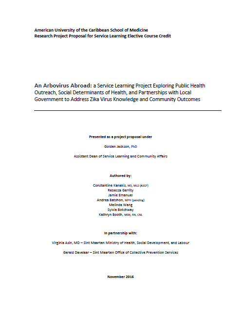
Figure 1. Title Page of original IRB/Research Project Proposal under G. Jackson, Ph.D., Assistant Dean, Community Affairs and Service Learning at AUC School of Medicine

After we secured IRB approval, we began work quickly. Holding meetings with the Ministry’s representative consultant for their office of Collective Prevention Services (i.e. vector control) and scheduling the remaining work for the semester. With five new members of the “Z-Pack” we established a loose timeline with our advisor. Our new goal: integrate what we learned last semester and bring it to a conclusive change within the community.

irb2

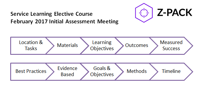
Figure 2. Title card from initial briefing meeting with members of the Z-Pack, including coordinating partner from the Ministry of Health (CPS office) Mr. G. Davelaar.

This integration of knowledge from literature review/research, evidence-based best practices, and forward moving progress are all things those of us in the medical laboratory profession are quite familiar with. Getting IRB approval for a lab-centric project is quite involved and requires meticulous proof and substantial support to posit any claim to the benefit/risk ratio involved with human or animal subjects. I remember from my own graduate and undergraduate research that without heaps of evidence, you will be hard pressed to continue in any direction. While public health is a different science, the basis on evidence-based research is still present. During our initial assessments, literature reviews, and brainstorming, the “Z-Pack” went through hundreds of scientific articles covering everything from infection control precedents, to social behavioral change, and even the use of media and fear to illicit change.

Laboratory scientists know the impact of their work, though it may not always be the most evident to the general public. The near 70% of diagnostic information that comes from our work, and the virtual entirety of neoplastic diagnoses rely heavily on our training, skill, and certified competency in evidence-based practices. ASCP has a long-standing mission of advocacy for patients in the way its members and affiliates represent the profession at large. I believe that having those years of experience under my belt and those letters behind my name give me a head start when executing translational research. Going from raw data, analyzing it, and bringing it to life is something we all inherently train to do—and do well!

So, up to date, my team has secured two measures to contribute to our research. First, we gave an educational presentation to a community after-school program in one of Sint Maarten’s endemic regions. We had tailored a wonderful presentation I discussed in a previous post which caught the eye of the Ministry of Health and has spread to numerous places around the island under their sponsorship. With the same success, we managed to reach school-aged children in an engaging way about Zika, their health, and source reduction. Our second event is slated for this weekend where we have partnered with the Muslim student-interest group (MSA) on campus to go with them on their routine visit to a local mosque on a school-sponsored student service day we call “Community Action Day.” While the MSA students engage with their local community, the “Z-Pack” will conduct a two-part effort: to conduct a grounds-inspection for source/vector control around the mosque, and deliver a presentation for both children and adults regarding Zika prevention behavior.

How do those two events connect with my theme of evidence-based lab scientists? Well, one of my engagements when at Northwestern Medicine was to teach a course discussing transfusion protocols and laboratory information to clinical nursing staff. Presenting information, or teaching people, new ways to think about their environment at work or home is a part of being interdisciplinary. I was able to speak with medical jargon to the clinical staff, but with the children I have to use my ability in translating medical knowledge to understandable facts while also keeping the audience interested. My team proved in our last school-aged project, that when children are engaged and enthusiastic about something they have learned, they will take those messages home with them and hopefully contribute to a positive outcome. As for the second example, what could be more directly appropriate for lab folks to understand here: a surprise inspection! Sure, no one’s losing any accreditation points here, but the fact remains that we all have experience from one side or another making sure that things are up to code on pre-determined conditions and protocols. We have an SOP from the Ministry regarding the items of inspections as they relate to source control, so translating them to a new site should prove interesting.

I’ll close this post off with an interesting piece recently posted by Ms. Susan M. Lehman, MA, MT (ASCP)SM where she discussed learner (i.e. student) experiences. She talked briefly about how online curriculums and other lab-skills courses may rely on more independent learning, changing the expectations of students. One of her students summarized it positively saying, “you get what you put into it.” That’s what I think about the service elective my work is associated with. It could be simple directed readings with great discussions, but what my “Z-Pack” team has and the skills we each bring to it have made the project and its partnerships exciting.

Thanks for reading!


ckanakisheadshot_small

Constantine E. Kanakis MSc, MLS (ASCP)CM graduated from Loyola University Chicago with a BS in Molecular Biology and Bioethics and then Rush University with an MS in Medical Laboratory Science. He is currently a medical student at the American University of the Caribbean and actively involved with local public health.

Microbiology Case Study: A 25 Year Old Male with Fevers, Headache, and Neck Pain

Case History

A 25 year old Caucasian male with no significant past medical history presented to the emergency department (ED) with a several day history of persistent fevers, headache (pain 10/10), dizziness and neck pain. He also reported facial and hand numbness and difficulty focusing at work.  His laboratory values at that time showed a normal white blood cell count (5.3 TH/cm2), normal hemoglobin (13.8 g/dL), slightly decreased platelet count (142,000 TH/cm2) and slightly elevated liver enzymes. A computed tomography (CT) of his head showed no abnormalities, and a lumbar puncture was performed that was suggestive of viral meningitis (92% lymphocytes). After obtaining blood cultures, the patient received a dose of vancomycin and ceftriaxone and was discharged home.

Two days later he returned to the ED with complaints of worsening neck pain, photophobia, decreased appetite, and fevers reaching 105°F. He reported fevers of such intensity that he resorted to soaking himself in ice baths. On further questioning, he reported working in a microbiology lab that handles cytomegalovirus (CMV) and attenuated mycobacterium, but was unaware of any exposures or sick contacts. He has 2 dogs he rescued (one with a history of heartworm), 3 cats (one with a history of tapeworms) and a mouse. Infectious disease was consulted and a thorough workup was initiated, which included repeat blood cultures and testing for hepatitis, human immunodeficiency virus (HIV), respiratory pathogens and syphilis. He was started on cefepime initially and then later changed to meropenem and levofloxacin.

Laboratory identification

brucan1

Figure 1. Gram stain from a positive blood culture illustrating small Gram negative coccobacilli (100x, oil immersion).

The microbiology laboratory reported the blood cultures collected during his second hospital visit were positive with small gram negative coccobacilli after approximately 60 hours on the automated instrument (Figure 1). No growth was noted after 24 hours incubation at 35°C in 5% CO2 on standard media. After 48 hours, small white colonies grew on sheep blood and chocolate agars but failed to grow on MacConkey agar. Biochemical tests revealed the organism was positive for catalase, oxidase and urease. In accordance with the suspected agents of bioterrorism manual, the culture was sent to the State Department of Health for further classification. The organism was identified by PCR as Brucella spp.  Subsequently, the Centers for Disease Control and Prevention (CDC) performed species level PCR and identified the isolate as B. canis.

On further questioning, the patient denied consuming unpasteurized milk products but reported recently adopting a pregnant dog from a local shelter, who had subsequently delivered stillborn puppies of which the patient had been in close proximity. At this point, the patient’s antibiotics were switched to a 6 week course of oral doxycycline and rifampin. On follow up visits, he was doing well and symptom free. Unfortunately, the dog also tested positive for B. canis and had to be euthanized.

Discussion

Brucella spp. are common zoonoses among wildlife and domestic animals including cattle (B. abortus), pigs (B. suis), goats (B. melitensis) and dogs (B. canis) who are usually asymptomatic carriers. While rare in the United States due to vaccination of livestock, Brucella spp. is considered endemic in areas of the Middle East, Central and South America and the Indian subcontinent. Symptoms of infection generally occur during an infectious abortion in which the placenta, fetal tissues and secretions contain high levels of the bacteria which can survive in the environment under various conditions for long periods of time. Humans are usually infected due to consumption of unpasteurized milk and cheeses. High risk professions such as veterinarians and slaughterhouse workers can also be infected by direct contract with contaminated materials or inhalation of aerosolized particles. Symptoms generally appear 1 to 2 weeks after infection with remittent/undulant fever the characteristic feature of the illness, in addition to arthralgias, fatigue, weight loss and hepatomegaly.

Laboratory identification of Brucella spp. is the gold standard but can be challenging as it is a slow growing organism and can infect personnel leading to laboratory acquired infections (LAI). When small Gram negative coccobacilli are identified that fail to grow on MacConkey agar, this should alert the laboratory worker of a potential agent of bioterrorism and work up should be performed in appropriate biosafety cabinets. Brucella spp. grows as small, smooth white colonies that appear after 24 to 48 hours incubation. It is catalase, oxidase and urease positive. Automated systems and MALDI-TOF mass spectrometry are not terribly reliable or recommended for identification of this organism due potential aerosolization events. When Brucella spp. is suspected, the level A clinical laboratory (a sentinel lab) should notify and send samples to a Level B/C lab (state health department) for confirmation.  Subsequently, confirmed isolates can be forwarded to a level D lab (CDC) for speciation.

While overall, the mortality for Brucella spp. is very low, significant morbidity can result with long term non-specific symptoms and cardiac and osteoarticular complications. Good outcomes result when acute presentations are treated with combined regimens of antibiotics. The World Health Organization (WHO) recommends the use of oral doxycycline and rifampin for 6 to 8 weeks. Susceptibility testing is not recommended as resistance is rare and the concern for laboratory safety. In the case of laboratory exposures, prophylaxis with doxycycline and rifampin for 3 weeks is recommended for high risk workers. In the case of low risk employees, temperature monitoring for 6 months and serologic testing at defined time points is standard as the incubation period for Brucella can be this long.


mb

-Melissa Brents, MD, is a 4rd year Anatomic and Clinical Pathology resident at the University of Mississippi Medical Center.

Stempak

-Lisa Stempak, MD, is an Assistant Professor of Pathology at the University of Mississippi Medical Center in Jackson, MS. She is certified by the American Board of Pathology in Anatomic and Clinical Pathology as well as Medical Microbiology. She is the director of the Microbiology and Serology Laboratories.  Her interests include infectious disease histology, process and quality improvement and resident education. 


The Learner Experience in a Blended Model of Curriculum Delivery

Key to successful delivery of an online course (or as in our case, a blended model of online and traditional), along with achievement of the learning objectives, is the learner experience. I’ll never forget the feelings of trepidation I had on our first day with our inaugural class, piloting this new model of curriculum delivery with our bacteriology course.

Our lesson plan requires that the students prepare for class by studying the online lecture material as homework, prior to the next day’s laboratory section. Our students were excited about starting our program and eager to learn, yet some were hesitant. I remember one student stating that they “might not be so sure about this new format.” After all, we hadn’t tried it before, and to be frank, it was scary. I remember thinking to myself, “What are we going to do if they do not study the online content? What if they do not prepare for class? What if they dislike this format? What will we do if they flunk their first exam?
Fortunately that was not the case, and our student’s performance in our program has been and continues to be highly successful.

Alex, a student in our current class put it this way:

“It is worth noting that this is not your typical college course. The program here really emphasizes the “reverse classroom” technique. For those unfamiliar, this term means that one will read about the lesson the night before and come to class the next day and perform a laboratory assignment based on that reading.

I came into the program experiencing nothing like this before, so I wasn’t sure how this learning strategy would work for me. After completing our didactic schedule, many of my peers would agree with me that this learning technique is fantastic and is very beneficial to the overall learning experience.

However, to maximize this benefit, time management is vital. Simply reading the lesson at the last minute does not cut it. Whether it helps you to take notes as you go, doing a re-read, or fill out a study guide, this style of learning is a classic example of getting out what you put into it.”

I loved hearing our student reflect that “you get out of it what you put into it.” To me, that is the ultimate goal of education, to prepare our students to be able to think critically and self-direct their learning. In this regard, our inaugural class was a success.


Lehman_small

-Susan M. Lehman, MA, MT(ASCP)SM graduated from the University of Wisconsin-Madison in 1983 with a BS in medical technology. She is program director for the Medical Laboratory Science Program and course director for Clinical Microbiology I and II; her areas of interest include distance education and education methodology.

Microbiology Case Study: A 71 Year Old Man with a dehised Corneal Wound

Case History

A 71 year old man presented with a dehisced corneal wound status post corneal transplant.

Laboratory Diagnosis

Corneal scrapings from the ulcer were submitted for interpretation and two separate organisms were isolated from the blood agar plates. The first was a non-motile gram negative rod that grew on 5% horse blood agar and MaConkeys agar. The organism was oxidase negative and spot indole negative and was identified as Klebsiella pneumoniae.

The second organism grew in bright yellow-pigmented colonies on 5% horse blood agar (Image 1) but did not grow on MacConkeys. The organism was oxidase positive and spot indole positive and was identified as Chryseobacterium indologenes by MALDI.

chryseo1
Image 1.

Discussion

Chryseobacterium indologenes, previously known as Flavobacterium indologenes is a yellow pigmented, gram-negative filamentous, non-motile rod that is a non-glucose fermenter and can be found in soil, plants, foodstuffs and water sources including those found in hospitals. It produces a water-insoluble pigment, flexirubin which gives it its characteristic color. It was first isolated from a clinical specimen in 1983 however there have been more recent reports of bacteremia related to C. indologenes related to use of indwelling devices, such as a catheters.

  1. indologenes typically exhibits resistance to multiple antibiotics, however, a case series of 16 patients with C. indologenes infections, all nosocomial and in patients with comorbidities, showed no clear relationship between antibiotic susceptibility and response to treatment (1).


Reference:

  1. Lin Y-T, Jeng Y-Y, Lin M-L, Yu K-W, Wang F-D, Liu C-L. 2010. Clinical and Microbiological Characteristics of Chryseobacterium indologenes Bacteremia. J. Microbiol. Immunol. Infection.43:498-505.


-Agnes Balla, MD is a 3rd year anatomic and clinical pathology resident at the University of Vermont Medical Center.

Wojewoda-small

-Christi Wojewoda, MD, is the Director of Clinical Microbiology at the University of Vermont Medical Center and an Assistant Professor at the University of Vermont.


Zika Virus: The Struggle is Still Real

Well, even though the groundhog has predicted another 6 weeks of winter, it’s not too early to start thinking about those summer pests – mosquitos – and the diseases they bring with them. Zika virus, in particular. Even though the winter weather has dulled our sensitivity to this emerging threat, it remains a significant problem. The virus is now circulating in 65 countries, mostly in the Americas (1). Currently, the CDC has issued travel alerts to areas where Zika is spreading including Miami-Dade, Florida, Puerto Rico, American Samoa, and the US Virgin Islands (2).  Laboratory professionals should be aware of Zika virus because the diagnosis of Zika Virus Disease (ZKD) relies on the laboratory, and many healthcare professionals require guidance from the laboratory on how to proceed with diagnostic testing.

Zika is a flavivirus borne by the Aedes mosquito. Symptoms of ZKD usually last 2-7 days and include mild fever, skin rash, conjunctivitis, muscle and joint pain, malaise or headache. There is scientific consensus that Zika virus infection during pregnancy is the cause of congenital brain abnormalities of the fetus, including microcephaly. This devastating effect of the virus sets it apart from the other Aedes-borne viruses dengue, chikungunya, and yellow fever.

Diagnosis of Zika virus relies on laboratory testing, and yet, there are no FDA approved assays for Zika virus currently available. There are however a number of assays that have been given approval for emergency use, including: Real-Time PCR, MAC-ELISA, and a plaque neutralization reduction test. In the United States, these tests are available from the CDC and some state health labs. An algorithm describing the appropriate use of these tests can be found here. Unfortunately, in developing countries where Zika is endemic, access to the appropriate diagnostic test can be very difficult.

Impediments to accurate diagnosis of ZKD in developing countries include lack of education and access to quality laboratories that offer the right test. Lack of education encompasses not only transmission and prevention of the virus, but also who should seek medical attention and when and who should be tested and when. In many countries where visiting a medical professional is a financial burden to a family, it is less likely that a family will seek medical attention for a disease that has such mild symptoms symptoms as ZKD. While most cases of ZKD don’t require medical attention beyond comfort care, if patients don’t report to clinics or health centers, it is difficult to track and confirm cases if no one presents with a suspected case! Also, there is a need for consensus about what should be called a suspected ZKD case and then how to proceed with confirmatory testing. Some countries, including Brazil, the respective Ministries of Health have issued definitions of a suspected Zika case. The Brazilian definition includes: “patients who present with pruriginous maculopapular exanthema accompanied by two or more of the following signs and symptoms: fever, conjunctival hyperemia without secretion and pruritus, polyathralgia, and periarticular edema” (3). Suspected cases can be confirmed with diagnostic testing, but this is another challenge. The easiest and least expensive test for a clinic in the developing world is a Ig-M based rapid diagnostic test. There are several of these available commercially, mostly from European markets. However, these demonstrate significant cross-reactivity with other flaviruses such as dengue and chikungunya, which are also endemic in areas where Zika is now circulating. The most appropriate tests – RT-PCR, MAC-ELISAs, and plaque reduction tests – are only available in national laboratories it at all. The combination of lack of patients reporting Zika-like symptoms, lack of consensus of what constitutes a suspected case, and limited availability of confirmatory testing means that there is a significant likelihood that the number of Zika cases in many developing countries are underreported.

In January 2016, the WHO presented the Strategic Response Framework and Joint Operations Plan in response to the growing Zika virus epidemic. In October 2016, a quarterly update was released that described the goals and scope of the plan through December 2017. The plan is Strategic Response Plan comprised of four areas: 1) Detection, 2) Prevention, 3) Care and Support, and 4) Research. $10.9 million are dedicated to the detection arm of the strategic plan, which in addition to laboratory testing and diagnosis includes assessment and implementation of preparedness measures, and surveillance and monitoring in it’s scope. $41.2 million are dedicated to the research arm of the plan, which includes the “fast track and scale up of research development and availability of diagnostic tests.”

Hopefully in the next year we will see not only new diagnostic testing, but also medical interventions such as vaccines. In the meantime, it is important that we as laboratory professionals continue to be apprised of available testing, to educate our healthcare partners on the use of lab testing for ZKD, and to support research and development of Zika diagnostics.

References

  1. Falcao et al. Ann Clin Microbioal Antimicrob (2016) 15:57
  2. MMWR, February 12, 2016: 65(05); 122-127
  3. Zika Strategic Response Plan Quarterly Update. 25 October 2016.


Sarah Brown Headshot_small

Sarah Riley, PhD, DABCC, is passionate about bringing the lab out of the basement and into the forefront of global health.  

Microbiology Case Study: A 14 Year Old Boy with Cystic Fibrosis

Case History

A 14 year-old-boy with Cystic Fibrosis had a respiratory culture collected at his routine clinic visit. It grew abundant mixed respiratory flora, and rare Gram-negative coccobacilli. This organism grew as non-lactose fermenting colonies on MacConkey agar (Figure 1) in approximately 36 hours and was oxidase and catalase positive. The isolate was identified by MALDI-TOF MS with a score of 2.39, which is acceptable for species-level identification.

bord1

Discussion

Our isolate was identified as Bordetella bronchiseptica. Bordetella spp. are small Gram-negative rods that often appear as coccobacilli. Like other Bordetella spp., our isolate was catalase positive. Oxidase results vary across the genus, but B. bronchiseptica is oxidase positive. Some Bordetella spp. including B. pertussis and B. parapertussis, are very sensitive to metabolites and toxic substances found in many types of microbiological media. For the best chance of recovering these fastidious Bordetella spp. in culture, specialized agar such as Regan-Lowe, Bordet-Gengou, or Stainer-Scholte medium along with extended incubation periods are used. Due to the difficulty of culturing B. pertussis and B. parapertussis, these days culture is only performed at large reference laboratories or public health facilities. Testing by PCR is the current clinical practice and has greatly improved the sensitivity of detection for these fastidious organisms. In contrast, other Bordetella spp. including B. bronchiseptica are routinely recovered in culture using standard laboratory methods.

The most clinically relevant Bordetella spp. in humans are B. pertussis, the infectious agent of whooping cough, and B. parapertussis, which causes a similar illness to B. pertussis but generally symptoms are less severe. B. bronchiseptica causes respiratory infections in many animals including cats and dogs, and is the infectious agent of kennel cough.  B. bronchiseptica can cause respiratory infection in humans who acquire the bacterium primarily from their infected pets. Human infection is rare, and most likely to occur in immunocompromised patients such as those with poorly controlled HIV or Cystic Fibrosis.

B. bronchiseptica produces a β-lactamase making the organism resistant to penicillin and many cephalosporins. Most strains are resistant to trimethoprim-sulfamethoxazole as well. In contrast, strains of B. bronchiseptica are generally susceptible to β-lactam/ β-lactamase inhibitor combinations, quinolones, aminoglycosides, and tetracycline.

Our patient was not having an exacerbation at the time of specimen collection, so he continues to do well. We expect to find B. bronchiseptica in his future sputum specimens, but the pathogenicity of B. bronchiseptica in such a low amount compared to respiratory flora is unclear.

Erin McElvania TeKippe, PhD, D(ABMM), is the Director of Clinical Microbiology at Children’s Medical Center in Dallas Texas and an Assistant Professor of Pathology and Pediatrics at University of Texas Southwestern Medical Center.

ASCP Annual Meeting Call for Abstracts Now Open

The 2017 ASCP Annual Meeting is in Chicago, IL September 6-8. If you’d like to present your research at the meeting, the call for abstracts is now open. Summarize your work in 300 words or less and submit it through the online portal by March 20th, 2017.

Spread the word, and good luck!


Managing the Emergency in Your Lab

So far in 2017, the United States has seen blizzards, fires, tornadoes, floods, and other disasters. Have any of these disasters struck near you or affected your laboratory? If it did strike your area, would you be prepared? Would your lab staff know what to do and how to work to continue lab operations? A comprehensive emergency operations plan is not something that should be dusted off and considered when an emergency situation occurs. It should be reviewed and tested on a regular basis, and all lab staff should know how to put it into action easily.

The College of American Pathologists (CAP) requires laboratories to have “written policies and procedures defining the role and responsibilities of the laboratory in internal and external disaster preparedness.”  A second related standard also requires that labs have a functional evacuation plan in case work can no longer be performed in the department because of unsafe conditions. These policies should be developed with input from lab leaders, medical directors, and other key hospital or facility emergency management personnel. The disaster plan for the lab must work for the department, but consideration must be given to other areas if the lab does not stand alone in the building.

As with many lab safety guidelines and rules, regulatory agencies often put forth changes or updates as they deem necessary. At the end of 2016, the U.S. Centers for Medicare & Medicaid Services (CMS) published an updated final rule for healthcare providers- and that includes labs- regarding Emergency Preparedness. The purpose of the rule was to establish national emergency preparedness requirements, to ensure adequate planning for both natural and man-made disasters, and to provide coordination with federal, state, tribal, regional and local emergency preparedness systems.

The CMS requirements can be broken down into four elements, Policies and Procedures, Risk Assessment and Emergency Planning, Communication Plans, and Training and Testing. First, all lab and hospital emergency management policies or procedures need to comply with federal and state laws. As stated earlier, these policies need to be easily understood so that any staff member can put them into motion. There may be disaster scenarios in which lab leadership may not be able to get to the site. Lab emergency operations plans should be reviewed or updated annually.

Hospitals and labs should review the hazards in the local areas and assess what disaster types are most likely. Consider situations like equipment or power failures, and even an interruption in communications, including cyber-attacks. CMS also wants facilities to plan for the loss of all or a portion of a facility, or even the loss of supplies.

Laboratories should have a plan to contact staff, including physicians or other necessary persons. This communication system should be well-coordinated within the facility and across health care providers. The state and local public health departments and emergency management agencies need to be included in the facility communication plan as well.

The final CMS-required core element for emergency response includes testing and training. All staff needs to be familiar with the contents of the response plan, and the plan should be well-maintained through regular training of staff and testing. That testing can include the use of table-top drills or even assessing how the plan worked in a real disaster scenario. While CAP allows many lab policies to be reviewed once every other year, CMS requires an annual review or update of these disaster policies and procedures.

Developing a comprehensive emergency management plan is no small undertaking, and if you don’t have one in place already, make sure you gather a team to help with that project since there is much to consider. If you belong to a system of laboratories, you also need to consider how the plan will connect the actions of multiple sites. If you have a plan in place, make sure you assess it regularly for ease of use and the ability to achieve its goals. Those goals should include the safety of staff, the continued delivery of services (if possible), and recovery to normal operation. We know that emergency situations aren’t all that rare, and following this pathway can help your lab be ready when the next disaster strikes.


Scungio 1

Dan Scungio, MT(ASCP), SLS, CQA (ASQ) has over 25 years experience as a certified medical technologist. Today he is the Laboratory Safety Officer for Sentara Healthcare, a system of seven hospitals and over 20 laboratories and draw sites in the Tidewater area of Virginia. He is also known as Dan the Lab Safety Man, a lab safety consultant, educator, and trainer.


Microbiology Case Study: A 3 Year Old Girl with Abdominal Pain and Fever

Case History

A 3 year old girl initially presented with abdominal pain and fevers. Ultrasound identified a left kidney mass, leading to a left radical nephrectomy and excision of retroperitoneal mass. Pathology showed a Wilms’ tumor, diffuse anaplasia type. Staging uncovered multiple pulmonary metastases and involvement of a supraclavicular lymph node. She received chemotherapy and radiation without regression of disease.  She then presented to the ED and was admitted for neutropenic fever. She was started on broad-spectrum antibiotics. She continued to spike fevers so an antifungal, micafungin, was added.  While admitted, she developed scattered erythematous papules. Infectious disease was consulted and a skin biopsy from the left forearm was obtained.

Laboratory Results

  • Bacterial cultures, blood: negative
  • Fungal cultures, blood: negative
  • Blastomyces urine antigen: negative
  • Skin biopsy: slight epidermal hyperplasia with follicular dilatation, mild vascular ectasia, and focal erythrocyte extravasation. Negative for organisms.
  • Bacterial culture, tissue: no growth.
  • Gram stain: rare budding yeast forms seen
  • Fungal culture, tissue: no growth to date
blasto-1
The gram smear made from skin biopsy tissue for bacterial culture displayed rare broad-based budding yeast forms, consistent with Blastomyces dermatiditis.

blasto-2

The gram smear made from skin biopsy tissue for bacterial culture displayed rare broad-based budding yeast forms, consistent with Blastomyces dermatiditis.


Discussion

Blastomyces dermatitidis is a dimorphic fungus found in moist soil and decomposing matter. It is endemic within the Mississippi and Ohio River valleys as well as the Great Lakes region and Southern United States.  There are reports of infection in Africa and India. The fungus is transmitted by inhalation of as few as 10-100 conidia. Once in the lungs, the spores convert to yeast and multiply. Infection usually results in a flu-like illness with pulmonary involvement 3-15 weeks post-exposure. Hematogenous spread can further result in involvement of the skin, bone, genitourinary tract, and central nervous system.

The gold standard for diagnosis is culture or cytopathology/histology. However, the organism is a slow grower, which can take 2-4 weeks, and may fail to grow in one-third of cases. On culture at room temperatures (25-30°C), the mold form appears wrinkled and waxy and is cream to tan in color. Microscopically, they form septate hyphae with short or long conidiophores bearing small round to pear-shaped conidia (2-10 microns). This arrangement of the mold is described as a “lollipop” appearance. At 35-37°C, the fungus is a yeast (8-10 microns) with classic broad-based budding and double contoured walls.

Antigen testing is available on urine, serum, bronchoalveolar lavage fluid, and CSF. Antigen testing is more rapid, utilizing enzyme immunoassay, but has a lower sensitivity. Antigen testing is most sensitive in patients with isolated pulmonary disease. Serial urine antigen testing can be used to indicate disease regression or relapse.

A real-time PCR assay is available for confirmation of B. dermatitidis. The probe targets the promoter region of the BAD1 gene, which encodes an adhesin molecule and virulence factor. This method can be performed in five hours, but is only available at reference laboratories.

Mild to moderate pulmonary and extrapulmonary blastomycosis can be treated with oral itraconazole. Severe cases, CNS involvement, or infection of immunosuppressed patients, pregnant women, or children require amphotericin B followed by step-down therapy with itraconazole for 6-12 months.

Upon report of the mold on gram smear, micafungin was discontinued and amphotericin B treatment initiated. Her fever and rash resolved. The patient was transitioned to oral itraconazole prior to discharge and will remain on therapy for 12 months.

REFERENCES

  1. https://www.cdc.gov/fungal/diseases/blastomycosis/index.html
  2. Frost HM, Novicki TJ. Blastomyces Antigen Detection for Diagnosis and Management of Blastomycosis. Journal of Clinical Microbiology. 2015;53(11):3660-3662. doi:10.1128/jcm.02352-15.
  3. Sidamonidze K, Peck MK, Perez M, et al. Real-Time PCR Assay for Identification of Blastomyces dermatitidis in Culture and in Tissue. Journal of Clinical Microbiology. 2012;50(5):1783-1786. doi:10.1128/jcm.00310-12.
  4. Chapman SCAW, Dismukes WE, Proia LA, et al. Clinical Practice Guidelines for the Management of Blastomycosis: 2008 Update by the Infectious Diseases Society of America. Clinical Infectious Diseases. 2008;46(12):1801-1812. doi:10.1086/588300.


-Prajesh Adhikari, MD is a 2nd year anatomic and clinical pathology resident at the University of Vermont Medical Center.

Wojewoda-small

-Christi Wojewoda, MD, is the Director of Clinical Microbiology at the University of Vermont Medical Center and an Assistant Professor at the University of Vermont.