Lab Value Changes in Transgender Males

For patients with gender dysphoria, the Endocrine Society has endorsed the use of hormone therapy to promote secondary sexual characteristics of the desired gender. These guidelines were first established in 2007 and revised last year, and gave the first evidence guided recommendations for clinicians treating transgender patients.

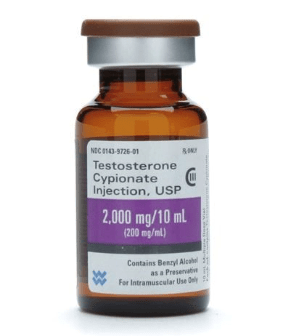
For transgender males, testosterone by itself is prescribed as an injectable oil-based solution. These doses are given as intramuscular injections- usually into the thigh. If that’s too painful, subcutaneous injections have been shown to have similar efficacy. The doses given to transgender males is much higher (50-100mg/ injection) than that given to men with testosterone deficiency (30-50 mg/ injection). Primarily because the men have more testosterone to start with. Also, whereas topical testosterone gel may be sufficient for men with “low T,” it doesn’t seem to provide enough testosterone to transgender males and is quite expensive, so it is generally not used.

Nov1.png
Image 1. Picture of testosterone cypionate vial from mcguffmedical.com. This is used for intramuscular injections.

Upon starting testosterone injections, the frequency of injections is every one to two weeks. However, the onset of physical secondary sexual characteristics takes 3-6 months to begin. After about 3 years, most of the changes to occur will have manifested. These physical changes are outlined in the table below. You’ll notice how certain traits like cessation of menses and fat redistribution start within the first 6 months whereas muscle growth and voice change take effect after 6 months. Also, the time certain effects take maximal effect varies; the voice doesn’t deepen further after 2 years, but hair growth continues to increase through 5 years.

Physical Effect Begins Maximal Effect
Facial/body hair growth 6-12 mo 4-5y
Skin oiliness/acne 1-6mo 1-2y
Scalp hair loss 6-12 mo
Increased muscle mass 6-12 mo 2-5y
Fat redistribution 1-6mo 2-5y
Cessation of menses 1-6mo
Deepening of voice 6-12 mo 1-2y

Table 1. Timeframe of physical traits that manifest in transgender males while taking testosterone hormone therapy. Based on Hembree et al. 2017 (1).


Just as hormone therapy induces physical manifestations of secondary sexual characteristics for transgender men, we would suspect that internal aspects of physiology are affected too.  Values measured by the laboratory provide meaningful insight into how our body and its different organ systems are functioning. Accordingly, the Endocrine Society also recommended laboratory monitoring of transgender patients starting hormone therapy.

  1. Measure Testosterone and hemogoblin/ hematocrit every 3 months for the 1st year, then 1-2x/ year afterwards.
  2. Monitor Lipids at regular intervals

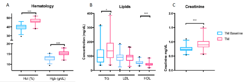
Previous studies have monitoring these lab values found consistent increases in hemoglobin and hematocrit (2,3). This is due to the stimulation of erythropoiesis by testosterone (4).  While excessive testosterone could lead to polycythemia (excessive RBCs in the blood), it is not a commonly described complication in transgender patients. Some summary results from our study for hemoglobin and hematocrit are shown in Figure 1A, which shows a clear shift in levels.

However, reports on lipids have been varied LDL and triglyceride changes (2,3). The only consistent finding was that HDL decreased in transgender males taking testosterone (2,3). In our study, we found triglycerides were increased with decreased HDL (Figure 1B). The take-away is that because cardiovascular cut-offs are based on risk and not a reference range, patients and clinicians will have to be aware of these possible metabolic changes.

Creatinine, when it was checked, increases for transgender males (5). We found creatinine was strongly increased in our study to become similar to baseline creatinine in transgender women before taking hormone therapy (Figure 1C). This topic as it relates to glomerular filtration rate is very complex and will be discussed further in a future post.

To illustrate lab value changes in transgender men, I’ll direct you to data that I found in a large study of over 300 transgender patients including about 80 transgender men. The completed manuscript is not currently available but will be printed soon:

Nov 2

However, this does not mean Cisgender male reference intervals are adequate for transgender men. This topic needs further exploration and ideally a prospective trial to be performed in a controlled manner. A double-blind study would not be possible as it would be unethical to perform.

References

  1. Hembree WC, Cohen-Kettenis PT, Gooren L, Hannema SE, Meyer WJ, Murad MH, et al. Endocrine Treatment of Gender-Dysphoric/Gender-Incongruent Persons: An Endocrine Society* Clinical Practice Guideline. J Clin Endocrinol Metab. 2017
  2. Wierkx K, et al. Cross-Sex Hormone Therapy in Trans Persons is Safe and Effective at Short-Time Follow-Up: Results from the European Network for the Investigation of Gender Incongruence. J Sex Med, 2014. 11(8):1999-2011.
  3. Mueller A, Kiesswetter F, Binder H, Beckmann MW, Dittrich R. Longer-term administration of testosterone undecanoate every 3 months for testosterone supplementation in female-to-male transsexuals. J Clin Endocrinol Metab. 2007
  4. Paller CJ, Shiels MS, Rohrmann S, Menke A, Rifai N, Nelson WG, et al. Association Between Sex Steroid Hormones and Hematocrit in a Nationally Representative Sample of Men. J Androl. 2012 33(6): 1332-1341.
  5. Fernandez JD, Tannock LR. Metabolic Effects of Hormone Therapy in Transgender Patients. Endocr Pract. 2016;22:383–8.


SoRelle Picture

-Jeff SoRelle, MD is a Molecular Genetic Pathology fellow at the University of Texas Southwestern Medical Center in Dallas, TX. His clinical research interests include understanding how the lab intersects with transgender healthcare and advancing quality in molecular diagnostics.

Hematopathology Case Study: A 56 Year Old Man with Sinus Congestion and Axillary Adenopathy

Case History

A 56 year old male presented to his PCP complaining of sinus congestion, rhinorrhea, night sweats, decreased appetite and fevers of up to 101-102 every evening. Hematologic evaluation revealed a neutropenia and a lymphopenia. An infectious disease work up was negative. His LDH was elevated. Physical examination reveals an enlarged left axillary lymph node. An excisional biopsy was performed.

Biopsy Findings

Figure 1.jpg

Figure 2.jpg

H&E stained sections demonstrate an enlarged node with effaced architecture and scattered residual follicles with small, mature cells. There is a proliferation of intermediate to large, to very large, atypical and highly pleomorphic cells many of which demonstrate bizarre forms, irregular nuclear morphology and acidophilic nucleoli. The lymphoma cells are noted to focally traverse through adipose tissue. Occasional hallmark cells are appreciated.

To further characterize the infiltrate, immunohistochemical stains were performed and interpreted with appropriate controls. The lymphoma cells were diffusely positive for CD45 (LCA), CD43, and CD30 (membranous and Golgi) with a Ki-67 of 80-90%. These cells were negative for CD20, PAX-5, CD3, CD4, CD8 (mostly), CD5, D10, BCl-2, BCl-6 and ALK1.

The morphologic features and immunophenotype of the cells was diagnostic of anaplastic large cell lymphoma, ALK negative.

Discussion

Anaplastic Large Cell Lymphoma (ALCL), ALK-negative (ALK-) is defined as a CD30+ T-cell neoplasm that morphologically resembles ALK-positive ALCL, but lacks ALK protein expression. It most commonly affects adults (aged 40-65 years), and has a slight male preponderance with a male-to-female ratio of 1.5:1. T. Most patients present with advanced disease (stage III-IV), lymphadenopathy and B symptoms. The most common differential diagnosis is ALK-positive ALCL.

The molecular deciphering of ALCL began in the 1990s with the discovery of a recurrent t(2;5) (p23;q35) translocation fusing the ALK gene and the nucleophosmin gene generating a NPM-ALK fusion protein, as well as other ALK translocations resulting in a high ALK kinase activity. This triggers the major oncogenic pathway in ALK-positive ALCL. Pharmacologic therapy has been developed to target ALK, and has shown efficacy. Thus, compared with ALK-negative cases, ALK-positive occurs in younger patients and has a better prognosis. ALK-negative ALCL also tends to involve both lymph nodes and extranodal tissues, although extranodal sites are less commonly involved than in ALK+ ALCL.

The other differential diagnoses of ALK- ALCL includes, primary cutaneous ALCL (C-ALCL), other subtypes of CD30+ T-cell or B-cell lymphoma with anaplastic features and classic Hodgkin Lymphoma. If a single lymph node or cutaneous cases are suggestive of ALK- ALCL, C-ALCL needs to be considered. Any cases that involve the gastrointestinal tract need to be distinguished from CD30+ enteropathy-associated and other intestinal T-cell lymphomas.

Molecular analysis of ALK- ALCL shows characteristic strong expression of CD30, in equal intensity in all the cells. Loss of T-cell markers is frequently seen, however, more than half of all cases express one or more T-cell markers. CD2 and CD3 are more commonly expressed than CD5, and CD43 is almost always expressed. CD4+ is frequently positive, while CD8+ is rare. Many cases also express cytotoxic markers TIA1, granzyme B, and/or perforin.

The genetic profile in ALK-negative ALCL has been found to be pretty heterogenous. Most notably, activating mutations of JAK1 and/or STAT3 have been shown to lead to activation of the JAK/STAT3 pathway. Chromosomal rearrangements of DUSP22 (i.e. chromosomal rearrangements in or near the DUSP22-IRF4 locus on 6p25.3) occur in 30% of the cases, and rearrangements of TP63 occur in about 8% of cases. Neither of the rearrangements have been reported in ALK+ ALCL.

From a prognostic standpoint, studies have shown that the rearrangements have effects on the survival rate. TP63-rearranged cases were shown to have an unfavorable prognosis worse than ALK- ALCL with neither rearrangement, while DUSP22-rearranged cases were shown to have favorable outcomes similar to ALK-positive ALCLs.

References

  1. Gaulard P, de Leval L. ALK-negative anaplastic large-cell lymphoma. 2016 Jan 14;127(2):175-7.
  1. Edgardo R. Parrilla Castellar et al., ALK-negative anaplastic large cell lymphoma is a genetically heterogeneous disease with widely disparate clinical outcomes Blood. 2014 Aug 28; 124(9): 1473–1480.


Bradon Zelman

-Brandon Zelman is 4th year medical student at the Philadelphia College of Osteopathic Medicine and an aspiring pathologist. You can follow Brandon on Twitter @ZelmanBrandon.

Mirza-small

-Kamran M. Mirza, MD PhD is an Assistant Professor of Pathology and Medical Director of Molecular Pathology at Loyola University Medical Center. He was a top 5 honoree in ASCP’s Forty Under 40 2017. Follow Dr. Mirza on twitter @kmirza.

Hematopathology Case Study: A Newborn Infant with a High White Blood Cell Count

Case History

The patient is a 1 day old baby boy born at 39 weeks to a 44 year old woman. On physical examination, the baby had a mildly flattened occiput with thickened nuchal skin, downward slant of palpebral fissures with epichanthal folds and slightly low set ears. On imaging, he had a ventricular septal defect. A CBC was performed which revealed a white count of 34.2 K/uL with a differential that included 37 blasts.

Peripheral Blood Smear 

TAM1.jpg

TAM2

Cytogenetics

TAM3

TAM4.JPG

Diagnosis

The peripheral blood showed an increased white count, many nucleated red blood cells as well as a population of blasts. The cytogenetic analysis confirmed the suspicion of trisomy 21. Flow cytometry showed that the population of blasts expressed myeloid as well as erythroid and megakaryocytic lineage specific antigens. The patient was found to have a GATA1 mutation. Approximately one month after birth, the patient’s white count normalized to 7.2 K/uL with only 4 circulating blasts counted.

Discussion

In a patient with trisomy 21, this presentation is consistent with a diagnosis of transient abnormal myelopoiesis associated with Down syndrome (TAM). TAM occurs in 10% of newborns with Down syndrome. Patients typically present with cytopenias, leukocytosis, an increase in blasts and hepatosplenomegaly. Less commonly, patients can have respiratory distress, bleeding, skin rash or jaundice. The blasts are morphologically and immunophenotypically similar to those seen in acute myeloid leukemia. They often have basophilic cytoplasm, coarse basophilic granules and cytoplasmic blebbing, which suggests a megakaryocytic lineage. The immunophenotype generally includes expression of myeloid markers such as CD117, CD13 and CD33 plus erythroid and megakaryocytic markers like CD41, CD42b and CD61.1

In addition to trisomy 21, mutations in GATA1 are almost always seen in the blast cells of patients with TAM. GATA1 is a hematopoetic transcription factor. Bhatnagar, et al. (see diagram below) describe a three step model to explain the evolution of TAM. The initial event is abnormal hematopoesis in the fetal liver caused by trisomy 21. This causes an increase in megakaryocyte-erythroid progenitors in the hematopoetic stem cell compartment.  The second step is the acquisition of an N-terminal truncating GATA1 mutation before birth. GATA1 is a regulator of normal megakaryocyte and erythroid differentiation. The truncated mutation causes marked expansion of megakaryoblastic progenitors.

TAM5
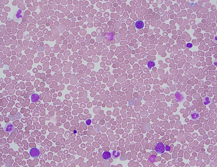
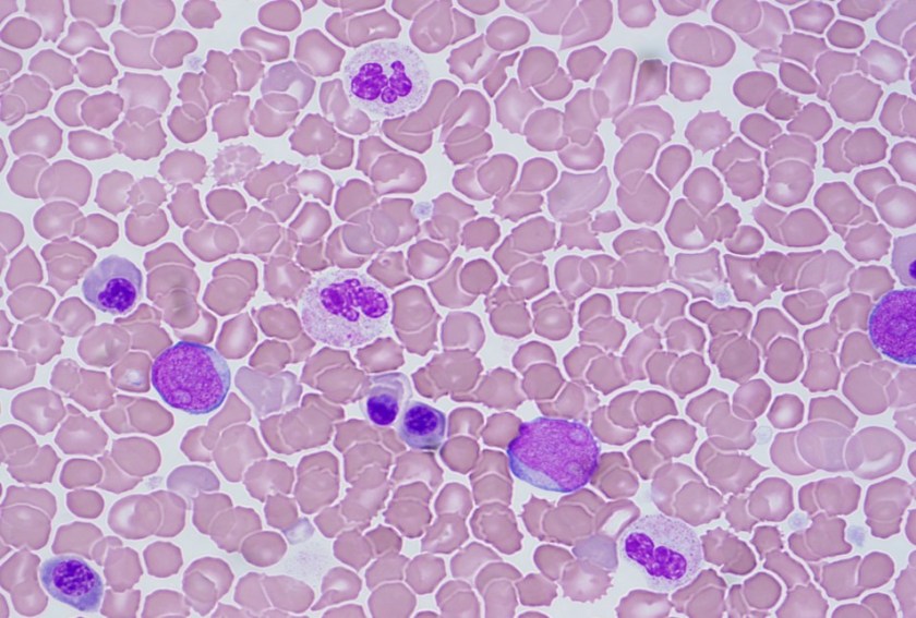
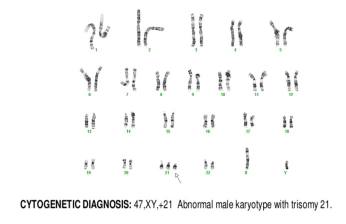
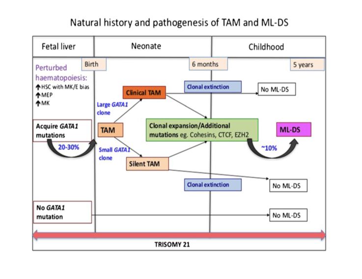
TAM has a high rate of spontaneous remission and typically resolves spontaneously in 90% of patients over several weeks to 6 months. This coincides with extinction of the GATA1 clone. However, in around 10% of these patients, myeloid leukemia of Down syndrome (ML-DS) develops within 5 years of the initial presentation. Additional mutations in cohesion component genes and epigenetic regulators occur in these patients that result in clonal expansion and non-transient leukemia. 2 Children who develop ML-DS generally have a good response to chemotherapy and a have a better prognosis than children without Down syndrome who develop AML.

References

  1. Swerdlow SH, Campo E, Harris NL, et al. WHO Classification of Tumours of Haematopoetic and Lymphoid Tissues (Revised 4th edition). IARC: Lyon 2017.
  2. Bhatnagar, Neha et al. “Transient Abnormal Myelopoiesis and AML in Down Syndrome: An Update.” Current Hematologic Malignancy Reports5 (2016): 333–341. PMC. Web. 21 Oct. 2018.


Marcus, Chelsea_099-Edit

Chelsea Marcus, MD is a third year resident in anatomic and clinical pathology at Beth Israel Deaconess Medical Center in Boston, MA and will be starting her fellowship in Hematopathology at BIDMC in July. She has a particular interest in High-grade B-Cell lymphomas and the genetic alterations of these lymphomas.


Investing in the Best Testing

“Damn that q-tip goes in deep!

But it lit up negative so d/c to street

But it was flu, cuz he bounced back again

And now my Press Gayney’s a minus ten…”

Video 1. Another classic excerpt from a favorite: ZDoggMD, singing about this year’s flu season and available testing options on the horizon—because, let’s face it—rapid flu tests aren’t quite cutting it anymore.

Hello again everyone! Back again to talk about a new set of recommendations from last month’s post. This time it’s about influenza. Recommendation: get vaccinated. Thank you. See you next time…

Seriously, as the 2018-2019 flu season dawns upon us, it’s time to talk about vaccines, tests, prevention, and health literacy. I’m sure many of your social media pages are filled with various debates, articles, and fake news stories on one side or another pitting science, pseudo-science, and non-science all against each other for public spectacle. In the lens of laboratory science and medicine at large, I think most if not all of us agree that preventable diseases should be prevented, and if not, at the very least detected accurately, sensitively, and early. Influenza A/B is a prime example of a consistent threat to our health and safety that has wavered responses in various socio-medical circles.

Official communication and guidance from the Centers for Disease Control and Prevention (CDC) clearly tells those of us in health-care to embrace a multi-tiered approach to protecting public health regarding the flu. That approach includes vaccination, testing, infection control, anti-viral treatment, and anti-viral prophylaxis. And why such a fuss over the flu? It’s a big deal! Last year, the CDC reported approximately 80,000 deaths associated with influenza as a primary cause. 80,000 deaths! That’s almost 7 times as many that died from H1N1/Swine Flu complications back in 2009, where only 12,000 patients were killed by the virus. And even more so, in the terrifying Ebola epidemic of 2016—in which there was a staggering 1 recorded death in the US—nearly 29,000 people were infected globally and only 11,300 died (despite under-reporting). I’m being dramatic, I know. But it’s important for us to recognize true epidemics when they happen, and even more important for societies like ours to be at the forefront of preventing them from developing any further.

flu1.jpg
Image 1. The CDC recommends you get your flu shot every year, because obviously.

flu2.jpg

Image 2. I’m not here to talk about the anti-vax elephant in the room. That’s not fair to elephants. But imagine if the CDC reported 44% of flu vaccine misconceptions were addressed!

As an aside, I’ll probably recommend that you get your annual flu shot a hundred times in this post alone. But just to have a clear reference, please look at the following table. It’s critical to be able to both distinguish common cold versus influenza symptoms for yourself, as well as educate your patients and peers about the differences between the two. This information can change the way people perceive treatments (i.e. why the doctor only recommended rest/Tylenol and didn’t give out antibiotics for their symptoms) and why it’s absolutely crucial to protect vulnerable populations from an otherwise fatal virus. So, micro-rant aside, it should be clear that by now we should be working on a way to both improve our prophylaxis with vaccines and medications as they always leave room for improvement—I’m looking at you Tamiflu and Relenza! Notwithstanding any analysis of efficacy for the flu vaccine, the CDC reports a variable and transparent success rate of vaccines. It can be difficult to predict and assess epidemiologic trends and mutations as the influenza virus continues to change annually.

flu3.JPG
Table 1. Distinguishing the common cold, and “flu-like symptoms” from a proper influenza viral infection.
flu4.jpg
Image 3. CDC Report on seasonal flu vaccine effectiveness since 2004.

So, what was the deal with ZDoggMD plugging some PCR testing in the opening credits here? That’s a good question and one that inspired this article in the first place. Obviously, if you follow my posts you know I follow his, and at the end of this latest video he discusses new available options for influenza point-of-care testing (POCT) for clinics and emergency rooms. This was a partnership with the company Cepheid and linked with their promoting their POCT PCR-based FluA-B testing. Here’s a quick paraphrasing of the CDC recommendations on influenza testing: because of the numerous false negative tests every season, the bests tests in order of preference are RT-PCR, immunofluorescence, and rapid antigen testing. Did you catch that? Rapid Flu swabs are bottom of the barrel stuff here. UpToDate, the clinical resource for current practices and standards discusses rapid influenza tests as sacrificing turn-around-time (TAT) for accuracy: “commercially available rapid antigen tests for influenza virus yield results in approximately 15 minutes or less but have much lower sensitivity than RT-PCR, rapid molecular assays, and viral culture.” (I didn’t bold those words, they did). Most of the places I’ve worked run through boxes of rapid flu swab kits ALL DAY LONG. But what are we missing? Clinically, this is supposed to be an important “no miss” diagnosis—it’s dangerous, it’s contagious, it’s mutatable…

Who remembers learning biostatistics in school? Remember SPIN and SNOUT? “Specificity is used to rule IN, Sensitivity is used to rule OUT” So why are we relying on the LOWEST sensitivity available to us for ruling out influenza? Probably because of technological/practical limitations up to this point in time, and of course the most glaring limiting reagent of all: funding, also known as “administrative buy-in.” Have I hit enough lab management buzz words in this post? Not yet.

flu5.JPG
Table 2. Per UpToDate, these are the quick and dirty details on our favorite available flu tests currently on lab benches across the country. I’d say there’s got to be a better way, but there already is.

Sweet. So, it’s a little expensive but ultimately better for our patients, right? Done and done, whip up a cost-benefit-ratio report for the suits upstairs and let’s start a validation project! Well, yes and no. I’m a big proponent of utilizing MALDI-TOF—the mass spectrometry based system to replace traditional bacterial identifications. A 2015 study published in the Journal of Clinical Microbiology stated, “The use of MALDI-TOF MS equated to a net savings of 87.8%, in reagent costs annually compared to traditional methods. …The initial cost of the instrument at our usage level would be offset in about 3 years. MALDI-TOF MS not only represents an innovative technology for the rapid and accurate identification of bacterial and fungal isolates, it also provides a significant cost savings for the laboratory.” What promise! Cepheid’s ED POCT PCR Flu test promises 18% fewer tests needed, 17% fewer antibiotics prescribed, and overall savings per patient visit of up to $700. But this sounds like another, too familiar, recent promise from another voice in our profession. Something about quick, easy, and accurate testing on chips with micro-laboratories available commercially and only using microliters of whole blood for analysis. “Unfortunately, none of those leads has materialized into a transaction. We are now out of time,” read the goodbye letter to the company’s stockholders—Theranos, that’s the one. The moral of the story here: it’s good to remain fiscally prudent when deciding what your clinic or hospital should invest in with regard to testing. However, when something has been a proven and successful replacement which ultimately is recommended by multiple societies within the field then something’s got to give.

flu6
Image 4. MALDI-TOF saves money! You spend a little upfront, but then your hospital can write articles about how your bacteriology department has a swab-to-sensitivity TAT of a few hours. Less errors, less antibiotics, more likes on social media!

What do you see in your practice or laboratory as far as influenza testing? Are there issues I missed? What is your experience with rapid tests, or PCR testing? Is anyone else as big a fan of MALDI-TOF as I am? Did you get your flu shots yet? Leave your comments and questions below! Share with a colleague today!

See you next time!

I have absolutely no affiliation with Cepheid, financial or otherwise, but as an educational/professional resource read more information about Cepheid’s molecular rapid flu tests, read their literature at www.GetTheRightTest.com

flu7

References

  1. Carreyrou J. (2018) Blood Testing Firm Theranos to Dissolve. Wall Street Journal. Health: Theranos Co. Letter to Shareholders. Accessed at: http://online.wsj.com/public/resources/documents/Theranos_Stockholders_Letter_2018.pdf?mod=article_inline
  2. Cephid (2018) Is it really flu? Cutting emergency department costs with bedside rapid molecular tests. Accessed at: http://www.cepheid.com/images/Cepheid-WP-ED-Cost-FINAL.pdf
  3. CDC (2017) Interim guidance for influenza outbreak—management in long-term care faciltites. e Recommendations of the Advisory Committee on Immunization Practices – United States, 2016-17 Season. Accessed at: http://cepheid.com/images/CDC-interim-guidance-outbreak-management.pdf
  4. CDC (2018) Seasonal Influenza Vaccine Effectiveness, 2004-2008. Accessed at: https://www.cdc.gov/flu/professionals/vaccination/effectiveness-studies.htm
  5. CDC (2018) Flu Symptoms and Complications. https://www.cdc.gov/flu/consumer/symptoms.htm
  6. Dayhoff-Brannigan, M (2018) To Tamiflu or Not to Tamilflu? National Center for Health Research. Accessed at: http://www.center4research.org/tamiflu-not-tamiflu/
  7. Dolin, R. (2018) Diagnosis of Seasonal Influenza in Adults. UpToDate. https://www.uptodate.com/contents/diagnosis-of-seasonal-influenza-in-adults?search=influenza&source=search_result&selectedTitle=6~150&usage_type=default&display_rank=6#H1289544319
  8. McNeil, D. (2015). “Over 80,000 Americans Died of Flu Last Winter, Highest Toll in Years” The New York Times.
  9. McNeil, D. (2015). “Fewer Ebola cases go unreported than thought, study finds”. The New York Times
  10. ZDoggMD (2018) This Flu Test https://www.youtube.com/watch?v=YKTYw-7ikJQ#action=share
  11. Tran A, et al. (2015) Cost Savings Realized by Implementation of Routine Microbiological Identification by Matrix-Assisted Laser Desorption Ionization–Time of Flight Mass Spectrometry. Journal of Clinical Microbiology. DOI:1128/JCM.00833-15

ckanakisheadshot_small

–Constantine E. Kanakis MSc, MLS (ASCP)CM graduated from Loyola University Chicago with a BS in Molecular Biology and Bioethics and then Rush University with an MS in Medical Laboratory Science. He is currently a medical student actively involved in public health and laboratory medicine, conducting clinicals at Bronx-Care Hospital Center in New York City.

Hematopathology Case Study: A 63 Year Old Man with Fatigue

Case history

A 63 year old male presented with extreme fatigue and weakness of unknown duration. Physical examination revealed scattered petechiae and mildly decreased muscle strength. His past medical history included a one year history of cough that had recently improved. Laboratory investigation demonstrated severe anemia and thrombocytopenia with a mild leukopenia.

Review of the peripheral blood smear showed smudge cells, circulating neutrophils with Döhle bodies and toxic granulation. CT scan of the chest showed upper/anterior mediastinal lymphadenopathy without hilar lymphadenopathy.

A biopsy of the bone marrow was performed.

Microscopic Findings

hod1.jpg


hod 2.jpg

The bone core biopsy revealed a hypercellular marrow for the patient’s age with a pronounced lymphohistiocytic infiltrate involving 30-40% of the biopsied marrow space. Interspersed along the infiltrate were large, atypical lymphoid cells with pleomorphic nuclei and prominent nucleoli. The marrow aspirate smear reveals progressive trilineage hematopoiesis with scattered hemophagocytic histiocytes.

Immunophenotype

hod3.jpg

The large atypical lymphoid cells were positive for CD30 and EBER, while being dimly positive for PAX5 and negative for CD20.

Diagnosis

The detection of mononuclear Hodgkin cells staining for CD30 along with a characteristic reactive infiltrate, together with dim PAX-5 staining, positive EBER, and negative CD20 is sufficient to diagnose involvement of a secondary site by Hodgkin lymphoma. The lymphoma was associated with a secondary hemophagocytic lymphohistiocytosis.

Discussion

Hodgkin lymphoma (HL) is a B-cell derived monoclonal lymphoid neoplasm. HL has a bimodal age distribution, with teenagers or patients in their early 20s and patients older than 55 years having the highest incidence. Although the typical presentation is with peripheral lymph node involvement, extranodal sites may be involved by either direct invasion or hematogenous dissemination. These sites include the spleen, liver, lung and bone marrow. About one third of patients have constitutional symptoms such as high fevers, night sweats, and weight loss.

Two broader forms of Hodgkin lymphoma exist: Classic Hodgkin lymphoma (CHL) and the less common nodular lymphocyte-predominant Hodgkin lymphoma (NLPHL). NLPHL tends to preserve the entire B-cell transcriptional phenotype, while the neoplastic cells in CHL fail to do so.

CHL is composed of mononuclear Hodgkin cells and multinucleated Reed-Sternberg cells surrounded by an infiltrate of non-neoplastic reactive cells that might encompass small lymphocytes, plasma cells, eosinophils, neutrophils, and histiocytes. Fibrosis may also be present in the form of bands or may be more diffusely spread. The four histological subtypes: nodular sclerosis CHL, lymphocyte-rich CHL, mixed cellularity CHL, and lymphocyte-depleted CHL are based on the composition and characteristic of the reactive infiltrate, and the cytological features of the neoplastic cells.

The classic Reed-Sternberg cell is binucleated, with prominent eosinophilic nucleoli, often referred to as having an “owl’s eye” appearance. However many neoplastic cells are not of the typical Reed-Sternberg variant, and can be mononuclear, termed Hodgkin cells, or cells with more condensed cytoplasm and pyknotic reddish nuclei known as mummified cells.

Hodgkin/Reed-Sternberg cells (HRS) in Classic Hodgkin Lymphoma fail to preserve their B-cell traits, and this is reflected by their immunophenotype. The majority of cases are negative for CD45, and although CD20 may be expressed, it is usually present only on a minority of the neoplastic cells and stain with varied intensity. The HRS cells stain with PAX5 with a lower intensity than the surrounding reactive cells, making them easily detectable. The HRS cell stains positive for CD30 and CD15 in nearly all cases. Both of them stain the membrane with accentuation around the Golgi apparatus. EBV associated Hodgkin Lymphoma will stain positive with EBER, detecting EBV-encoding small RNA.

Bone marrow involvement is rare, ~5-10% of cases, and suggest vascular dissemination of the disease. Bone marrow trephine biopsies are commonly performed in the staging of patients with newly diagnosed CHL which guides the further treatment and gives us information about prognosis. Involvement of the bone marrow represents stage IV disease (advanced stage) in the Ann Arbor staging classification and patients with advanced stage disease typically receive a more prolonged course of chemotherapy. The 5-year survival rate of stage IV Hodgkin lymphoma is ~65%,  a much worse prognosis when compared with stage I, stage II, and stage III with ~90%, ~90%, and ~80% 5-year survival rates respectively.

References

  1. Stein H, Pileri SA, Weiss LM, et al. Hodgkin Lymphomas. In Swerdlow SH, Campo E, Harris NL, Jaffe ES, Pileri SA, Stein H, Thiele J, editors: WHO classification of tumours of haematopoietic and lymphoid tissues, revised ed 4, Lyon, France, 2017, IARC Press, pp 423-464
  2. Ansell SM. Hodgkin Lymphoma: Diagnosis and Treatment. Mayo Clin Proc. 2015 Nov;90(11):1574-83.
  3. Howell SJ, Grey M, Chang J, Morgenstern GR, Cowan RA, Deakin DP, Radford JA. The value of bone marrow examination in the staging of Hodgkin’s lymphoma: a review of 955 cases seen in a regional cancer centre. Br J Haematol. 2002 Nov;119(2):408-11.
  4. Clarke C, O’Malley C, Glaser S. Hodgkin lymphoma. In: Ries LAG, Young JL, Keel GE, Eisner MP, Lin YD, Horner M-J, eds. SEER Survival Monograph: Cancer Survival Among Adults: U.S. SEER Program, 1988-2001, Patient and Tumor Characteristics. National Cancer Institute, SEER Program, NIH Pub. No. 07-6215, Bethesda, MD, 2007.


Hans.jpg

-Hans Magne is a 6th- year medical student at Poznan University of Medical Sciences. Follow Hans on Twitter @HHamnvag

Mirza-small

-Kamran M. Mirza, MD PhD is an Assistant Professor of Pathology and Medical Director of Molecular Pathology at Loyola University Medical Center. He was a top 5 honoree in ASCP’s Forty Under 40 2017. Follow Dr. Mirza on twitter @kmirza.

Regulatory Inspections: Are You Ready?

Part Two: The inspectors are here!

In last month’s post we reviewed the importance of being prepared for your regulatory inspections, as well as some tips for how to accomplish this task. This month we’ll focus on the inspection process itself – areas the inspectors may focus on, and how your preparation work last month will make the process go smoothly.

Once your formal inspection is underway, don’t panic. Based on all of your preparation work in the months prior, you should be organized and ready to answer any question the inspectors may have regarding your current procedures and policies. The focus of each inspection will vary based on the regulatory agency it is being performed by, but the following areas have a high likelihood of being reviewed.

Utilize a Patient Tracer.  One of the easiest ways to evaluate your laboratory processes from pre-analytical, through analytical phase, and finally the post-analytical phase is to utilize a patient tracer. Inspectors may pick a specific date or date range, and ask to see all associated documents for a particular test.

Pre-Analytic. Gather a copy of the test requisition indicating the patient information, ordering physician, and specific tests requested. Ensure your phlebotomy team knows where your policies related to patient identification and specimen collection are located, and ensure they are following the requirements within these procedures. Inspectors may ask to observe the phlebotomy collection process, so prepare your staff ahead of time to reduce the potential for nervousness.

Analytic. All records related to the testing of the sample will need to be produced. This includes the actual instrument print-out for the sample in question and/or worksheets used to document results, quality control records for the day of testing, instrument maintenance and calibration records, as well as the training and competency records for the technologist who performed the test that day. Training and competency assessment are two different tasks – ensure you have documentation to support both of these activities at the required intervals for the staff member in question. Depending upon the actual circumstances surrounding that particular sample, there may be additional documentation requested such as corrective action logs, critical call notification records, or confirmation testing records.

Post-Analytic. Inspectors will also need to review how your results are being documented and displayed on the patient charts. They will be focusing on accurate transcription of results including units of measure and reference ranges; the correct timing of sample collection, receipt in the lab, and result reporting times; in addition to all of the patient demographics properly being displayed. They may also ask to see a corrected report to see how clinicians are notified about any changes, so patient treatment can be adjusted accordingly in a timely manner.

Proficiency Testing (PT) Results. One of the common requirements of a laboratory accreditation program is the participation in a proficiency testing program for all regulated analytes. Since the intent of this program is to ensure accuracy in your patient testing results, inspectors will be focused on any unsuccessful PT surveys, and the root cause analysis you performed to investigate the occurrence. Was this an isolated and random error, or is there a systemic quality issue which caused the inaccurate result? Did you perform a look-back to confirm accuracy of patient results being reported between the time of PT analysis and when the laboratory was notified of the unsuccessful event? Were your preventive actions implemented and sustained, or are you still continuing to experience accuracy problems with your testing? Be sure to document all steps of your investigation, and have that documentation available to inspectors for review.

Quality Metrics. Laboratory directors have a responsibility to provide oversight of their laboratory’s quality program, and to ensure that medically reliable data is being generated. There are many ways to monitor the quality of your laboratory program, and you should be prepared to speak on your methods in use to the inspectors. Although labs are not expected to be perfect, there is a responsibility to monitor for issues and initiate appropriate corrective and preventive actions when they are identified. Ensure that your monthly performance improvement metrics are reviewed and signed by your laboratory director, and any metrics not meeting performance goals have documented corrective action initiatives. Metrics should be meaningful and demonstrate continuous monitoring and improvements within the laboratory.

Be Honest and Transparent. If an inspector asks for specific documentation which you do not have, be upfront and let them know. Trying to hide a problem or misdirect an inspector away from a problem area can result in even more citations as it creates an environment of mistrust. Inspections are an opportunity to identify and improve upon the weak points in your laboratory program, and the inspectors themselves can offer ideas and suggestions on better ways to meet certain requirements that you may be struggling with. Some regulations can be interpreted differently by different individuals – ensure that your staff can speak to your practices in use and explain to the inspectors how you are satisfying the requirements.

Coming up in part 3, we’ll discuss what to do next – how to address any issues identified during the inspection process, and how to keep the overall experience positive and beneficial to your staff.


Nevins-small

-Kyle Nevins, MS, MLS(ASCP)CM is one of ASCP’s 2018 Top 5 in the 40 Under Forty recognition program. She has worked in the medical laboratory profession for over 18 years. In her current position, she transitions between performing laboratory audits across the entire Northwell Health System on Long Island, NY, consulting for at-risk laboratories outside of Northwell Health, bringing laboratories up to regulatory standards, and acting as supervisor and mentor in labs with management gaps.

The Best Laid Plans: A “Trial by Fire”

From around 2009 to 2016, I worked very closely with a USA-trained surgeon, Dr. Brian Camazine of Earthwide Surgical Foundation, who visits Nigerian Christian Hospital in Aba, Nigeria for one month every quarter. He performs between 200 to 300 surgeries, which produce 40 to 60 surgical pathology specimens each visit.  Dr. Camazine has invested time, energy, and money into training local Nigerians in surgical skills, acquiring surgical and medical supplies to support his patient population, and following up all of his patients with Skype clinics after he returns.

My role in Dr. Camazine’s activities was to receive the surgical pathology samples, process them, and return results for him as quickly as possible. When Dr. Camazine contacted me, there was no pathology laboratory at NCH. Dr. Camazine uses a heavily subsidized model for all of the services provided at NCH such that a patient may pay ~$200 for a surgery (complete care including pathology) that would have cost them $2,000 to $4000 elsewhere in Nigeria. My hospital at the time had an ongoing project of a similar fashion with several sites in Africa but the costs of that program were growing. Dr. Camazine agreed to pay a fee of $25 per sample to my hospital to offset the technical costs of our laboratory processing the samples, and I provided all diagnostic results pro bono. Dr. Camazine was only charging patients $20 per case for pathology; thus, he subsidized the service further.

I had many long and difficult discussions with Dr. Camazine about this program and how we needed to focus on a sustainable solution that did not involve transport to the US for processing for many reasons including (but not limited to): a) danger and difficulty with sending tissue, b) long turnaround time because of shipping delays, c) chain of custody and requisition challenges, and d) capacity building in pathology. We kept at it with this long-term plan in mind but, as I departed my hospital to join ASCP in 2016, a drastic decision had to be made because I would no longer be able to shepherd this service. Dr. Camazine reached out locally to Nigerian laboratories and was fortunate to meet Dr. Chidi Onwuka from the Department of Histopathology at the University of Uyo Teaching Hospital. Brian and Chidi came to a feasible financial arrangement and, with the closeness of the laboratory, Chidi can return results to Brian in about 1 week (Meet Chidi and read Brian’s Blog here). This was a great success for Brian and Chidi because it represented moving from a non-sustainable, bridging program (i.e., what I had set up with Brian) to a permanent solution with the local laboratory. For over two years, Chidi has provided high quality service with quick turnaround time and massively improved the patient care journey for NCH patients.

On June 27th, 2018, however, that complete pathology solution came to a screeching halt when a fire swept through the laboratory and destroyed all of the equipment and reagents. The laboratory in question had just been completely updated with 40 Million Naira (~$115,000 USD) worth of equipment and upgrades, but it was all lost. Dr. Chidi reached out to Brian, myself, and many others with an urgent request to help him get a replacement laboratory up and running. After so much success, it was heartbreaking to hear such a loss had occurred.

IMG_20180627_143654 (1).jpg

The ASCP Partners for Cancer Diagnosis and Treatment in Africa Initiative was launched in 2015 with a goal of bringing 100% access to cancer diagnostics services to all patients. Although the population of patients Brian cares for and Chidi diagnoses are within Africa and within the scope of the Partners Initiative, at the time of the fire, there were at least 10 laboratory projects (including equipment, training, IHC, telepathology, etc.) in process through the Partners project. We were seemingly “at capacity” to help. What could we do? Although we have ASCP member volunteers that donate equipment, we have a waiting list of labs wanting to receive the equipment. Although Brian and Chidi are my colleagues and friends, the distribution of global health resources, assistance, and capacity should always be done with equity. As part of the Partners Initiative, ASCP Center for Global Health acquires equipment (typically through donation which means donor requirements of the local countries) and covers shipping costs to move the equipment to the recipient sites but we had not yet formalized this process. But, for Chidi, I simply didn’t have the equipment available to send.

Then, I received a WhatsApp message from Chidi on August 3rd with a small bit of good news. He had located a microtome in the USA that he could purchase; however, he did not have sufficient funds to ship the equipment. Now, finally, ASCP could help him! But it was not quite that easy!

ASCP staff member Dr. Debby Basu got the microtome in the USA to Chidi in Nigeria. This was not an easy task. Debby faced two major challenges for organizing Chidi’s shipment. First, she had to establish key templates and tools necessary to facilitate donation. Although we have several sets of donated equipment that are to be shipped from ASCP to other sites, Chidi’s microtome was the first actual piece of equipment that would go with our new shipping agent. As this was our first shipment with Bollore, she first had to work with Bollore to determine what documentation ASCP was responsible for providing. She then developed the in-house documents, templates and tools needed to facilitate shipment using Bollore’s services (e.g. commercial invoice, packing list, Shipper’s Letter of Instructions (SLI) Form (customs information), donor letters, etc.). She served as the liaison between the original vendor, recipient and shipper to make sure that donation and shipping documentation was consistent, and that information was clear and available to all parties. The second challenge was understanding the complex international shipping guidelines for exporting scientific instruments and goods on US side and importing donation on receiving end. To address this on the domestic side, she worked closely with the shipper directly to clarify domestic customs guidelines specific to the context of the items being shipped and ensure customs documentation was completed appropriately. On the Nigerian side, she connected Chidi to Bollore’s Nigeria-based shipping team to establish a local point of contact for him. She then coordinated with both the US-based and Nigeria-based shipping teams to clarify country-specific importation requirements and provide Chidi with necessary documentation to ensure smooth receipt of instrument. It had been ASCP’s intention to use Bollore for the donation program but Chidi’s emergency pushed our agenda forward and Debby was able to race into action to make the process go. Now, Chidi has his microtome (and is replacing his other equipment) and ASCP’s shipping donation program has its process finalized for the next series of donations.

SPC L4 (1).jpg

ASCP is so grateful to all of our members and member volunteers who have made the Partners Initiative a functional and impactful global health program. We are careful in our assessments, planning, and development of implementation plans with each of our sites and their leadership. However, terrible things happen unexpectantly. We hope that ASCP can always be a light in the dark when all others have gone out.


milner-small

-Dan Milner, MD, MSc, spent 10 years at Harvard where he taught pathology, microbiology, and infectious disease. He began working in Africa in 1997 as a medical student and has built an international reputation as an expert in cerebral malaria. In his current role as Chief Medical officer of ASCP, he leads all PEPFAR activities as well as the Partners for Cancer Diagnosis and Treatment in Africa Initiative.

Vending Laboratory Safety

When you put your money into a vending machine, there is always a gamble. There is a risk of the machine not working- it will take your money but not dispense any products, or the item might just get stuck inside the machine and no amount of banging or tipping will help. As humans, though, we take that risk, and the “danger” is only the loss of some money.

The potential danger for a patient in the hospital can be higher. For years, healthcare organizations have been working with other agencies to improve patient safety. Two professions that often serve as the gold standards of safety culture are the airline and nuclear industries. I have seen many speakers over the years from those agencies give amazing speeches on attaining such high safety ratings. On my more cynical days, I often think that hospital caregivers will probably never reach the same level of safety that is seen in the nuclear and airline industries, and I feel there is a “logical” reason for that. If a pilot or an employee at a nuclear plant makes an error, it potentially places his or her own life at risk, so more attention is paid and fewer errors are made. If an employee makes a mistake when treating patients, the error affects the patient and not the employee, so paying constant attention may not seem as urgent to the worker (I told you these were cynical thoughts).

Now let’s go back to the vending machine. There is some risk to take when putting money into the machine, but once the money is accepted, we feel free to make our selection. Now, if you’ve ever watched someone make such a selection, you may notice that they will not risk making a mistake- they will check, double-check, and even triple-check to make sure they press the right button combination so they get the correct item. The outcome of any mistake made here directly affects the person craving that specific soda or candy bar, so the caution taken to ensure a proper selection is greater. Is that just human behavior? Do we make safer choices if the risk directly affects us?

If that theory is true, then laboratory employees should always work safely. They should always wear proper PPE, they should never eat or drink in the labs, and they would never use their cell phones in the department. Yet many lab safety professionals know that these unsafe behaviors still exist, even in today’s world where we handle highly infectious organisms and deal with bloodborne pathogens daily. If unsafe behaviors lead to exposure- to harm that directly affects the employee- why do these behaviors remain? What’s missing from the picture? I believe the answer lies somewhere between complacency and education, but I also believe both can be handled with increased safety awareness.

Staff who have been in the lab for many years can lose their respect for the chemicals and samples they handle every day. They know that they have worked with them for many years with no negative outcomes, and older lab employees remember the days when all of those unsafe behaviors ran rampantly. Ask a mature lab tech about smoking in the lab, placing party casseroles in the microbiology incubator to keep it warm for the party, and even mouth pipetting. Many laboratory employees worked in environments like that and came out unscathed. But not everyone did.

The reason OSHA and other lab accrediting agencies put forth more stringent safety regulations over the years is because so many lab employees were infected, injured, or killed as a direct result of those unsafe actions. Even in the span of my ten years in lab safety, I can tell a different horror story to each person who says they are fine not paying attention to safety rules. It’s important to do that. Injuries and exposures occur every day in labs, and if they happen in your lab, it is vital the story is told to other staff. Transparency and discussing methods of prevention with staff makes an impact because it makes the danger real and more personal. If you’re in a lab where accidents are rare, that’s great- but make sure you continually raise awareness of the inherent dangers in the lab work place by finding stories of events in other labs and talking about them. Tell stories of near miss events as well. It is good to discuss events that were averted through solid safety practices as well.

Lab safety education, both initial and on-going, are key to helping staff understand the environments in which they work. Safety competencies, drills, and tests are good tools to keep awareness of the lab’s safety issues on the minds of employees every day. Telling safety stories and sharing incidents are other actions that can also reduce safety complacency. Every day our employees come to work, and the potential dangerous possibilities are always there in the lab “vending machine.” Help them to be careful to make the correct selection so they can remain healthy and happy with the career choice they have made.


Scungio 1

Dan Scungio, MT(ASCP), SLS, CQA (ASQ) has over 25 years experience as a certified medical technologist. Today he is the Laboratory Safety Officer for Sentara Healthcare, a system of seven hospitals and over 20 laboratories and draw sites in the Tidewater area of Virginia. He is also known as Dan the Lab Safety Man, a lab safety consultant, educator, and trainer.

Laboratory Medicine for Transgender Patients: An Introduction

Welcome to a new series where I’ll explore the role of lab medicine in the care of transgender patients! Many of you may be asking yourself, “Why should I care? I’m in the lab far separated from these dicey patient care issues.” However, the lab plays important roles as the patient moves through the healthcare setting. Everywhere from name confirmation by phlebotomists and before blood transfusion to sex-specific reference intervals, the lab interacts with the healthcare of transgender patients in important ways. With more transgender patients presenting for clinical management, and more clinicians armed with hormone therapy guidelines created and endorsed by the Endocrine Society, it will be expected for laboratory professionals to know how to manage these patients too.

For me, my first encounter with transgender healthcare through the laboratory was during my clinical chemistry rotation when the lab paged me about a very high estradiol value 10 times higher than the upper limit of normal. I found that the patient was a transgender woman taking excessive hormone doses. Their doctor counseled them and persuaded them to stick with their prescribed dose, because the risks of supraphysiologic estrogen is not known. While we were glad the patient didn’t have an estrogen secreting tumor, I wondered how this hormone therapy may affect other aspects of their health and physiology as reflected by lab values.

After a literature review, I found there were few studies that addressed changes in lab values with hormone therapy. Those papers I found had limited numbers of patients, so I decided to find the answers for myself. Subsequently, I (along with two medical students) studied a large number of patients attending transgender specific clinics.  I’ll discuss our findings as a part of this series.

For now, I’ll go over terminology so everyone can be on the same page. Many of us are likely unfamiliar with the experiences of transgender individuals and don’t realize how what appears to be a verbal misstep can be offensive. The first distinction to make is the difference between sex assigned at birth and gender. Sex is assigned at birth to a child, often based on external anatomy. Gender is the set of behaviors and roles that society or culture assigns to a person that ranges from masculine to feminine. However, gender identity is a deeply held internal sense of whether you consider yourself male, female, both or neither. This is distinct from sexual orientation, which one colleague explains: “orientation is who you go to bed with, gender expression is what you go to bed wearing, and gender is who you go to bed as.” When one’s gender identity is concordant with their sex assigned at birth, they are called cisgender; whereas, discordance between sex assigned at birth and gender identity is termed transgender (I think of cis and trans stereochemistry in organic chemistry). The process of using medical or surgical interventions to transition is referred to as gender-affirming hormone therapy or gender-affirming surgery.

The easiest way to address someone whose preferred name doesn’t match their sex in their record is to address them as they appear: use female pronouns if they are dressed as a woman and male pronouns if they are dressed as a man. And if you’re not comfortable with that, a simple “How would you like to be addressed?” is appreciated. I will go into the importance and challenges of legal sex/name and pronouns in the electronic health record in a later discussion.

To round out the topic of terminology, I’d also like to mention a few terms that should be avoided. “Transgendered” adds an unnecessary “-ed” as transgender is already an adjective. It is further confusing, because it makes the word sound past tense (we wouldn’t say “lesbianed,” for example). Rather, a person undergoes gender transition as they accept and express their gender identity through a set of social, physical, medical or legal changes (sometimes call gender affirmation process). Using terms like pre-op/ post-op/ sex change overly emphasizes the role of surgery in the process, and thus gender transition is more inclusive. Similarly, asking for someone’s “real name” overly emphasizes their legal name and there are limited situations where that would be necessary to use. Derogatory terms include tranny, hermaphrodite, or transvestite and shouldn’t be used even when referring to people who are intersex or wear clothes of the opposite sex.

Thanks for making it all the way through this first post, I look forward to hearing any questions you have and exploring this topic together further!

References

  1. Goldstein Z, Corneil TA, Greene DN. When Gender Identity Doesn’t Equal Sex Recorded at Birth: The Role of the Laboratory in Providing Effective Healthcare to the Transgender Community. Clinical Chemistry 2017; 63(8):1342-1352.
  2. Rosendale N, Goldman S, Ortiz GM et al. Acute Clinical Care of Transgender Patients. JAMA Intern Med. Published online August 27, 2018.
  3. Roberts TK, Kraft CS, French D et al. Interpreting laboratory results in transgender patients on hormone therapy. Am J Med. 2014;127(2):159-62.

SoRelle Picture

-Jeff SoRelle, MD is a Molecular Genetic Pathology fellow at the University of Texas Southwestern Medical Center in Dallas, TX. His clinical research interests include understanding how the lab intersects with transgender healthcare and advancing quality in molecular diagnostics.

In Favor of Co-Testing

Recently, Lablogatory interviewed R. Marshall Austin, MD, PhD, in regards to the benefits of using both liquid-based cytology and HPV testing to screen for cervical cancer. The interview below has been lightly edited for brevity and clarity.


Hi, Dr. Austin. Thanks for joining us today. Can you tells us a bit about your background?

I consider myself a gynecological pathologist, which includes surgical pathology and cytology. I’ve been involved with cervical screening issues for quite some time. Going back to the 90s, and even before that with CLIA ’88. My PhD is in virology, which is relevant now with the all the HPV issues. I did my subspecialty training in GYN and breast pathology and cytology at the armed forces institute of pathology.

Over your career you’ve authored or co-authored over 80 papers relating to cervical cancer screening. What made you so interested in this field of study?

It was an area that became a hot topic with CLIA ’88. CLIA ’88 was precipitated by a Wall Street Journal expose on Pap smear screening in the United States, which was ironic because the Pap smear has been the most effective cancer screening test in the history of medicine. This drew me in, since it was my subspecialty area of interest. There had been technological advances in the field even though the Pap smear itself hadn’t changed that much since it was introduced during World War II. Computer-assisted screening, liquid-based cytology, HPV testing all really have dramatically changed the field.

What was your initial reaction when the US preventative services task force released their draft document on cervical cancer screening recommendation in September of 2017?

I thought it was a mistake. I wrote a letter why I thought so, and apparently a lot of other people did, too.

How integral was the pathology and medical community’s reaction to this draft document in changing the USPSTF’s recommendations to include co-testing?

I’m sure that the feedback had a cumulative impact. I’ve heard different views on what components were most instrumental.

What made you decide to perform the recent study that appears in AJCP?

I had read online at the end of last year a pre-publication paper published by the Journal of the National Cancer Institute. I had seen their figures presented by Walter Kinney as early as October at a meeting in Amsterdam. I asked him where these figures available, and he said they were going to be published. I thought their results were probably different than what we would have seen in our own lab. So I thought we really need to look at our own data in the exact same format as the data presented in the JNCI. We were able to do that by about March.

Wow! That seems really fast, considering how large your data set is.

We have kind of an unusual set up here because I work with two information scientists here at the University of Pittsburg. We automatically have all of our data being taken from our LIS into a cervical screening model which we call the Pittsburgh Cervical Cancer Screening Model and we have over 13 years of data. So we’re in kind of a unique position to very quickly put our data into different types of formats. Agnieszka Onisko, the information scientist on the publication, was able to quickly look at our data and get it into the same format as the paper from Kaiser. Once I saw how different our results actually were, my goal was to get the paper out before the USPSTF report came out. We had our tables and figures by March and I submitted the manuscript to AJCP in early May.

Let’s talk a little bit about the benefits of cotesting, and some of the downsides.

Well, the reason I always tell people, the reason that women get screened is because they don’t want to get cancer and they don’t want to die of cancer. Getting screened isn’t a pleasant experience, necessarily, but women don’t get screened because they’re worried about dysplasia or some other condition. They’re worried about cancer. The other thing that’s always been misunderstood is the limitations of screening. Screening was effectively sold to the American public by the American Cancer Society and the National Cancer Institute, and while it was an effective campaign, it basically left women with the impression that if they get screened, they won’t get cancer. Although cervical cancer screening has been the most effective cancer screening program ever, it’s never been perfect. A paper out of England in 2016 had a sophisticated analysis about the effects of cytology screening on cancer rates in England. It estimated that about 70% of cancer mortality was being eliminated with screening, and could potentially be as high as 83%, which still isn’t 100%. So when women get cancer, they get upset. My general philosophy has always been that we should do as much as we can to minimize cancer in the screened population because that’s what the public wants and expects.

The disadvantages of cotesting is one, it adds costs. Two tests cost more than one, after all. And also, cotesting adds the potential for more red flags that require potential investigation that can increase the number of procedures. Having said that, and having been involved in a number of years especially in cases where litigation is involved the public wants and expects the most protection possible. So, to me, the extra cost is still in line with what the public wants: the maximal possible protection.