In the Line of Fire: Transforming Data into Decisions

Hello everyone. Back again!

This time, I’ve got something to talk about that’s a little more serious. I don’t like to deviate from fun lab-related memes and insights,but every now and then something really strikes a chord. Enough so to talk to all of you about it. Some of you reached out to me after my post discussing clinician burnout and suicide in healthcare and that felt great; connecting with people who had some powerful stories to share really validated that conversation. Today, I want to talk about guns. Specifically, the public health epidemic of gun violence, the current conversation about whose “lane” (read:responsibility) belongs to whom, and what role those of us in laboratory medicine play.

I was horrified to see the recent shooting and murder of three in my Chicago home at Mercy hospital in the Near South Side. I won’t rehash the details that are on the news. Emergency resident physician Dr.Tamara O’Neal, newly minted Chicago Police Officer Samuel Jimenez, and pharmacy resident Dayna Less were all shot and killed point-blank by a gunman in the Mercy Hospital emergency department. A place that is supposed to be for healing, safety, and hope. Senseless.

Image 1. Victims of the Mercy Hospital shooting: (L-R) emergency room Dr. Tamara O’Neal, Chicago police Officer Samuel Jimenez, and pharmacy resident Dayna Less. These were all new to their careers, whose lives were abruptly ended by senseless gun violence. Source: ABC7 Chicago.

This now presses the start button on America’s newest tradition: a very short-lived, ill-timed, and often tone-deaf debate about the firearm subculture in our nation. Okay, bias check: you should know that I am not a fan of guns of any kind. If it were up to me, they would either belong in museums or find more useful lives melted and repurposed as metal used to reinforce hurricane-prone buildings or safe hypodermic needles for patients in need. That said, this isn’t a gun debate article; nor is it an open forum to discuss gun control, the second amendment, the NRA, or anything political. I respect opinions and educated civil discourse, but this piece today is focused on health—public health.

The epidemic of gun violence in America is a problem. The American Public Health Association (APHA) posted on their website extensively on the topic of gun related deaths which “kill more than 38,000 people and cause nearly 85,000 injuries each year. As a longtime advocate for violence prevention policies, APHA recognizes a comprehensive public health approach to addressing this growing crisis is necessary.” (Read their fact sheet here)Furthermore, the American College of Physicians (ACP) published a position paper on the topic in the Annals of Internal Medicine journal (read it here)where they establish a comprehensive set of recommendation from a conglomerate of clinical medical specialty organizations. Increasingly now more than ever does this prevalence of gun related injury and death present itself as a major health concern: a public health epidemic. I could talk to you about the number of mass shootings in our country, or the epidemiologic incidence of gun-related deaths compared to other countries, even the policy discussion around gun ownership and regional policies regarding safety and gun control—it doesn’t matter. All the charts and graphs any recycled article on the subject will just fade into the mist of “yet another shooting.” That’s not okay. I don’t want to drown you in data. Better put, I can’t. See, the problem is you’ll see the same pieces of information regarding the gun debate as you scroll through the news on your social media. Something new I want to add to this conversation is the overwhelming emphasis on the simple truth that this is a public health issue.

Image 2. In the US, we fund approximately as much research for gun violence as we do for drowning and falls. Of the three, mortality related to gun violence is about the same as sepsis—and that’s heavily documented. Source: Journal of the American Medical Association.

This unfortunate new reality is no different from other public health programs that have addressed various issues over the past decades. What do deaths from motor vehicle accidents, fires, smoking-related ung cancer, obesity and type 2 diabetes, heart attacks, antibiotic resistant bacterial infections, and traumatic brain injuries have in common? Per the American Foundation for Firearm Injury Reduction in Medicine (AFFIRM), they were all public health crises that pushed medicine past a breaking point in clinical burden and forced us to invest in research which conclusively provided results to address related mortality and morbidity. AFFIRM is a non-profit organization which is building a coalition in medicine for the purpose of researching and addressing this newest public health issue. They argue that,without medical evidence we won’t be able to find solutions to the senseless loss of life from gun violence. Death from car accidents gave us the seatbelt and tickets for disobeying its required legal usage. Death in home fires got us the smoke detector and regulations surrounding their installation. Lung cancer deaths led to smoking cessation programs, increased taxation, and policy changes regarding access to cigarettes. Sugar-related morbidities created a conversation about healthy diets, public policies addressing food deserts, and taxation programs for drinks with added sugar. Heart attack deaths gave us longitudinal studies for best care practices and lifestyle recommendations.Resistant bugs established a new discussion on antimicrobial stewardship. Brain injuries gave us new guidelines for concussions. I could go on. That’s only the tip of the public health iceberg. The point is that if there is an epidemiological trend where people are literally dying, data married with health metric-oriented research create solutions!  

But let’s add deaths from gun violence to that list. What then do they all have in common, besides the concern for improving public health? Save for the tragically evident lack of a solution, the similarity becomes clear: there is lobby, interest, power, and support. Cars didn’t always have seat belts, cigarettes used to be cheap and doctors used to smoke at work,no one talked about cheeseburgers giving you heart attacks and diabetes decades ago, and helmet-clashing football players didn’t always receive the treatment they needed. Why? Because some entity—corporate, societal, etc.—wasn’t keen on“buying in.” Much like it takes justification and convincing for administration to buy your fancy chemistry analyzer, so do the public and oppositional lobby groups which require swaying toward the intervention(s) being proposed.

Image 3. The Dickey Amendment passed in 1996 was a small rider in a bill that was part of a larger budgetary spending bill. Effectively, it completely disallows public health research into gun related deaths, gun violence, or any publication that would endorse gun control or limitations. Source: 104th Congress.

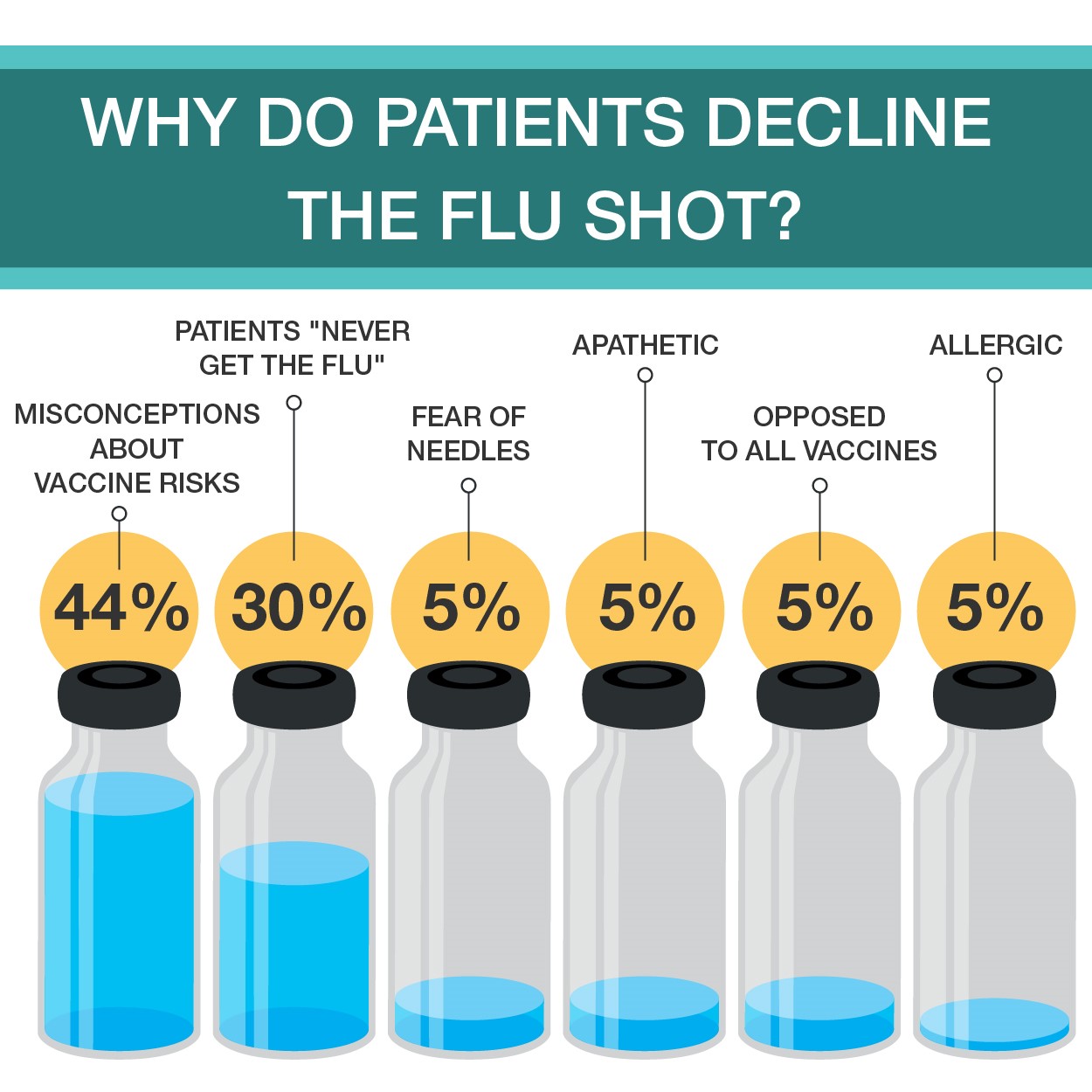
Often, the data stacks high enough to influence decisions on its own. But that isn’t the case with gun related mortality. I see gun related violence as sort of the opposite of the vaccine debate: with the flu shot there seems to be too much data and not enough stories to convince the anti-vax movement to realize the significant threat being addressed. On the other hand, gun related violence exhibits far too many stories without any significant amounts of data. Possibly, this might be related to the limitations placed upon the CDC since the mid-1990’s that forbid them from using funds “to advocate or promote gun control.” Yes, really. Just last month, I wrote about the newest advancements in influenza testing and the best practice of vaccinating annually.I cited thousands of deaths related to vaccine-preventable or epidemiologic illness; 80,000 dead from influenza last year, thousands from swine flu over a decade ago, etc. But when you try and cite proper, medical data regarding guns in public health, its … not so easy. No data, no research. No research, no change.

Many of you have undoubtedly read about the current social media “discussion” regarding whose “lane” gun violence is to navigate: The National Rifle Association (NRA) asserted in a tweet that doctors, discussing the issue only within their field should leave it to more “qualified” groups like them. That’s been a tinder box of vitriol the medical community, for lack of a better term, is up in arms about. I followed and read tons of comments about this as it unfolded, hearing from endless doctors, nurses, and laboratorians posting with blood spattered scrubs, decimated trauma bays, and emptied blood bank refrigerators that this growing epidemic is enraging clinicians about. Earlier, I highlighted similarities between public health problems and their respective solutions citing that they all shared oppositional lobby groups. What better profession to handle the topic in question than medicine—whose associated lobby power from professional societies like ours to Big Pharma amass one of the largest voices in policy making in America. And another thing, as gun violence is a public health concern, whose literal job is it to address health, mortality, and morbidity? All of ours. Nurse educators lead patients through lifestyle modifications they can employ to curtail some effects of diabetes, physicians manage patient treatment regimens balancing input from pharmaceutical tools to professional guidelines,clinicians like us strive to provide the best resources available by advancing hemoglobin A1c levels or point of care testing. We all play roles in every single healthcare matter that translates to life or death, so why not this one?

So, I touched on it a little here, but what role does the medical laboratory professional play? Besides bullets in tissue section, how does the public health epidemic of gun violence reach the lab? I wasn’t so sure, until I read a story about Dr. Julie Melinek, a forensic pathologist with UC Davis and the Alameda County Sheriff’s Department. In response to the NRA’s“stay in your lane” tweet regarding gun deaths, Dr. Melinek tweeted “Do you have any idea how many bullets I pull out of corpses weekly? This isn’t my lane. It’s my [expletive] highway.” She proceeded to turn her phone off and work for a few hours. When she returned, things were viral. In an interview with Medscape, she discussed this story and the topic at large with editor-in-chief Dr. Eric Topol. She talked about the epidemiologic role clinicians of all specialties play in risk assessment and harm reduction,saying “…if we see something that’s dangerous for the pediatric population,like a toy that breaks apart or is a choking hazard, we report it to the Consumer Product Safety Commission and it gets recalled because it’s a hazard.”She and Dr. Topol explored the ways clinicians can advocate for patients and public health at large, concluding with some poignant words, encouraging those of us in medicine to reach out to elected officials. The internet facilitates such an easy way to communicate, she says that it becomes paramount to voice the opinions held within the medical community to those in policy-making; especially clinicians who may own guns or be active NRA members! Because, ultimately, this isn’t about gun ownership or second amendment rights—its about the health, well-being, and safety of our patients.

Dr. Melinek represents a single voice within the pathology community. You’ve read my posts about lab management values, interdisciplinary team work, attainable goals, and utilization of data to make clinical decisions. Those of us in lab medicine find ourselves at the forefront of translating data into decisions. When quality control measures on instrumentation fail to correct after countless interventions, do we continue running assays? No! We work-up and investigate what root cause is the problem and fix that if possible; thinking outside the box, looking at lesser-than-obvious causes, investigating all possible solutions, etc. In pathology we’re the first to implement new, highly advanced tests and corroborate with other specialties about what the new changes mean for patient care and management of diseases (i.e. 5thgeneration high-sensitivity troponins and evolving to a new standard of care for acute coronary syndromes). We’re also the first to notice trends that impact patient outcomes and the first to provide solutions: think back to the last time you spent a few minutes reading your labs metrics and goals posted somewhere at work. Dr. Melinek collecting bullets from her autopsy patients is no different than forensic pathologists historically noting trends in mortality statistics, iatrogenic, environmental, and other causes of death.And, when those trends get published and presented, they call for further research and investment into public health interventions that may prevent those deaths in the first place. Pathology, public health, epidemiology, and laboratory medicine are built for this. We’re the tangible bridge between what gets discovered and what gets researched. We’re also in a privileged position to have a bird’s eye view of a larger clinical, epidemiologic picture as pathologists see populations of patients.

In a recent Lablogatory post, ASCP’s Lotte Mulder (ASCP Leadership Institute and Patient Champions programs) wrote about Moral Capacity, Courage, and Resiliency. Specifically, she said “It is not enough to understand and recognize a moral dilemma, it is important to act on it… it is critical for leaders to understand that culture influences moral and ethical behavior.” If America’s gun violence problem is one that desperately needs data, then why shouldn’t we, then, be professional and cultural leaders and advocate through data collection, analysis, and translation like we always do? Let’s use our tools and our talent for lab medicine, in partnership with the growing coalition of clinical professional specialties, and cultural humility for the populations we protect, and address this once and for all.

Thank you.

–Constantine E. Kanakis MSc, MLS (ASCP)CM graduated from Loyola University Chicago with a BS in Molecular Biology and Bioethics and then Rush University with an MS in Medical Laboratory Science. He is currently a medical student actively involved in public health and laboratory medicine, conducting clinicals at Bronx-Care Hospital Center in New York City.

Regulatory Inspections: Are You Ready?

Part Three: The inspectors have left – now what?

In the previous 2 blog posts we discussed how to prepare for your inspection, and what to expect during the inspection itself. In the last of our 3 part series on regulatory inspection preparedness, today we’ll be covering what to do after the inspection ends.

Throughout the inspection itself, the inspectors should be communicating any issues or citations they uncover; ensure that your management staff is taking notes on any of these potential findings. Based on these notes, you should start working to address and correct any issues right away. Formal documentation regarding the nature of any official citations can take several weeks to receive back, depending upon the regulatory agency performing the inspection. Waiting for the formal report to begin making corrections will reduce the time you have to form a plan of correction, and can further impact patient care depending upon the citation received.

Have a Plan. Draft a spreadsheet to record: 1) each issue identified, 2) laboratory department(s) it was found in, 3) associated risk factor (patient care or safety issues = 1, regulatory requirements = 2, recommendations = 3), 4) staff member assigned to investigate and correct the issue, 5) due date for investigation response, and 6) status of the investigation (in progress, on hold, completed). Share this spreadsheet with your management team, and review at weekly/monthly staff meetings for updates on progress completion.

Risk 1 Issues. The safety of your patients and staff, along with ensuring accuracy in testing results is the number one priority of a laboratory. If the inspectors identified any weaknesses in these areas, they should be addressed first. This would include items such as staff not adhering to required safety precautions, not following manufacturer requirements for quality control testing or instrument maintenance/calibration, lack of follow-up for QC or proficiency testing failures, along with any other finding which questions the integrity and accuracy of the testing being performed.

Risk 2 Issues. Double check the regulatory standard to ensure you fully understand the requirements, and that you have appropriate evidence of compliance. As the testing activity menu and complexity of testing increases, the amount of documentation requirements can increase as well. Even with a paperless system, it is easy to overlook a signature of review or checkmark on a log. “If it’s not documented, it wasn’t done.” For simple administrative oversights, review your current processes to identify any gaps or areas that can be improved upon to ensure all documentation is properly filled out each month. If the inspectors noted a discrepancy between your current policy and how staff are actually performing a test, review the testing process to see where the true discrepancy is – is the policy outdated and needs to be revised, or do staff need to be retrained on the current policy with competency assessed for compliance?

Risk 3 Issues. Inspections are a great opportunity for further education for all those involved, both the inspector and staff being inspected as well. For some regulations, there is no one set way that must be followed in order to demonstrate compliance with a requirement. Hearing how someone else is meeting the requirements may spark an innovative idea from your own staff on how your current processes can be improved. Be open to hearing new ideas, and find ways to implement those which you feel would be successful at your institution.

Evaluate All Sections of the Lab. When investigating a finding in one laboratory department, ensure that any process improvements are shared across all areas of the lab. Just because microbiology didn’t get caught with expired reagents like hematology did this inspection, doesn’t mean that they aren’t at risk for future inspections.

Focus on the Positives. Congratulate and recognize your staff on their successes in the areas you performed exceptionally well in. It’s a joint effort to ensure the lab is inspection ready; be sure to pass along any compliments received throughout the inspection process to all levels of staffing. Focus on what you’re doing well and how you can continue to maintain those processes and implement them in additional areas.

A little bit of preparation ahead of time will make the inspection process smoother and less stressful for all involved. When viewed as a learning experience and opportunity for improvement rather than a visit from the “lab police”, laboratory inspections can be a useful tool to confirm the quality of your overall laboratory program.


Nevins-small

-Kyle Nevins, MS, MLS(ASCP)CM is one of ASCP’s 2018 Top 5 in the 40 Under Forty recognition program. She has worked in the medical laboratory profession for over 18 years. In her current position, she transitions between performing laboratory audits across the entire Northwell Health System on Long Island, NY, consulting for at-risk laboratories outside of Northwell Health, bringing laboratories up to regulatory standards, and acting as supervisor and mentor in labs with management gaps.

Moral Capacity, Courage, and Resiliency

Leaders’ decisions and actions have moral ramifications, both on an individual and an organizational level. There are three factors of moral development, namely moral capacity, moral courage, and moral resiliency.

Being able to recognize a dilemma as a moral issue is one of the critical aspects of leadership. Such awareness is referred to as moral capacity, which influences the characteristics and recognition of a moral issue. There are multiple aspects that influence a leaders’ moral capacity. The first aspect is their previous experiences with moral dilemmas and how much they learned from them. The second is to what extent a leader is able to see and understand the multiple perspectives of an issue. The third is how leaders view their role and whether or not that incorporates a moral view.

It is not enough to understand and recognize a moral dilemma, it is important to act on it. Such moral courage is especially important when under pressure to act immorally. Such pressure can come from peers, supervisors, or the entire organization. Therefore, the more moral courage someone has, the more likely it is that they take a moral action or make a moral decision. One important aspect of moral courage is the notion of willpower. Willpower is a muscle that people can practice with small tasks, such as drinking a glass of water before breakfast. The more people practice it on small tasks, the more likely they are to use it during challenging situation, such as making a moral decision when pressured to do otherwise.

Moral resiliency is an extension of moral courage. While moral courage focuses on the strength to make moral decisions in the short-term, moral resiliency is a process through which leaders continuously adapt their moral compass and actions. Moral resiliency is this what creates sustainable moral decision-making.

Depending on how leadership effectiveness is defined, moral behavior can either make leaders more or less effective. When looking at effectiveness in the short term, it is possible that moral behavior can impede effectiveness if measured in terms of money or short-term success. However, when looking at effectiveness in the long term, moral behavior increases leaders’ effectiveness. The more honest, and thus morally, people behave, the more effective they are. In a world that is becoming more globalized, it is critical for leaders to understand that culture influences moral and ethical behavior. In other words, what is moral in one culture might be immoral in another. To increase leaders’ effectives it is therefore important to understand the cultural implications of behavior and to be aware of the differences in appropriate and effective behavior.


lotte-small

-Lotte Mulder earned her Master’s of Education from the Harvard Graduate School of Education in 2013, where she focused on Leadership and Group Development. She’s currently working toward a PhD in Organizational Leadership. At ASCP, Lotte designs and facilitates the ASCP Leadership Institute, an online leadership certificate program. She has also built ASCP’s first patient ambassador program, called Patient Champions, which leverages patient stories as they relate to the value of the lab.

What’s “In” for Thrombocytopenia Diagnosis? Advanced Platelet Parameters: The Immature Platelet Fraction (IPF%) and the Immature Platelet Count (IPF#)

Platelets are our first line of defense in controlling bleeding. Abnormally low numbers of platelets can lead to easy bruising, tiny leaks from capillaries into the skin and mucous membranes, causing petechiae, and bleeding. The platelet count is a significant parameter in the CBC and it is therefore vital to be able to report accurate and precise platelet counts. Furthermore, physicians must be able to use this information to diagnose the cause of the thrombocytopenia in order to recommend treatment.

What a platelet count alone cannot tell us is the reason for thrombocytopenia. Just as there can be many reasons for a low hemoglobin, and many causes for an increased or decreased WBC, there are numerous causes for a decreased platelet count. After ordering a CBC, the next steps in determining etiology of thrombocytopenia have historically been a thorough physical with attention to any bleeding symptoms and organ enlargements, and a medical history. The medical history should include family history, and notation of recent viruses or drug therapies. After these tests, a bone marrow aspirate and biopsy may also be necessary to clarify etiology. While modern, automated hematology analyzers produce reliable platelet counts, measuring only the circulating platelet count does not give us any information as to the etiology, so there is a need for further testing. With thrombocytopenia, platelet counts can be less reliable than with normal counts.Platelet counts were originally performed by impedance methods, then better accuracy and precision was obtained with optical platelet counts. Physicians rely on precision with very low platelet counts to make informed decisions about when to transfuse patients. The problem with the impedance counts at the low end, is that RBC fragments, schistocytes and microcytic RBCs can be counted as platelets, giving a falsely high count. On the other hand, measuring platelets by size can miss large platelets leading to a falsely low count.

Historically, the mean platelet volume (MPV) has been used along with the platelet count to aid in making a differential diagnosis. The MPV is analogous to the red cell distribution width (RDW) for red cells, and can be used to as an indicator of the maturity of platelets. Young platelets are the largest, and as they age, the size decreases. The normal ranges for MPV are generally about 9-12 femtoliters (fl).The MPV will be higher if more platelets are being released from the bone marrow, and lower if fewer are being newly released and we are counting mature platelets. Thus, the MPV can be used as an indirect marker for platelet production. However, an inherent problem with the MPV is that, similarly to the impedance platelet count, this count can be unreliable because any RBC fragments or particles may interfere with the measurement.

So, what is a physician to do?And how can the lab provide information to help them make the best differential diagnosis and transfusion decisions? In an effort to provide a parameter that could help differentiate causes of thrombocytopenia, the concept of reticulated platelet counts (retPLT) was first introduced in research in the late 1960s. The term is used to describe immature, functional platelets in the peripheral blood.Reticulated platelets are to mature platelets as reticulocytes are to mature red blood cells. These are the youngest platelets, within 24 hours of being released from the bone marrow. Reticulated platelets are large, with increased amounts of RNA, and the number in the circulation can be used to provide an estimate of the rate of thrombopoiesis. Originally, these were stained with new methylene blue and manual counts were done, much like a manual reticulocyte count; tedious,and imprecise. It wasn’t until about 30 years later that a flow cytometry method was described for measuring retPLT. Using traditional flow cytometry, reticulated platelets can be stained with a Thiazole Orange dye and passed through a flow cytometer. This method, however, has been shown to have wide normal ranges from 1-15% because of the lack of analytical standardization. Variations in the concentration of the thiazole dye used, the timing, and the gate settings all make it difficult to compare results obtained from one laboratory to another. In addition, flow cytometry is time consuming, labor intensive and costly.

Newer flow cytometry methods are now available on select hematology analyzers. There are currently 2 companies that have analyzers that can report retPLT using routine CBC reagents and controls. Reticulated platelets can be measured with the same K2 EDTA tube used for the CBC. The test is automated, simple to perform, fast, and gives standardized results with tighter normal ranges. The Abbott CELL-DYN Sapphire measures the retPLT using a fluorescent dye and flow cytometry with 2 dimensional gating. Sysmex XE and XN analyzers offer several Advanced Clinical Parameters including measures of reticulated platelets, expressedas the Immature Platelet Fraction (IPF%) and the Absolute Immature Platelet Fraction Count (IPF#). Sysmex offers a fluorescent platelet count (PLT-F) as an addition to impedance counting (PLT-I) and optical counting (PLT-O). PLT-F is more reliable because it uses a platelet specific dye which eliminates noninterference seen with other methods. The fluorescent dye labels the RNA and forward scatter is used to determine size while fluorescence is used to measure RNA content. With gating set based on cell volume and RNA content, the PLT-Fcan be measured. When there is an abnormal scattergram or a low platelet count,the PLT-F is reflexed and the IPF% and IPF# are also reported.

What’s the clinical utility of the IPF? Thrombocytopenia can have many causes.Immature platelets are functioning platelets, and an increased IPF means that we have more newly formed immature platelets circulating. The IPF helps physicians to differentiate thrombocytopenia caused by platelet destruction or consumption versus thrombocytopenia caused by deficient platelet production in bone marrow failure. It is vital to know the pathogenesis of thrombocytopenia in making decisions about treatment. With these advanced parameters, these decisions can often be made without costly, time consuming flow cytometry,without an invasive bone marrow biopsy and without waiting for the results of such biopsy. This can often save a patient an unnecessary platelet transfusion.

The reference range for IPF% in healthy individuals is1.0-7.0%. Together with a low platelet count, an increased IPF indicates an increase in platelet production. This is seen in patients with excessive destruction of platelets. An example of the clinical utility of the IPF can be seen in the diagnosis of immune thrombocytopenic purpura (ITP). ITP is an autoimmune bleeding disorder in which the immune system makes anti-platelet antibodies which destroy platelets. Acute forms occur more often in children while adults can have chronic ITP. ITP can be diagnosed on clinical findings but laboratory confirmation is often necessary. This can be expensive with long turnaround times using traditional flow cytometry and/or bone marrow aspirates.An IPF reported with a CBC is fast, inexpensive, and be extremely beneficial in aiding a timely diagnosis. Patients with ITP have been shown to have the consistently highest IPF values with ranges from 7-28%.1 As their platelet counts recover, the IPF% returns to the normal range, without the need for transfusions. Thus, the IPF can be used not only to help diagnose but also as an indicator of remission.

Figure1. Platelet scattergrams from a healthy individual with a normal IPF (a) and a patient with a high IPF (b). Mature platelets appear as blue dots, green dots represent the IPF with increased cell volume and higher fluorescence intensity compared to mature platelets1

In contrast to what we see with ITP, thrombocytopenia with alow normal or decreased IPF indicates decreased bone marrow production of platelets. Patients with bone marrow failure are more likely to have bleeding episodes with low platelet counts and may need transfusion. Rapid differential diagnosis using the IPF can help physicians help these patients get early treatment.

IPF may also be a reliable indicator of bone marrow recovery. Traditionally, neutrophil counts have been used as an indicator of recovery after a bone marrow transplant. IPF can be used as an indicator of imminent platelet recovery. It has been shown that,post-transplant, the IPF% increases before the platelet count. In a study done with stem cell transplant patients, it was shown that the absolute neutrophil count took an average of 13 days to recover, compared to 9 days for the IPF. The IPF was shown to recover before the Immature reticulocyte count, platelet count and absolute neutrophil count, giving physicians earlier indication that the transplant was successful.2 This is significant because it can eliminate the need for bone marrow biopsies and platelet transfusions.  

Thrombocytopenia is not an uncommon finding in neonates, particularly in the neonatal intensive care unit (NICU). There are various causes for this, including sepsis, placental insufficiency and immune thrombocytopenia. The IPF% and IPF# can be used to diagnose and distinguish the cause of thrombocytopenia in neonates, and direct the treatment. When platelet count platelet count drops below 50 x 103/Lin an otherwise healthy appearing infant in the first 72 hours of life, neonatal alloimmune thrombocytopenia (NAIT) can be suspected. This condition is similar in pathogenesis to hemolytic disease of the fetus and newborn (HDFN), and is caused by an incompatibility in human platelet antigens between mother and baby. This occurs most often when the mother is HPA-1b and the father and baby are HPA-1a. The mother forms anti-HPA-1a which crosses the placenta and destroys the fetus’ platelets.This is a thrombocytopenia caused by platelet destruction, and the IPF% is high. The condition is self-limiting and resolves in 1-4 weeks. Neonatal sepsis can also present with a high IPF, but typically is found in very sick or premature babies and the degree of thrombocytopenia is not as severe as with NAIT. In contrast, neonatal thrombocytopenia due to placental insufficiency would exhibit a decreased IPF due to a deficiency in platelet production. Using the IPF% and IPF# to help differentiate the causes of neonatal thrombocytopenia can help steer the treatment and save infants from unnecessary invasive procedures and transfusions.

TheIPF has proved to be very valuable in the clinical setting. It has been used in the investigation of etiology in secondary thrombocytopenias due to chronichepatitis C, liver disease and HIV. It has been used to guide treatment in thrombocytopenias such as thrombotic thrombocytopenic purpura (TTP). IPF can also be useful in evaluation of hereditary platelet thrombocytopenias. The IPF% and IPF# can be compared after transfusion to support the theory that, after platelet transfusion, theIPF% will decrease due to the newly increased platelet count, but the IPF#remains the same. This validates that the IPF is a reflection of continual platelet production by the bone marrow.4

IPF%and IPF# are expanded CBC parameters that physicians can use to aid in differentiation of various thrombocytopenic states. Treatment for the different classes of thrombocytopenia can differ drastically, and knowing the class of thrombocytopenia helps direct the management. The IPF parameters are automated,easy to perform at the same time as the CBC, and provide standardized results that are inexpensive and available 24 hours a day in the hospital setting. Using the IPF can also reduce diagnostic costs for the patient. Many studies have been conducted on the varied applications of the IPF and research continues investigating possible further uses of this advanced clinical parameter. This is the new hematology, constantly providing the clinician with better tools for making diagnoses and treating patients. Platelet counts alone and MPVs are out. Make room for the new kid on the block; the IPF is in.

References

  1. Arshi Naz et al. Importance of Immatureplatelet Fraction as a predictor of immune thrombocytopenic purpura. Pak J MedSci 2016 Vol 32 No 3:575-579
  2. Zucker ML et al. Immature Platelet fraction asa predictor of platelet recovery following hematopoietic progenitor celltransplanatation. Lab Hematol 2006 12(3):125-30
  3. Briggs,C. Assessment of an immature plateletfraction (IPF) in peripheral thrombocytopenia. Br J Haematol 2004Jul;126(1):93-9
  4. Sysmex White Paper. The role of the ImmaturePlatelet Fraction(IPF) in the differential diagnosis of thrombocytopenia. www.sysmex.com/us
  5. Fujii,T et al.. A new approach to detectreticulated platelets stained with thiazole orange in thrombocytopenicpatients. Thromb Res. 2000 Mar 15;97(6):431-40
  6. Cremer Malte The immature platelet fraction(IPF) in neonates. Diagnostic Perspectives 2011 Vol1:36-42
  7. Cremer M. et al. Immature platelet values indicateimpaired megakaryopoietic activity in neonatal early-onset thrombocytopenia.Thrombosis and Haemostasis 2010; May;103(5):1016-21

-Becky Socha, MS, MLS(ASCP)CM BB CM graduated from Merrimack College in N. Andover, Massachusetts with a BS in Medical Technology and completed her MS in Clinical Laboratory Sciences at the University of Massachusetts, Lowell. She has worked as a Medical Technologist for over 30 years. She’s worked in all areas of the clinical laboratory, but has a special interest in Hematology and Blood Banking. When she’s not busy being a mad scientist, she can be found outside riding her bicycle.

The Transparent Injury

While Maria was working in Microbiology, she cut her finger while pulling reports off of the printer. It was a minor paper cut, so she ignored it, put her gloves on and continued her work back on the bench. A week later, the tiny cut was swollen and red. She decided to report the incident to her manager since it wasn’t healing. The manager asked Maria to report to the Occupational Health department, but was unsure if any treatment would be covered since the incident was not reported while she was at work.

Steve and Josh were bored during the night shift and they created a ball made from rubber bands to toss around. When Josh didn’t catch the ball, it hit the open tray of formaldehyde on the gross bench, and it splashed into Josh’s eye. He rinsed his eyes in the eyewash station for a couple of minutes, but both men were afraid to report the incident for fear of getting in trouble. Josh’s eye irritation continued to worsen, and he had to go to the eye doctor for treatment.

There are obvious consequences for injuries that occur in the laboratory, and reporting them is important for many reasons. Staff may be motivated in some instances to not report, but that creates problems for the individual, the department, for the facility, and even for other labs across the country! That may seem like a stretch, but it will become clearer with exploration.

The value in injury and accident reporting starts with medical follow-up. Those incidents which require treatment or abatement of infection can and should be dealt with quickly, and appropriate monitoring can be done if necessary. Some injuries may require immediate first aid, and a trip to the emergency department may even be necessary. Not reporting those types of injuries can be very dangerous for staff. Other incidents may require physician office visits or other monitoring, and employees who need it should be encouraged to comply.

In many work places the injury follow-up visits and treatment are covered financially by the institution, either via a structured occupational health program or through reimbursement. Some organizations may not offer financial coverage, however, if the incident that occurred at work is not reported as soon as possible. That reporting delay can raise suspicion as to whether or not the injury actually did occur while on the job, and since the written reporting protocol was not followed, there may also be no obligation for employer medical coverage.

Departmental issues will arise when incident reporting in not part of the overall lab safety culture. Sometimes there can be reprisals for unsafe behaviors which lead to accidents, but if the safety culture is good and if managers and employees coach against such practices, then there should be fewer overall incidents to report. That said, a culture of secrecy regarding injuries or exposures can also be dangerous. There is value in talking to all staff about an incident that occurred within the department. Staff can learn from the event and have a healthy discussion about how to keep it from reoccurring. A discussion of events can bring important safety issues to light, particularly if similar incidents happen with multiple people. This sharing of information can also promote awareness of good safety practices that can aid in the prevention of further incidents for all who work in the department.

OSHA requires the reporting of certain work place injuries, those that may have led to time away from work or that need medical follow up, for example. This injury data is compiled and reported nation-wide. It becomes a good source for benchmark data, a way to be able to compare your lab injury rates with others across the country. The U.S. Bureau of Labor Statistics provides this data as information labs can use. One way to utilize the information is to see if the number of reportable injuries you are seeing in your lab is comparable to a national average. That assessment can give you a starting point in determining whether or not your lab’s safety incidents are at typical levels. Of course, lab safety professionals want to see zero injuries, but if you see your lab injury numbers are very high compared to benchmark data, you can begin to see where to focus in on fixes for the lab physical environment or on creating specific safety training.

There is great value in talking about safety incidents that may result in injury or exposure in the lab setting. These “safety stories” raise awareness of safety issues, and they can act as a deterrent for repeat incidents. Create a culture where staff feel free and comfortable to report incidents, and be sure to discuss them with all staff, and record reportable injuries as well. Having reliable national data also provides helpful information to other labs, and better information can help to improve safety in laboratories everywhere!


Scungio 1

Dan Scungio, MT(ASCP), SLS, CQA (ASQ) has over 25 years experience as a certified medical technologist. Today he is the Laboratory Safety Officer for Sentara Healthcare, a system of seven hospitals and over 20 laboratories and draw sites in the Tidewater area of Virginia. He is also known as Dan the Lab Safety Man, a lab safety consultant, educator, and trainer.

Lab Value Changes in Transgender Males

For patients with gender dysphoria, the Endocrine Society has endorsed the use of hormone therapy to promote secondary sexual characteristics of the desired gender. These guidelines were first established in 2007 and revised last year, and gave the first evidence guided recommendations for clinicians treating transgender patients.

For transgender males, testosterone by itself is prescribed as an injectable oil-based solution. These doses are given as intramuscular injections- usually into the thigh. If that’s too painful, subcutaneous injections have been shown to have similar efficacy. The doses given to transgender males is much higher (50-100mg/ injection) than that given to men with testosterone deficiency (30-50 mg/ injection). Primarily because the men have more testosterone to start with. Also, whereas topical testosterone gel may be sufficient for men with “low T,” it doesn’t seem to provide enough testosterone to transgender males and is quite expensive, so it is generally not used.

Nov1.png
Image 1. Picture of testosterone cypionate vial from mcguffmedical.com. This is used for intramuscular injections.

Upon starting testosterone injections, the frequency of injections is every one to two weeks. However, the onset of physical secondary sexual characteristics takes 3-6 months to begin. After about 3 years, most of the changes to occur will have manifested. These physical changes are outlined in the table below. You’ll notice how certain traits like cessation of menses and fat redistribution start within the first 6 months whereas muscle growth and voice change take effect after 6 months. Also, the time certain effects take maximal effect varies; the voice doesn’t deepen further after 2 years, but hair growth continues to increase through 5 years.

Physical Effect Begins Maximal Effect
Facial/body hair growth 6-12 mo 4-5y
Skin oiliness/acne 1-6mo 1-2y
Scalp hair loss 6-12 mo
Increased muscle mass 6-12 mo 2-5y
Fat redistribution 1-6mo 2-5y
Cessation of menses 1-6mo
Deepening of voice 6-12 mo 1-2y

Table 1. Timeframe of physical traits that manifest in transgender males while taking testosterone hormone therapy. Based on Hembree et al. 2017 (1).


Just as hormone therapy induces physical manifestations of secondary sexual characteristics for transgender men, we would suspect that internal aspects of physiology are affected too.  Values measured by the laboratory provide meaningful insight into how our body and its different organ systems are functioning. Accordingly, the Endocrine Society also recommended laboratory monitoring of transgender patients starting hormone therapy.

  1. Measure Testosterone and hemogoblin/ hematocrit every 3 months for the 1st year, then 1-2x/ year afterwards.
  2. Monitor Lipids at regular intervals

Previous studies have monitoring these lab values found consistent increases in hemoglobin and hematocrit (2,3). This is due to the stimulation of erythropoiesis by testosterone (4).  While excessive testosterone could lead to polycythemia (excessive RBCs in the blood), it is not a commonly described complication in transgender patients. Some summary results from our study for hemoglobin and hematocrit are shown in Figure 1A, which shows a clear shift in levels.

However, reports on lipids have been varied LDL and triglyceride changes (2,3). The only consistent finding was that HDL decreased in transgender males taking testosterone (2,3). In our study, we found triglycerides were increased with decreased HDL (Figure 1B). The take-away is that because cardiovascular cut-offs are based on risk and not a reference range, patients and clinicians will have to be aware of these possible metabolic changes.

Creatinine, when it was checked, increases for transgender males (5). We found creatinine was strongly increased in our study to become similar to baseline creatinine in transgender women before taking hormone therapy (Figure 1C). This topic as it relates to glomerular filtration rate is very complex and will be discussed further in a future post.

To illustrate lab value changes in transgender men, I’ll direct you to data that I found in a large study of over 300 transgender patients including about 80 transgender men. The completed manuscript is not currently available but will be printed soon:

Nov 2

However, this does not mean Cisgender male reference intervals are adequate for transgender men. This topic needs further exploration and ideally a prospective trial to be performed in a controlled manner. A double-blind study would not be possible as it would be unethical to perform.

References

  1. Hembree WC, Cohen-Kettenis PT, Gooren L, Hannema SE, Meyer WJ, Murad MH, et al. Endocrine Treatment of Gender-Dysphoric/Gender-Incongruent Persons: An Endocrine Society* Clinical Practice Guideline. J Clin Endocrinol Metab. 2017
  2. Wierkx K, et al. Cross-Sex Hormone Therapy in Trans Persons is Safe and Effective at Short-Time Follow-Up: Results from the European Network for the Investigation of Gender Incongruence. J Sex Med, 2014. 11(8):1999-2011.
  3. Mueller A, Kiesswetter F, Binder H, Beckmann MW, Dittrich R. Longer-term administration of testosterone undecanoate every 3 months for testosterone supplementation in female-to-male transsexuals. J Clin Endocrinol Metab. 2007
  4. Paller CJ, Shiels MS, Rohrmann S, Menke A, Rifai N, Nelson WG, et al. Association Between Sex Steroid Hormones and Hematocrit in a Nationally Representative Sample of Men. J Androl. 2012 33(6): 1332-1341.
  5. Fernandez JD, Tannock LR. Metabolic Effects of Hormone Therapy in Transgender Patients. Endocr Pract. 2016;22:383–8.


SoRelle Picture

-Jeff SoRelle, MD is a Molecular Genetic Pathology fellow at the University of Texas Southwestern Medical Center in Dallas, TX. His clinical research interests include understanding how the lab intersects with transgender healthcare and advancing quality in molecular diagnostics.

Gram Stain Examination – Beyond Infectious Organisms

Case History

A 72 year old female with past medical history of stage IV ovarian adenocarcinoma treated with chemotherapy and interval debulking surgery, presented to emergency room with a one week history of confusion and worsening balance.

CT scan of the head showed new communicating hydrocephalus.  A magnetic resonance imaging couldn’t be performed initially because of patient’s uncontrolled agitation.  Lumbar puncture (LP) was performed.  Following this procedure the patient’s mental status showed some improvement and therefore neurosurgery team decided to insert ventriculoperitoneal (VP) shunt to treat her hydrocephalus and prevent recurrence of seizures.

It was Friday afternoon when a microbiology technologist brought the patient’s cerebrospinal fluid (CSF) gram stain to be reviewed.  It was confirmed that no inflammatory cells and organisms were present.  However, cells in the background looked very atypical (Image 1a, b).

CSF1
Image 1:  Gram stain of CSF showing atypical epithelial cells at (a) 40x and (b) 100x with oil.

CSF2
Image 1b.

Discussion

The gram stain is used to provide preliminary information about the microorganism present in the specimen.  Gram stain differentiates bacteria into two fundamental varieties of cells.  Bacteria that retain the initial crystal violet stain (purple) are said to be “Gram-positive,” whereas those that are decolorized and stain red with carbol fuchsin (or safranin) are said to be “Gram-negative” (1).  An adequate examination of a gram-stained smear includes observing numerous representative fields and the fields containing neutrophils yield the most information (2).  Gram stain provides information about number of bacteria present, gram reaction and shape of the bacteria.  In background we can also see epithelial cells and inflammatory cells.  However, it’s a good practice to also appreciate and investigate any odd looking findings.

To investigate further, we visited the hematology laboratory to view their CSF slide to determine if these cells were a processing artifact.  After it was confirmed that hematopathology saw the same atypical cells, a cytopathologist was requested to review the gram stain since patient’s CSF cytology specimen was to be processed after the weekend.  Cytopathologist favored our suspicion and decided to process the cytology specimen late in the day on Friday and it was confirmed that those atypical cells were consistent with the metastatic adenocarcinoma.

Neurosurgery team was immediately contacted to reconsider insertion of the VP shunt as the shunt would drain fluid from the CSF into the peritoneal cavity and thus there was concern for transferring of malignant cells from central nervous system into abdomen/pelvis. However, after consulting oncology team it was later decided to proceed with the procedure since patient’s primary tumor originated in abdomen/pelvis and current intraabdominal tumor burden was not significant as compared to the symptoms driven by CNS involvement. Proceeding with this procedure was considered to be palliative and the best course of action to improve the patient’s quality of life.

References

  1. Beveridge TJ. Use of the gram stain in microbiology. Biotech Histochem.2001 May;76(3):111-8.
  2. Barenfanger J, Drake C. Interpretation of gram stains for the nonmicrobiologist. 2001 July;32(7):368–375.

KM

-Kiran Manjee, MD, is a 1st year anatomic and clinical pathology resident at University of Chicago (NorthShore).

-Erin McElvania, PhD, D(ABMM), is the Director of Clinical Microbiology NorthShore University Health System in Evanston, Illinois. Follow Dr. McElvania on twitter @E-McElvania. 


Overview of Working with Different Generations: Composite of Current Workforce

There are currently five different generations at work today: Traditionalists, Baby Boomers, Gen Xers, Millennials, and Generation Z. This means that in any work environment, you can have a group of people between the ages of 15-80. This is an exciting time to be working because we can all learn from many different generational experiences, values, and communication styles.

The two largest generations in the work place are the Baby Boomers and the Millennials. This is because these are both the largest generations in terms of population. However, with the Baby Boomers slowly moving into retirement, the Millennials are about to take over.

Traditionalists are still present in the workforce for a few reasons. First, they have tremendous experience and organizational knowledge and many organizations are trying to keep them around so that they do not lose that information. This means that Traditionalists are often Presidents of organizations or members of their Board of Directors. Secondly, Traditionalists are loyal to their organizations and they generally keep working as long as they can because of their values of security and getting the job done.

Generation X and Z are also in the workplace, but neither is very large. However, Gen Xers serve an important purpose because they are flexible and adaptable and because they value work-life balance and constructive feedback. They understand both the world without technology, so that can relate to Baby Boomers, and the world of the internet and social media, so they learn technology fast, which is appreciated by Millennials. Generation Z is only now starting to enter the workforce, so little is known about their work styles. However, they are expected to be independent, entrepreneurial, determined, and loyal.

The key to working with multiple generations is respect. Everyone wants to be respected and appreciated for what they bring to an organization. Being open and flexible to learning about different generational values and communication styles, will set any leader and employee up for success. Provide everyone with positive and constructive feedback and create a work environment that allows for more flexibility in terms of work hours, work location, and dress code whenever possible. Finally, realize that what motivates you personally is not necessarily what motivates other, especially if they are from different generations. Working with a diverse group of generational workers is a great benefit, to both the organization and to individuals.


lotte-small

-Lotte Mulder earned her Master’s of Education from the Harvard Graduate School of Education in 2013, where she focused on Leadership and Group Development. She’s currently working toward a PhD in Organizational Leadership. At ASCP, Lotte designs and facilitates the ASCP Leadership Institute, an online leadership certificate program. She has also built ASCP’s first patient ambassador program, called Patient Champions, which leverages patient stories as they relate to the value of the lab.

Investing in the Best Testing

“Damn that q-tip goes in deep!

But it lit up negative so d/c to street

But it was flu, cuz he bounced back again

And now my Press Gayney’s a minus ten…”

Video 1. Another classic excerpt from a favorite: ZDoggMD, singing about this year’s flu season and available testing options on the horizon—because, let’s face it—rapid flu tests aren’t quite cutting it anymore.

Hello again everyone! Back again to talk about a new set of recommendations from last month’s post. This time it’s about influenza. Recommendation: get vaccinated. Thank you. See you next time…

Seriously, as the 2018-2019 flu season dawns upon us, it’s time to talk about vaccines, tests, prevention, and health literacy. I’m sure many of your social media pages are filled with various debates, articles, and fake news stories on one side or another pitting science, pseudo-science, and non-science all against each other for public spectacle. In the lens of laboratory science and medicine at large, I think most if not all of us agree that preventable diseases should be prevented, and if not, at the very least detected accurately, sensitively, and early. Influenza A/B is a prime example of a consistent threat to our health and safety that has wavered responses in various socio-medical circles.

Official communication and guidance from the Centers for Disease Control and Prevention (CDC) clearly tells those of us in health-care to embrace a multi-tiered approach to protecting public health regarding the flu. That approach includes vaccination, testing, infection control, anti-viral treatment, and anti-viral prophylaxis. And why such a fuss over the flu? It’s a big deal! Last year, the CDC reported approximately 80,000 deaths associated with influenza as a primary cause. 80,000 deaths! That’s almost 7 times as many that died from H1N1/Swine Flu complications back in 2009, where only 12,000 patients were killed by the virus. And even more so, in the terrifying Ebola epidemic of 2016—in which there was a staggering 1 recorded death in the US—nearly 29,000 people were infected globally and only 11,300 died (despite under-reporting). I’m being dramatic, I know. But it’s important for us to recognize true epidemics when they happen, and even more important for societies like ours to be at the forefront of preventing them from developing any further.

flu1.jpg
Image 1. The CDC recommends you get your flu shot every year, because obviously.

flu2.jpg

Image 2. I’m not here to talk about the anti-vax elephant in the room. That’s not fair to elephants. But imagine if the CDC reported 44% of flu vaccine misconceptions were addressed!

As an aside, I’ll probably recommend that you get your annual flu shot a hundred times in this post alone. But just to have a clear reference, please look at the following table. It’s critical to be able to both distinguish common cold versus influenza symptoms for yourself, as well as educate your patients and peers about the differences between the two. This information can change the way people perceive treatments (i.e. why the doctor only recommended rest/Tylenol and didn’t give out antibiotics for their symptoms) and why it’s absolutely crucial to protect vulnerable populations from an otherwise fatal virus. So, micro-rant aside, it should be clear that by now we should be working on a way to both improve our prophylaxis with vaccines and medications as they always leave room for improvement—I’m looking at you Tamiflu and Relenza! Notwithstanding any analysis of efficacy for the flu vaccine, the CDC reports a variable and transparent success rate of vaccines. It can be difficult to predict and assess epidemiologic trends and mutations as the influenza virus continues to change annually.

flu3.JPG
Table 1. Distinguishing the common cold, and “flu-like symptoms” from a proper influenza viral infection.

flu4.jpg
Image 3. CDC Report on seasonal flu vaccine effectiveness since 2004.

So, what was the deal with ZDoggMD plugging some PCR testing in the opening credits here? That’s a good question and one that inspired this article in the first place. Obviously, if you follow my posts you know I follow his, and at the end of this latest video he discusses new available options for influenza point-of-care testing (POCT) for clinics and emergency rooms. This was a partnership with the company Cepheid and linked with their promoting their POCT PCR-based FluA-B testing. Here’s a quick paraphrasing of the CDC recommendations on influenza testing: because of the numerous false negative tests every season, the bests tests in order of preference are RT-PCR, immunofluorescence, and rapid antigen testing. Did you catch that? Rapid Flu swabs are bottom of the barrel stuff here. UpToDate, the clinical resource for current practices and standards discusses rapid influenza tests as sacrificing turn-around-time (TAT) for accuracy: “commercially available rapid antigen tests for influenza virus yield results in approximately 15 minutes or less but have much lower sensitivity than RT-PCR, rapid molecular assays, and viral culture.” (I didn’t bold those words, they did). Most of the places I’ve worked run through boxes of rapid flu swab kits ALL DAY LONG. But what are we missing? Clinically, this is supposed to be an important “no miss” diagnosis—it’s dangerous, it’s contagious, it’s mutatable…

Who remembers learning biostatistics in school? Remember SPIN and SNOUT? “Specificity is used to rule IN, Sensitivity is used to rule OUT” So why are we relying on the LOWEST sensitivity available to us for ruling out influenza? Probably because of technological/practical limitations up to this point in time, and of course the most glaring limiting reagent of all: funding, also known as “administrative buy-in.” Have I hit enough lab management buzz words in this post? Not yet.

flu5.JPG
Table 2. Per UpToDate, these are the quick and dirty details on our favorite available flu tests currently on lab benches across the country. I’d say there’s got to be a better way, but there already is.

Sweet. So, it’s a little expensive but ultimately better for our patients, right? Done and done, whip up a cost-benefit-ratio report for the suits upstairs and let’s start a validation project! Well, yes and no. I’m a big proponent of utilizing MALDI-TOF—the mass spectrometry based system to replace traditional bacterial identifications. A 2015 study published in the Journal of Clinical Microbiology stated, “The use of MALDI-TOF MS equated to a net savings of 87.8%, in reagent costs annually compared to traditional methods. …The initial cost of the instrument at our usage level would be offset in about 3 years. MALDI-TOF MS not only represents an innovative technology for the rapid and accurate identification of bacterial and fungal isolates, it also provides a significant cost savings for the laboratory.” What promise! Cepheid’s ED POCT PCR Flu test promises 18% fewer tests needed, 17% fewer antibiotics prescribed, and overall savings per patient visit of up to $700. But this sounds like another, too familiar, recent promise from another voice in our profession. Something about quick, easy, and accurate testing on chips with micro-laboratories available commercially and only using microliters of whole blood for analysis. “Unfortunately, none of those leads has materialized into a transaction. We are now out of time,” read the goodbye letter to the company’s stockholders—Theranos, that’s the one. The moral of the story here: it’s good to remain fiscally prudent when deciding what your clinic or hospital should invest in with regard to testing. However, when something has been a proven and successful replacement which ultimately is recommended by multiple societies within the field then something’s got to give.

flu6
Image 4. MALDI-TOF saves money! You spend a little upfront, but then your hospital can write articles about how your bacteriology department has a swab-to-sensitivity TAT of a few hours. Less errors, less antibiotics, more likes on social media!

What do you see in your practice or laboratory as far as influenza testing? Are there issues I missed? What is your experience with rapid tests, or PCR testing? Is anyone else as big a fan of MALDI-TOF as I am? Did you get your flu shots yet? Leave your comments and questions below! Share with a colleague today!

See you next time!

I have absolutely no affiliation with Cepheid, financial or otherwise, but as an educational/professional resource read more information about Cepheid’s molecular rapid flu tests, read their literature at www.GetTheRightTest.com

flu7

References

  1. Carreyrou J. (2018) Blood Testing Firm Theranos to Dissolve. Wall Street Journal. Health: Theranos Co. Letter to Shareholders. Accessed at: http://online.wsj.com/public/resources/documents/Theranos_Stockholders_Letter_2018.pdf?mod=article_inline
  2. Cephid (2018) Is it really flu? Cutting emergency department costs with bedside rapid molecular tests. Accessed at: http://www.cepheid.com/images/Cepheid-WP-ED-Cost-FINAL.pdf
  3. CDC (2017) Interim guidance for influenza outbreak—management in long-term care faciltites. e Recommendations of the Advisory Committee on Immunization Practices – United States, 2016-17 Season. Accessed at: http://cepheid.com/images/CDC-interim-guidance-outbreak-management.pdf
  4. CDC (2018) Seasonal Influenza Vaccine Effectiveness, 2004-2008. Accessed at: https://www.cdc.gov/flu/professionals/vaccination/effectiveness-studies.htm
  5. CDC (2018) Flu Symptoms and Complications. https://www.cdc.gov/flu/consumer/symptoms.htm
  6. Dayhoff-Brannigan, M (2018) To Tamiflu or Not to Tamilflu? National Center for Health Research. Accessed at: http://www.center4research.org/tamiflu-not-tamiflu/
  7. Dolin, R. (2018) Diagnosis of Seasonal Influenza in Adults. UpToDate. https://www.uptodate.com/contents/diagnosis-of-seasonal-influenza-in-adults?search=influenza&source=search_result&selectedTitle=6~150&usage_type=default&display_rank=6#H1289544319
  8. McNeil, D. (2015). “Over 80,000 Americans Died of Flu Last Winter, Highest Toll in Years” The New York Times.
  9. McNeil, D. (2015). “Fewer Ebola cases go unreported than thought, study finds”. The New York Times
  10. ZDoggMD (2018) This Flu Test https://www.youtube.com/watch?v=YKTYw-7ikJQ#action=share
  11. Tran A, et al. (2015) Cost Savings Realized by Implementation of Routine Microbiological Identification by Matrix-Assisted Laser Desorption Ionization–Time of Flight Mass Spectrometry. Journal of Clinical Microbiology. DOI:1128/JCM.00833-15

ckanakisheadshot_small

–Constantine E. Kanakis MSc, MLS (ASCP)CM graduated from Loyola University Chicago with a BS in Molecular Biology and Bioethics and then Rush University with an MS in Medical Laboratory Science. He is currently a medical student actively involved in public health and laboratory medicine, conducting clinicals at Bronx-Care Hospital Center in New York City.

Reflective Judgment

Reflective judgment—the ability to evaluate and process information in order to draw plausible conclusions—is one of the most critical leadership skills. People move through three different phases (pre-reflective judgment, quasi-reflective judgment, and reflective thinking) as they learn to develop their judgment skills.

During the pre-reflective thinking period, people acquire knowledge through experience, observations, evaluation, and authority figures. All information collected is seen as absolute and the truthfulness of the data is not questioned. Problems and challenges views through this lens are resolved with well-structured assumptions and people are certain about the effect of their solutions. This phase has three stages itself. People in the first stage see knowledge as absolute and concrete. Different beliefs are not seen and/or believed. The second stage is exemplified by a similar view, namely that knowledge is certain and absolute, but it is understood that knowledge is not always immediately available. People in this stage also heavily rely on authority figures to get their knowledge from and there is no critical thinking. The third stage embodies knowledge that is either certain or uncertain for a short period of time. If knowledge is uncertain, people in this stage rely on personal beliefs. Authority figures still play an essential role in providing information and all their information is seen as absolute.

The second developmental phase for reflective judgment is the quasi-reflective thinking phase. During this period, people start to recognize and understand that not all information is completely accurate or absolute. Additionally, people acknowledge that claims from authority figures also contain some uncertainty. This uncertainty can be caused by missing or incomplete information or by the methods that were used to gather information. People in this phase use evidence to draw conclusions, but they do not yet understand to process of how to get to a sound conclusion. In this phase, there are two different stages. During the first stage, people understand that knowledge is not absolute or certain. People also acknowledge that there is always a sense of ambiguity in knowledge. In the next stage, context starts playing a significant role. People start to understand that knowledge is bound by a certain context and that it is therefore more subjective than initially assumed. Conclusions are sometimes delayed, because knowledge is created through individual perceptions of reality. During both these stages, information from authority figures are thus not seen and absolute, but as more subjective and uncertain.

The third developmental period of reflective judgment is reflective thinking. In this phase, people understand that knowledge is never certain or absolute. However, instead of becoming stuck because of this understanding, people in this phase work to make conclusions that are reasonable. Knowledge and knowledge claims are actively evaluated based on their context. Critical thinking is an important part of this phase, as knowledge is judged and reflected upon prior to making conclusions. This phase also has two different stages: in the first stage, people understand that knowledge is not absolute and certain. However, information is evaluated and judged by comparing evidence and different opinions about the information. During this stage, the solutions people create are based on certain criteria, for example if the solution is pragmatic. During the final stage of reflective judgment, knowledge is created and accepted through a reasonable inquiry process that promotes critical thinking. The solutions are evaluated based on their probability and reasonableness.


lotte-small

-Lotte Mulder earned her Master’s of Education from the Harvard Graduate School of Education in 2013, where she focused on Leadership and Group Development. She’s currently working toward a PhD in Organizational Leadership. At ASCP, Lotte designs and facilitates the ASCP Leadership Institute, an online leadership certificate program. She has also built ASCP’s first patient ambassador program, called Patient Champions, which leverages patient stories as they relate to the value of the lab.