Microbiology Case Study: An 83 Year Old Male with Fever

Case History

The infectious disease service was consulted on an 83 year old male for fever.

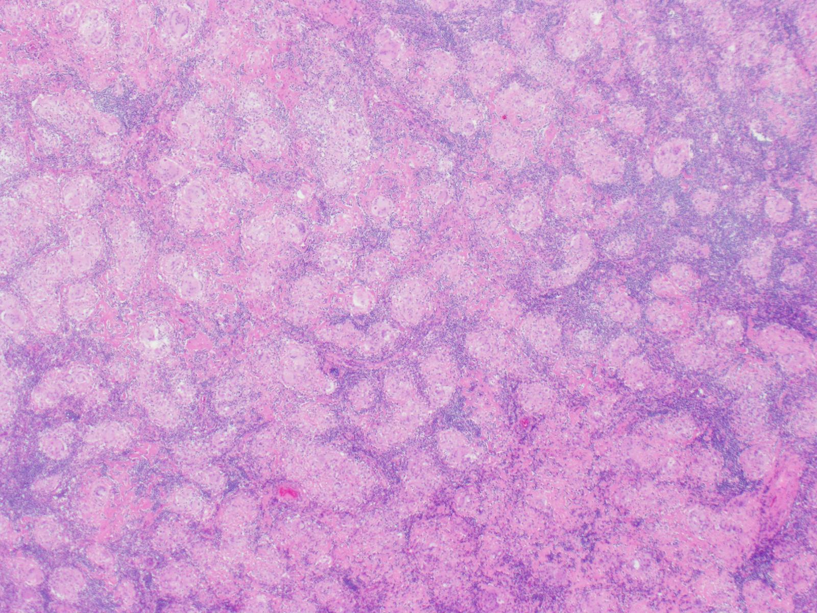
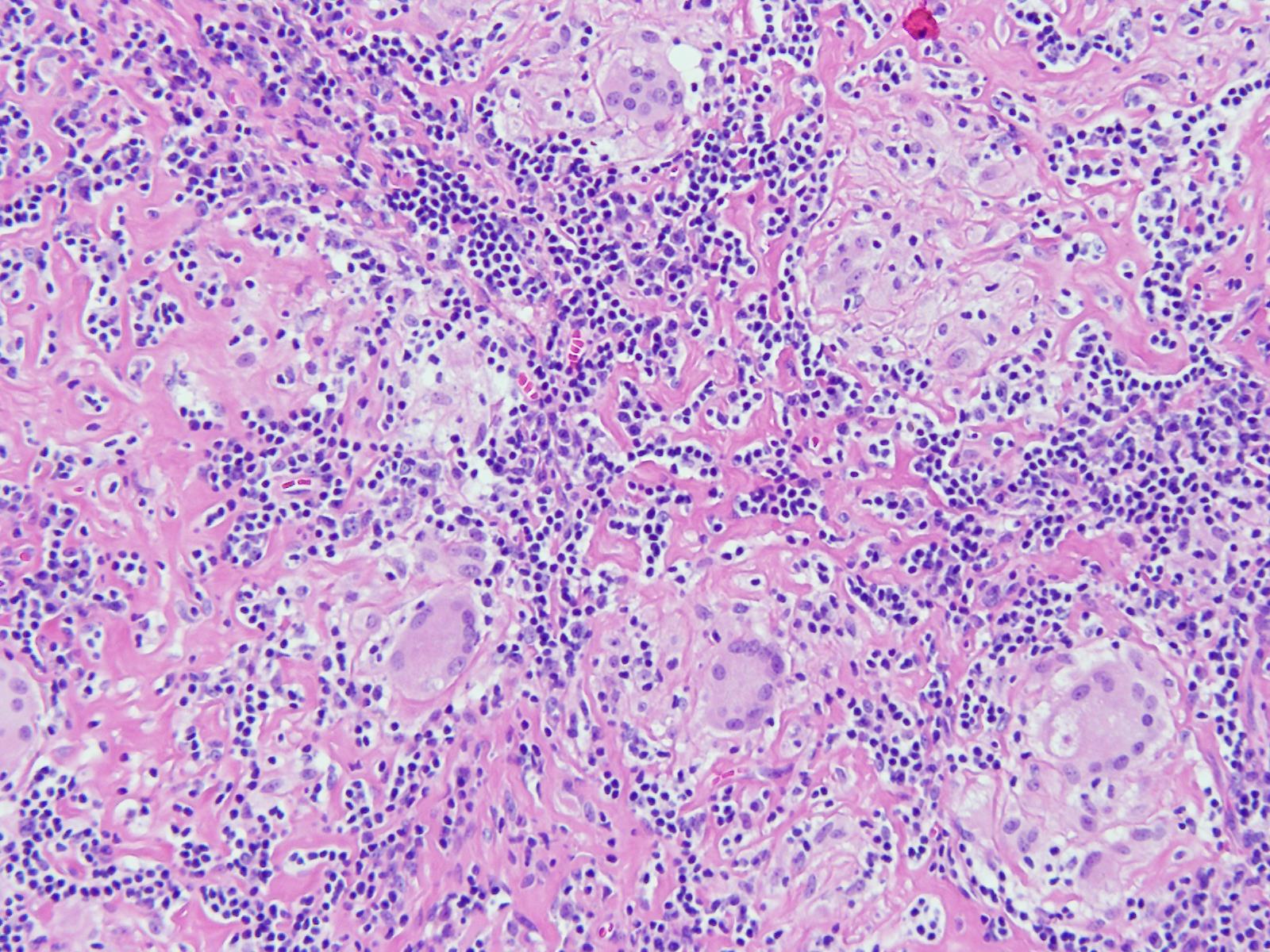
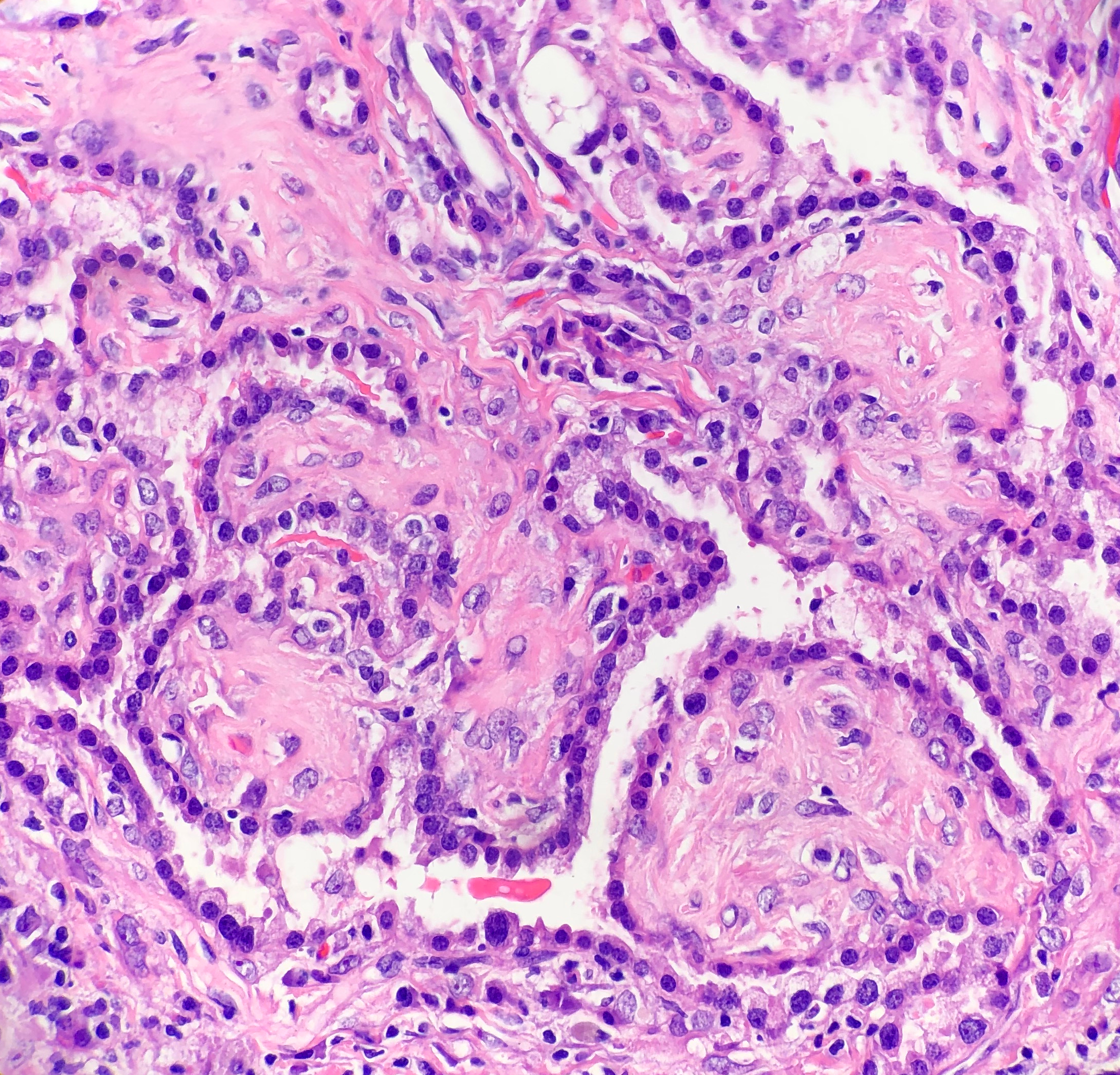
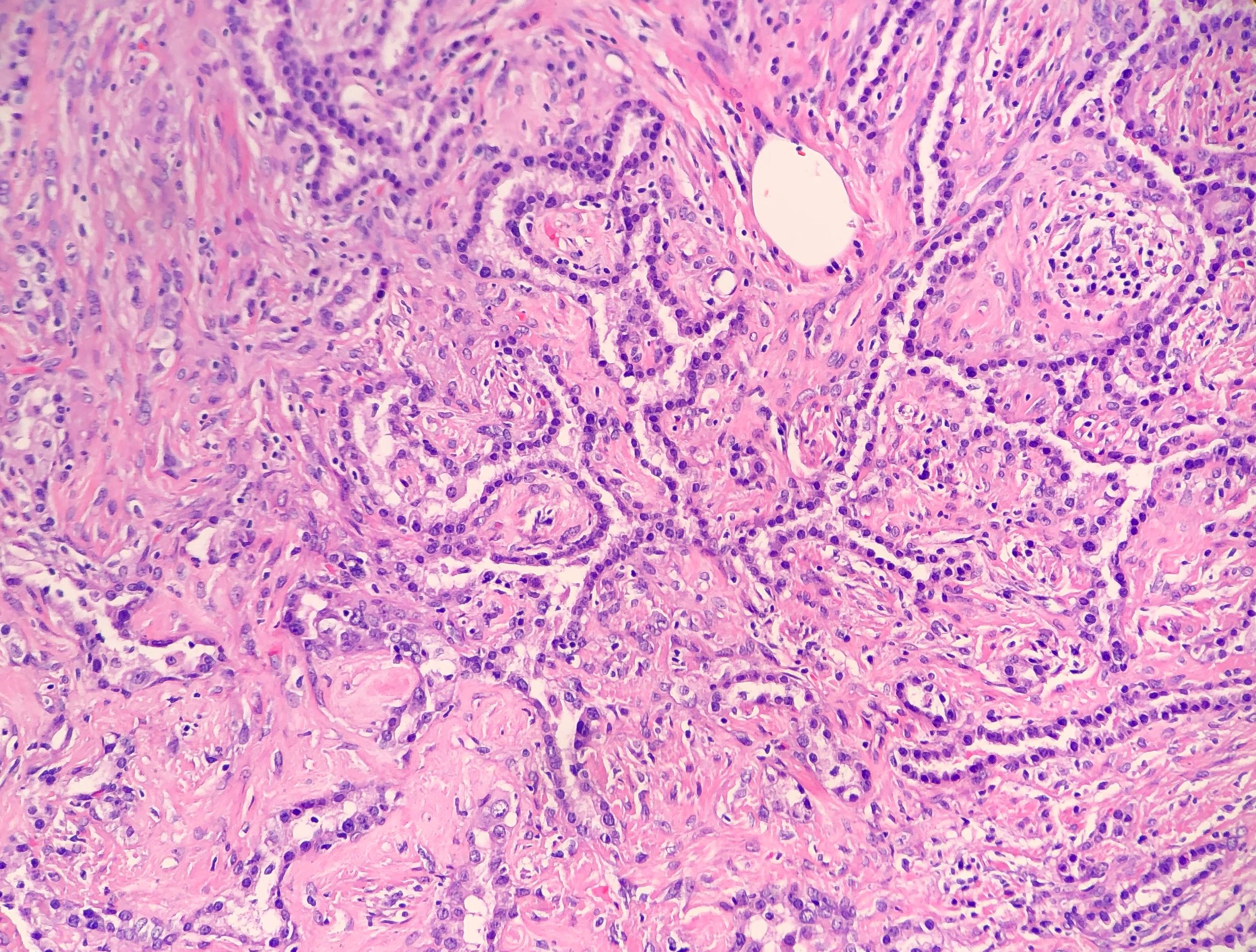
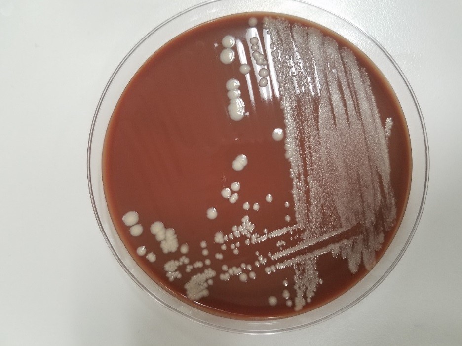
His past medical history was significant for diabetes mellitus, anemia and renal insufficiency. He initially presented 3 weeks ago with chills, rigors and fever to 103 degrees Fahrenheit. For the past several months, the patient has had weight loss (10-20 pounds over an unspecified timeframe), fatigue and new iron deficiency anemia. A heart murmur was heard on physical exam. The patient was admitted for suspicion of sepsis and he was started on empiric antibiotics vancomycin and ceftriaxone. Three sets of blood cultures were drawn prior to initiation of antibiotics, which were all positive for gram positive cocci in pairs and chains. Transesophageal echocardiogram (TEE). TEE showed large vegetation on posterior mitral leaflet measuring 1cm x 1.8 cm, and a smaller mass on the anterior leaflet. A week after admission, a mitral valve replacement was performed followed and a portion of the valve was sent for culture (Figure 1).

Laboratory Identification

Image 1. Gram stain from mitral valve specimen showing a large accumulation of gram positive cocci in chains (100x magnification).
Image 2. Brown-Hopps stain of the surgically resected mitral valve tissue vegetation showing sheets of gram positive bacteria (40x magnification).

Discussion

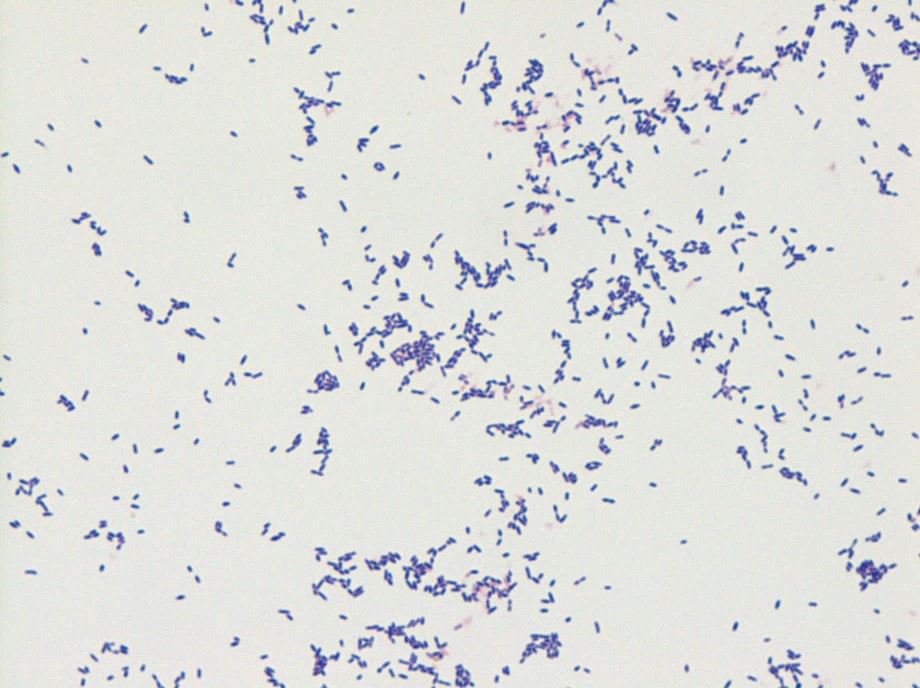
The gram positive organism from blood and mitral valve culture was identified as Streptococcus mitis by MALDI-TOF mass spectrometry. S. mitis is a member of the Streptococcus genus. Streptococci have a number of features that aid in laboratory identification: they are Gram positive, catalase-negative, spherical/ovoid, with organisms that are usually found in chains. They are facultative anaerobes.

More specifically, S. mitis belongs to the viridans streptococci group which includes Streptococcus mutans, Streptococcus sanguis, and Streptococcus salivarius, among many others. The most common infection caused by viridans streptococci is bacterial endocarditis, as in the case of this patient. Other infections can include brain abscesses, liver abscesses, dental caries, and bacteremia.

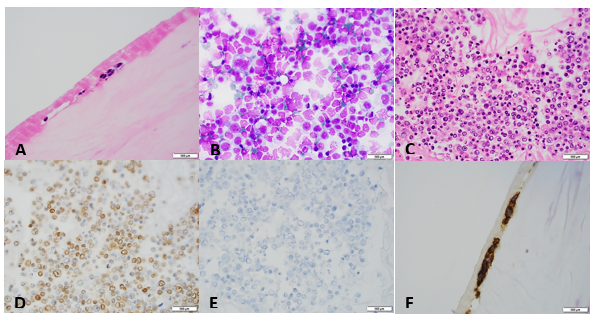
Patients with bacterial endocarditis have an infection of the heart valves or the endoecardial wall that leads to formations of vegetations. These vegetations are composed of thrombotic debris and organisms (Image 2), often associated with destruction of cardiac tissue. Its onset often involves severe symptoms including fever, chills, and weakness. Fever is the most consistent symptom of infective endocarditis, but it may be subtle or even absent in some cases, especially in older adults. Weight loss and flu-like symptoms may also be seen. Left-sided infective endocarditis, as in the case of our patient, will present with murmur in 90% of cases. In long-standing infective endocarditis, patients may present with Roth spots (retinal hemorrhages), Osler nodes (subcutaneous nodules in the digits), microthromboemboli (which appear as splinter hemorrhages under fingernails and toenails), and Janeway lesions (red nontender lesions on the palms or soles).

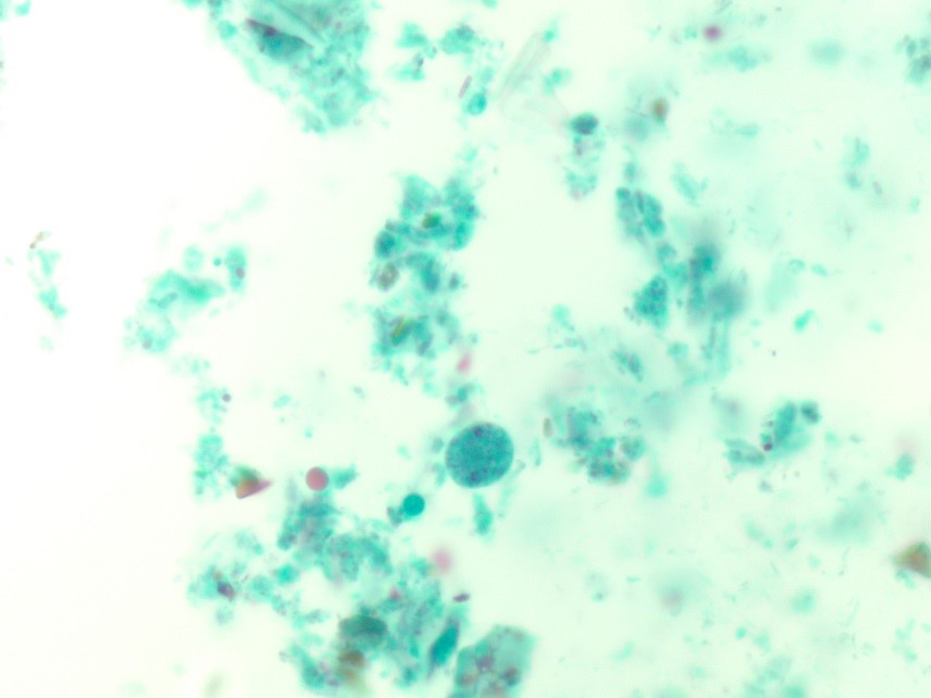
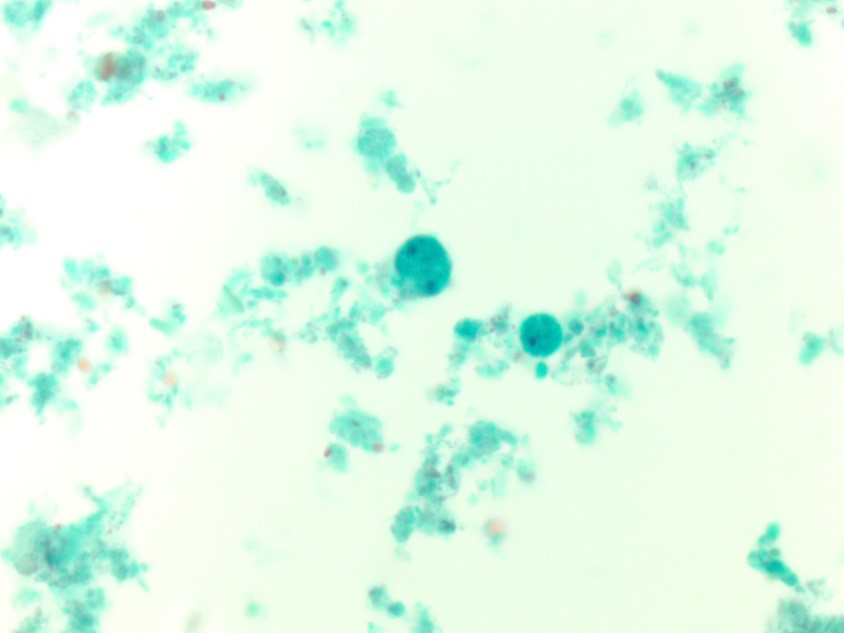
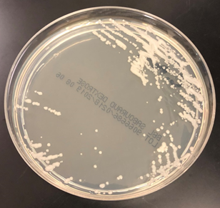
In the laboratory, the diagnosis of S. mitis and other viridians streptococci is often detected via blood culture as in the case of this patient. Once the blood culture bottle becomes positive, a Gram stain is performed, which shows Gram positive cocci in chains (Image 1). These features are helpful in differentiating Streptococcus from Staphylococcus (which appears as clusters instead of chains). Biochemical testing can be done to narrow down the species and identify S. mitis, which is optochin resistant (as opposed to S. pneumonia), acetoin negative (in contrast to most other viridans organisms), and urease negative (which differentiates it from S. vestibularis which is urease positive).

Surgical pathology can also aid in diagnosis by microscopically identifying vegetations on the affected valve (Image 2). Treatment of bacterial endocarditis is usually with penicillin or ceftriaxone, however susceptibility testing should be performed on S. mitis and other viridians streptococci because resistance can occur to penicillin. Blood cultures are followed until they are negative for 72 hours. In the case of our patient, his cultures became negative shortly after he started treatment. Susceptibility testing showed that the organism is sensitive to penicillin and ceftriaxone. The patient was continued on ceftriaxone and is clinically improving.

-Haytham Hasan, MD, is an Anatomic and Clinical Pathology resident at NorthShore Evanston Hospital (University of Chicago).

-Erin McElvania, PhD, D(ABMM), is the Director of Clinical Microbiology NorthShore University Health System in Evanston, Illinois. Follow Dr. McElvania on twitter @E-McElvania. 

Microbiology Case Study: An 8 Year Old Male with Left Knee Pain

Case History

An 8 year old male with no significant medical history presented with left knee pain and swelling for one week. Physical examination revealed a temperature of 101.2°F and a left swollen, tender knee with reduced range of motion. A joint aspirate was performed, and synovial fluid and blood were sent for microbiological analysis.

Laboratory Findings

Synovial fluid analysis showed increased neutrophils, a nucleated cell count of 90,840 cells/cmm, and no crystals.

Blood cultures were negative. Gram smear of the joint fluid showed many neutrophils and no bacteria. Fluid culture grew convex tan-yellow colonies on blood and chocolate plates at 48 hours (Image 1). Gram smear revealed gram-negative cocci (Image 2). The organism was identified by MALDI-TOF as Aggregatibacter aphrophilus. Antibiotic susceptibility testing showed susceptibility to augmentin, ampicillin, ceftriaxone, and levofloxacin.

Image 1. Growth on anaerobic chocolate plate.
Image 2. Gram stain from anaerobic culture showing gram negative cocci.

Discussion

Aggregatibacter aphrophilus is a gram negative coccobacillus that requires 5% CO2 and grows best on blood agar. It is oxidase negative and catalase negative. It is categorized as a HACEK organism, being a cause of culture-negative endocarditis. It is considered normal oral flora, and dental procedures can be a source of infection. Aggregatibacter endocarditis can cause a positive P-ANCA and be misdiagnosed as a vasculitis. It has also been reported as causes of sacroiliitis, bartholinitis, endophthalmitis, and brain abscesses. Treatment is generally ceftriaxone for 8 weeks. Identification is by biochemical methods or MALDI-TOF. Broad range PCR (br-PCR) has also been described, which targets a highly-conserved region of 16S rDNA, and then compares the sequences to database sequences.

The patient was given cefazolin, and his temperature downtrended. He was discharged prior to results but placed on oral augmentin. After susceptibility testing, infectious disease was consulted and he was placed on ceftriaxone for 8 weeks. He continued to improve and subsequent cultures were negative.

References

  1. Ratnayake L, Olver WJ, Fardon T. Aggregatibacter aphrophilus in a patient with recurrent empyema: a case report. J Med Case Rep. 2011;5:448. Published 2011 Sep 12. doi:10.1186/1752-1947-5-448
  2. Hirano K, Tokui T, Inagaki M, Fujii T, Maze Y, Toyoshima H. Aggregatibacter aphrophilus infective endocarditis confirmed by broad-range PCR diagnosis: A case report. Int J Surg Case Rep. 2017;31:150–153. doi:10.1016/j.ijscr.2017.01.041

-Jonathan Wilcock, MD is a 1st year anatomic and clinical pathology resident at the University of Vermont Medical Center.

-Christi Wojewoda, MD, is the Director of Clinical Microbiology at the University of Vermont Medical Center and an Associate Professor at the University of Vermont.

Hematopathology Case Study: A 36 Year Old Woman with an Incidental Neck Mass

Case History

A 36 year old female underwent thyroidectomy for multinodular goitre that led to the fortuitous discovery of a neck mass. The neck mass specimen submitted comprised two lymph nodes measuring 2.2 cm and 1.3 cm in the greatest dimensions, with a fleshy tan cut surface.

Biopsy Findings

H&E stained sections revealed numerous non-necrotizing granulomas effacing and replacing normal lymph node architecture. These consisted of pale epithelioid histiocytes and Langhans type of giant cells. The granulomas lacked a peripheral rim of lymphocytes. AFB and GMS stains were negative for microorganisms

Diagnosis

A diagnosis of non-necrotizing granulomatous lymphadenitis was rendered noting that in the correct clinical context the findings could represent sarcoidosis.

Discussion

Granulomatous inflammation is a special type of chronic inflammatory response characterised by the formation of discrete collections of histiocytes called granulomas. Activated histiocytes appear as epithelioid cells with round to oval nuclei, often with irregular contours and abundant granular eosinophilic cytoplasm with indistinct cell borders. They may coalesce to form multinucleated giant cells. When found in the lymph node, the reaction pattern is called granulomatous lymphadenitis. It can be caused by a variety of different conditions, and therefore, requires thorough workup to come to a conclusive diagnosis.

On the basis of presence or absence of necrosis, granulomatous lymphadenitis can be classified as necrotizing or non-necrotizing. Additionally, the presence of an abscess, usually central, indicates a suppurative lymphadenitis.

Non-necrotizing granulomatous lymphadenitis:

Sarcoidosis lymphadenitis is the prototype of non-necrotizing granulomatous lymphadenitis. It shows the presence of discrete granulomas without a peripheral rim of lymphocytes, called “naked granulomas”. The early phase shows follicular hyperplasia and sinus histiocytosis, followed by appearance of epithelioid cell nodules toward the end of this phase. The peak phase shows well-demarcated granulomas composed of epithelioid cells with scattered multinucleated giant cells observed throughout the lymph node. Granulomas may occasionally coalesce. In the late phase, increased collagen fibers result in fibrosis and hyalinization. There are no neutrophils and it is uncommon to find small foci of central necrosis. Numerous inclusions such as asteroid, Schaumann, or Hamazaki-Wesenberg bodies can be seen. In this case, we observed well-demarcated granulomas throughout the lymph node, typical of the peak phase without any caseous necrosis or suppuration.

Other causes of granulomatous lymphadenitis can be ruled out as follows.

Sarcoid-like lymphadenitis: It shows a similar pattern of non-necrotizing lymphadenitis like sarcoidosis. However, classically sarcoid like reaction shows scattered small epithelioid granulomas with sparsely arranged epithelioid cells. The border of the granulomas is usually obscure. The CD4:CD8 ratio ranges from 0.8 to 2.25 while in sarcoidosis, it is >3.5. These findings help distinguish sarcoid-like lymphadenitis from sarcoidosis.

Sarcoid-like adenitis may be seen in numerous conditions such as carcinoma, Toxoplasmosis, fungal infections, tuberculosis, immunocompromised states, pneumoconiosis etc. The fact that tuberculosis and fungal infections can present with a non-necrotizing granulomatous lymphadenitis highlights the importance of performing fungal (PAS & GMS) and AFB (Ziehl Neelson) stains in non-necrotizing lymphadenitis as well. In this case, the granulomas had distinct borders, numerous epithelioid cells, no organisms were identified on special stains, nor was there any history of immune compromise; ruling out a sarcoid-like reaction.

Berylliosis: The lymph node picture in Berylliosis is identical to that of sarcoidosis. We may even see asteroid bodies or Schaumann bodies. A diagnosis can be established by eliciting a history of chronic exposure to Beryllium. Beryllium lymphocyte proliferation test (BeLPT) is a test that measures Beryllium sensitization and is very specific for Beryllium exposure. There was no known history of exposure to Beryllium in this case.

Toxoplasmosis: A classic triad of follicular hyperplasia, small granulomas composed of epithelioid cells within and around hyperplastic follicles and, monocytoid B cell hyperplasia, is observed in toxoplasmosis lymphadenitis. This case did not show follicular hyperplasia, ruling out toxoplasmosis.

Necrotizing granulomatous lymphadenitis

Even though we did not find any necrosis in this case, yet, it is worthwhile to review briefly the various causes of necrotizing lymphadenitis.

  • Non-suppurative

Tuberculosis: Histology of a tuberculous lymph node is characterised by central caseous necrosis surrounded by an epithelioid cell layer. The outermost layer is comprised of lymphocytes and fibrosis. Plasma cells are not observed. Diagnosis can be established by performing an AFB stain that demonstrates acid fast rod shaped bacteria in the areas of necrosis. Organisms can also be detected by PCR.

BCG lymphadenitis: About 0.7 to 2.3% of BCG vaccinated children may develop BCG lymphadenitis that is smaller than tuberculous lymphadenitis. Early phase shows follicular hyperplasia and sinus histiocytosis. Later, there is development of micronodules of epithelioid granulomas without necrosis and epithelioid cell granulomas with central caseous necrosis. Langhans giant cells are rare.

Fungal infections: Fungal infections by Histoplasma, Cryptococcus, coccidiodomycosis, pneumocystis may also cause a necrotizing granulomatous inflammation. There are numerous neutrophils, and fungal structures can be seen. GMS and PAS can be used in cases where it is difficult to the find the fungal elements on H&E.

  • Suppurative

Tularemia: There are three forms of histological changes, Abscess form, showing abscess with central necrosis and mononuclear cells, Abscess-granulomatous form with granulomas with central necrosis, which form large lesions with central abscesses, and granulomatous form with caseating necrosis at the centre of the granulomas.

Cat Scratch disease: Similar to tularemia, there are three phases of histologic presentation, an early phase of follicular hyperplasia, intermediate phase of microabscess, and a late phase of granulomatous inflammation. Monocytoid B cell clusters are observed close to the abscess.

Conclusion

Sarcoidosis is usually diagnosed by excluding other causes of granulomatous inflammation, as we did in this case. Characteristic non-necrotizing, discrete granulomas were seen throughout the lymph node. The age of the patient and female gender epidemiologically support the diagnosis. This case reflects an example work up of a granulomatous lymphadenitis that is a morphologic presentation of myriad diseases.

-Swati Bhardwaj, MD has a special interest in surgical pathology and hematopathology. Follow her on Twitter at @Bhardwaj_swat.

Kamran M. Mirza, MD, PhD, MLS(ASCP)CM is an Assistant Professor of Pathology and Medical Education at Loyola University Health System. A past top 5 honoree in ASCP’s Forty Under 40, Dr. Mirza was named to The Pathologist’s Power List of 2018. Follow him on twitter @kmirza.

Surgical Pathology Case Study: A 43 Year Old Female with a Lung Nodule Noted on Imaging Following Chest Congestion

Case History

The patient is a 43 year old woman who experienced chest congestion and presented to her local physicians office. A chest X-ray was ordered and demonstrated a lung abnormality. A follow-up CT scan confirmed a 1.9 cm smoothly marginated nodule in the upper lobe with no adenopathy and a normal liver and adrenal glands. The nodule was mildly hypermetabolic on PET scan. A bronchoscopy was performed, which was non-diagnostic. Two subsequent CT scans demonstrated no change in the size of the nodule. Overall, the patient feels well and denies cough, hemoptysis, dyspnea on exertion, and weight loss. Due to the suspicion of cancer, the patient has decided to undergo a lung lobectomy.

Diagnosis

Received in the Surgical Pathology lab for intraoperative consultation is a 30.0 x 7.2 x 2.2 cm lung lobectomy specimen. There is an attached 6.2 cm staple line, which is removed and the subjacent resection margin is inked blue. The entire pleural surface is inked black. The specimen is sectioned revealing a 2.1 x 1.7 x 1.0 cm white-tan, firm, round nodule that is 0.5 cm from the blue inked resection margin and 0.2 cm from the black inked pleural surface. The remainder of the specimen is composed of red-tan, spongy, grossly unremarkable lung parenchyma without nodules or other lesions. Photographs of the specimen are taken (Figure 1). A representative section of the nodule is submitted for frozen section and read out as “diagnosis deferred”. Representative sections of the specimen are submitted as follows:

A1FS:   Frozen section remnant

A2-A7:   Nodule, entirely submitted

A8-A10:   Grossly unremarkable lung parenchyma

Immunohistochemical stains show the epithelial cells in the lesion to be positive for CK7, TTF-1, and surfactant proteins A and B which supports these cells to be type 2 pneumocytes (all controls are appropriate). Based on the immunohistochemical stains and routine H&E slides, the case was signed out as a sclerosing pneumocytoma

Image 1. Gross presentation of the well-defined, round sclerosing pneumocytoma.

Discussion

Sclerosing pneumocytoma (SP) is a rare, benign pulmonary tumor that was first described in 1956 as a vascular tumor, but has since been found to be of primitive respiratory epithelium origin. In the past, SP has also been referred to as sclerosing hemangioma, pneumocytoma, and papillary pneumocytoma, but the 2015 World Health Organization classification of lung tumors states that the agreed upon term for this tumor should be a sclerosing pneumocytoma. SP is commonly seen in middle aged adults, with a female to male ratio of 5:1. There is no racial bias. Patients are usually asymptomatic, with the tumor incidentally found on screening chest radiographs. If the patient was to present with any symptoms, they would usually include a cough, hemoptysis and chest pain. Radiographically, SP appears as a solitary, well-defined, homogenous nodule along the periphery of the lung.

Grossly, most SPs appear as a solitary, firm, well-circumscribed, yellow-tan mass generally arising along the periphery of the lung. The majority of these tumors appear within the lung parenchyma, but there have been cases reported of endobronchial and pleural based SP tumors. Multifocal unilateral tumors and bilateral tumors are uncommon.

Histologically, SP consists of two epithelial cell types: surface cells and round cells. Surface cells are cuboidal, resembling type II pneumocytes, with finely stippled nuclear chromatin, indistinct nuclei, occasional nuclear grooves, and inclusions. The stromal round cells will have bland oval nuclei with coarse chromatin and eosinophilic cytoplasm (Figure 2). Both the surface cells and round cells will have a low mitotic rate, but can have moderate to marked nuclear atypia. Ciliated bronchial epithelium is often identified in the tumor. There are four architectural patterns identified within SP: papillary, sclerotic, solid and hemorrhagic, with over 90% of SPs displaying three of the patterns, and all of the tumors containing at least two of the patterns.

  • Papillary pattern: Complex papillae composed of surface cells covering a stroma of round cells
  • Sclerotic pattern: Papillae containing hyalinized collagen, either in solid areas or along the periphery of hemorrhagic areas (Figure 3)
  • Solid pattern: Sheets of round cells bordered by surface cells
  • Hemorrhagic pattern: Large blood filled spaces
Image 2. Photomicrograph demonstrating the cuboidal surface cells and round stromal cells.
Image 3. Photomicrograph of the papillary and sclerotic architectural patterns.

Immunohistochemical stains can be helpful in the diagnosis of SP, with both the surface cells and round cells exhibiting expression of thyroid transcription factor 1 (TTF-1) and epithelial membrane antigen (EMA). It should be noted that TTF-1 is also used for the diagnosis of pulmonary adenocarcinoma, increasing the risk of misdiagnosing SP. The surface cells will also express both pancytokeratin (AE1/AE3) and Napsin A, with the round cells being negative for AE1/AE3, but having a variable expression of cytokeratin 7 and the low molecular weight cytokeratin (CAM 5.2). Molecular pathology has demonstrated a frequent loss of heterozygosity at 5q, 10q and 9p, and an allelic loses at p16 in the surface and rounds cells. Although the immunohistochemical stains and molecular pathology results can be very helpful, diagnosis of a SP is still largely based on routine H&E slides showing the two epithelial cell types and four architectural patterns.

Electron microscopy will show abundant lamellar bodies similar to those in type II pneumocytes in the surface cells. Round cells will lack the lamellar bodies and instead will contain variably-sized electron-dense bodies that have been thought to represent the different stages of lamellar body maturation.

The differential diagnosis for SP includes a variety of benign and malignant neoplasms, which can be difficult to distinguish on cytology, small biopsies and intraoperative consultations. The cytologic features include moderate to high cellularity with a bloody background and foamy macrophages, occasional nuclear pleomorphism in the round cells, absent mitotic figures, and occasional necrosis with cholesterol clefts and calcifications. In the case of small biopsies, making a diagnosis of SP can be difficult if the papillary pattern is highly prevalent without one of the other three patterns present. With intraoperative consultations, the frozen section artifact can make it difficult to appreciate the two epithelial cell types or the four architectural patterns. The gross examination, as well as the radiographic findings of a well-circumscribed tumor can help point the Pathologist to favoring a benign neoplasm over a malignant one. The benign neoplasms that should be considered in the differential diagnosis include:

  • Clear cell tumor, which will have clear cells with scant stroma, thin-walled vessels and a strong expression of HMB-45
  • Pulmonary hamartoma, which will have a combination of cartilage, myxoid stroma, adipose tissue and trapped respiratory epithelium
  • Hemangiomas, which are rare in the lung, and will lack epithelial cells and contain either a cavernous or capillary morphology

The malignant neoplasms that should be considered in the differential diagnosis include:

  • Bronchioalveolar carcinoma, which can have a papillary pattern, but will not contain the two epithelial cell types and combination of the four architectural patterns
  • Metastatic papillary thyroid carcinoma, which is distinguished from SP by the presence of the characteristic Orphan Annie nuclei
  • Metastatic renal cell carcinoma, which will contain nuclear atypia and striking vascularity
  • Carcinoid, which will contain organoid and ribbon-like growth patterns

Currently, with the benign nature of SP, surgical excision is the preferred treatment choice to cure the patient. There have been cases reported of lymph node metastasis and recurrence, but neither of these appear to effect the prognosis. This just helps to highlight the need for a multidisciplinary approach to this benign tumor.

References

  1. Hisson E, Rao R. Pneumocytoma (sclerosing hemangioma), a Potential Pitfall. Diagn Cytopathol. 2017;45(8):744-749
  2. Keylock JB, Galvin JR, Franks TJ. Sclerosing Hemangioma of the Lung. Arch Pathol Lab Med. 2009;133(5):820-825.
  3. Travis WD, Brambilla E, Nicholson AG, et al. The 2015 World Health Organization Classification of Lung Tumors: Impact of Genetic, Clinical and Radiologic Advances Since the 2004 Classification. J Thorac Oncol. 2015;10(9):1243-1260.
  4. Wu R. Sclerosing Pneumocytoma (Sclerosing Hemangioma). Pathology Outlines. http://www.pathologyoutlines.com/topic/lungtumorsclerosingheman.html. Revised February 19, 2019. Accessed June 6, 2019.

-Cory Nash is a board certified Pathologists’ Assistant, specializing in surgical and gross pathology. He currently works as a Pathologists’ Assistant at the University of Chicago Medical Center. His job involves the macroscopic examination, dissection and tissue submission of surgical specimens, ranging from biopsies to multi-organ resections. Cory has a special interest in head and neck pathology, as well as bone and soft tissue pathology. Cory can be followed on twitter at @iplaywithorgans.

Microbiology Case Study: A 58 Year Old Female with Abdominal Pain

Clinical History

A 58 year old female with no significant past medical history presented her primary care physician with chief complaint of abdominal pain. She reported continued vague abdominal symptoms for the past two months, with intermittent diarrhea and increased flatulence. No recent travel history or significant exposures were identified. An ultrasound of the right upper quadrant was unremarkable and no gallstones were present. The patient was scheduled for a screening colonoscopy. A stool specimen was submitted to the microbiology laboratory for stool culture and ova & parasite exam.  

Laboratory Identification

Image 1. Trichrome stained fecal smear illustrating a binucleated trophozoite with fragmented karyosomal material from a stool ova & parasite exam.
Image 2. Additional trichrome fecal smear image highlighting both uninucleate and binucleate trophozoites that range in size from 5 to 15 um.

The findings from the ova and parasite exam were consistent with Dientamoeba fragilis, an intestinal flagellate. The stool culture was negative for Salmonella, Shigella, and Escherichia coli 0157:H7. Stool enzyme immunoassays were negative for Campylobacter spp.and Shiga toxin 1 and 2.

Discussion

Dientamoeba fragilis is an intestinal flagellate with worldwide distribution and causes asymptomatic and symptomatic infections, predominantly in small children. Symptoms of infection may include intermittent diarrhea, abdominal pain, anorexia, weight loss, and flatulence.  While the pathogenesis is not completely understood, transmission is thought to occur via the fecal oral route and it is hypothesized that the trophozoites are transmitted via the eggs of nematodes, Enterobius vermicularis and Ascaris lumbricoides, due to a higher incidence of co-infections between these organisms than expected.

In the laboratory, the diagnosis of D. fragilis is made by ova and parasite exam. The trophozoite resembles amebae and is typically 9-12 µm. Most trophozoites are binucleate with finely granular cytoplasm and the within the nuclei there are 4-8 fragments of karyosomal granules (Figure 1). Due to the fact that 30-40% of D. fragilis trophozoites are uninucleate (Figure 2) and they lack external flagella, they must be differentiated from Endolimax nana and Entamoeba hartmanni, which are both non-pathogenic amebae. Historically, no cyst phase was known for D. fragilis; however, recent studies have identified precyst forms or putative cysts. Permanently trichrome stained slides are essential to diagnosing D. fragilis infection, as the organism is hard to detect in concentrated smears.

Since our patient was symptomatic, she was treated with iodoquinol, the drug of choice for D. fragilis infections. Her symptoms resolved and colonoscopy did not reveal additional pathology.  

-Debbie Walley, MD, is a 4th year Anatomic and Clinical Pathology chief resident at the University of Mississippi Medical Center. 

-Lisa Stempak, MD, is an Assistant Professor of Pathology at the University of Mississippi Medical Center in Jackson, MS. She is certified by the American Board of Pathology in Anatomic and Clinical Pathology as well as Medical Microbiology. She is the Director of Clinical Pathology as well as the Microbiology and Serology Laboratories. Her interests include infectious disease histology, process and quality improvement, and resident education.

Microbiology Case Study: A 15 Year Old Male with Endocarditis

Case History

A 15 year old male with a past medical history significant for Tetralogy of Fallot (congenital heart defect), multiple valve replacements, chronic kidney disease, and prior Bartonella endocarditis. He presented with a “flu-like” illness including muscle aches, fevers, fatigue, and night sweats. His symptoms slowly dissipated after about three days. However, he had labs drawn including multiple blood culture sets which were all positive for growth.

Laboratory Findings

Gram stain showed gram positive bacilli and culture plates grew two morphologies of slow growing gray, granular and opaque colonies.This organism was identified by MALDI-TOF as Corynebacterium pseudodiphtheriticum.

Image 1. Gram stain with gram positive bacilli .
Image 2. Culture with small, grayish colonies with granular appearance and opaque centers (growth at day 2).

Discussion

The genus Corynebacterium comprises a collection of irregular-formed, rod-shaped or coccoid bacteria that are non-motile, catalase-positive, and non-spore-forming.

Corynebacterium pseudodiphtheriticum (previously designated as Corynebacterium hofmannii) is a nonlipophilic, nonfermentive, urease- and nitrate-positive Corynebacterium species.1 C. pseudodiphtheriticum is part of the usual oropharyngeal bacterial flora, including the nares and throat. It appears to play a role in preventing colonization of oropharyngeal epithelia by pathogenic bacteria.

Most commonly, C. pseduodiptheriticum is a pathogen of the respiratory tract with cases of nosocomial and community-acquired pneumonia, bronchitis, tracheitis, pharyngitis, and rhinosinusitis. Endocarditis is the second most common infection site, although very rare. Cases of urinary tract and wound infections have also been reported.

Treatment is usually with penicillin alone or in combination with aminoglycosides. Antibiotic susceptibility profiling of C. pseudodiphtheriticum isolates showed that resistance to oxacillin, erythromycin, clindamycin, and macrolides are common.1

References

  1. Burkovski A. Corynebacterium pseudodiphtheriticum: Putative probiotic, opportunistic infector, emerging pathogen. Virulence. 2015;6(7):673–674. doi:10.1080/21505594.2015.1067747

-Nicole Mendelson, MD is a 1st year Anatomic and Clinical Pathology resident at the University of Vermont Medical Center.

-Christi Wojewoda, MD, is the Director of Clinical Microbiology at the University of Vermont Medical Center and an Associate Professor at the University of Vermont.

Hematology Case Study: A 69 Year Old Female with Breast Implants

Case History

A sixty nine year old female who underwent right breast reconstruction about 13 years ago due to breast cancer presents to the doctor office with right breast pain and right breast enlargement over the last two months. She has lost some weight and does not recall any trauma to this area. She had a textured saline implant. Examination reveals no definite palpable masses. MRI of right breast showed intact saline implant with moderate amount of fluid surrounding the implant within the intact external capsule. No adenopathy was noted. Right breast implant was removed and complete capsulectomy was performed.

Image 1. A. Section of breast capsule with rare atypical hyperchromatic cells (arrow). B. Cytospin preparation of the fluid surrounding the implant with numerous atypical lymphocytes. C. Cell block of the fluid with large atypical lymphocytes. D, E. Lymphocytes are positive for CD30 (image D) and negative for ALK-1 (image E). F. CD30 positive cells in the section of the implant.

Diagnosis

Breast implant-associated anaplastic large cell lymphoma.

Discussion

Breast implant associated anaplastic large cell lymphoma is a provisional entity that is morphologically and immunophenotypically similar to ALK-negative anaplastic large cell lymphoma. It arises primarily in association with a breast implant. It is a very rare entity with an incidence of 1 in 500,000 to 3 million women with implants. Tumor cells may be localized to the seroma cavity or may involve pericapsular fibrous tissue. Sometimes it can form a mass lesion. Locoregional lymph node may be involved. The mean patient age is 50 years. Most patient presents with stage 1 disease, usually with peri-implant effusion. The mean interval from implant placement to lymphoma diagnosis is 10.9 years. There is no association with the type of implant. Histologic examination shows two different types of proliferations. In patients with seroma, the proliferation is confined to the fibrous capsule (“in situ” iALCL). However, the distribution of neoplastic lymphocytes could be heterogeneous with some cellular areas with numerous large pleomorphic cells of varying size and some fibrotic areas with rare atypical lymphocytes. It is beneficial to look at the seroma fluid in addition to capsule sections, because sometimes the neoplastic lymphocytes are predominantly present in fluid (as in our case). Patients presenting with tumor mass show more heterogeneous proliferations infiltrating surrounding tissues (“infiltrative” iALCL). They consists of either sheets are clusters of large neoplastic cells accompanied by a large number of eosinophils. By immunohistochemistry, the tumor cells are strongly positive for CD30. CD2 and CD3 are more often positive than CD5. CD43 is almost always expressed. Most cases are CD4 positive. The prognosis is very good in patients with disease confined to the capsule. The median overall survival is 12 years. However, patients with a tumor mass could have a more aggressive clinical outcome.

References

1. Swerdlow SH, Campo E, Harris NL, et al. WHO Classification of Tumours of Haematopoetic and Lymphoid Tissues (Revised 4th edition). IARC: Lyon 2017.

2. Jaffe, E , Arber, D, et al. Hematopathology (second edition) 2017.

-Junaid Baqai, MD, was born in Chicago, IL but spent most of his life in Karachi, Pakistan. He graduated from DOW Medical College in Pakistan and did his residency in anatomic and clinical pathology at Danbury Hospital, CT followed by hematopathology fellowship from William Beaumont Hospital, Michigan and oncologic-surgical pathology fellowship from Roswell Park Cancer Institute, New York. He currently serves as Medical Director of hematology, coagulation and flow cytometry at Memorial Medical Center and Medical Director of Laboratory at Taylorville Memorial Hospital.

Microbiology Case Study: A 24 Year Old Male with No Past Medical History Returning from Guatemala with Fevers, Myalgia, and Cough

Case History

A 24 year old male with no past medical history presented with fevers, myalgia, and cough following return from a 1-week trip to Guatemala where he spent significant time within caves. The patient described his cough as persistent, non-productive, and associated with mild shortness of breath at rest that significantly worsens with activity. In the emergency department, the patient was afebrile with a WBC of 10.2, Transaminitis, and chest X-ray showed diffuse reticular pattern. He underwent a bronchoscopy and BAL washout.

Laboratory Findings

Histoplasmosis Urine Antigen test came back positive.

Image 1. Fungal culture with white/tan, fluffy mold (growth at day 7).
Image 2. Scotch tape prep with tuberculate macroconidium. This mold was morphologically identified as Histoplasma capsulatum and sent to Mayo Laboratories for further confirmatory testing.

Discussion

Histoplasma capsulatum is an intracellular, thermally dimorphic fungus (grows as a yeast at body temperature/37°C in humans or culture media and as mold at 25°C in the environment/culture media). Histoplasma is found in soil, particularly in areas containing bird and bat droppings, such as caves. Within the United States Histoplasma in found in central and eastern states with a predominance in the Ohio and Mississippi River Valleys. This fungus is also found in parts of Central and South America, Africa, Asia, and Australia.

Infection with Histoplasma capsulatum causes significant morbidity and mortality worldwide. Upon inhalation of conidia, H. capsulatum transforms into the pathogenic yeast phase. This form replicates within macrophages that carry the yeast from the lungs to other organs. Histoplasmosis has three main forms:

  • Acute primary histoplasmosis which presents as a pneumonia with fever, cough, myalgia.
  • Chronic cavitary histoplasmosis which is characterized by pulmonary lesions that often resemble cavitary tuberculosis.
  • Progressive disseminated histoplamosis that spreads to infect many organs in immunocomprimised patients.

In the laboratory, culture of blood, tissue and respiratory specimens may be completed. In addition, a test for H. capsulatum antigen is sensitive and specific when simultaneous serum and urine specimens are tested. It is important to note that cross-reactivity with other fungi (Coccidioides immitis, Blastomyces dermatitidis, Paracoccidioides brasiliensis, Penicillium marneffei) has been identified.

Growth on fungal culture shows white/tan, fluffy mold that turns to brown to buff with age. The organism may also produce wrinkled, moist, or heaped yeast-like colonies that are soft and cream when grown at 37°C on certain media. Scotch tape preparation of the mold form shows tuberculate macroconidia, a diagnostic structure of Histoplasma capsulatum. The mycelia are septate and produce microconidia and macroconidia. Yeast forms of Histoplasma capsulatum are small (2 to 4 μm) and reproduce by budding. These budding forms may be seen on histology specimens. A commercially available DNA probe can be performed on culture material to confirm identification.

Patients with mild-moderate histoplasmosis can often have resolution of their symptoms without treatment. Those with more moderate-severe disease require antifungal agents including amphotericin B or itraconazole.

-Nicole Mendelson, MD is a 1st year Anatomic and Clinical Pathology resident at the University of Vermont Medical Center.

-Christi Wojewoda, MD, is the Director of Clinical Microbiology at the University of Vermont Medical Center and an Associate Professor at the University of Vermont.

Hematopathology Case Study: A 76 Year Old Man with Lymphadenopathy

Case History

76 year old man with a history of chronic lymphocytic leukemia/small lymphocytic lymphoma (CLL/SLL) with new anterior mediastinal mass and increasing lymphadenopathy.

Lymph Node Biopsy

H&E

Diagnosis

Tissue sections show a diffuse atypical lymphoid infiltrate that completely effaces the normal nodal architecture. The infiltrate is composed of numerous small lymphocytes with round to mildly irregular nuclei, clumped chromatin, inconspicuous nucleoli and scant cytoplasm. There are also expanded pale areas that contain intermediate sized cells with more open chromatin and distinct single to multiple nucleoli. These cells are most consistent with prolymphocytes/paraimmunoblasts and form the proliferation centers characteristic of CLL/SLL. Occasional centroblastic-type B-cells are noted within these proliferation centers. In addition, there are scattered single to multinucleated cells that have irregular nuclear membranes with pale, vesicular chromatin and prominent inclusion-like, eosinophilic nucleoli. These cells morphologically resemble Hodgkin cells, Reed-Sternberg cells, mummified forms and other variants. These large cells are more evident in areas with a histiocyte rich background and around foci of necrosis. Occasionally, apoptotic bodies and mitotic figures are seen.

 Immunohistochemical studies show that the vast majority of the small-intermediate lymphocytes express B-cell markers CD20 (dim) and PAX5 and co-express CD5 and CD23 (subset). This is consistent with a background of CLL/SLL. The large atypical cells are positive for CD30, PAX5 and CD20 (variable). CD3 highlights numerous scattered background small T-cells, which are increased in the areas with the large cells. In situ hybridization for Epstein Barr viral RNA (EBER ISH) is mainly staining the large atypical cells. By Ki-67, the proliferation fraction is overall increased (40%) with increased uptake by the large atypical cells.

The morphologic and immunophenotypic findings are consistent with involvement by the patient’s known small lymphocytic lymphoma/chronic lymphocytic leukemia (SLL/CLL) with aggressive morphological features. The aggressive features include expanded proliferation centers and an elevated Ki-67 proliferative index (40%). Additionally there are histiocyte/T-cell rich areas composed of multiple EBV positive large atypical cells with morphologic and immunophenotypic features compatible with Hodgkin/ Reed-Sternberg cells. These areas are most in keeping with evolving classic Hodgkin lymphoma. Sheets of large cells indicative of large cell transformation are not seen, although increased scattered large centroblastic-type B cells are present.

Discussion

Lymph node involvement by CLL/SLL will typically show a diffuse proliferation of small lymphocytes with effacement of the normal nodal architecture.  The small lymphocytes have round nuclei, clumped chromatin and scant cytoplasm. Scattered paler areas known as proliferation centers are characteristic of this entity. The proliferation centers are composed of a mixture of cell types including small lymphocytes, prolymphocytes and paraimmunoblasts. Prolymphocytes are small to medium in size with relatively clumped chromatin, whereas paraimmunoblasts are larger cells with round to oval nuclei, dispersed chromatin, eosinophilic nucleoli and slightly basophilic cytoplasm. Some cases show increased and enlarged proliferation centers with a higher proliferation rate. This must be distinguished from large cell transformation.1

Aggressive features of CLL/SLL include proliferation centers that are broader than a 20x field or becoming confluent. An increased Ki-67 proliferation >40% or >2.4 mitoses in the proliferation centers can also portend a more aggressive course. These cases tend to have worse outcomes than typical CLL/SLL and better outcomes than cases that have undergone Richter transformation to diffuse large B-cell lymphoma (DLBCL). Transformation to DLBCL occurs in 2-8% of patients with CLL/SLL. Less than 1% of patients with CLL/SLL develop classic Hodgkin lymphoma (CHL). In order to diagnose CHL in the setting of CLL/SLL, classic Reed-Sternberg cells need to be found in a background appropriate for CHL, which includes a mixed inflammatory background. The majority of these CHL cases will be positive for EBV.1

Richter’s transformation is defined as an aggressive evolution of CLL. While the most common type of transformation is to a high-grade B-cell Non-Hodgkin lymphoma, other histological transformations have been described. This includes CHL, lymphoblastic lymphoma, hairy cell leukemia and high-grade T-cell lymphomas. The prognosis for patients who present with transformation to CHL is poor compared to de novo CHL.2 A large study from the M.D. Anderson Cancer Center described 4121 patients with CLL/SLL and found that only 18 patients or 0.4% developed CHL. The median time from CLL to CHL diagnosis was 4.6 years. Fourteen of the patients received chemotherapy. The overall response rate was 44% with a complete response rate of 19%. The median overall survival was 0.8 years and all patients eventually died from disease recurrence or progressive disease.3 This dismal prognosis is similar to patients with Richter transformation to DLBCL and much worse than patients with de novo CHL, which is curable in >85% of cases.1

References

  1. Swerdlow SH, Campo E, Harris NL, et al. WHO Classification of Tumours of Haematopoetic and Lymphoid Tissues (Revised 4th edition). IARC: Lyon 2017.
  2. Janjetovic S, Bernd HW, Bokemeyer C, Fiedler W. Hodgkin’s lymphoma as a rare variant of Richter’s transformation in chronic lymphocytic leukemia: A case report and review of the literature. Mol Clin Oncol. 2016;4(3):390–392.doi:10.3892/mco.2016.727.
  3. Tsimberidou, AM, O’Brien, S and Kantarjian, HM, et. al. Hodgkin transformation of chronic lymphocytic leukemia. Cancer. 2006;107(6).doi.org/10.1002/cncr.22121.

Chelsea Marcus, MD is a Hematopathology Fellow at Beth Israel Deaconess Medical Center in Boston, MA. She has a particular interest in High-grade B-Cell lymphomas and the genetic alterations of these lymphomas.

Microbiology Case Study: A 30 Year Old Male with Fevers and Cough

Clinical History

A 30 year old African American male presented to the emergency department (ED) with fevers and cough. His past medical history was significant for type 1 diabetes & diabetic nephropathy requiring a kidney/pancreas transplant three years prior. He is compliant with his immunosuppressant regimen. He described the cough as non-productive and denied shortness of breath or chest pain. He denied sick contacts, recent travel, and has no pets. After hospital admission, he became septic, developed severe hypotension (70/30s), and was transferred to the intensive care unit (ICU). Chest x-ray showed multifocal consolidations in bilateral lung fields and a small pleural effusion consistent with pneumonia. He was empirically started on vancomycin, piperacillin-tazobactam, azithromycin, and micafungin. Infectious diseases was consulted and recommended a board variety of tests and cultures given the patient immunosuppressed status.

Laboratory Identification

The following results were obtained:

Sputum culture: normal respiratory flora, negative for fungi and acid fast bacilli

Streptococcus pneumoniae & Legionella pneumophila antigens: negative

Histoplasma & Blastomyces urinary antigens: negative

Fungitell: negative

Respiratory viral PCR panel: positive for adenovirus, coronavirus, and rhinovirus

Image 1. Cryptococcal lateral flow assay showing positive (left) and negative (right) results. The patient had a positive result from the serum with a titer of 1:20.

Given this positive serum result for Cryptococcus neoformans despite multiple negative sputum cultures, a bronchoalveolar lavage & lumbar puncture were performed and bacterial & fungal cultures were performed. 

Image 2. Discrete, mucoid, cream colored colonies of Cryptococcus neoformans growing on Sabouraud dextrose agar after the third week of incubation at 25°C from the bronchoalveolar lavage specimen.

Discussion

Cryptococcus neoformans is an encapsulated yeast that is most commonly acquired through inhalation and can infect & disseminate to multiple organ systems including the lungs, central nervous system, skin and bones, especially in immunocompromised patients such as those with HIV or organ transplant patients. The thick polysaccharide capsule gives colonies of C. neoformans a mucoid appearance, serves as a major virulence factor, and also plays an important part in various laboratory identification methods.

In the lab, C. neoformans will grow a variety of selective and non-selective agars including blood, chocolate, Sabouraud dextrose, and cornmeal agars as discrete, cream colored colonies (Image 2). On microscopic examination, the C. neoformans yeast are gram positive with narrow based budding and a thick capsule. The yeast vary in size from 2-20 µm and are evenly spaced from one another due to the capsule. C. neoformans is positive for both urease and phenoloxidase. Historically, India ink stain was performed on CSF specimens to highlight the capsule using direct microscopy. Grocott’s methenamine silver (GMS), mucicarmine, and Fontana-Masson histochemical stains are all positive for C. neoformans.

Cryptococcal antigen tests directed to the capsular polysaccharide can also be used to diagnosis C. neoformans infections from both serum and CSF specimens. Common methods include immunochromatographic lateral flow assays or particle agglutination. Advantages to these methods include increased sensitivity and the ability to provide semi-quantitative titer results which can be used to monitor the patient’s response to therapy. Rarely, false negative results can occur due to extremely high concentrations of the cryptococcal antigen. In order to combat the prozone effect, the sample should be diluted prior to repeating the test if there is a high suspicion of cryptococcal infection. False positive results may also occur when macroglobulins are present in the sample due to disease states such as rheumatoid arthritis or lupus. Use of pronase can prevent the interference of macroglobulins on serum test results. False positive test results have also be documented due to interferences from various collection devices such as anaerobic vials.

In the case of our patient, as C. neoformans is intrinsically resistant to echinocandins, he was switched from micafungin to fluconazole. He responded well and after completing the therapeutic course, he continued on a prophylactic dose of fluconazole. His cerebrospinal fluid culture showed no growth and the cryptococcal lateral flow assay was negative on the CSF specimen.

-Charles Middleton, MD, is a first year Anatomic and Clinical Pathology resident at the University of Mississippi Medical Center.


-Lisa Stempak, MD, is an Assistant Professor of Pathology at the University of Mississippi Medical Center in Jackson, MS. She is certified by the American Board of Pathology in Anatomic and Clinical Pathology as well as Medical Microbiology. She is the Director of Clinical Pathology as well as the Microbiology and Serology Laboratories. Her interests include infectious disease histology, process and quality improvement, and resident education.