Up in Smoke

Hello again everybody, and welcome back! Last month, I was flattered by a double feature with my post about giving a TEDx talk and Dr. Razzano interviewing me for her global health series. This month, I’d like to address a topic that’s been literally everywhere lately and is just as hard to ignore as…well, second-hand smoke. So, fasten your seatbelts, ensure your seats and tray tables are in the upright position, make sure your biases are stowed in the seat before you, and (of course) please note the no smoking sign as we take off on the topic of vaping!

Image 1. What? I’ve been traveling a lot. There’s inspiration everywhere!

The Smoking Gun

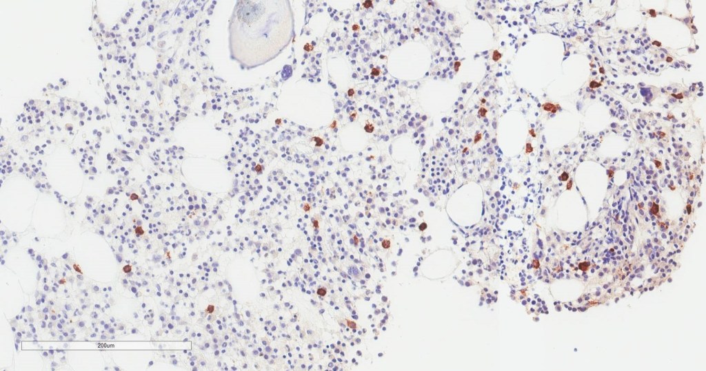
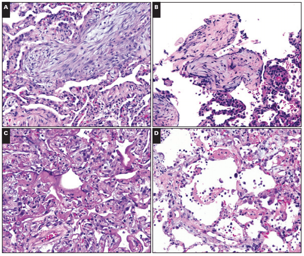
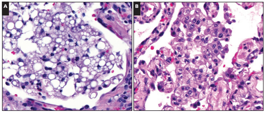
You may have noted that in the past few weeks or months the topic of vaping has been a mainstay of nighttime news stories and front-page print articles. That’s because there’s a lot happening, and from a lot of different angles. It can be messy and confusing, especially because there’s a scientific and non-scientific debate: availability, marketing, health risk, research, and more—all happening at once. I’m going to talk a little bit about all of this, but mostly we’ll look at the medical aspect of vaping as some fantastic publications are making their way into medical journals, including our very own American Journal of Clinical Pathology (AJCP). Recently, friend, colleague, and fellow member of the ASCP Social Media Team and pulmonary pathologist at the Cleveland Clinic, Dr. Sanjay Mukhopadhyay (@smlungpathguy on Twitter) published a noteworthy article with AJCP demonstrating the histopathologic findings of vaping associated lung injury. In essence, vaping causes acute lung injury which is recognized in tissue, supporting the case that both further studies are mandated for health and safety and that vaping should currently be considered a potential critical health risk.

Image 2. About half of the official ASCP Social Media Team (#ASCPSoMeTeam!) from left to right Lab scientist and educator Aaron Odegard (@odie0222), myself (@CEKanakisMD), Dr. Sanjay Mukhopadhyay (@smlungpathguy), famous resident Dr. Adam Booth (@ALBoothMD), and Dr. Kamran Mirza (@Kmirza). If you want updates with great pathology and lab medicine stories and content—follow ALL OF THESE twitter handles!

In this paper, Dr. Mukhopadhyay, et al, tried to capture the direct tissue-related effects of vaping. EVALI, Electronic-Cigarette or Vaping use Associated Lung Injury, has received quite a bit of spotlight in the media as I mentioned. Case series featured in the New England Journal of Medicine (NEJM) highlighted patients in the Midwest with EVALI-type pulmonary disease, but the number of publications on the topic is currently scarce—let alone ones that demonstrate the actual pathophysiology in-process in those affected patients. In the AJCP paper, lung biopsies from a small number of male patients who havdrespiratory illness and concurrent histories of vaping were examined. With all other pulmonary pathology worked up and negative, their biopsies showed various patterns of acute lung injury. The NEJM cases were also worked up and found to be negative for the differentials of pulmonary disease whether infectious, inflammatory, or otherwise; adding credence to a developing body of research supporting the connection between vaping and EVALI.

Image 3. Here’s the mainstay paper I keep referencing. It’s part of a growing number of published works on the topic and part of our expanding understanding of EVALI and its health implications from both public health and pathologic/diagnostic viewpoints.

Where There’s Smoke, There’s…a Lot of Stuff, Actually

There are a ton of stories in the lay-press about vaping-related illnesses. The surveillance data from those NEJM case series and the CDC show a median age of 19 with an overwhelming 94% being hospitalized and roughly two-thirds of those requiring ICU intervention and one-third having to be placed on mechanical ventilation. Of note, 11% of these patients claimed that they vaped pure nicotine product, while 89% smoked cannabinoids/THC in their vape products. Most of them presented to medical care with oxygen saturations <89% on room air (normal O2 sats are variable by patient, but they should be above 95% in ideally healthy individuals). This is neither an endorsement or comment on the medical uses of cannabinoids or a statement on their health effects. Instead, it should be worth mentioning that not only are electronic-cigarette products a new way of smoking higher concentrations of tobacco-obtained or synthetic nicotine but also other products, which have very little data with regard to their associated health risks.

Image 4a. If you haven’t been able to read Dr. Mukhopadhyay’s paper yet, don’t worry I got you. Here are a few cases’ computed tomography (CT) scans that show clinically diagnostic evidence of pulmonary disease visible as (A) ground-glass opacity, GGO, with a pattern that mimics a peripheral eosinophilic pneumonia, (B) more GGOs with areas of consolidation, or solid-looking lung tissue, (C) lower lung GGOs and consolidation with some thickened tissue, and (D) patchy GGOs. All of these cases and more demonstrated some kind of pneumonia and lung tissue pathology but had been worked up and found negative for other causes of disease aside from their shared history of vaping.
Image 4b. Okay, this is a blog for medical laboratory professionals, right? So here’s some slides for the glass pushers! PLEASE NOTE: this is just a sample of a number of histopathologic findings published in the paper, so to see the rest go to the primary source. I’ve highlighted these images as they demonstrate the two major lung injury patterns seen in the EVALI entity: organizing pneumonia seen in (A) & (B) and diffuse alveolar damage (DAD) seen in figures (C) & (D).

Put This in Your Pipe and (please don’t) Smoke It

Okay, I mentioned cannabinoids. Now that I have your attention, I want to walk you through a unique piece of the EVALI discussion you may have seen in the media: the implication of Vitamin-E substances as a potential culprit for these lung-related injuries. The New York Times recently published a piece that cites the CDC’s consideration of Vit-E Acetate as a “a very strong culprit.” Think about it this way: the aerosol generated by vaping devices can reach very high temperatures (higher than traditional cigarettes), if a substance is inhaled at this temperature, and contains lipid-soluble-contents like Vitamin-E acetate, you’re breathing in a grease fire! Here’s an oversimplification: some studies of vaping came up with a theory that a grease fire would cause injury in the lungs similar to a pattern caused by inadvertent inhalation of mineral oil into the lungs known as “exogenous lipoid pneumonia”. However, when expert lung pathologists including Dr. Mukhopadhyay looked at lung biopsies from EVALI patients, they didn’t find even a single case of exogenous lipoid pneumonia. What does this mean? Not much at this point. It’s certainly possible that vitamin E acetate causes lung damage but not in the way mineral oil does. As the CDC materials state, this is early days if it is indeed a health epidemic (it probably is though, please stop vaping). More research is needed, as always, but you can read the NYT article and CDC primer article here.

Image 5. Not all that glitters is…Vitamin-E Acetate. The paper includes images of exogenous lipoid pneumonia (not from the cases studied) and endogenous lipoid pneumonia (from an EVALI case) as a comparison. Note that from a tissue standpoint, the lipid- filled macrophages on the right from an EVALI patient do not resemble the lipid-filled macrophages on the left (caused by mineral oil). Sure, there’s lipid in macrophages in the EVALI lung, but is that because a lipid is causing the damage, or because lipid from the membranes of injured cells is being cleaned up by macrophages? Lung pathologists think that the latter is more likely.

Fired Up, Ready to Go and Sending Smoke Signals

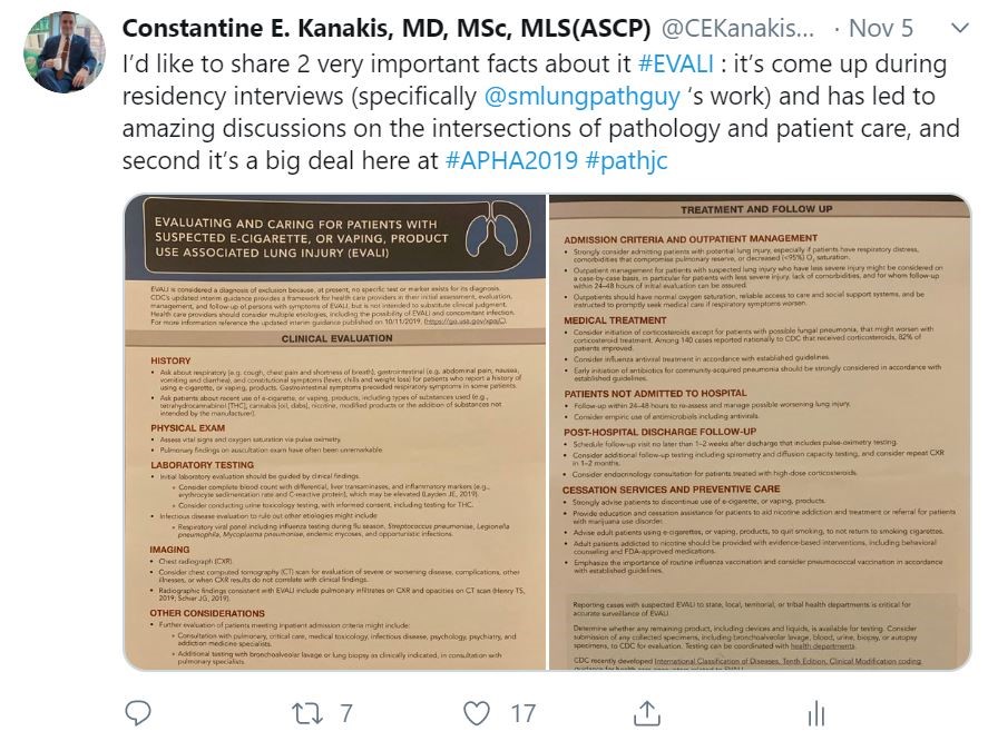
So, imagine you’re a vaper. Imagine you started because it helped you quit traditional cigarettes. That’s fantastic, good for you. You’re on the road to smoking cessation and better health! But perhaps the vaping-associated lung injury cases has made you a little defensive. Trust me I learned the hard way as I joined in the discussion earlier this month on a live-tweet pathology journal club on the AJCP article featured here. They happen under the hashtag #PathJC and lots of folks jump into the discussion from different places, institutions, time zones, and across disciplines—but its not just a bunch of pathologists analyzing an article in an academic bubble. Twitter is a public forum and that brings with it public scrutiny and commentary. As such, there were lots of lay people participating in the discussion and many individuals who held a positive opinion of electronic cigarettes. So not only did we have a very comprehensive discussion in the merits and shortcomings of published literature on the topic of EVALI, we also had to field questions and engage in non-jargon conversations with concerned (and sometimes passionate) members of the non-scientific community. Suffice it to say, it’s a tricky tightrope to walk when you’re trying to balance your anti-smoking public health crusade with some good old-fashioned medical education challenged with a sprinkle of vitriol on the most open of forums, the internet. But that’s okay! I strongly think, that in the future of medical practice, those of us in any discipline (but especially pathology and lab medicine) should lead the charge as champions of truth to connect our revered medical data to people in real terms—basically translate translational medicine.

Image 6. Why am I showing you my twitter profile picture? Easy: one of those “incendiary” comments in discussing smoking and vaping in a public forum actually included someone screen capturing my profile and accosting my “smug” pose and taste for esophageal damage in drinking hot coffee, citing poor data references for caffeine related deaths versus that of smoking. How do you deal with this? Calmly, with open honest information and, most importantly, with humility to address the barriers in communication between opposing points of view. Champions of truth, remember? But once you notice you’re talking to folks online who represent companies in the tobacco industry, ABORT MISSION, you went too far, haha! (True story, yikes!)

Once the Smoke Settles

Basically, everything’s going to be okay. There’s always a crisis or an epidemic happening that we have to address with limited data, developing knowledge, and some cohort of representative push back. That’s the nature of public health. But I’ll pull straight from the authors’ conclusion in the AJCP paper and remind you that not only is this just one, single study with very small number of cases to measure clinical outcomes, but further study is needed to support what is just beginning to be a correlation between vaping and lung injury.

TL;DR – it might seem obvious to some that hot smoke burns your lungs, but we’ve got to prove it and take steps to protect our patients everywhere.

And the good news is there are lots of us working on this. Scientists, public health officials, researchers, reporters, medical professionals, and especially pathologists are here collecting data and adding knowledge to that growing body of evidence to address this …hot topic.

Image 7. Here’s at least two of those people. Spoiler: it’s my wife and me. Here we are the recent American Public Health Association (APHA) conference in Philadelphia where the topic of vaping, smoking, and lung injury were very much in the forefront of public health research as it fits into the context of social determinants of health, medical literacy campaigns, and other concurrently related health issues like asthma and COPD.
Image 8. I actually joined that live tweet #PathJC journal club discussion from the APHA 2019 conference and was lucky enough to have, in-hand, the official EVALI clinical information release from the CDC booth in the expo floor. Check it out on my Twitter feed.

Breathe Easy

What’s past this smokescreen challenge? The same thing as always: hard work, collaboration, innovation, and paradigm shifting. If you’ve read my previous posts, you know I like to wax a bit about the future of medicine and the humanity behind our profession. Taking everything into consideration with this newest and hottest of public health concerns, our role as diagnosticians and translational representatives is as important as ever. And, if we want to ensure the recognized contributions of pathology in the wider field of medicine (and health-at-large) we should work with our colleagues in and out of the medical profession to demystify this kind of research, cleanly communicate health data to the public, and push the boundaries of personalized health and improved patient outcomes. But beware: when you address big topics like smoking, vaping, EVALI, and THC use, it can be easy to get too hot, and even burn out.

Thanks again, see you next time, and hope you had a Happy Thanksgiving!

(This is absolutely stolen from @iHeartHisto on Twitter, but enjoy a slice of pump-skin pie!)

Constantine E. Kanakis MD, MSc, MLS (ASCP)CM completed his BS at Loyola University Chicago and his MS at Rush University. He writes about experiences through medical school through the lens of a medical lab scientist with interests in hematopathology, molecular, bioethics, transfusion medicine, and graphic medicine. He is currently a 2020 AP/CP Residency Applicant and actively involved in public health and education, advocating for visibility and advancement of pathology and lab medicine. Follow him on Twitter @CEKanakisMD

Hematopathology and Molecular Diagnostics Case Study: A 63 Year Old Man with Fatigue

The following case is an interesting overlap of Hematopathology and Molecular Diagnostics, and shows the utility of sequencing to detect a cancer before biopsy could.

A 63 year old gentleman presented to a heme/onc physician with six months of intractable anasarca, fatigue, and a recent mild thrombocytopenia (Table 1). They were otherwise in healthy condition. The physician initiated a lymphoma work-up that included a bone marrow biopsy. The tests were negative for M-protein.

Table 1. Summary of symptoms and relevant abnormal labs.

The bone marrow biopsy was somewhat limited, but the core contained multiple marrow elements. After a thorough review by a Hematopathologist, no evidence of dysplasia or other irregularities could be detected (Image 1). Flow cytometry detected no aberrant blast population. Cytogenetics detected 20del [16/20] and 5del [3/20]. These findings did not clearly indicate a specific diagnosis.

Image 1. 40x view of the bone marrow specimen at the initial presentation. No evidence of dysplasia was found.

As the clinical suspicion for a malignancy was high, the bone marrow specimen was sent for sequencing on a 1385-gene panel test. The test included tumor-normal matched DNA sequencing (“tumor” sample: bone marrow, normal: saliva), RNA whole transcriptome sequencing on the bone marrow, and Copy Number Variant (CNV) analysis. Tumor-normal matched sequencing helps rule out variants that are normal and present in the patient.

Somatic mutations were determined as those that were present in the “tumor” sample and not in the matched normal sample. The somatic variants found are listed below with their variant allele frequency (VAF) in parenthesis. Recall that a VAF of 40% means that a mutation is present in the heterozygous state in 80% of cells.

  • IDH2 (p.R140Q, 46%)
  • SRSF2 (p.P95T, 51%)
  • CBL (p.R499*, 47%)
  • KRAS (p.K117N, 12%)
Figure 1. View of IGV, which displays the NGS reads for IDH1 along with the variant allele highlighted in red. The color of the bars indicates the direction of the reads (forward in red and reverse in blue). This reflects the allele frequency of approximately 50%.

The mutations in these genes are commonly found in myeloid cancers including myselodysplastic syndrome. Activating mutation in IDH2 (isocitrate dehydrogenase 2) increase the production of the oncometabolite 2-HG, which alters methylation in cells taking them to an undiffereitiated state. SRSF2 (Serine And Arginine Rich Splicing Factor 2) is a part of the spliceosome complex, which regulates how sister chromatids separate from each other. Failures in the proper function of the complex creates genomic instability. CBL (Casitas B-lineage Lymphoma) is a negative regulator of multiple signaling pathways, and loss of function mutations (as seen here) lead to increased growth signals through several tyrosine kinase receptors. KRAS (Kirsten RAt Sarcoma virus) is an upstream mediator of the RAS pathway, which acquires mutations that lead to constitutive activation and sends growth signals to cells causing them to proliferate.

Furthermore the CNV analysis also found the heterozygous loss of chromosome 20 as reported in cytogenetics. CNV analysis did not detect chromosome 5 deletion, as it was below the limit of detection (20% for CNV analysis).

Figure 2. This plot shows the normalized read frequency of genes across each of the chromosomes is shown here. The drop at chromosome 20 is shown in a pale brown color on the right side of the graph. This is consistent with the cytogenetic findings. The loss of 5q isn’t seen as it is below the limit of detection of 30%.

These mutations are all individually common in MDS, but the co-occurance of each gives very strong evidence that MDS is the diagnosis (Figure 3). There have also been studies that provide prognostic implications for several of the genetic mutations present. Some mutations like SRSF2 or CBL at high VAF (>10%) indicate a poor prognosis, but mutations in IDH2 or TP53 at any frequency have not only a high chance of progression, but also a faster time to onset of disease. Another non-genetic risk factor for developing MDS is an elevated RDW, which we saw in our patient.

Figure 3. From Becker et al 2016.

All of these high-risk factors together led us to push for a diagnosis of MDS based off of molecular findings, and the patient was started on treatment with Azacitadine. Our assessment was confirmed 3 months later when, the patient’s follow up bone marrow biopsy showed significant progression with megakaryocytic and erythroid dysplasia and hyperplasia and reticulin fibrosis MF2 (Image 2). Aberrant blasts were detected (1-2%), but not elevated. This demonstrates how molecular findings predicted and predated the patient’s rapid progression to morphologic disease.

Image 2. Dysplastic, hyperplastic megakaryocytes and erythroid lineage.

In summary, multiple molecular mutations indicative of MDS were found in a symptomatic patient’s unremarkable bone marrow biopsy months before a rapid progression to MDS.

References

  1. Steensma DP, Bejar R, Jaiswal S et al. Blood 2015;126(1):9-16.
  2. Sellar RS, Jaiswal S, and Ebert BL. Predicting progression to AML. Nature Medicine 2018; 24:904-6.
  3. Abelson S, Collord G et al. Prediction of acute myeloid leukemia risk in healthy individuals. Nature 2018; 559:400-404.
  4. Desai P, Mencia-Trinchant N, Savenkov O et al. Nature Medicine 2018; 24:1015-23.
  5. Becker PM. Clonal Hematopoiesis: The Seeds of Leukemia or Innocuous Bystander? Blood.2016 13(1)

-Jeff SoRelle, MD is a Chief Resident of Pathology at the University of Texas Southwestern Medical Center in Dallas, TX. His clinical research interests include understanding how the lab intersects with transgender healthcare and improving genetic variant interpretation.

A 66 Year Old Male with Diarrhea, Weight Loss, and Night Sweats

Case History

A 66 year old man with past medical history of recently diagnosed Clostridioides difficile colitis presented to emergency department with diarrhea, weight loss of 52 pounds in 4 months, and occasional night sweats. CT imaging revealed dilation of small bowel with thickened mucosal folds. The duodenum was subsequently biopsied to reveal diffuse intestinal lymphangiectasia containing PAS positive and Congo red negative eosinophilic material and lamina propria foamy macrophages. Laboratory investigations revealed normocytic anemia, proteinuria, and peripheral IgM kappa monoclonal gammopathy.

Biopsy Findings

Image 1. Aspirate.
Image 2. Core biopsy.
Image 3. CD138.
Image 4. Kappa ISH.
Image 5. Lambda ISH.

Bone marrow aspirate shows increased plasma cells and mast cells. H&E stained sections demonstrate a normocellular bone marrow with trilineage hematopoiesis and involvement by 35% plasma cells. By immunohistochemistry, CD138 highlights clusters of plasma cells that predominantly express kappa light chain restriction.

FISH and Mutation Analysis

FISH demonstrated loss of chromosome 11 and gain of chromosome 15, which was consistent with plasma cell dyscrasia. MYD88 mutation analysis did not detect the mutation.

Diagnosis

The findings of the patient’s normocytic anemia, IgM monoclonal gammopathy, and intestinal lymphangectasia with an associated plasma cell dyscrasia involving the bone marrow favor a lymphoplasmacytic lymphoma/Waldenström macroglobulinemia.

Discussion

Waldenstrom macroglobulinemia (WM) is a malignant B-cell lymphoproliferative disorder characterized by lymphoplasmacytic infiltration of the bone marrow and peripheral IgM monoclonal gammopathy.1 It is rare with an overall incidence of 3 per million persons per year, accounting for 1-2% of hematologic cancers.1 It occurs predominantly in Caucasian males, with a median age of 63-68 years old at diagnosis.1-3

Patient may be asymptomatic for years and require observation or experience a broad spectrum of signs and symptoms. These symptoms may be attributable to the tumor infiltration of the bone marrow and lymphoid tissues, IgM circulating in the blood, and IgM depositing into tissues. The most common clinical presentation of WM is fatigue and nonspecific constitutional symptoms, such as fever, night sweats, and weight loss, due to normochromic, normocytic anemia. 20-30% of patients may exhibit lymphadenopathy and hepatosplenomegaly due to infiltration of peripheral tissues. High concentration of IgM in the circulation may lead to hyperviscosity, resulting in oronasal bleeding, gingival bleeding, blurred vision due to retinal hemorrhages, and neurological symptoms, including headache, ataxia, light-headedness, dizziness, and rarely, stroke.2-3 The gastrointestinal manifestations are rare; however, IgM monoclonal protein may deposit into the lamina propria of the GI tract, causing diarrhea, steatorrhea, and GI bleeding.4 Other IgM-related manifestations include cold agglutinin hemolytic anemia, cryoglobulin, and amyloid deposition in tissues.3

Diagnosis of WM includes evidence of IgM monoclonal gammopathy and at least 10% of bone marrow infiltration by lymphoplasmacytic cells.5 Monoclonal gammopathy can be detected by the monoclonal spike, or M-spike, on serum protein electrophoresis.3 Serum immunofixation may be performed to identify the type of monoclonal protein and the type of light chain involved.3 In terms of immunophenotype, neoplastic cells express surface IgM, cytoplasmic Igs, CD38, CD79a, and pan B-cell markers (CD19, CD20, and CD22). CD10 and CD23 are absent. Expression of CD5 occurs in approximately 5-20% of cases.6 Recent studies have reported two most common somatic mutations in WM, which are MYD88 L265P mutations (90-95% of cases) and CXCR4 (30–40% of cases).7 Absence of these mutations, however, do not completely exclude the diagnosis of WM.

The International Staging System for WM identifies five factors associated with adverse prognosis, including age older than 65, hemoglobin < 11.5g/dL, platelet count < 100K/μL, beta-2-microglobulin > 3mg/L, and monoclonal IgM concentration > 7g/L.3 Patients younger than the age of 65 years with 0 or 1 of these factors are in the low-risk category with a median survival of 12 years.3 In contrast, patients with 2 or more risk factors are in the intermediate- and high-risk categories and have a median survival of almost 4 years. 3

Management of WM depends on the patient’s clinical manifestations.Furthermore, patients with minimal symptoms should be managed with rituximab, whereas patients with severe symptoms related to WM should receive more aggressive treatment, including dexamethasone, rituximab and cyclophosphamide. Hyperviscosity syndrome is an oncologic emergency that requires removal of excess IgM from the circulation via plasmapheresis.8

References

  1. Neparidze N, Dhodapkar MV. Waldenstrom’s Macroglobulinemia: Recent advances in biology and therapy. Clin Adv Hematol Onco. 2009 Oct;7(10): 677-690.
  2. Leleu X, Roccaro AM, Moreau AS, Dupire S, Robu D, et al. Waldenstrom Macroglobulinemia. Cancer Lett. 2008 Oct;270(1):095-107.
  3. Tran T. Waldenstrom’s macroglobulinemia: a review of laboratory findings and clinical aspects. Laboratory Medicine. 2013 May;44(2):e19-e21.
  4. Kantamaneni V, Gurram K, Khehra R, Koneru G, Kulkarni A. Distal illeal ulcers as gastrointestinal manifestation of Waldenstrom Macroglbulinemia. 2019 Apr; 6(4):pe00058.
  5. Grunenberg A, Buske C. Monoclonal IgM gammopathy and Waldenstrom’s macroglobulinemia. Dtsch Arztebl Int. 2017 Nov;114(44):745-751.
  6. Bhawna S, Butola KS, Kumar Y. A diagnostic dilemma: Waldenstrom’s macroglobulinemia/plasma cell leukemia. Case Rep Pathol. 2012;2012:271407.
  7. Varettoni M, Zibellini S, Defrancesco I, Ferretti VV, Rizzo E, et all. Pattern of somatic mutations in patients with Waldenstrom macroglobulinemia or IgM monoclonal gammopathy of undetermined significance.
  8. Oza A, Rajkumar SV. Waldenstrom macroglobulinemia: prognosis and management. Blood Cancer Journal. 2015;5:e394.

-Jasmine Saleh, MD MPH is a pathology resident at Loyola University Medical Center with an interest in dermatopathology and hematopathology. Follow Dr. Saleh on Twitter @JasmineSaleh.

–Kamran M. Mirza, MD, PhD, MLS(ASCP)CM is an Assistant Professor of Pathology and Laboratory Medicine, Medical Education and Applied Health Sciences at Loyola University Chicago Stritch School of Medicine and Parkinson School for Health Sciences and Public Health. A past top 5 honoree in ASCP’s Forty Under 40, Dr. Mirza was named to The Pathologist’s Power List of 2018 and placed #5 in the #PathPower List 2019. Follow him on twitter @kmirza.

A Med Tech Gives a TEDx Talk

Hello again everyone!

After a lot of positive responses and sharing on social media, my article last month got lots of people talking about annual meetings and how great they are for networking, learning, and advancing our profession. Not too long after the ASCP Annual Meeting in Phoenix, I was back in my Manhattan apartment working on my speech and graphics for a real life TEDx session hosted at my medical school.

Let’s pause here: if you either haven’t heard of the TED/TEDx brand or if you binge watch their 18 minute videos and want more links to watch now, now, now!

TED is a non-profit organization whose mission is to share “ideas worth spreading.” They’re about 35 years old and based in NYC stateside, and Vancouver in Canada. Basically, over the last few decades they hold conferences at those flagship sites called “TED talks” where selected speakers present on a myriad of topics. TEDx conferences are officially licensed but off-site events which operate under TED protocol and guidelines. There have even been spin-off conferences like TED MED, which focus solely on healthcare.

Image 1. What’s a TEDx talk? Basically, an off-site, officially sanctioned, “idea sharing” conference.

Some of the students at AUC School of Medicine, organized such a conference with official TED licensing and recruited me to join their list of speakers to deliver talks on their chosen theme: resilience. Officially called TEDxAUCMed, this conference included community members, students, artists, activists, and more discussing the human capacity for resilience in ways not commonly discussed. “Weathering the Storm” was the official event title, as the school located in the island nation of St. Maarten displays daily resilience especially since being hit by Hurricane Irma in 2016. Among their list of incredible speakers, I was humbled to be included! I titled my talk “Unrecognizable Medicine” and wanted to deliver a talk to students, clinicians, and those of us in medicine witnessing first-hand a tidal wave of new technologies and paradigms that redefine the way we discuss health. Oh, and since I’m a huge fan of #GraphicMedicine more and more each day, I hit that hashtag hard and decided to illustrate my whole talk!

Image 2. Title Card from my TEDx talk.

So what did I talk about, exactly…and what’s the big deal? I’m not going to re-hash my presentation for you in text—that’d be boring, and I’m obviously going to put a link at the bottom for you to watch it yourself. I got you, lab fam! But essentially, what I set up was a three-tiered template to assess and navigate that tidal wave of tech. Tools, skills, and strengths—three things inherent to the practice of medicine in any specialty.

Image 3. Red back-ligting. So intense. Thanks for coming to my TEDx Talk, literally!

There are untapped topics in medicine which are looming over the horizon. As medicine continues to evolve and change, the problems we face and the needs we must meet will become moving targets. New specialties will emerge, and new technologies will replace centuries old tools we cling to today. A shift in thinking is both proactive and healthy in a profession that mandates our commitment to preserving health and quality of life. I have spent years battling stereotypes in medicine and hope to challenge the fabric that places individuals in professional or academic boxes. Fresh first-years at some schools are already using point-of-care ultrasounds (POCUSes) instead of stethoscopes—which student sounds like they have better info on morning rounds, a student who maybe kinda-sorta heard some non-descript murmur, or a mini-pocket echocardiogram with an ejection fraction of 45%? Stereotypes have too long shaped the way students choose specialties, equating some areas to colloquial high school cliques! No offense to orthopedics or dermatology. Troponins used to be something you could hang your white coat on, but not anymore. What do you do with a new 5th generation Trop of 39 with a delta of 18? ACS or acute MI? Cancer therapy is exploding with personalized treatments being added every day! Any student right now would impress their heme/onc attending on rounds if they suggested PDL-1 and other immunotherapy testing for patients with newly diagnosed lung cancers. *Deep breath*

Ok. My point is, tomorrow’s medicine is going to have a lot of different therapies, tools, and even vocabulary that schools may never catch up with. How do you prepare for this explosion of knowledge? You look to yourself to take an inventory of your strengths and use those to guide your clinical sails. Addressing stereotypes head-on, learning on the spot, dealing with complex identities in your patients, and always practicing with compassion will lend itself to staying ahead and staying fulfilled.

Image 4. If you’re drawing cartoons of pathologists for an educational series, you probably make them look like you. Or in this case me, I guess. Keep an eye out for my #PathDoodles on social media!

Pretty heavy stuff right? But there’s something else that caught my attention in reflection on the TEDx talk… I’ve searched the TED library of videos, and while there are plenty of doctors, scientists, and pioneers in research discussing medical ideas, I haven’t seen any medical laboratory scientists. If you find any, please correct me. But, as I understand it, it’s just me. And that’s something special.

Image 5. My wife and I check-in for rehearsal at the TEDxAUCMed conference in sunny St. Maarten.

There’s a culture shift in our profession, and a lot of us are talking about it. Pathology and laboratory medicine are stepping out from behind the healthcare curtain and asserting itself as a champion for patients, truth, and the importance of data-driven medicine. Not only do I talk to groups of folks every time I get a stage, but I use social media to reach clinicians and patients! Yes, I’m one of few medical students-turned-residency applicants who didn’t change their name to hide their online presence for the winter. But instead of a secret twitter hibernation, I’ve used social media as a tool to network, engage, and connect.

One of my favorite new projects is something I call #PathDoodles where I break down the aspects of pathology and some specialty topics for those outside of medicine (and sometimes just outside our profession). I’ve already covered things like “what is pathology?” and the importance of autopsies, the role of medical laboratory scientists, and I continue to add more regularly!

Image 6. One of a growing list of #PathDoodles.

There’s a culture shift in our profession, and a lot of us are talking about it. Pathology and laboratory medicine are stepping out from behind the healthcare curtain and asserting itself as a champion for patients, truth, and the importance of data-driven medicine. Not only do I talk to groups of folks every time I get a stage, but I use social media to reach clinicians and patients! Yes, I’m one of few medical students-turned-residency applicants who didn’t change their name to hide their online presence for the winter. But instead of a secret twitter hibernation, I’ve used social media as a tool to network, engage, and connect.

One of my favorite new projects is something I call #PathDoodles where I break down the aspects of pathology and some specialty topics for those outside of medicine (and sometimes just outside our profession). I’ve already covered things like “what is pathology?” and the importance of autopsies, the role of medical laboratory scientists, and I continue to add more regularly!

Follow me on Twitter (@CEKanakisMD) and check out my TEDx talk:

My talk begins at 5:00:00. Enjoy!

Constantine E. Kanakis MD, MSc, MLS (ASCP)CM completed his BS at Loyola University Chicago and his MS at Rush University. He writes about experiences through medical school through the lens of a medical lab scientist with interests in hematopathology, molecular, bioethics, transfusion medicine, and graphic medicine. He is currently a 2020 AP/CP Residency Applicant and actively involved in public health and education, advocating for visibility and advancement of pathology and lab medicine. Follow him on Twitter @CEKanakisMD

Hemoglobin Electorphoresis in Children

This last month, I rotated through our Children’s hospital, which included reviewing hemoglobin electrophoresis tests. I’d learned about them before in residency, but they can be quite more interesting (complicated) than I expected.

Hemoglobin electrophoresis is a blood test to look at different types of hemoglobin to determine if there are any abnormalities. In a children’s hospital it is frequently ordered as a reflex for an abnormal newborn screen or when a child is incidentally found to be anemic. The test is performed in 2 stages. 1st lysed blood samples are run on gel electrophoresis and different types of hemoglobin are separated as they move at different speeds. Several types of hemoglobin will run within the same region, so a secondary method of separation is always employed.

Below, you can see how some bands in the same area of an acidic gel (agarose) are actually very different on the alkaline gel (cellulose acetate) and vice versa.

At our hospital, we use HPLC and measure retention times of the hemolysate to quantify and identify different hemoglobin types present. As a basic primer you should recall that hemoglobin is a tetramer with a pair of alpha globin + a pair of either beta, delta or gamma globin (each separate genes).

Alternative hemoglobins are enriched in populations where malaria is endemic as these variants may provide improved fitness by promoting resistance to the malarial parasite that reproduces inside red blood cells. Thus, many people of African or south east Asian descent may carry these variants.

Our case is that of a 2 year old girl with anemia who had testing sent by her primary care doctor for the following CBC:

This is indicative of microcytic anemia, but unlike some Thalessemias the RBC isn’t very high. More on this later.

Looking at the gel result, there is a large band in the area coinciding with Hgb C. We also see the normal Hgb A2 and a small amount of Hgb F. We know Hgb F can be increased in Hgb SS and thus could also be present if she had Hgb C trait or disease.

InkedBlog 1B_LI

Looking at the next HPLC result, we see there is a similar very high level of Hgb C (68%) with corresponding levels of Hgb F and Hgb A2 (note: acetylated Hgb F and Hgb F are added together). Thus, this fits with a homozygous C with some compensatory A1 and F, right?

Remember Hgb C is a β -globin variant and you only have 2 β -globin genes, so if you are homozygous for the C variant on the β-globin gene (HBB), then Hgb A1, which is made of normal β-globin would be impossible to produce. Also you might be bothered by all of these small peaks. However, there are often small peaks that can’t be definitively identified and are likely post-translationally modified hemoglobin. But in the context of an abnormal Hgb A1 that shouldn’t be there, we dug deeper.

One of the most common hemoglobinopathies is Beta Thalassemia (β-Thal), which clinically manifests when less of the beta hemoglobin protein is produced. Heterozygous mutations lead to Beta Thalassemia minor with minimal symptoms, while homozygous mutations lead to β-thal major with symptoms of anemia. Mutations in the β -globin gene, HBB, can lead to complete loss of β-globin (β0 variant) or partial of β-globin (β+ variant).

As this patient has less than 50% of Hgb A present (expected amount), they could also have a β+ variant as well. This would make them compound heterozygous for C and β+.

One of the hallmarks of Thalassemia is an increase in Hgb A2 (normal 2.5-3.5%). Hemoglobin A2 is a normal variant of A that is composed of two alpha and two delta chains (δ2α2). We see in our case that the Hgb A2 is normal at 2.5%. So it seems the patient doesn’t display a typical Thalassemia picture.

One condition that could create this scenario is if there is a variant in the delta chain of A2 that causes it to elute differently. Indeed, there is a delta variant that creates hemoglobin A2 prime (A2’) that moves near the S region of the HPLC. And when we look back at our unknown hemoglobins, Hgb X is marked at 1.03 of the S region and has an abundance of 3.9%. This supports it being the Hgb A2’ and if we add this together with the Hgb A2 we get an elevated 6.6% A2 total, which would be consistent with Beta Thalassemia. Lastly, one would wonder if we could find this third hemoglobin variant A2’ on the alkaline gel. Previous studies have shown the A2’ variant is more negatively charged, so on a basic gel, it should move further from the negative anode than the other hemoglobins. We don’t see anything to the left of the HgbC, but if we flip the gel over and look under the patient label, you can see a faint band that is likely the A2’!

In summary this case arose from 3 separate mutations in a single patient. She was compound heterozygous for a Hgb C and β+ variants in the β-globin gene and she was heterozygous for an A2’ variant on the delta-globin gene.  This was certainly a case where paying close attention mattered.

References:

  1. Abdel-Gadir D, Phelan L, and Bain BJ. Haemoglobin A2′ and its significance in beta thalassaemia diagnosis. Int J Lab Hematol. 2009 Jun;31(3):315-9. doi: 10.1111/j.1751-553X.2008.01038.x. Epub 2008 Feb 21.
  2. https://ghr.nlm.nih.gov/condition/beta-thalassemia

-Dr. Charles Timmons MD PhD is a pediatric pathologist at Children’s Medical Center in Dallas, TX. His responsibilities include signing out hemoglobin electrophoresis, HPLC and globin sequencing, and has been residency director for 17 years.

-Jeff SoRelle, MD is a Chief Resident of Pathology at the University of Texas Southwestern Medical Center in Dallas, TX. His clinical research interests include understanding how the lab intersects with transgender healthcare and improving genetic variant interpretation.

Surgical Pathology Case Study: An Elderly Patient with Unexplained Pain, an Unremarkable CT Scan, and Enlarged Rugal Folds on EGD

Case History

The patient is a 72 year old woman who presented to her physician’s office with postprandial pain and unintentional weight loss. A CT scan was performed that showed no obvious abnormality or cause for the patient’s abdominal pain. The patient subsequently underwent an EGD and EUS which revealed enlarged gastric folds without hemorrhage. In addition, there was wall thickening seen in the body of the stomach within the luminal interface, superficial mucosa, deep mucosa and submucosa consistent with possible gastritis versus an infiltrative process. The remainder of the EGD and EUS was grossly unremarkable. These findings were concerning for possible linitis plastica. Pathology on the samples taken from the EGD were consistent with poorly differentiated adenocarcinoma that was invasive in both the gastric fundus and gastric body. The patient was initially taken to the operating room for a staging laparoscopy to ensure that there was no metastatic disease before beginning a preoperative chemotherapy regimen. The staging laparoscopy revealed a thickened gastric wall from the fundus to the antrum, consistent with linitis plastica, and no obvious evidence of metastatic disease. The patient then underwent peritoneal washings which showed no evidence of positive cytology. Based on these findings, the patient was started on a chemotherapy regimen of epirubicin, cisplatin and fluorouracil (5-FU), which she tolerated well. The patient was then taken to the operating room for a total gastrectomy procedure with Roux-en-Y esophagojejunostomy.

Diagnosis

Received fresh for intraoperative consultation is a total gastrectomy specimen with a black stitch designating the proximal side. It was requested by the surgical team to have the proximal esophageal margin frozen to ensure that esophageal tissue was indeed present, as well as to exclude the presence of any carcinoma. The proximal margin was negative for carcinoma with squamous mucosa present. The stomach measures 17.0 cm in length with an internal circumference ranging from 14.7 cm proximally to 9.0 cm distally. There is a 1.0 cm long portion of attached duodenum with an internal circumference of 5.8 cm. The serosal surface of the stomach is glistening, pink-tan and smooth with a scant amount of attached yellow, lobulated adipose tissue and omentum along the length of one entire edge measuring 26.0 x 13.0 x 1.0 cm. The stomach is opened to reveal glistening, tan mucosa with irregular rugal folds which are diffusely nodular, predominantly in the body of the stomach. There is a 6.5 x 5.0 cm are of flattened mucosa in the pyloric region (Image 1). The wall thickness measures 0.5 cm throughout. There are no grossly identifiable masses or nodules. Gross images are taken and the serosal surface is inked entirely in black. The adipose tissue is examined for candidate lymph nodes. Representative sections are submitted as follows:

B1 FS: Frozen section remnants

B2-B6:     multiple representative sections from the cardia

B7-B10:   multiple representative sections from the body

B11-B12:   multiple representative sections from the pylorus

B13:     representative perpendicular section through the distal resection margin

B14:     seven putative lymph nodes

B15:     five putative lymph nodes

B16:     three putative lymph nodes

B17:     seven putative lymph nodes

B18:     six putative lymph nodes

B19:     three putative lymph nodes

B20:     six putative lymph nodes

Histologically, the specimen consisted of diffuse, poorly differentiated, discohesive cells throughout all the layers of the stomach, penetrating into the serosa, with fibrosis, inflammation and signet ring cells present. In addition, angiolymphatic invasion was present. Based on the gross presentation and histologic appearance, the specimen was signed out as a diffuse gastric adenocarcinoma with a stage of T3.

Image 1.

Discussion

As of 2018, gastric cancer is the sixth most common cancer with approximately 1.03 million cases, and the third leading cause of cancer deaths worldwide, resulting in 783,000 deaths. Due to a better understanding of epidemiology, pathology, and molecular testing, as well as advances in new forms of treatments, the incidence and mortality in gastric cancer has been declining over the years. Of the gastric cancer types, rates of intestinal type carcinoma have been decreasing, however, the incidence of poorly cohesive gastric carcinoma (PCGC) and signet ring cell carcinoma (SRC) has increased. In order to accurately discuss PCGC, there must first be a discussion about the standardization of gastric cancer subtype definitions. Poorly cohesive, signet ring cell, and diffuse gastric carcinomas have commonly been used interchangeably. In 2010, the World Health Organization defined poorly cohesive gastric carcinoma as being composed of isolated or small groups of tumor cells. If there was a predominance of signet ring cells, then it would be termed a signet ring cell carcinoma. Mariette et al. proposed that a PCGC composed of 90% or more signet ring cells should be classified as SRC. The term “diffuse” corresponds to the same term “poorly cohesive”, and because of this, I will be using the term “poorly cohesive” solely going forward. In addition to this, the term “linitis plastica” would commonly be used interchangeably, but is best used as a term to describe the macroscopic appearance of PCGC or SRC.

Gastric carcinoma is classified as either early or advanced stage to help determine the appropriate type of intervention. Early gastric carcinoma is defined as invasive carcinoma confined to the mucosa and/or submucosa, regardless of lymph node metastases or tumor size. These tumors are generally smaller, measuring less than 5 cm in size, and found most commonly on the lesser curvature of the stomach at the angularis. Histologically, early gastric carcinoma will commonly present as well differentiated, mostly with tubular and papillary architecture. If the biopsies are composed of only mucosa, then distinguishing between well-differentiated carcinoma and carcinoma in situ or high grade dysplasia can be difficult. The presence of stromal desmoplasia in invasive carcinoma can help differentiate it from intramucosal invasion, which can contain single tumor cells within the lamina propria. This is an important distinction to make as intramucosal carcinoma does metastasize. Advanced gastric carcinomas will present grossly as either exophytic, ulcerated, or infiltrative tumors. Histologically, advanced gastric carcinomas will invade the muscularis propria and demonstrate cytologic and architectural heterogeneity, with a combination of patterns.

The 2010 World Health Organization classification determined four major histologic patterns of gastric cancer, which will often present with a combination of elements from the other patterns:

  1. Tubular: Most common pattern in early gastric carcinoma, with branching, distended or fused tubules containing intraluminal mucus, and nuclear and inflammatory debris
  2. Papillary: Most common in the proximal stomach with epithelial projections containing an underlying fibrovascular core. Also, it is frequently associated with liver metastases and an increased risk of lymph node involvement.
  3. Mucinous: Extracellular mucin makes up at least 50% of the tumor volume
  4. Poorly cohesive (including SRC): Mixture of signet ring and non-signet ring cells. Signet ring cells will have mucin pushing the nucleus to the periphery of the cell.

Helicobacter pylori (H. pylori) is a gram negative infectious bacteria that has been linked to gastric cancer. H. pylori is present in about half of the world’s population and other than gastric cancer, it is also associated with chronic gastritis, peptic ulcer disease, and gastric lymphomas. The bacteria is typically acquired during infancy and will remain for life if left untreated, with reactive oxygen species being generated that are capable of causing DNA damage due to the chronic infection. In addition, H. pylori can induce hypermethylation, resulting in the inactivation of tumor suppressor genes. Although H. pylori infection is considered a strong risk factor for developing gastric cancer, more commonly in intestinal type than diffuse type gastric cancer, only a small portion of those infected with the bacteria actually develop the malignancy. It is believed that approximately 80% of distal gastric cancers are due to a H. pylori infection, whereas there is little association between H. pylori and cardia gastric cancers.

In PCGC, such as this case, it is generally diagnosed in younger patients without a gender bias. Although PCGC can be associated with an H. pylori infection, it is more commonly related to a mutation in the tumor suppressor gene epithelial cadherin, also known as E-cadherin and CDH1. PCGC presents as an infiltrative growth of poorly differentiated, discohesive malignant cells that appear to arise from the middle layer of the mucosa. These cells can infiltrate as individual cells or as small clusters, but usually do not form glands (Image 2). If the gastric wall becomes extensively infiltrated by malignancy, the wall can be thickened and rigid, a macroscopic presentation termed as linitis plastica, which can lead to pyloric obstruction. Within PCGC, numerous signet ring cells can be present, leading to SRC. There is also a hereditary form of poorly cohesive gastric cancer referred to as hereditary diffuse gastric carcinoma, with an autosomal dominant pattern of inheritance. Histologically, it will include hyperchromatic nuclei, occasional mitoses, patchy intramucosal signet ring cells in the lamina propria, and carcinoma in situ associated with pagetoid spread of tumor cells along the preserved basement membrane. Hereditary diffuse gastric carcinoma will present with multifocal tumors under an intact mucosal surface, making diagnosis difficult. In patients with a CDH1 mutation and a family history of gastric carcinoma, a prophylactic gastrectomy is often the recommended treatment option.

Image 2.

References

  1. Adachi Y, Yasuda K, Inomata M, et al. Pathology and prognosis of gastric carcinoma well versus poorly differentiated type. Cancer. 2000;89(7)1218-24.
  2. Cancer. World Health Organization. Who.int. https://www.who.int/news-room/fact-sheets/detail/cancer. Published September 20, 2018. Accessed September 18, 2019.
  3. Carcas LP. Gastric cancer review. J Carcinog. 2014;13:14. Published 2014 Dec 19. doi:10.4103/1477-3163.146506
  4. Hu B, El Hajj N, Sittler S, Lammert N, Barnes R, Meloni-Ehrig A. Gastric cancer: Classification, histology and application of molecular pathology. J Gastrointest Oncol. 2012;3(3):251–261. doi:10.3978/j.issn.2078-6891.2012.021
  5. Mariette C, Carneiro F, Grabsch HI, et al. Consensus on the pathological definition and classification of poorly cohesive gastric carcinoma. Gastric Cancer. 2019;22(1):1-9 https://doi.org/10.1007/s10120-018-0868-0-
  6. Pernot S, Voron T, Perkins G, Lagorce-Pages C, Berger A, Taieb J. Signet-ring cell carcinoma of the stomach: Impact on prognosis and specific therapeutic challenge. World J Gastroenterol. 2015;21(40):11428–11438. doi:10.3748/wjg.v21.i40.11428
  7. Van Cutsem E, Sagaert X, Topal B, et al. Gastric Cancer. Lancet. 2016;388(10060):2654-64. https://doi.org/10.1016/S0140-6736(16)30354-3
  8. Weisenberg E. Diffuse (poorly cohesive) type carcinoma. Pathology Outlines. http://www.pathologyoutlines.com/topic/stomachdiffuse.html. Revised August 22, 2019. Accessed September 18, 2019

-Cory Nash is a board certified Pathologists’ Assistant, specializing in surgical and gross pathology. He currently works as a Pathologists’ Assistant at the University of Chicago Medical Center. His job involves the macroscopic examination, dissection and tissue submission of surgical specimens, ranging from biopsies to multi-organ resections. Cory has a special interest in head and neck pathology, as well as bone and soft tissue pathology. Cory can be followed on twitter at @iplaywithorgans.

Tackling the Testosterones: Total, Free, and Bioavailable

When a patient gets their “testosterone test” at the doctor to assess their libido, do they really know what they’re getting? Does your lab test for testosterone, and are you confused about which of these confusingly-named tests are in-house versus send-out? Do you need a refresher on the types of testosterone tests out there and the clinical significance of each?

A Primer on Testosterone

Testosterone, being a fairly hydrophobic member of the steroid-ring family, is the major androgen in males. Apart from its well-known function in promoting the development of primary male reproductive organs and secondary male sex characteristics, it also has important anabolic effects in maintaining muscle mass, bone maturation, regulation of the hypothalamic-pituitary-adrenal axis under stress, and even in promoting platelet aggregation through enhancing platelet thromboxane A2 expression.1 In females, testosterone increases sexual arousal, and is in fact used clinically as treatment for female sexual arousal disorders. So, clearly an important member of the steroid family.

Being hydrophobic, much of the testosterone in the human body is not freely available, but rather bound. Total testosterone signifies the total pool of testosterone available in the human body, and is largely encompassed by the majority of bound testosterone with a small (usually 1.5-2.0%) proportion of free testosterone, which is biologically active. The bound testosterone can further be subdivided into testosterone bound to sex-hormone binding globulin (SHBG), a small glycoprotein that strongly binds various androgens and estrogens, and testosterone bound toalbumin, which is a relatively weak interaction.

Recently, the concept of bioavailable testosterone has been defined,2 based on the understanding that testosterone bound to SHBG (around 2/3rd of the bound proportion) is relatively inaccessible, while testosterone bound to albumin is weakly interacting, and thus potentially bioactive. Therefore, the definition of bioavailable testosteroneincludes both free and albumin-bound testosterone, which comprise the non-SHBG bound proportion.

How is testosterone measured?

Conventionally, total testosterone is measured through either immunoassays (both radioimmunoassays, or more commonly, chemiluminescent immunoassays) or mass spectrometry coupled with gas chromatography (GC/MS) or liquid chromatography (LC-MS/MS). Isotope dilution mass spectrometry (IDMS) is the reference method for testosterone measurement,3 but due to cost and convenience, most labs utilize immunoassays. Sex hormone binding globulin (SHBG) is commonly measured through chemiluminescent immunoassays, and also available for many platforms.4

There are two main approaches to the measurement of free testosterone, which is significantly more challenging. The gold standard for free testosterone measurement is equilibrium dialysis (see inset), a time consuming, expensive, and laborious assay that uses semi-permeable membranes to measure antibody-bound fractions of testosterone. Moreover, results can vary with pH, temperature, and methods of dilution.5 Due to these complications, calculated free testosterone is an attractive alternative used by many laboratories.

What is equilibrium dialysis? Equilibrium dialysis and ultrafiltration are reference methods used to determine true free testosterone calculation. Briefly, a relatively large quantity of serum (500 to 1000 uL) is placed in one chamber of an equilibrium dialysis apparatus, which is comprised of two fluid chambers separated by a semi-permeable membrane. Free-labeled testosterone passes through the membrane, while testosterone bound to SHBG does not. The radioactivity in the free chamber is quantified as a proportion of the total testosterone level, as measured by another assay, such as LC/MS-MS.

What is calculated free testosterone, and how is it calculated?

Recognizing the difficulty of performing equilibrium dialysis on large volumes of testosterone specimens, several researchers have looked into devising good approximations of free testosterone through mathematical expressions modeling the distribution of testosterone among its various compartments. One of the most popular approximations, the Vermeulen equation developed by Dr. Alex Vermeulen,6 models the distribution of testosterone among the SHBG-bound, albumin-bound, and free component through association constants of testosterone among these compartments, and can be modeled by the equation in Figure 1, which depends on the total testosterone, SHBG concentration, and concentration of albumin (although this will be discussed below). The overall concordance of this method with apparent free testosterone obtained through equilibrium dialysis (AFTC), the reference method, is very good, with a correlation coefficient of 0.987 and mean values well within the SEM between the two methods.6

Figure 1. The Vermeulen equation for calculated free testosterone.

In studies of the variation of calculated free testosterone values to the albumin concentration, Vermeulen et al. demonstrated that between “normal” albumin concentrations ranging from 5.8–7.2 × 10⁻⁴ mol/L (40 to 50 g/L), the mean calculated free testosterone varied from 340 ± 40.9 pmol/L assuming an albumin concentration of 40 g/L, to 303 ± 35.4 pmol/L assuming a concentration of 50 g/L albumin. Moreover, the concordance of calculated FT results to AFTC concentrations remained very good (correlation coefficient of 0.992) when an intermediate fixed albumin concentration (43 g/L) was used in this calculation, compared to actual albumin levels. Overall, these calculations suggest that for healthy individuals without marked abnormalities in plasma protein composition, such as in nephrotic syndrome or cirrhosis of the liver, or pregnant patients, a fixed albumin concentration could be used without significantly affecting calculated FT results. Of course, in individuals with marked changes in plasma proteins, the actual albumin concentration should be accounted for.

Willem de Ronde et al5 compared five different algorithms for calculating free or bioavailable, which includes the Vermeulen and Sodergard method (which use similar parameters), as well as methods by Emadi-Konjin et al, Morris et al, and Ly et al. In general, there was high concordance between the Vermeulen and Sodergard methods (r=0.98) for measuring free testosterone, and lower, but still reasonable (r=0.88) concordance between Vermeulen and other methods. Fundamentally, the Vermeulen and Sodergard equations were derived from experimentally derived association constants from the law of mass action, as opposed to the other algorithms, which rely on experimentally derived free and bioavailable testosterone measurements that was modeled by regression equations, and thus depends on the accuracy of these measurements. Though the experimental basis underlying the Vermeulen and Sodergard equations is stronger, it is known that supraphysiologic concentrations of other steroid hormones (estradiol or dihydrotestosterone), in competition for binding sites to SHBG, can significantly underestimate free testosterone by any of these methods. Of course, inaccuracies in the measurement of total testosterone or SHBG can significantly affect results, as well as significant perturbations in total serum protein concentrations (as mentioned above).

Since the publication of the above work, additional calculations for free testosterone accounting for other modes of interaction of SHBG such as allostery and dimerization have been published that may further improve concordance with AFTC;7,8 however, further study is needed to determine if these methods actually result in superior calculated FT measurement for clinical decision making, as well as changes in sensitivity to interference.

Why do accurate free testosterone measurements matter?

Testosterone bound to serum albumin is essentially inactive; therefore, the only testosterone that is biologically relevant is free (and to a lesser extent, bound to SHBG). Current consensus guidelines still support the use of total testosterone for defining hypogonadism in men,9,10 although emerging studies and newer task-force consensus groups11,12 highlight an emerging role for both calculated and free testosterone measurements in addition to total testosterone. The role of direct free testosterone measurement is still hotly debated; a recent analysis of CAP proficiency data indicates considerable heterogeneity among laboratories using the reference methods described above, and suggests considerable cost savings without significant loss of reliability can be achieved by using calculated or FT bioavailable T over direct FT measurement.13 Further standardization of these assays is needed to better understand the tradeoffs here.

References

  1. Ajayi A a. L, Halushka PV. Castration reduces platelet thromboxane A2 receptor density and aggregability. QJM. 2005;98(5):349-356. doi:10.1093/qjmed/hci054
  2. Shea JL, Wong P-Y, Chen Y. Free testosterone: clinical utility and important analytical aspects of measurement. Adv Clin Chem. 2014;63:59-84.
  3. Botelho JC, Shacklady C, Cooper HC, et al. Isotope-Dilution Liquid Chromatography–Tandem Mass Spectrometry Candidate Reference Method for Total Testosterone in Human Serum. Clinical Chemistry. 2013;59(2):372-380. doi:10.1373/clinchem.2012.190934
  4. Dittadi R, Fabricio ASC, Michilin S, Gion M. Evaluation of a sex hormone-binding globulin automated chemiluminescent assay. Scand J Clin Lab Invest. 2013;73(6):480-484. doi:10.3109/00365513.2013.805807
  5. Ronde W de, Schouw YT van der, Pols HAP, et al. Calculation of Bioavailable and Free Testosterone in Men: A Comparison of 5 Published Algorithms. Clinical Chemistry. 2006;52(9):1777-1784. doi:10.1373/clinchem.2005.063354
  6. Vermeulen A, Verdonck L, Kaufman JM. A Critical Evaluation of Simple Methods for the Estimation of Free Testosterone in Serum. None. 1999;84(10):3666-3672. doi:10.1210/jcem.84.10.6079
  7. Heinrich-Balard L, Zeinyeh W, Déchaud H, et al. Inverse relationship between hSHBG affinity for testosterone and hSHBG concentration revealed by surface plasmon resonance. Molecular and Cellular Endocrinology. 2015;399:201-207. doi:10.1016/j.mce.2014.10.002
  8. Zakharov MN, Bhasin S, Travison TG, et al. A multi-step, dynamic allosteric model of testosterone’s binding to sex hormone binding globulin. Mol Cell Endocrinol. 2015;399:190-200. doi:10.1016/j.mce.2014.09.001
  9. Margo KL, Winn R. Testosterone Treatments: Why, When, and How? AFP. 2006;73(9):1591-1598.
  10. American Association of Clinical Endocrinologists Medical Guidelines for Clinical Practice for the Evaluation and Treatment of Hypogonadism in Adult Male Patients—2002 Update. Endocrine Practice. 2002;8(6):439-456. doi:10.4158/EP.8.6.439
  11. Bhasin S, Cunningham GR, Hayes FJ, et al. Testosterone therapy in men with androgen deficiency syndromes: an Endocrine Society clinical practice guideline. J Clin Endocrinol Metab. 2010;95(6):2536-2559. doi:10.1210/jc.2009-2354
  12. Liu Z, Liu J, Shi X, et al. Comparing calculated free testosterone with total testosterone for screening and diagnosing late-onset hypogonadism in aged males: A cross-sectional study. J Clin Lab Anal. 2017;31(5). doi:10.1002/jcla.22073
  13. Morales A, Collier CP, Clark AF. A critical appraisal of accuracy and cost of laboratory methodologies for the diagnosis of hypogonadism:  the role of free testosterone assays. Can J Urol. 2012;19(3):6314-6318.

-Dr. Jim Hsu is a 2nd year pathology resident currently in training at Houston Methodist Hospital. After completing a M.D./Ph.D at the University of Texas Medical Branch in Galveston, he realized his passions remained in the lab, but wanted to bring that passion into patient care, and soon realized that pathology was the key to achieving both. His love for all things data drew him to pathology informatics, and with the suggestion of his mentor Dr. Wesley Long, to API. In particular, he is interested in the transformative power of data analysis in improving best practices, reducing error, and combating bias. Outside of the lab, he is interested in financial markets, algorithms, neuroscience, reading, and traveling (for the food, of course).

Hematopathology Case Study: An 80 Year Old Man with Rapid Onset Cervical Adenopathy

Case History

An 80 year old man presented with rapid onset of cervical adenopathy over a period of few months. The largest lymph node measuring 6 cm was biopsied and sent for histopathological evaluation.

Biopsy Findings

Sections from the lymph node showed effacement of the lymph node architecture by a fairly monotonous population of medium to large sized lymphoid cells arranged in vague nodular pattern. Focally, a starry sky pattern was observed. The cells were 1.5-2 times the size of an RBC, with high N:C ratio, irregular angulated nuclei and small nucleoli. A high mitotic rate of 2-3 mitoses/hpf was seen.

Immunohistochemistry

Immunohistochemical stains showed that the lymphoma cells were positive for CD20, CD5, SOX-11, and negative for Cyclin D1, CD10, CD23, CD30, BCL-1, and BCL-6. Ki67 index was about 70%.

Diagnosis

A diagnosis of Mantle cell lymphoma, pleomorphic variant was made.

Discussion

Mantle cell lymphoma is a peripheral B cell lymphoma, occurring in middle aged or older adults, with a male: female ratio of 7:1. Although Cyclin D1 expression is considered a hallmark of mantle cell lymphoma, yet about 7% cases are known to be Cyclin D1 negative. In these cases, morphological features and SOX-11 positivity helps in establishing a definitive diagnosis.

Differential Diagnosis

In the assessment of morphological features of lymphoma, the cell size is an important starting point. In this case, the lymphoma cells ranged from medium to large sized. The following differential diagnoses were considered:

  • Burkitt lymphoma

This case showed a “starry sky” pattern focally. A medium sized population of cells, high mitotic rate and a high Ki67 index (70%) favoured a Burkitt lymphoma. However, although commonly seen in Burkitt lymphoma, a “starry sky” pattern is not specific for this type of lymphoma. Also, the lack of typical “squaring off” of nuclei, basophilic cytoplasmic rim were against the diagnosis of Burkitt lymphoma. The nuclei in this case showed 0-1 small nucleoli, unlike the typical basophilic 2-3 prominent nucleoli of Burkitt lymphoma. Moreover, Ki67 index, even though high was not enough for Burkitt lymphoma where it approaches 100%. The cells were negative for CD10 and Bcl-6, which are almost always found in a Burkitt lymphoma. Hence, a diagnosis of Burkitt lymphoma was ruled out.

  • Diffuse Large B cell Lymphoma

The presence of interspersed large cells with nucleoli, irregular nuclei, high mitotic rate, and a high Ki67 index with a history of very rapid enlargement of lymph node suggested a diagnosis of Diffuse Large B cell lymphoma. However, the scant cytoplasm, lack of bizarre cells, and absence of CD10, BCl-2, BCl-6 were against a diagnosis of DLBCL.

  • Lymphoblastic lymphoma

A diagnosis of lymphoblastic lymphoma was favoured by the irregularly angulated nuclei, and presence of nucleoli. However, the cells of lymphoblastic lymphoma have a more delicate nuclear chromatin, higher mitotic rate as against the relatively condensed chromatin and the low to high variable mitotic rate of Mantle cell lymphoma. Also, lymphoblastic lymphomas are more commonly of the T cell subtype and occur commonly in younger individuals. In this case, B cell markers were positive (CD 20), and the patient was 80 year old, disfavouring a lymphoblastic lymphoma. The blastoid variant of mantle cell lymphoma is practically indistinguishable from lymphoblastic lymphoma, except that it is Tdt negative.

Cyclin D1 negativity in Mantle cell lymphoma

In the cases of Cyclin D1 negative mantle cell lymphomas, morphology plays a critical role in coming to a diagnosis of mantle cell lymphomas. In this case, points that favoured the diagnosis of mantle cell lymphoma were clinical features such as older age (80 years), and male gender, and morphological features such as a vaguely nodular pattern of growth, irregular nuclei, and 0-1 small nucleoli. Due to the presence of variably sized cells with distinct nucleoli, a pleomorphic variant was considered. Even though Cyclin D1 was found to be negative, the cells were positive for SOX-11.

SOX-11 is a transcription factor that is not normally expressed in B cells, but is sensitive and fairly specific for mantle cell lymphomas. It is important to note that SOX-11 is also positive in 25% Burkitt lymphoma, 100% lymphoblastic lymphoma, and 66% T-prolymphocytic leukemia. Herein lies the importance of recognising morphological features, as all of these lymphomas that may express SOX-11 were ruled on the basis of morphology. A more specific antibody, MRQ-58 may be used for greater specificity. The presence of SOX-11 is considered a specific biomarker for Cyclin-D1 negative mantle cell lymphomas. In these cases, there is upregulation of Cyclin D2 or D3 that may substitute for Cyclin D1 upregulation. But, immunohistochemical detection of Cyclin D2 or D3 is not helpful for establishing a diagnosis, as other lymphomas are commonly positive for these markers. Hence, it is important to perform SOX-11 immunohistochemistry to diagnose the Cyclin D1 negative variant of mantle cell lymphoma.

SOX-11 can be used not just for the diagnosis, but also for determining prognosis of mantle cell lymphoma. Indolent MCL usually lack SOX-11 expression. The pattern of SOX-11 staining has also been used a marker of prognosis. Cytoplasmic expression of MCl, seen in only a few cases was associated with a shorter survival as compared to the more common nuclear staining of SOX-11.

Conclusion

In this age, lymphoma diagnosis relies heavily on the use of immunohistochemical markers. However, this case highlights the importance of morphological features in diagnosing lymphomas with unusual immunohistochemical marker profile. Although, this case was negative for Cyclin D1, considered a hallmark of Mantle cell lymphoma, yet, the combination of morphological features with SOX-11 staining helped in clinching the diagnosis. To avoid a misdiagnosis, it would be prudent to perform SOX-11 staining in all lymphoma cases morphologically resembling MCL, but lacking Cyclin-D1.

-Swati Bhardwaj, MD has a special interest in surgical pathology and hematopathology. Follow her on Twitter at @Bhardwaj_swat.

–Kamran M. Mirza, MD, PhD, MLS(ASCP)CM is an Assistant Professor of Pathology and Laboratory Medicine, Medical Education and Applied Health Sciences at Loyola University Chicago Stritch School of Medicine and Parkinson School for Health Sciences and Public Health. A past top 5 honoree in ASCP’s Forty Under 40, Dr. Mirza was named to The Pathologist’s Power List of 2018 and placed #5 in the #PathPower List 2019. Follow him on twitter @kmirza.

Making Meetings Matter

Hello again everyone!

I’m writing to you now back in Manhattan after visiting sunny Phoenix, AZ for this year’s ASCP Annual Meeting. Last month I talked about downtime, pathology emergencies, and introduced you all to our insightful and dynamic colleague, Jalissa Hall. It was great working with her and one of the last things we talked about was getting to go to professional society meetings. We also talked about the upcoming meeting next year in Austin, TX! And that’s exactly what I’d like to talk about with you this time: why going to meetings like ASCP is not only educational, but an excellent way to network with your laboratorian peers from around the country.

Image 1a. My wife and I made it to the Phoenix Hyatt Regency on registration day! ASCP swag on, obviously.
Image 1b. Behind the Scenes – Hosting the ASCP 2019 Facebook Live broadcast with two fantastic colleagues, Dr. K. Mirza and Dr. A. Booth! Did you catch us? But more about social media later…

I couldn’t go to every single session—there’s just too many—but I did learn so much valuable, practical information at the educational sessions. Here are just a mere few insights from the long list of fantastic speakers I had the chance to visit!

I participated in an interactive session on the ASCP/CAP/ASH guidelines for lymphoma workup…

Figure 1. All the multidisciplinary expertise must go through rigorous adjustment and evaluation all the way throughout the process of seeking out and publishing proper guidelines. (Source: ASCP 2019 session 5007-19; Kroft, S., Sever, C., and Cheung, M.)

Drs. Kroft, Sever, and Cheung discussed updates from the WHO 2016 guidelines as well as relating any changes in concurrent literature to appropriate diagnostic accuracy with evidence-based guidelines. If it sounds familiar, it’s because I talked about these guidelines a few months ago! In my month clerkship at The Mayo Clinic in Rochester, MN I presented a therapy-related AML case in the setting of Li-Fraumeni disorder. In my discussion I stressed the utility and importance of having organized and algorithmic guidelines to diagnose patients accurately, effectively, and timely. This time, instead of just talking about the guidelines, I got to listen to some of the folks who actually put them together—and, according to them, it’s no easy task!

I learned about culturally appropriate leadership training…

Figure 2. The panelists each had something insightful and moving to contribute to this wonderful discussion on female empowerment in our profession, and ultimately how it relates to improving patient care! (Source: ASCP 2019 session 8012-19; Mulder, L., Upton, M., Vuhahula, E., Abedl AlThagafi, M., Papas, F., and Sanford, K.)

This year’s ASCP president, Dr. Melissa Upton moderated this fantastic panel and opened with an old proverb: “If you want to go fast, go alone. If you want to go far, go together.” This was definitely a theme for each of the mini-sessions’ discussions. ASCP’s own Lotte Mulder discussed her research on culturally applicable leadership training using her Leadership Institute Initiative. She talked about countries that are culturally different and developmentally different up and down the spectrum can all benefit from leadership development and opportunity. Next came Dr. Edda Vuhahula, an accomplished physician, educator, and advocate in Tanzania. She related her experiences of women in leadership roles, and challenges on the horizon as more women rise to these positions every day. Dr. Malak Abed AlThagafi talked about her “hats:” as an entrepreneur, a medical director, and a researcher in her whirlwind story of empowerment and accomplishment. Finally, medical laboratory scientist and former Philippine Army colonel, Filipinas Papas gave her personal perspectives on sexism, education, bias, and opportunity.

Celebrated my colleagues and my contributions to the 6th Choosing Wisely list of recommendations…

Figure 3. My totally biased favorite slide from Dr. Lee H. Hilbourne, chair of the ASCP Effective Test Utilization Steering Committee. It’s an honor to be included in this year’s list, alongside so many accomplished contributors.

The Choosing Wisely initiative, partnering with the American Board of Internal Medicine and many other specialty organizations, is one of my favorite programs at ASCP. To date, our lab medicine organization has the highest number of effective test utilization recommendations. ASCP seeks active contributions to our expanding lists of recommendations to eliminate wasteful, unnecessary testing and to improve patient outcomes. This talk was also a great opportunity to honor the ASCP 2019 Choosing Wisely Champions: Dr. Gary W. Procop from the Cleveland Clinic, Dr. Lucy Nam from the Inova Lab best practice team, and Dr. Alyssa Ziman from UCLA Health. Want to read the most updated list of recommendations ASCP made to the Choosing Wisely initiative?

Check it out here: https://www.ascp.org/content/docs/default-source/get-involved-pdfs/istp_choosingwisely/2019_ascp-30-things-list.pdf

I watched some cutting-edge exchanges about cellular therapy…

Image 2. Here I am with laboratorian S. Malakian and Dr. Gastineau with The Mayo Clinic after they discussed the future of complex cell therapies.

One really effective take-home message from this seminar was that, if we’re going to rely on cellular therapy in the future—especially as it relates to “individualized medicine”—then who do you think should be in charge? Who’s got the most experience and knowledge when it comes to cell storage, transfusion protocol, patient outcomes, and high reliability? Short answer: it’s us. Long answer: go back and check out a piece I wrote about high-stakes responsibility in and out of the lab!

Popped into fascinating hematologic cases at our neighboring SHEAHP2019 meeting…

Listen, I like hematopathology, I’ll be the first to tell you that. There were so many people giving presentations in this near standing-room-only meeting, that I recognized from papers, abstracts, and journals that I’ve read in the past year alone! There were so many interesting sessions at this meeting, I wish I could have seen more…

Image 3. Here’s Dr. J. Dalland from Mayo Clinic Pathology discussing a lymphoproliferative disorder with associated eosinophilia. These talks go deep into morphology and photypic patterns, so that Hemepath colleagues have a chance to assess their workup and protocols. It’s also great learning for avoiding pitfalls—this case shows architectural changes in lymph nodes which could cause someone to misdiagnose!

Learned how to create an impactful dialogue with patients directly…

What do you do as a pathologist when a patient wants to speak to you? Yes, you. Not a typo! This was the last talk I went to and it was a great way to close out this awesome conference.

Image 4. Me with (left to right) Dr. K. Sanford from VCU, Patient Champion Anthony Reed, Dr. M. Sitorius from the University of Nebraska, and M. Mitchell. All of these individuals had amazing things to say about bridging the gap between the bench and the bedside!

In their own ways these patient advocates demonstrated that if you want to represent our lab profession as one of accuracy, answers, and hope, we’ve got the skills and resources to do it! Dr. Sanford sees so many patients in her transfusion services and discusses their care plans regularly. Mr. Reed is an ASCP patient champion who, after being diagnosed with ESRD, became a learned lab ally. Dr. Sitorius is a family medicine physician at a pathology conference, talking about empathy and connection! Ms. Mitchell has done fantastic work with her pathology colleagues after beating cancer and fighting for patient education every day! These folks have taken our field of laboratory medicine to its outer edges, touching patients’ lives directly—and I left energized to take it further in the future.

And of course, I learned so much about the utilization of social media as a practical tool for education, advocacy, and outreach…

I can’t list every single session, lecture, keynote, presentation, or panel in this article. This was just a glimpse of what meetings like this have to offer. You will learn, obviously, but you’ll also gain access to new perspectives and meet people who reinvigorate your passion for your profession in ways you didn’t even consider. One of the most fulfilling experiences of this meeting was being on the ASCP Social Media Team! Posting to Instagram, Facebook, and Twitter with the hashtags #ASCP2019, #ASCPSoMeTeam, or the scavenger hunt #ASCPiSpy was a great way to bolster our enthusiastic network. This was my third ASCP Annual Meeting, and I met so many wonderful people I can’t wait for the next one! Here’s a few of my favorite snaps from the meeting:

Image 5. Here’s part of our amazing #SocialMediaTeam: (left to right) A. Odegard from Baptist Health, myself, Dr. S. Mukhopadhyay from the Cleveland Clinic, Dr. A. Booth from the University of Texas, and Dr. K. Mirza from Loyola Chicago!
Image 6. At my first ASCP meeting in California, Jeff Jacobs, ASCP’s Chief Science Officer, gave me some of the best advice for my own personal and professional growth, “Stay Humble” he told me. Nearly 5 years later, he added “Don’t Give Up” on goals, yourself, or anything in life. You can’t pick that up in a path review book. I feel lucky to know people like him.
Image 7. #SoMe FTW (Social Media for the win!) At this great talk, Dr. C. Arnold, Dr. L. Shirley, and Dr. D. Gray III, all from the Ohio State University discussed how to use social media to build a reputation and expand your impact as a pathologist, educator, and advocate!
Image 8: Conferences are a great time to run into old friends and colleagues whom you may have spent a month rotating with! If you read about my time at Danbury Hospital in Connecticut, Drs. O. Olayinka and G. Kuar were part of it and I’m glad to call them friends!
Image 9: Presented by the ASCP Resident and Pathologist Councils, this was a great networking session to discuss fellowships, employment, and how to plan for the first 100 days of working in laboratory medicine from PGY-1 and on! I certainly learned a lot!
Image 10: (left to right) Dr. K. Chaztopoulos from the Mayo Clinic, myself, and K.C. Booth, RN in front of his finalist poster in the scientific category! Another valuable professional connection and friend made through my experiences in laboratory medicine.
Image 11. When one of your mentors (Dr. K. Mirza) is signing copies of The Pathologist magazine that featured him on the cover, you get in line for one …obviously.
Image 12. Dr. M. Upton is an inspirational speaker and insightful individual both on stage and in person. She had words of encouragement for my upcoming residency interview season and made sure I felt I could rely on ASCP for whatever I needed professionally. Thank you, Dr. Upton!
Image 13. Some more colleagues from Mayo Clinic Pathology (left to right): Dr. A. Ravindran, Dr. D. Larson, Dr. J. Dalland, and myself. These folks were very busy with all the great hematology sessions at the SHEAHP2019 meeting.
Image 14: No ASCP Annual Meeting would be complete without the leadership, passion, and vision of our CEO Dr. Blair Holladay. He, his leadership team, and this organization have been integral in my path to pathology and I can’t wait to see what’s in store for the future!

Social media has become so valuable in our field. Not just for networking, but sharing cases, impressions, publications, and more! It’s so easy to rally behind a hashtag and support a cause in so many instances—why not in our profession? Get involved, be an active voice for your own practice as well as your colleagues.

If you want to learn more about the sessions you may have missed, download the ASCP2019 app from the Apple App Store or Google App Store!

Thanks for reading! See you on social media, because when we communicate and collaborate, we are #StrongerTogether! I’m on twitter at @CKanakis, until next time!

–Constantine E. Kanakis MSc, MLS (ASCP)CM graduated from Loyola University Chicago with a BS in Molecular Biology and Bioethics and then Rush University with an MS in Medical Laboratory Science. He is currently a medical student actively involved in public health and laboratory medicine, conducting clinicals at Bronx-Care Hospital Center in New York City.