A 55 year old patient with past medical history of stage IV non-Hodgkin’s lymphoma on rituximab and Campylobacter jejuni bacteremia 1 year prior presented to the Emergency Department on the orders of their primary care provider, after outpatient blood cultures grew gram negative bacilli resembling Campylobacter species. Their symptoms included a 1-2 month history of fatigue and weakness and a 3 week history of intermittent fevers and chills with developing productive cough, sinus pressure, sore throat, progressive dyspnea on exertion, nausea, and decreased appetite.
Laboratory Findings
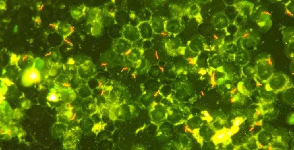
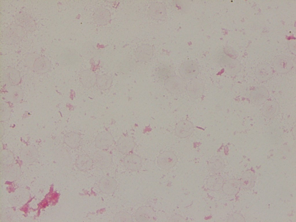
Initial (outpatient) blood culture was positive in the aerobic bottle at 60.1 hours, with the initial gram stain showing no organisms. The bottle was placed back in the analyzer and flagged positive again, at which point a second gram stain was performed, which again showed no organisms. An acridine orange stain was performed (Image 1), revealing multiple spiral/”gull shaped” rods. A third gram stain (Image 2) with more time allowed for safranin staining revealed faint gram negative rods. MALDI-TOF MS was attempted with no identification. The culture growth was sent to a reference laboratory and was identified via sequencing as Helicobacter species. The organism was not viable for susceptibility testing.
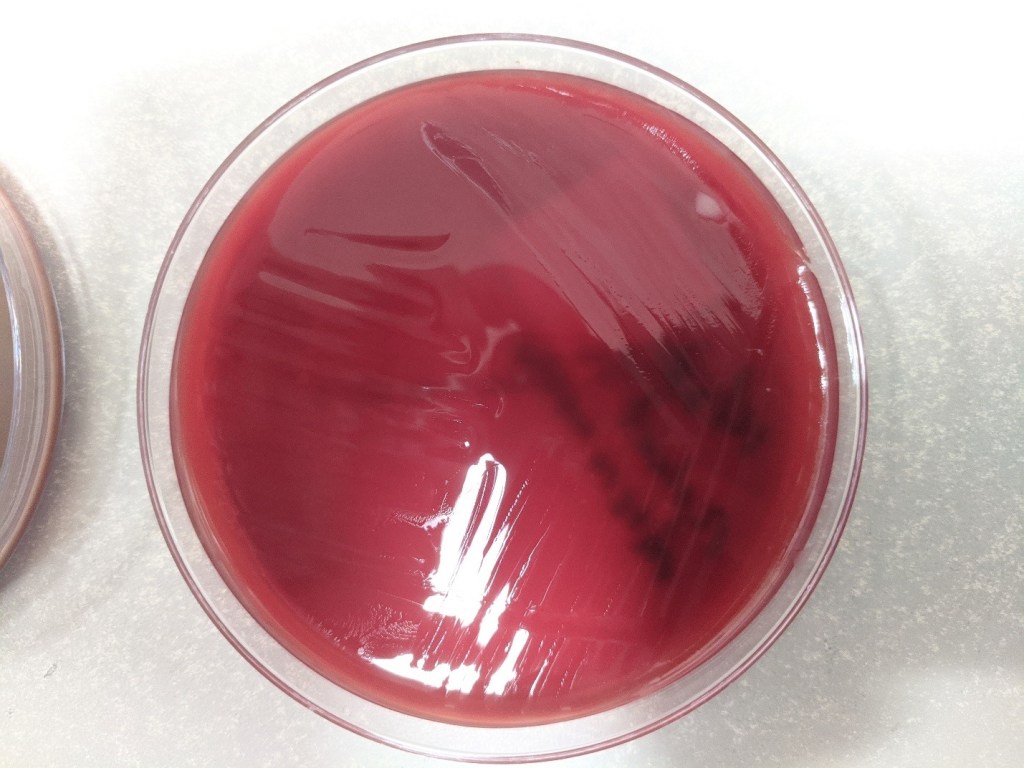
Image 1. Acridine orange stain of blood culture sample: this stain causes the nucleic acids to fluoresce orange, highlighting the bacteria against the background of blood.Image 2. Gram stain after allowing extra time for safranin staining, showing few gram negative rods.Image 3. A replating on blood agar showed difficult to discern, thin spreading colonies.
Two sets of subsequent blood cultures also grew gram negative bacilli at 65 and 67 hours. The blood culture broth from one of these cultures was also sent to the reference lab, but again did not have viable growth for susceptibility testing.
Discussion
The genus Helicobacter includes 35 species, consisting of gram negative spiral bacilli, previously considered to be part of the Campylobacter genus. Pathogenic species are classically associated with the gastrointestinal tract as they are able to survive in the harsh acidic conditions of the human stomach. The most common clinically relevant species is H. pylori, which is associated with gastric ulcers as well as other inflammatory processes in the stomach and duodenum. In prior reports, bacteremia caused by Helicobacter species is typically associated with some other underlying disease process, such as malignancy, immunocompromised state, or disruption of the GI mucosal barrier (1, 2, 3, 4, 5, 6).
Helicobacterspp. are similar in morphology to Campylobacterspp. on a gram stain; given the patient’s prior history of C. jejuni bacteremia, it was not unreasonable for the gram smear to initially be called consistent with Campylobacterspp. However, the clinical course and antibiotic susceptibility profiles of Helicobacter and Campylobacter bacteremia cases can differ in important ways. Further, susceptibilities can differ between different species of Helicobacter. There are no established guidelines for the treatment of Helicobacter spp. bacteremia and breakpoints for antibiotic susceptibility testing for some Helicobacter species have not been established. (7)
The patient in this case was discharged on a course of azithromycin with clinical improvement: at that time, the sequencing result revealing Helicobacter had not yet been received, and the clinical team was acting on the belief that the organism in the patient’s blood was a recurrence of the previous Campylobacter infection. On a follow up outpatient appointment with Infectious Disease, wherein sequencing results were available, tetracycline was prescribed due to concern about the possibility of resistance or relapsing infection.
References
Abidi, Maheen Z., et al. “Helicobacter Canis Bacteremia in a Patient with Fever of Unknown Origin.” Journal of Clinical Microbiology, vol. 51, no. 3, 2013, pp. 1046–1048.
Araoka, Hideki, et al. “Clinical Characteristics of Bacteremia Caused by Helicobacter Cinaedi and Time Required for Blood Cultures To Become Positive.” Journal of Clinical Microbiology, vol. 52, no. 7, 2014, pp. 2745–2745.
De Luca, et al. “Helicobacter Pylori Bacteremia: An Unusual Finding.” Infectious Disease Reports, vol. 8, no. 3, 2016, pp. 74–75.
Han, Xiang Y., et al. “Helicobacter Pylori Bacteremia with Sepsis Syndrome.” Journal of Clinical Microbiology, vol. 48, no. 12, 2010, pp. 4661–4663.
Imataki, Osamu, et al. “Enteral Malakoplakia Prior to Helicobacter Cinaedi Bacteremia.” American Journal of Gastroenterology, vol. 112, no. 1, 2017, pp. 187–188.
Saito, Sho, et al. “Helicobacter Fennelliae Bacteremia: Three Case Reports and Literature Review.” Medicine, vol. 95, no. 18, 2016, p. e3556.
Yamamoto, Kei, et al. “Comparison of the Clinical and Microbiological Characteristics of Campylobacter and Helicobacter Bacteremia: the Importance of Time to Blood Culture Positivity Using the BACTEC Blood Culture Systems.” BMC Research Notes, vol. 10, no. 1, 2017, pp. 1–6.
-Tom Koster, DO is a 1st year Anatomic and Clinical Pathology Resident at the University of Vermont Medical Center.
-Christi Wojewoda, MD, is the Director of Clinical Microbiology at the University of Vermont Medical Center and an Associate Professor at the University of Vermont.
Medical school councilors have good intentions in mind when they steer medical students who realize that direct patient care isn’t their strong suit into pathology. But I am different kind of pathologist – the one who sees (or talks to) patients every day. I am a member of unique subspecialty – Transfusion Medicine – which is the most patient-centric subspecialty of all pathology subspecialties. And, contrary to the popular wisdom, I like seeing patients.
Don’t get me wrong though, my heart and soul still live in the lab, deeply rooted in understanding test performance, troubleshooting and quality control. But direct patient care helps to put all the work I have done in the lab into a perspective.
One program that became especially dear to my heart is our chronic RBC exchange program for the kids and adults with sickle cell anemia who have high risk of developing serious complications from the disease, such as stroke, acute chest syndrome, and severe iron overload. As an apheresis physician I see these patients quite frequently due to the nature of the program – chronic RBC exchanges every 4 to 6 weeks. This also means that I quickly had to learn quite a lot not only about managing the exchanges, but also about patients’ success and failures, spend time explaining to parents the benefits of the program and engaging them to maintain compliance with rigorous schedule. The work is not immediately rewarding. All the adjustments I do to the plan of care show changes in lab values in a month or two at best. But it is not entirely about numbers. Another aspect that makes this program special is when you notice that the kids you treat are doing better at school, have less ED visits and overall live a more fulfilling life.
Sometimes the patient interaction is not as direct as in the case of the sickle cell RBC exchange program. For example, being part of the obstetric team that cares for the patient with severe hemolytic disease of fetus and newborn is also extremely rewarding. And the more challenging clinical question is the more rewarding it is in the end. Just this summer we had a patient who developed an antibody to very high frequency antigen that is present in 99.7% of the population and finding the right donor for intrauterine transfusion involved quite a few people in at least 3 cities. When all the pages, phone calls, emails, and personal conversations between me and residents, obstetricians, anesthesiologists, pediatricians, and blood suppliers result in a positive outcome for mom and baby – I feel elated. And who wouldn’t?! That is why I enjoy what I do!
-Aleh Bobr MD is currently the medical director of blood bank and tissue services at University of Nebraska Medical Center in Omaha, NE. He did his residency in Anatomic and Clinical pathology and Fellowship in Transfusion Medicine at Mayo Clinic Rochester, MN. Prior to that he did his post-doctoral research fellowship in Immunology with focus on dendritic cell biology at University of Minnesota and Yale University. He received his medical degree from Vitebsk State Medical University in Vitebsk, Belarus. Current interests include application of apheresis, platelet refractoriness.
I often have an argument or discussion with my spouse about facts versus opinions. Although both concepts represent information, my brain is mostly concerned with facts as a scientist and a problem solver. My spouse, having spent years in the hospitality, banking, and real estate business, is “all about the customer,” with success rooted largely in recognition of and alignment to their opinions. In my current role as CMO at ASCP where we have “concierge customer service” as one of our principles, I have adapted to listening, understanding, and operationalizing the opinions of a diverse group of individuals. However, when it comes to science, I feel that it is important to remain with facts until the point where science runs out of answers, and we have to guess about something. Where I run into trouble is that I have the opinion that people who do not understand the facts should not necessarily espouse their opinions. Someone once said an expert is someone who knows everything about a topic as well as everything that is wrong about a topic—referring to common misunderstandings that flow through our common knowledge. Opinions are like belly buttons—everyone has one, but some are cleaner than others.
When it comes to journalism, I am unfortunately a purest. I just want the facts. I was, therefore, a bit taken aback by a New York Times article discussing an innovative technology in neurosurgery for intraoperative consultations for brain tumors. My visceral negative response emerged from the surgeon discussed in the article owning part of the company that built the device for the study. My secondary concerns stem from the article discussing intraoperative consultations done by pathologists and, yet, not a single pathologist was interviewed for the article. But the biggest concern I had which made me delve deeper into the topic was the enormous number of inaccurate facts or complete untruths presented in the article. It is hard to say it wasn’t poor journalism but, as a scientist, I had to go to the source.
The scientific article in question was “Near real-time intraoperative brain tumor diagnosis using stimulated Raman histology and deep neural networks” published on Jan 6, 2020. The New York Times article, “A.I. Comes to the Operating Room” was published the same day. I read the article myself, and when it didn’t quite pass my sniff test, asked three of my colleagues who are experts in this area to also read the paper and the news report. Jane Brock, a breast pathologist who is truly an innovative thinker and dreams of the day when pathologists can study tissue immediately with confocal laser imaging and rapid molecular testing—part of her research—said of the technology, “this is a great paper and a great microscope.” She further mentioned how brain is ideal because it is homogenous, easily flattened, and amenable to artificial intelligence (AI) review because of the limited parameters needed to be evaluated for clinical decision making intraoperatively. “This is definitely the future of pathology—getting rid of frozen sections in favor of fresh tissue imaging,” Dr. Brock said. “It also means you can take tissue for research/molecular diagnostics, image it, and not waste it just by imagining it [on frozen section]. The time savings are huge.” It was exciting to hear Dr. Brock’s enthusiasm for the technology conceptually and how it could be a boon for pathologists’ ability to consult during surgery.
Dr. Rebecca Folkerth, a surgical neuropathologist for more than 20 years before she became the neuropathologist for the Office of the Medical Examiner of New York, had some concerns about the science in the paper and the maturity of the technology to replace a consultation with a pathologist today. “The questions asked [in the paper] were of necessity extremely basic, and hardly represent the real world as encountered in the operating room and neuropathology laboratory,” said Dr. Folkerth. She was concerned in the news article with the statement “Final pathologic diagnosis is increasingly driven by molecular rather than morphologic criteria.” “That is actually true for a minority of nervous system lesions, such that the ‘gold standard’ for diagnosis remains ‘macroscopic (gross) pathology’ [essentially imaging] and ‘cytologic and histoarchitectural features’ as well as clinical and laboratory findings. In other words,” concluded Dr. Folkerth, “the training and experience of a physician [pathologist] is what allows the synthesis of all data points to arrive at a comprehensive interpretation.” With regard to the immediate application to real world practice, Dr. Folkerth said, “It is telling that no ‘gliosis/treatment effect’ cases were analyzed [because] distinction of post-treatment changes from a neoplastic process in the brain is one of the most difficult encountered in clinical neuropathology.” Other cellular processes that occur in the brain were also not in the study. “A glaring omission of this paper,” Dr. Folkerth concluded, “were the consequences of the errors in ‘predicting diagnosis’. Were these ‘class A’ [leading to radical changes]?” In the article, the surgeon states that neuropathologists “hate frozen sections” which Dr. Folkerth says is simply not true. Perhaps the author should have interviewed an actual neuropathologist for this piece. Both Dr. Brock and Dr. Folkerth had concerns about the practicality of the technology where Dr. Brock felt it was currently “too expensive” (relative to current practice which provides more information across the spectrum of neuropathology) while Dr Folkerth was not clear who this process could work in smaller or decentralizes or underserved surgical settings (as the new article suggests) without a lower cost. Dr. Folkerth agreed with Dr. Brock, however, that this technology “may well represent a revolution in intraoperative decision-making and outcome.”
Lastly, I spoke with Dr. Jason Hornick, who in addition to be an internationally renowned surgical pathologist, has been in charge of quality of intraoperative consultations at one of the premiere hospitals in the US. The “frozen section [procedure] does not often take longer than 30 minutes, and is not often ‘far less accurate’ than in the study,” Dr. Hornick began. “The rates of significant discordance between intraoperative diagnosis and the final diagnosis in published surveys (for all of surgical pathology) are generally less that 1.5%.” Dr. Hornick (and I) agree that the surgeon quoted in the article is bashing the practice of pathology without any accuracy to his statements. He is not quoting facts and he is not quoting opinions. He is simply saying things that are wrong and not supported by data or years of experience. Dr. Hornick also mentioned that the cost of frozen sections are trivial compared with the cost of this technology and, as pointed out by Dr. Folkerth, the pathologists’ intraoperative consultation is much broader than just reviewing a slide for signs of tumor. Dr. Hornick said it best: “The expert consultation provided intraoperatively by the pathologist to the surgeon is not restricted to making an accurate diagnosis; pathologists are uniquely suited to integrating the patient’s clinical history, imaging, prior pathology, and surgical findings to assist the surgeon in making surgical decisions.” He concluded, “The intraoperative consultation is not a laboratory test; it is a consultative opinion by an expert physician who often understands the patient’s disease better than the surgeon.”
With these three experts’ views including their clarification of the facts and their opinions, it seems pretty clear, in my opinion, that this news article is presenting an inappropriate picture of the practice of pathology and making claims about this technology which are not, in fact, accurate or fair. What struck me, however, was the comment by the surgeon that when he was working with pathologists, he may only ask two questions because of the very long time for the frozen section but with this technology he can ask six or seven questions and get the answers really quickly. If this surgeon needed to know the answers to those question, that is, if they were mission critical to patient care, why didn’t the surgeon work with the pathology team and demand higher quality and faster turnaround time rather than investing time and money in a novel, expensive technology (from which he profits) which can only provide a fraction of the answers that a pathologist can provide? Unless the technology completely replaces a pathologist—which it doesn’t seem to be able to do—the pathologists and frozen section labs still have to be available. Thus, costs are increased, not decreased. The only parameter for increased value for the patient would appear to be time savings; however, most clinicians and pathologists would agree that the value to a patient of a two-minute AI read versus less than 20-minute intraoperative consultation is minimal compared with the cost difference.
But outside of all of this, as the entire field of pathology faces pressure from technologies—largely driven by non-pathologists—we have to realize that pathologists’ consultations, whether intraoperative or on permanents, are a tool of quality directly for the patient. If a surgeon is able to perform a surgery and discard tissue, use a system from which he/she profits, or depends solely on a computer algorithm informed by a couple of hundred cases, where is the quality check for the patient? How do we know that was tumor that was removed? Pathologists are paid to perform intraoperative consultations and ASCP works very hard to ensure that pathologists are not only fairly paid for their work but that their involvement occurs whenever it can improve quality and care for the patient. Dr. Folkerth alone has seen thousands of cases on intraoperative consultation for neuropathology and the collective knowledge of currently living neuropathologists would be millions of cases. If such knowledge were captured by an AI across the full spectrum of neuropathology, the technology would truly be remarkable. But you can’t have such integration of knowledge without involving pathologists.
-Dan Milner, MD, MSc, spent 10 years at Harvard where he taught pathology, microbiology, and infectious disease. He began working in Africa in 1997 as a medical student and has built an international reputation as an expert in cerebral malaria. In his current role as Chief Medical officer of ASCP, he leads all PEPFAR activities as well as the Partners for Cancer Diagnosis and Treatment in Africa Initiative.
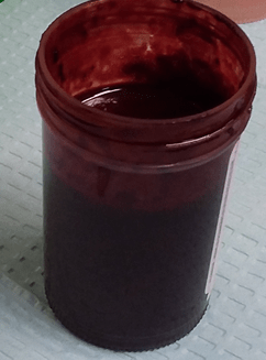
An 80 year old female had a history of chronic iron deficiency anemia with unknown cause and comorbidities included hypothyroidism, congestive heart failure (CHF), severe aortic stenosis and COPD. The patient presented at the ED with initial presentation with increasing shortness of breast, NYHA class 3-4. She was admitted to the hospital for further treatment for CHF, as well hyperventilation, sleep apnea and COPD. Her serum iron and iron saturation were tested and results were 2 umol/L (reference range for iron: 10-29 umol/L) and 7% (reference range: 14-51%), respectively. Part of her investigations included a qualitative fecal test to screen for gastrointestinal bleeding. The immunochemical fecal occult blood test was performed using a CLIA waived Hema Screen SpecificTM POCT test (Immunostics, Inc, USA) in the hospital lab. Hema Screen Specific test is a qualitative, sandwich dye conjugated immunoassay that uses a combination of monoclonal and polyclonal antibodies to detect the globin component of hemoglobin in the fecal samples. The manufacture recommended using Hema Screen Specific test in routine physical examines, hospital monitoring of bleeding in patients and for screening for colorectal cancer or gastrointestinal bleeding for any source (statement from the product package insert).
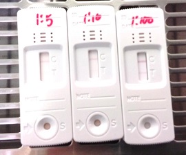
The specimen submitted to the lab was markedly red (Image 1), yet Hema screen test returned a negative result. Since this device is designed to detect occult blood in fecal samples, a prozone effect was suspected, as the stool appeared to contain overt hemorrhage. The specimen was reanalyzed with serial dilutions by a factor of 5, 10, and at 100 × dilution. The FIT result became clearly positive for blood (Image 2). The patient received a colonoscopy, which revealed internal hemorrhoids, severe diverticulosis in the left colon, as well as multiple angiodysplastic lesions. One such lesion was in the ascending colon and was actively bleeding at the time of colonoscopy. The others, which were not bleeding, were distributed in the proximal ascending colon, hepatic flexure, and proximal transverse colon. All angiodysplastic lesions were treated with argon plasma coagulation.
Image 1. Fecal specimen demonstrating overt hemorrhage.Image 2. Fecal immunochemical test performed on the patient sample submitted. Serial dilutions of fecal specimen were performed. At the dilution factor of 1:100, the result showed positive. Saline was used to dilute the fecal sample.
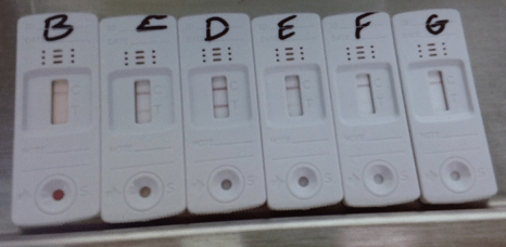
Moreover, we have tested the device with another bloody fecal sample during the initial evaluation. When an appropriate dilution factor was used, the prozone effect begins to lose its interference as show in Image 3.
Image 3. Fecal immunochemical test (FIT), showing control (C) and test (T) lines, applied to serial dilutions of fecal specimen (B ─ undiluted, C ─ 10 ×, D ─ 100 ×, E ─ 1 000 ×, F ─ 10 000 ×, G ─ 100 000 ×). At 10 × dilution, the prozone effect begins to lose its interference, and at 100 × the test is clearly positive. At dilutions higher than 1 000 ×, the concentration of blood becomes too low to return a positive result. (Image 3 provided by Dr. Andrew Lyon, PhD, DABCC, FCACB, Lab Medicine & Pathology, Saskatchewan Health Authority)
Discussion
The prozone effect (or Hook effect) has long been appreciated as a source of interference in immunoassays.1 It typically occurs in sandwich assays, of which the FIT test is an example.2 When the concentration of the analyte is excessively high, it oversaturates the capture and detection antibodies in favor of forming single antibody:analyte complexes, rather than sandwiches. This results in a false negative result where the assay is unable to detect the analyte. The solution to the prozone effect is serial dilution to lower the concentration of the analyte.
The FIT test is designed to detect microscopic amounts of blood, hence its function in screening for fecal occult blood. A number of hospital labs use this test in an acute care setting to screening bleeding in patients. However, its capacity is oversaturated in specimens containing overt hemorrhage, as in our patient. In these cases it is nevertheless important to prove that the red color of the specimen is truly due to blood, as bright red stool can be caused by a wide range of dietary factors. Some examples are red food coloring, beets, cranberries, and tomato juice.3 If these possibilities are not ruled out, the patient may become subject to the risks of unnecessary endoscopy. Serial dilution of the specimen is extremely useful in this type of situation.
References
Dasgupta A, Wahed A. Clinical Chemistry, Immunology and Laboratory Quality Control: A Comprehensive Review for Board Preparation, Certification and Clinical Practice. Amsterdam: Elsevier; 2014. 2.11.
Allison JE, Fraser CG, Halloran SP, Young GP. Population Screening for Colorectal Cancer Means Getting FIT: The Past, Present, and Future of Colorectal Cancer Screening Using the Fecal Immunochemical Test for Hemoglobin (FIT). Gut and Liver. 2014 Mar;8(2):117-30. https://doi.org/10.5009/gnl.2014.8.2.117
-Hao Li, MD is a currently a first year anatomical pathological resident at Western University, London ON, Canada. Prior to be a pathology resident, he was a neurosurgery resident at the University of Saskatchewan, Saskatoon SK, Canada. When he was at the University of Saskatchewan, he spent his third year primarily in neuropathology, with also some general anatomical pathology and clinical pathology. Through these experiences, he has come to realize that his passion and calling lay more in pathology than in surgery. He has successfully transferred into pathology, and started a new residency in anatomical pathology in July 2019. Having a background in the clinical neurosciences, he hopes to eventually pursue a fellowship in neuropathology, and possess the skill set to practice both anatomical pathology and neuropathology.
A 72 year old man was admitted to the hospital for an aneurysm repair. The physician ordered a type and crossmatch for 6 units of blood in preparation for surgery. The patient history included surgery in 2016 during which he was transfused with 4 units of RBCs.
patient’s blood type: A positive
antibody screen: negative
history: anti Jkb (2016)
6 Jkb negative units were found and full crossmatches were performed. One of the 6 donor units was incompatible. What is the most probable explanation for these findings?
If the patient has a negative antibody screen, and no history of an antibody, most facilities would do an electronic crossmatch or an immediate spin crossmatch. The immediate spin (abbreviated) crossmatch will simply verify ABO compatibility. However, if the patient has a positive antibody screen, we must identify the antibody, phenotype the patient, and do a full AHG crossmatch with donor units that are antigen negative for the corresponding antibody. In this case, the patient had a history of an antibody, so the antibody must be honored, and antigen negative units must be chosen for transfusion.
Kidd antibodies demonstrate dosage, are often weak, and can be found in combination with other antibodies. Because if this, they can be notoriously difficult to detect. They are usually IgG and are made in response to transfusion or pregnancy. Jkb has an antigen frequency of about 73% in the white population and about 43% in the black population. To find antigen negative blood, we consider that about 27% of units would be antigen negative. The tech working on the sample screened 21 units and found 6 that were Jkb negative.
AHG crossmatch results:
unit 1: compatible
unit 2: compatible
unit 3: compatible
unit 4: 3+ at AHG
unit 5: compatible
unit 6: compatible
There are 2 possible scenarios for the above results. A crossmatch is a test between donor’s red blood cells and patient’s plasma. Antigens, we know, are on red blood cells and antibodies are detected in the plasma. So, with a negative antibody screen, crossmatch incompatibility is due either to a patient antibody to a low incidence antigen on the donor red blood cells, or a donor cells with a positive direct antiglobulin test. We can easily rule in or out a positive donor DAT by performing a DAT on the segment. If the donor unit has a positive DAT, the unit should be quarantined and the positive DAT reported to the collecting facility. If the donor unit has a negative DAT, the patient likely has an antibody to a low incidence antigen.
Low frequency antigens are uncommon, but antibodies that recognize them are less rare. Fortunately, for patients with these antibodies to low frequency antigens, finding antigen negative compatible blood is easy. As we can see, 5 of the 6 chosen units were negative for the unknown low frequency antigen and were antiglobulin crossmatch compatible. The low prevalence of the antigen makes compatible blood readily available. If transfusion is necessary, it should not be delayed while waiting for identification of the antibody.
In this case, the antibody screen was repeated and the negative result was verified. In many cases, it may not be possible for a lab to identify the antibody because the lab may not have the necessary panel cells or typing reagents. Yet, these antibodies to low incidence antigens that react at AHG can be clinically significant and cause severe hemolytic transfusion reactions. To identify the antibody, you may need to send the sample to a reference lab for testing against a panel of reagent red cells that express low incidence antigens. Alternately, the donor red cells that were incompatible can be tested against known antibodies to low prevalence antigens to help identify the antibody.
In this patient, anti-Wra was identified. The incompatible donor unit was verified to be Wra positive. Wra is part of the Diego system, usually IgG, and has ben implicated in hemolytic transfusion reactions.
One of the reasons I have written up this case is questions my Transfusion Medicine students often ask about exam and exam prep questions concerning incompatibility. Below are 2 questions to give examples of the confusion.
“At the indirect antiglobulin phase of testing, there is no agglutination between patient serum and screening cells. One of 3 donor units was incompatible.. The most probable explanation for these findings is that the:
patient has an antibody directed against a high incidence antigen
patient has an antibody directed against a low incidence antigen
donor has an antibody directed against donor cells
donor has a positive antibody screen”5
answer: b
“Which of the following would most likely be responsible for an incompatible antiglobulin crossmatch?
recipient’s red cells possess a low incidence antigen
anti-K antibody in donor serum
recipient’s red cells are polyagglutinable
donor red cells have a positive direct antiglobulin test”4
answer: d
I am asked why is one answer “low prevalence antigen” and one answer “positive DAT”? I typically ask questions of my students to let them reason out the answer. Take a careful look at the words antigen and antibody. Remember that a DAT is a test of red cells, the IAT tests for antibodies in plasma. A crossmatch uses donor red cells against patient plasma. Therefore, even though these are both reasons for the incompatibility of one out of multiple units, each question only has one answer of a common reason for such incompatibility. Be sure to read questions and use your theory and knowledge of testing when encountering discrepancies and problems in Blood Bank. To all of my students: Happy Studying for your ASCP exam!
References
Fung, Mark K., Technical Manual 18th ed, Bethesda: AABB, 2014.
Harmening, Denise M. Modern Blood Banking and Transfusion Practices, 7th edition, 2019.
Schonewille, Henk, et al. “The importance of antibodies against low‐incidence RBC antigens in complete and abbreviated cross‐matching”. The Journal of AABB. 20 June 2003.
-Becky Socha, MS, MLS(ASCP)CM BB CM graduated from Merrimack College in N. Andover, Massachusetts with a BS in Medical Technology and completed her MS in Clinical Laboratory Sciences at the University of Massachusetts, Lowell. She has worked as a Medical Technologist for over 30 years. She’s worked in all areas of the clinical laboratory, but has a special interest in Hematology and Blood Banking. When she’s not busy being a mad scientist, she can be found outside riding her bicycle.
Welcome back everybody! Thank you for all the engagement on my last post, Up In Smoke¸ where I discussed the plenary publications surrounding the vaping crisis and EVALI as new pulmonary pathology entity. This month, let’s start 2020 off right. After the holiday break and going on some of my last pathology residency interviews, I’d like to reflect on this new year by taking a look at 20 exciting things on the horizon for those of us in pathology and laboratory medicine!
So, let’s take a look at 2020 with some 20/20 vision… (sorry, not sorry)
20. Big, big, big, big data
Image 1. Can’t mention databases without this invaluable website that has made me look somewhat semi-competent in many instances! Thanks Pathology Outlines (Source: um, pathologyoutlines.com)
Last year, Elsevier’s Clinical Solutions Director in China discussed three topics that would impact our profession in 2019—so let’s start there. These first three go hand-in-hand in prepping the stage for 2020. Up first: the never-ending explosion of biomedical information and the continuing tidal wave of health data we don’t even know what to do with just yet! It’s a very interesting estimate that, by 2020 (aka now!) the whole sum of medical knowledge will double every 73 days. How on earth are we to manage, when compared to 1950 it would have taken 50 years to double? Well, the argument in the linked Elsevier blogpost discusses how evidence-based inquiry databases will store and organize this knowledge for us: think UpToDate, or ExpertPath, or ImmunoQuery…some of you are nodding your heads in relief, great, I’ll move on.
19. Precision Medicine
Image 2. PD-L1, or programmed death ligand 1, is one of several new targets for cancer therapy that utilized cellular checkpoints in cell cycles alongside T cell and NK cell functional immunity to fight cancer a little more precisely than classic chemotherapy regimens. Look at you, all up to date, and stuff. (Source: AstraZeneca graphic, azimmuno-oncology.com, content)
The second topic last year’s Elsevier’s blogpost discussed was the growth and rapid development of highly specific, targeted, individualized treatment plans. The mainstay example is of course how oncology treatments are moving away from one-size-fits-all chemotherapies to individualized mutation-specific immunomodulating therapy. (We’re moving like melting glaciers but moving nonetheless.) I was definitely well equipped with my ASCP online CE credits as I found myself discussing testing patients during my heme/onc training for PDL-1 and other tailored targets. We’re just starting to ride this wave and it’s definitely growing fast.
18. AI in healthcare (part 1)
Image 3. Artificial Intelligence is getting really good at pattern recognition. Why did I choose this picture? Oh, because it’s a study of how China-based JF Healthcare, a Siemens off shoot start up AI group, designed an algorithm that beat radiologist at Stanford on precision, delivery, and accuracy. Woah. (Source: hitconsultant.net)
Yep. I went there—it’s exciting! But notice I’ll come around after some other topics to really get into the heart of AI in path. Basically, the last point in the blogpost discussed the way smart software has been growing in medicine; particularly with radiology and surgery, using advancements in robotics and detection software to predict and stratify clinical information for patient care. Within this context let me quote them directly for you, “…there remains some uncertainty around the role of AI and its true impact on pathology, it is important to recognize that AI-based technologies or machines will never replace pathologists. Instead, such innovations will play an assistive role, augmenting the decision-making capabilities of pathologists and helping them perform better and faster…” All my pathologist friends may now exhale. It’s going to be okay. We’ll talk more about this at #10.
17. New Tech, New Toys
Image 4. You don’t have to go far to read about new tools and new tech. How’s this: a saliva-based rapid Malaria detection assay, courtesy of our friends at ThePathologist.com. New, rapid, accurate and deliverable diagnostics…now within spitting distance. Nailed it.
Pathologists are like the 007’s of the clinical team…at least when it comes to developing tech. There are so many new gadgets and tools we clinicians have available to us today. I delivered a recent TEDx talk where I discussed the “unrecognizable future” of medicine—and obviously now look for new and exciting ways to tell people I gave a TEDx talk. The important thing is that 73 days of doubling medical knowledge is happening so fast we don’t even know what we have available to us! Finger-print drug tests, smartphone facial capillary blood pressures, liquid biopsies, virtual MS-based immunohistochemical stains that never actually stain a single cell, cytology AI, deep data mining of free text pathology reports…it’s not a short list. It’s exciting, and we should all be sharing and collaborating to use these exciting tools together in creative ways for positive outcomes!
16. No More Silos
Image 5. Business and management have long discussed the importance of tearing down silos of knowledge in order to improve workflow and outcomes. It’s a growing conversation in healthcare and we’ve got our own isolated pockets of data that need to come out to the forefront, too. (Source: ERP Consulting, Estes Group Image, estesgrp.com)
Last year, I wrote a few pieces here on Lablogatory that mentioned High-Reliability Organizations (HROs) which require absolutely full sharing of responsibility as well as knowledge in order to solve problems and improve patient-care outcomes. There are many ways siloed thinking can harm the progress of any institution. It takes leadership, creative problem-solving, transparency, and teamwork. In the coming months of 2020, keep an eye out for pockets within your organizational environment that act as black holes or veils to keep pieces of critical data from the rest of the team. Encourage discussions between you and your peers, check biases about what you think might be important for one team vs. another, and try to share successes and failures as a group.
15. New Types of Colleagues
Image 6. We’re all different. And that’s ok. Each one of us is a brain, and an athlete, and a basket-case, a princess, and a criminal. Okay maybe not the last one, but we can all contribute in some important meaningful way. (Source: The Breakfast Club, 1985)
What I just mentioned about engaging in new conversations with folks you might not have worked with before—its not groundbreaking, its just good practice! In order to tear down #16’s silos, we’ve got to seek out and explore new ways to collaborate with colleagues outside our everyday scope. There will always be discussions about generational divides and differences that create culture strife in the workplace, or political/opinionated schisms that divide even the most cohesive of medical specialties. (I’m looking at you ACOG, ACP, ASCCP, and others: it’s Cervical Cancer Awareness Month, can we just agree on some guidelines already…) Soapbox over. But seriously, this isn’t a new concept. Feel like a lab half filled with boomers and millennials can’t make the cut? Well, the Harvard Business Review gave us great recommendations for this exact type of interpersonal growth exercise—in the NINETIES! The take home message: having an open culture and proactive leadership allows for fruitful exchange and growth!
14. Digital Pathology!
Image 7. Bigger, I want these screens bigger! The desk of tomorrow’s anatomic pathologist might have less glass and more pixels, screens, and queued data with high-output servers that are stocked with smart software to sift out normal results so they can focus on really tough morphologies. Maybe even with augmented reality software, or other crazy stuff I can’t think of yet! (Source: Inspirata, digital pathology)
It’s coming. You can’t stop it. It’s exciting. I don’t care what you think. Well I actually do care, but don’t knock digi-path till it grows into whatever it’s going to become. The desk of the (anatomic) pathologist-of-the-future will look a lot different from today and that’s really cool. Once upon a time, a very long, long time ago—in the eighties maybe—radiologists still had films where we actually used radiation to change the exposure of images to be read over a light box. Classic scene, right? Doctor, the x-ray is ready! *THWIP* *CLICK* *BUZZ* and cue the contemplative stare on the wall light. Then, they went digital and get to hang out in the dark with four computer screens and coffee, and really comfy chairs. I mean what a form of progress, can’t deny.
13. MS Methodologies
Image 8. Okay, MSI crash-course time. All you really need to know is that this method allows for great specimen preservation on tiny samples, high resolution, the ability to combine with molecular testing, and fascinating implications for margin detection, mutation analyses, and more! (Source: https://blog.waters.com/molecular-visualization-ms-imaging-delivers-insights-for-cancer-research)
In my mailbox this month, is another excellent edition of The Pathologist and in it there’s a great article on Mass Spec imaging transitioning from a research tool to a clinical one. Woah. We’ve all talked about and praised MALDI-Tof methods for microbiologic assays and detection, but the expanse of mass spectrometry has developed rather quickly. Now, it’s looking for a niche in routine laboratory diagnostics outside of the old chemistry analyzer… It’s a new, non-destructive way of examining tissue and gleaning data from the smallest pieces of gross specimens. We’re onto something here, keep an eye on MSI.
12. Molecular—Need I say More?
Image 9. Move over International Space Station, the folks at Thermo Fischer Scientific want to share their take on the Next Generation of molecular testing. (Source: The Pathologist)
Same edition of The Pathologist, about 25 pages back: a discussion on the value of molecular Next Generation Sequencing. I’ve already bored half of you, wait! Come back. I agree with you, you can only call it Next-Gen so many times before a whole generation of laboratorians get bored of talking about new tumor markers or mutations. But what’s happening with NGS testing that you should know? Simply put, there are NGS analyzers that are faster, with smaller footprints, combined with smarter software that is making molecular more feasible for laboratories that used to shy away from the notion of including NGS or LDTs in their lab testing menus. This means more labs, running more molecular, for more specific populations, in real time that can collaborate with that many more new colleagues while breaking silos—well just look up at #18, 17, 16, and 15!
11. Global Health
Image 10. From Dr. Razzano’s post on Lablogatory
Dr. Dana Razzano recently interviewed me for her global health series, and we got the chance to talk about the important intersection of laboratory medicine and global public health. Getting involved in a community—especially for those of us in healthcare—often includes a survey of what kind of health challenges you face. For some it’s access to clean resources like water, for others it’s a complex system of reimbursement and billing issues that complicate delivery of care, or even more basic assessments reveal high rates of local infections with preventable illness. But you can’t tackle infrastructure change, political reform, or vaccine education single-handedly. Global health is an increasing part of our global world and, if we stay true to our professional values, we should be at the forefront.
10. AI in Healthcare (part 2)
Image 11. Drawing to represent AI from my TEDx talk, Unrecognizable Medicine 2019, TEDxAUCMED
Oh I told you I’d come back to this. Some folks are still apprehensive about AI—that’s ok—I am too, but only because I want to make sure it’s done right. Don’t expect any Skynet stuff, we’re not going that deep. So let me tell you some of the things I got to see on the residency interview trail that piqued my interests. At one hospital system, I saw plans for their anatomic pathology department to go fully digital with augmented AI software to help score mitoses and other morphologic traits by 2025. At another institution, I saw plans for data mining historical free text pathology reports to predict and stratify future specimens before they even got signed out! At a third system, I saw the utilization of smart software to predict clinical lab values for a patient’s personalized reference range…pre-analytically! This stuff is coming in hot so watch for it! What AI-related advancements are you seeing in your neck of the lab?
9. Patient Consultation
Image 12. Courtesy of SUNY Upstate Pathology Department via Twitter, a newly renovated pathology residency review room and patient consultation suite for the dedicated purpose of this invaluable interaction.
Another thing noteworthy of my residency trail are institutions which are championing the face-to-face consultative role of the clinical pathologist in patient care. We, at the end of the day, are consultants to all; physicians and patients alike. And many in our field are celebrating this role by pushing the envelope toward a progressive and effective future for pathology and laboratory medicine at large.
8. Graphic Medicine
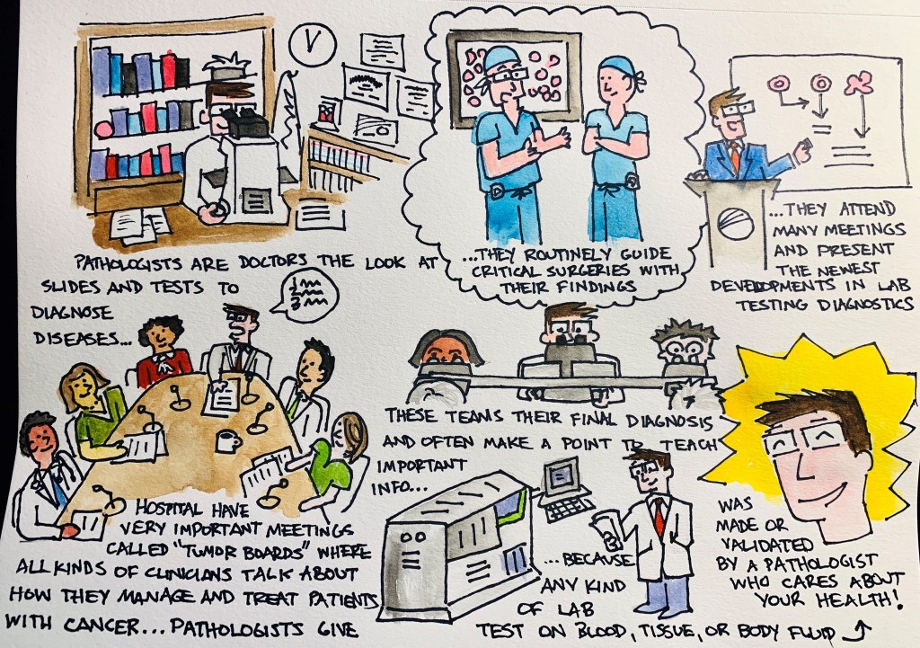
Image 13. My very first #pathdoodle – What do Pathologists do?
Graphic medicine is fantastic! I wish I could have discovered this years ago. Nevertheless, in the last two years I have sought out and read numerous pieces which bridge a significant gap between clinician and patient. And if we in laboratory medicine plan to reach patients more directly, we shouldn’t be afraid to come up with creative solutions to communicate with them. My (current) approach, #PathDoodles which I post occasionally on social media, attempts to explain concepts in pathology to the everyday lay person. What will you do to reach your patients?
7. Wellness Programs
Image 14. Anonymous survey responses to Dr. Saint Martin’s program from trainees at Loyola University Medical Center. (Source: The Pathologist)
Another interesting thing I encountered on the residency interview trail was the way in which different programs addressed the concept of wellness. Some did, some did not, but everyone discussed it. Because, after all, it is important; not just for residents and physicians, but medical lab scientists, support staff, and …yes… even administrators. Work and work-life stresses and balance take a psychological and emotional toll, and in order to be happy and healthy we need ways to strengthen our mental health along the way. Last year, Dr. Marissa Saint Martin won a 2018 award from the American Association for Physician Leadership for her work in preparing residency resilience and promoting wellness through structured curricula at Loyola Health System and Mount Sinai in Miami. She’s been featured with ASCP numerous times as well as in The Pathologist. It’s promising to see such strong support for residency trainees; keep an eye out for other praise-worthy programs this year!
6. Social Media
Image 15. It takes a new kind of clinician to serve a new kind of patient. How will you rise to meet the challenges on the horizon that we can’t predict yet?
You caught me. I can’t make any predictions about the future of health care delivery, education, or collaboration without touting the importance of social media. As a member of the official ASCP Social Media Team (Go #ASCPSoMeTeam!), I’m a proud contributor not only to the content that we publish online, but to the message that unifies and spreads our #StrongerTogether mantra. Social media is fantastic tool to reach new colleagues, spread new ideas, and make new advancements in both our field and in reaching people where they are.
5. Moving Out of the Basement
Image 16. Is this closer to a representative image of your lab than you’d wish? Don’t worry, you’re not alone. Basement labs are so last season. (Source: Seret Studios, Bridge Basement, Brooklyn NYC)
One of the most memorable interviews I had this season was with a transfusion specialist who recounted to me his memory of training in pathology during his early days of residency. Green and eager, he told me how he remembered being taken on a tour of the facilities to be shown his desk/work space and upon reaching an underground level of the hospital building a sign read ‘BASEMENT’ with an adjacent one ‘PATHOLOGY’ …he paused to say it out loud, “What’s a basement-pathologist?” We bonded over the stifled stereotypes in our work and shared stories of great and terrible lab spaces we’ve seen. Some programs are renovating, some (for better) are moving on up… How are your facilities?
4. The Pipeline Problem
Image 17. The …scope of a pathologist has changed over time (see what I did there?). Sure, lots of us push glass and diagnose entities on tissue samples with complex investigations and work up, but many more of us ensure quality laboratory efficacy, develop new cutting-edge tests, manage donor centers and transfusion protocol, address infectious or public health crises, and more! I think the more we celebrate our profession, the more will join and contribute to our #StrongerTogether culture!
Well I mentioned stereotypes. I’ve talked about it before. You already know about shortages in pathology from physicians to lab scientists. And don’t get me started on pay—especially for lab workers and those in forensic path! But this is a topic I think we’re going to see a boon in media coverage in the coming years, starting now. Some of our awesome colleagues are talking about the “pipeline problem” from a myriad of angles: addressing medical student clerkships, exposure to laboratory workflow in school curriculum, advertising the infinite possibilities of careers within our profession…and more. CAP, ASCP, USCAP, and other professional societies have done amazing work in their due diligence to represent our field and advocate for the advancement of our work and image. A former CAP president once said, “Pathology is what a pathologist does,” and well, it’s a good start. Where do you see solutions to our pipeline problem?
3. PA’s and… CPA’s?
Image 18. Pathologist assistants work directly under the supervision of a pathologist in a similar way that physician assistants work with physicians. Clinical pathologist assistants help to determine the cause of disease through the examination of blood and other bodily fluids and tissues. (Source: UAB)
Now this, this is a cool concept and it’s the first time I’ve seen it. We all know and love our pathology assistants (PAs)—especially resident pathologists—as they make the training, education, and workload a better process for learning. They have awesome training and operate essentially as highly specialized clinicians in anatomic pathology. But what about the clinical, laboratory medicine side of things…? I recently saw a program advertised at the University of Alabama at Birmingham that trains clinical PAs for consultative and ancillary support roles to the clinical pathologist! Fantastic! Read more about it here! There are a growing number of DCLS (doctor of clinical laboratory science) professionals, now we’ve got a new pipeline to invite talented folks to work with us in a new way.
2. I’ll Have a Residency!
Image 19. Need I say more? What does the paper say?? Where am I going to end up? It’s a whirlwind of a season and a crazy system—once in a lifetime sort of adventure! Stay tuned, I’m sure I’ll have a post or ten about the next chapter of my career… (Image source: AAMC)
I have seen so many fantastic programs across the US these past few months, and so many decisions go into the way medical graduates rank programs to match into. To learn how the AAMC/NRMP matching system works, watch this video (maybe with some calming tea). We’ll see where I end up matching to, but I’m excited for the next chapter and to really dive into pathology even more!
1. You!
Obviously, the most important thing to keep an eye out for this year is you! Your goals, your ambition, your plans, your ideas and thoughts, and all the ways you contribute to our fantastic profession! I encourage you all to share, collaborate, and be creative with the ways in which we advance the future of laboratory medicine and continue to keep in mind that we do these things for our patients everywhere.
Thanks for reading, see you next time!
–Constantine E. Kanakis MD, MSc, MLS (ASCP)CM completed his BS at Loyola University Chicago and his MS at Rush University. He writes about experiences through medical school through the lens of a medical lab scientist with interests in hematopathology, molecular, bioethics, transfusion medicine, and graphic medicine. He is currently a 2020 AP/CP Residency Applicant and actively involved in public health and education, advocating for visibility and advancement of pathology and lab medicine. Follow him on Twitter @CEKanakisMD
As you may recall last month I shared common barriers to biomarker testing for cancer patients in the community. I also began to dive-in to a few solutions that I have seen implemented to overcome the barriers. Last month I shared solutions that may help with high cost and long turnaround times for biomarker testing. This month I would like to discuss issues with tissue including quantity.
Here are the top 10 barriers that I’ve seen to biomarker testing in the community:
High cost of testing.
Long turnaround time for results.
Limited tissue quantity.
Preanalytical issues with tissue.
Low biomarker testing rates.
Lack of standardization in biomarker testing.
Siloed disciplines.
Low reimbursement.
Lengthy complex reports.
Lack of education on guidelines.
Sample quantity and quality are both important when considering biomarker testing. If we don’t have enough material we cannot perform the test (quantity not sufficient or QNS). If we have poor quality we cannot trust the results. The old adage of garbage in garbage out holds true for biomarker testing just as it does for all other lab tests.
I’ll start with sample quantity this month and cover quality issues next month. The issue here is that a variety of biopsy types are performed on patients depending on the location and size of a suspicious mass. Historically we only needed enough material for the pathologist to make a diagnosis. Now we often need enough material for diagnosis and biomarker testing. Some tumor types such as breast and ovarian cancers produce enough material in locations that are easily accessible that tissue quantity is rarely an issue, however other tumor types such as lung and pancreatic cancers there is often an issue with tissue quantity. These tumor types must be handled with care to ensure no tissue recovered is lost.
The first step in addressing tissue insufficiency is knowing where you are starting. Do you have an issue with quantity not sufficient (QNS) rate? If you don’t know how many of your cases are insufficient for biomarker testing, then you can’t determine if you have an issue. If your testing is performed at a reference laboratory, you can request your QNS rate from the lab. They may also be able to provide you with the national QNS rate and then you could benchmark yourself against your peers. It is important to have an accurate QNS rate, so if there are blocks that are not sent to the reference lab because the pathologist has determined the block to be exhausted (no tissue is left) then the QNS rate provided by the reference lab may be artificially low.
It is important to agree upon what is QNS. We consider a specimen to be QNS if we cannot perform biomarker testing on the block. Others may consider the block QNS only if there wasn’t sufficient material for diagnosis. We have to ensure there is enough tumor content in the tissue to proceed with biomarker testing, in our case 10% of the nucleated cells (not volume) must be tumor (determined by pathology review of an H&E slide). If we have enough tumor, we can still end up with a QNS block due to low DNA and RNA yield. So we need sufficient tumor and sufficient tissue.
Here is a brief overview of solutions I have seen work to address limited tissue that can lead to high QNS rates:
Education. The person collecting the biopsy needs to understand how much material is needed. Remember we have moved the goal post. Sufficient material for diagnosis was enough in the past, now we need more material to perform biomarker testing. Educating the team on why we need more material is valuable in ensuring sufficient material is collected.
ROSE. Rapid onsite evaluation (ROSE) by a pathologist in the procedure room to determine sufficiency has been shown to decrease the repeat biopsy rate [1]. The pathologist can ensure the biopsy is being collected in a tumor rich region and help ensure areas of necrosis are avoided.
Embedding cores separately. We often get core needle biopsies on lung cancer specimens. We prefer 3-5 cores. It is best practice to independently embed the cores in separate blocks. I have also seen labs that embed no more than 2 cores in one block. This would allow one block to be conserved for diagnosis and the other to be used for biomarker testing.
Visual cue for limited tissue. Someone far more creative than me developed a process in histology where in cases of limited tissue the tissue was embedded in a red cassette. This cassette color was a visual cue for everyone handling the block that the tissue was limited and care should be taken when facing into the block. This has evolved over time to a red bead being embedded beside the tissue. Any visual cue and an associated procedure to ensure tissue conservation can help ensure we are conserving tissue in cases where it matters.
Limited IHC Stains. The primary reason a biopsy is performed is for diagnosis. It is recommended that as few IHC stains as possible be used to make the diagnosis. This will conserve tissue for biomarker testing.
Unstained Slides. Cutting 15-20 unstained slides is considered best practices in tumor types such as lung where biomarker testing will be performed within 30 days. Long term storage of unstained slides is not recommended.
Reduce the number of times the block goes on the microtome, because every time the block is put back on the microtome it must be refaced. This results in wasted tissue. This can be prevented by thinking ahead and cutting everything you know will be needed while the block is on the microtome.
References
Collins BT, Murad FM, Wang JF, Bernadt CT. Rapid on-site evaluation for endoscopic ultrasound-guided fine-needle biopsy of the pancreas decreases the incidence of repeat biopsy procedures. Cancer Cytopathol. 2013;121:518-24.
-Tabetha Sundin, PhD, HCLD (ABB), MB (ASCP)CM, has over 10 years of laboratory experience in clinical molecular diagnostics including oncology, genetics, and infectious diseases. She is the Scientific Director of Molecular Diagnostics and Serology at Sentara Healthcare. Dr. Sundin holds appointments as Adjunct Associate Professor at Old Dominion University and Assistant Professor at Eastern Virginia Medical School and is involved with numerous efforts to support the molecular diagnostics field.
A 60 year old male with a past medical history of ulcerative colitis requiring total proctocolectomy and immunomodulatory therapy followed by an anti-Tumor Necrosis Factor α blocker for the last two years and primary sclerosing cholangitis with subsequent decompensated cirrhosis that ultimately required an orthotopic liver transplant on tacrolimus and prednisone for immunosuppression presents 17 days post-transplant with worsening headache for two weeks with associated word finding difficulty and expressive aphasia.
Laboratory and Diagnostic Findings
Brain magnetic resonance imaging demonstrated, a “Heterogeneous, partially hemorrhagic and centrally necrotic mass within the posterior left temporal lobe…infectious etiologies such as pyogenic/non-pyogenic abscesses to include fungal organisms, are highest on the differential” (Image 1). At the time of admission, his complete blood count demonstrated a leukocytosis (16.48×10⁹ cells/L), anemia (hemoglobin of 7.8 g/dL, hematocrit of 24.8%) and a normal platelet count (367×109 cells/L). The automated differential showed 82% neutrophils, 10% lymphocytes, 6% monocytes, 1% eosinophils, and 1% basophils. A lumbar puncture was performed to obtain cerebral spinal fluid (CSF) and the analysis showed a glucose of 60 mg/dL, protein of 34 mg/dL, nucleated cell count of <1, and 6 red blood cells (completely normal CSF indices). Broad spectrum antimicrobials (Vancomycin, Piperacillin/Tazobactam, Metronidazole and Micafungin) were initiated. A 1,3-β-D-glucan test had a result of >500 pg/mL in both serum and CSF. Galactomannan, Histoplasma urine antigen, Cryptococcus antigen and other fungal testing were negative. Antifungal therapy was changed to voriconazole. Craniotomy was determined to be the best course of action and the patient was taken to surgery for debridement and pathologic evaluation.
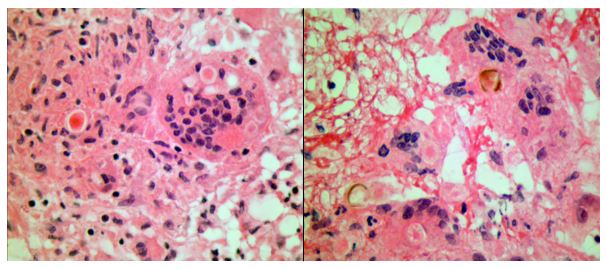
Frozen section evaluation during the time of surgery showed granulomatous inflammation. Septate hyphae were observed on the fungal smear. Following surgery, amphotericin was added. Histologic evaluation of the tissue submitted from surgery showed pyogranulomatous inflammation with pigmented, spore-like structures present in multinucleated giant cells on hematoxylin and eosin (H&E) stain (Image 2). Grocott’s methenamine silver (GMS) stain also highlighted short segments of septate hyphae (Image 3).
Cultures from the surgical debridement grew a mould with central pigmentation (Image 4). Direct microscopic examination of the mould revealed thick-walled, oblong conidia with 3-5 cells, and uniformly pigmented hyphae (Image 5). A germ tube test showed germ tubes originating from both ends of the conidia consistent with Bipolaris species.
Image 1. T1-weighted (left) and T2-weighted (right) magnetic resonance imaging of the brain demonstrating a left temporal lobe mass. Image 2. Hematoxylin and Eosin stained photomicrographs showing pyogranulomatous inflammation with giant cell formation and circular structures within them (left) (40x objective magnification). The right shows gold-brown pigmented structures within granulomatous inflammation (40x objective magnification). Image 3. Grocott’s methenamine silver stain highlighting short segments of irregular septate hyphae in the brain debridement specimen (10x objective magnification).Image 4. Mature wooly brown-black colony on potato dextrose agar. Image 5. Photomicrograph of a lactophenol blue tape prep of the mature fungal colony. Pigmented hyphae and short 3-4 cell conidia are readily identified (40x objective magnification). This specimen also tested germ tube positive (not shown), indicating that this dematiaceous fungus is Bipolaris spp.
The patient’s mental status significantly improved following surgical debridement, 2 weeks of liposomal Amphotericin B, as well as long term treatment with voriconazole. The voriconazole was later switched to posaconazole due to concerns for fluoride toxicity. He completed a year of posaconazole with significant improvement of the abscess observed on imaging and resolution of headaches with no other visual problems. He continued to recover cognitive function with some residual difficulty with reading, comprehension and speech that eventually resolved.
Discussion
Phaeohyphomycosis refers to infections caused by dematiaceous fungi that exist in a variety of forms when seen in tissues and commonly involves skin, soft tissue and nasal sinuses.1 In rare cases, central nervous system (CNS) involvement has been reported. CNS phaeohyphomycosis is predominantly seen in immunosuppressed patients; however, cases involving immunocompetent individuals do exist.2 In one case series from Houston, Texas, five of seven cases of cerebral mycosis were caused by a dematiaceous mould.3 Interestingly, the patient presented in this case came to medical attention around the Dallas-Fort Worth area of Texas.
Cladophialophora bantiana is the most common dematiaceous fungus associated with CNS phaeohyphomycosis, but rare cases of Bipolaris species have been reported previously in literature.4-6
We report a case of CNS phaeohyphomycosis by Bipolaris species following orthotopic liver transplant with an excellent patient outcome. This case is unusual, in part, because the typical hospital course of a patient with phaeohyphomycosis is generally dismal.7 The stories of successful treatment often involve complete debridement of discrete lesions.7-8 In our case, the patient underwent surgical debridement and treatment initially with liposomal Amphotericin B and later transitioned to long term therapy with newer azole antifungals.
References
Revankar SG, Sutton DA, & Rinaldi MG, (2004). Primary Central Nervous System Phaeohyphomycosis: A Review of 101 cases. CID, 38, 206-2016
Filizzola MJ, Martinez F, & Rauf SJ, (2003). Phaeohyphomycosis of the central nervous system in immunocompetent hosts: report of a case and review of the literature. Int J Infec Dis, 7, 282-286
Raparia K, Powell SZ, Cernoch P, Takei H, (2010). Cerebral mycosis: 7-year retrospective series in a tertiary center. Neuropathology, Jun; 30(3): 218-223.
Frank T, Esquenazi Y, Nigo M, Wanger A, Portnoy B, & Shepard S, (2016). Disseminated Phaeohyphomycosis with Brain Abscess and Biliary Invasion Due to Bipolarisspp. In an Immunocompetent Patient. Annals of Clinical & Laboratory Science, 46(4).
McGinnis MR, Campbell G, Gourley WK, & Lucia HL, (1992). Phaeohyphomycosis Caused by Bipolaris spicifera, An Informative Case. Eur. J. Epidemiol, 8(3), 383-386
Rosow L, Jiang JX, Deuel T, Lechpammer M, Zamani AA, Milner DA, Folkerth R, Marty FM, & Kesari S, (2011). Cerebral phaeohyphomycosis caused by Bipolaris spiciferaafter heart transplantation. Transpl Infect Dis, 13, 419-423.
Gadgil N, Kupfermen M, Smitherman S, Fuller GN, Rao G, (2013). Curvularia brain abscess. J Clin Neurosci, Jan;20(1): 172-175.
-John Markantonis, DO is a second year Clinical Pathology resident at UT Southwestern in Dallas. He has interests in Medical Microbiology and Transfusion Medicine.
-Dominick Cavuoti, DO is a Professor at UT Southwestern in the Department of Pathology. He is multifaceted and splits his time as the Medical Director of the Parkland Hospital Clinical Microbiology Laboratory and Parkland Cytology attending among other administrative and educational activities.
-Clare McCormick-Baw, MD, PhD is an Assistant Professor of Clinical Microbiology at UT Southwestern in Dallas, Texas. She has a passion for teaching about laboratory medicine in general and the best uses of the microbiology lab in particular.