Laying a Foundation for a Blended Curriculum Model

In the late 1990s—early 2000s—we were faced with a critical shortage of graduates in clinical/medical laboratory science, and we started evaluating the benefits of having our own program. Once we made the decision to seriously consider implementing our own program, I pitched a proposal for a blended model of curriculum delivery. My proposal was accepted by the Mayo Clinic School of Health Sciences as a pilot program to be supported by our newly created education technology center. Since our program and the education technology center were both new, we certainly experienced some growing pains. Also, had I known we’d have to reconstruct major elements of our online content every time we upgraded our content management system, I might have thought twice about it!

In the end, it was all worth it because today, we have an outstanding program built upon a solid foundation of both traditional and online content delivery that leverages our staff infrastructure and can be effectively managed and maintained over time.

In the curricular model developed for our academic program in Medical Laboratory Science (MLS), the didactic component is provided in an e-learning platform (Blackboard Learn) and is underscored by Transactional Distance Theory (Moore, 1991), in which the three modalities of learner interaction with content, instructor, and fellow students are integrated into the online module. Each lesson plan includes a laboratory module taught by traditional methods of interaction between the instructor and student in a classroom setting. A constructivist learning environment is facilitated, and each lesson plan is closely anchored in the context of the work the student will perform upon employment.

Here is a simple diagram of our curricular model:

mls-curricular-model_simple-diagram_1

The following learning theories define our program curricular model:

  1. Transactional Distance Theory (e-learning theory): The online lesson plan includes learner-content interaction, learner-learner interaction, and learner-instructor interaction.
  2. Constructivism: The roles of both the teacher and student are redefined in this educational model. The teacher moves away from the traditional role of “sage on stage” to that of a “facilitator” of the student’s acquisition of knowledge. The student becomes a more active learner in this model, moving away from the traditional role of passive learner.
  3. Anchored Learning:
  • Information is taught in the context of how the learner will apply it once he or she is working.
  • The online homework lesson correlates with a hands-on laboratory lesson designed to reinforce the e-learning content.
  1. Reversing the Lecture-Homework Paradigm (Moses, 2002): The traditionally taught lecture is provided as an online homework assignment.

Embracing technology has provided a means by which we can improve our teaching methods and promote change in our education infrastructure. More than half of our didactic courses in our MLS Program apply the new education strategy I learned about as “reversing the lecture-homework paradigm” (more commonly known as “flipping the classroom”). Instead of going to lecture, our students complete web-supported didactic modules asynchronously as “homework” assignments, allowing more classroom time for laboratory instruction.

By providing more hands-on laboratory lessons, we are giving our students the opportunity to practice laboratory procedures and apply new learning material in a way that corresponds more closely with what they will do for a living after they graduate. Instead of giving the typical “one-directional lecture” with limited opportunity for dialogue, our instructors are able to spend more time with our students, teaching practical applications of the content, answering questions, and helping problem-solve.

References

  1. Moore, M. G. (1991). Editorial: Distance education theory. American Journal of Distance Education, 5(3), 1-6.
  2. Moses, G. A. (2002). e-Technology must enable big education goals. Proceedings of the 2002 e-Technologies in Engineering Education (eTEE) Conference, Switzerland, Vol. P01, Article 20, 142-145.

Lehman_small

-Susan M. Lehman, MA, MT(ASCP)SM graduated from the University of Wisconsin-Madison in 1983 with a BS in medical technology. She is program director for the Medical Laboratory Science Program and course director for Clinical Microbiology I and II; her areas of interest include distance education and education methodology.

Laboratory Data and Global Health Security

Hello! If you’ve been following my posts these last few months you’ve seen my work here in the country of Sint Maarten regarding the Zika virus outbreak. I have been very fortunate to be involved at various levels of work with this new epidemic from lab testing and seroprevalence research to public health and policy initiatives. As 2016 comes to a close, I would like to celebrate that work by highlighting a few great milestones here and offer a few thoughts for the future starting next year.

I mentioned before that immunological lab testing has been ongoing, targeting IgM and IgG seroprevalence of Zika virus (along with other Arboviruses i.e. West Nile, Chikungunya, Dengue, and Yellow Fever etc) within the community around my medical school. Nearly two hundred samples later—and counting—the results point to a presence of nearly 10-15% of people are confirmed positive for an active (IgM) or resolved (IgG) infection with Zika. Despite the small sample size and ever-developing data sets, these numbers are highly suggestive of a correlation mimicking that presented to us by the Ministry of Health (see my Zika virus primer post from October of this year). This holds promise for future studies like these which depend heavily on that type of medical laboratory science and analytics. My involvement in the seroprevalence project has been very rewarding and I have been able to contribute my experience and skills as a laboratory professional in writing SOPs/protocols, ensuring proper laboratory safety, and highlighting specimen collection standards. With several years as a medical laboratory scientist, I have been lucky to find a place to do such good work. The opportunity to step up as a leader when it comes to laboratory skills is always fulfilling!

12_24-1

Figure 1: Seroprevalence research and testing publicity for the AUC campus community, collection and specimen processing. photo credit: A. Yancone, 2016.

Along with the success of the lab-driven research at school, the project I spend the most time with is aimed at public health. My last post highlighted some of the successes my team and I have had with creating and maintaining a good relationship between members of the local Ministry of Health and the school system. In February earlier this year, my school strengthened their partnership with the Ministry by establishing a mission of public health initiatives driven by students and faculty who were engaged in community affairs, service learning, and public health. This increased sustainability will surely create a more long-lived effort for translational research to have an impact on health. I couldn’t be prouder of the work my team has done to show the community that medical students here represent the very best ASCP qualities of advocacy, partnership, and outreach through science and education. It is an honor to have my contributions recognized by the local public health officials—even moreso when they have now incorporated and sponsored my teams’ message of vector control and source reduction to schools across the island. Having children aged 4-16 enthusiastically repeat back to us and representatives from the Ministry they would “throw standing water away!” made a strong impact on public health officials, students, and their parents/families. Setting goals for the community’s health metrics are supported by local epidemiologic data and pushed forward by policy initiatives, and always rooted in diagnostics and laboratory-confirmed cases. This means that all of our efforts, all of my presentations, and each step along the way holds a foundation in laboratory science and continues to motivate me as we continue forward. We all know the lab-week tag line that “nearly 70% of a patient’s chart is lab-data-driven,” and I can attest that for issues in public health and policy the same if not more applies!

12_24-2.jpg

Figure 2: American University School of Medicine Partners with Sint Maarten Ministry of Health, Social Development, and Labour to bolster public health efforts and community involvement. AUC 2016.

Last month, Sint Maarten’s Ministry of Health sent Dr. Virginia Asin, the section director of the Collective Prevention Services office, to a Global Health Security Meeting in Miami. In this meeting she presented the policy initiatives her country had in dealing with risk reduction in the Caribbean. As a leader in this field, she cited ongoing partnerships with NGOs, public health offices, and medical professionals. She also shared her strong support for a project she said was truly a foundation for success—my team’s public health outreach! Dr. Asin and other staff members from the Ministry were present at an end-of-the-quarter presentation and meeting regarding this community outreach work based from the school. She shared with us her thoughtful admiration and ongoing support for the work we do here and said she was proud to have us as colleagues. Strong words and poignant sentiment from a real public health physician working in the field. It is incredible to have these efforts noticed by the Global Health Security Agenda (GHSA)! The GHSA exists at the forefront of the CDCs efforts to address the spread of various diseases, the global preparedness for them, and their economic impact on the global community. Dr. Asin and the Ministry’s continued support continue to inspire me, as well as other students, to look toward the future in creating more opportunities to merge medicine, data analytics, and positive outcomes.

12_24-3

Figure 3: Global Health Security Agenda platforms. GHSA and the CDC 2016.

Slated to begin in January of next year, my team will begin conducting a new project aimed at improving local health literacy and source reduction. Under our IRB proposals, we hope to gather the data about the residents of this island and subsequently match them with correlated health statistics (i.e. confirmed/reported Zika cases) and actively engage with the Ministry in improving these numbers. With this current momentum, I would argue that even though a few successful highlights have come, the best is yet to come. If I have learned anything during my time as a medical laboratory scientist, there is a certain exciting progress to the dynamic way data is being used as we move forward. Breakthroughs in communication and analysis are allowing these advancements to take off: ASCP pathologists consulting on cases of chronic diseases in Africa, growing credibility surrounds our profession as our voice accomplishes great things in Washington, and advancing diagnostics are getting more efficient every day. Hopefully, projects like the ones I keep all of you updated on will continue to strengthen the connections between the labs, media, and public health officials as we continue to improve outcomes globally.

Until next time! Happy Holidays and Have a Happy New Year!

(…and don’t keep standing water around if you live in a warmer area, because…vector control!)


ckanakisheadshot_small

-Constantine E. Kanakis MSc, MLS (ASCP)CM graduated from Loyola University Chicago with a BS in Molecular Biology and Bioethics and then Rush University with an MS in Medical Laboratory Science. He is currently a medical student at the American University of the Caribbean and actively involved with local public health.

Microbiology Case Study: A 50 Year Old Male with High Fevers and Chronic Cough

Case History

A 50 year old male of Indian descent presents to the pulmonary clinic with complaints of high fevers and chronic cough. The cough has persisted for the past month and recently became productive with green sputum.  His fevers are cyclic in nature and reach 104°F.  He denies hemoptysis, unintentional weight loss or chest pain.  He has tried over the counter decongestants and cough suppressants as well as a course of levofloxacin, with minimal improvement. His past medical history is significant for rheumatoid arthritis, which is currently treated with methotrexate and prednisone. He works as a long distance truck driver and is a non-smoker.  A recent chest x-ray demonstrated a left hilar opacity with a nodular appearance. A computed tomography scan of the chest shows focal consolidation of the left lower lobe along with mediastinal and hilar adenopathy. Also, there are innumerable non-calcified nodules seen throughout bilateral lung fields. A bronchoscopy with bronchoalveolar lavage (BAL), transbronchial biopsy, and fine needle aspiration (FNA) of the enlarged lymph nodes were performed. BAL fluid was transported to the microbiology lab for bacterial, fungal and mycobacterial cultures.

Laboratory Identification

cocimm1

Figure 1. Histologic evaluation of the lung biopsy showed diffuse necrotizing granulomas which contained large yeast-like forms (red arrow) (H&E, 100x).

cocimm2

Figure 2. The large yeast-like forms measured between 10-25 µm in size and demonstrated a thick walled capsule (Grocott’s methenamine silver (GMS), 600x).

cocimm3

Figure 3. White mycelium with a downy texture and faint brown reverse grew on Mycosel agar after 28 days of incubation at 25°C.

cocimm4

Figure 4. Numerous coarse, septate hyphae producing thick walled, “barrel shaped” arthroconidia (lactophenol cotton blue stain, 1000x oil immerson).

Histology of the lung biopsy specimen showed necrotizing granulomas with occasional large, yeast-like spherules which measured between 10-25 µm in diameter (Figures 1 & 2). The spherules had a thickened capsule and endospores were not visualized. The fungal BAL cultures grew a white mycelium with a downy texture and light brown reverse after incubation for 28 days at 25°C on Mycosel agar (Figure 3). Microscopic morphology of a lactophenol cotton blue prep illustrated alternating thick walled arthroconidia suggestive of Coccidioides immitis/posadasii (Figure 4). The dimorphic mold was confirmed by DNA probe testing. Due to the findings on histology and the unusually slow growth of this particular isolate, Coccidioides IgM and IgG antibodies were performed by ELISA in the interim. They were found to be 5.4 and 4.4, respectively, suggestive of a current or recent infection. Laboratory studies for Aspergillus galactomannan, Fungitell, Cryptococcus antigen, and Histoplasma & Blastomyces urinary antigens were all negative. A Quantiferon Gold for Mycobacterium tuberculosis and all other cultures were also negative.

Discussion

Coccidioides immitis is often considered a thermally dimorphic mold geographically distributed to the arid climate of the southwestern United States and Mexico. It is morphologically identical to the C. posadasii, a species which is more widespread and endemic in South America. The two species can only be differentiated by molecular methods, although it is not routinely necessary as there is no difference in symptoms and treatment between the two.

Inhalation of infectious arthroconidia occurs as a result of environmental exposure to dust, sand and soil that has been disturbed. While many immunocompetent individuals who are exposed to C. immitis will show mild flu-like symptoms which resolve with no treatment, a portion of patients will go on to have pulmonary disease. A severe disseminated infection can occur in individuals with underlying immune system disorders, including rheumatologic diseases, HIV and transplant recipients on immunosuppression. C. immitis can have direct invasion of adjacent structures and can cause eruptive chronic granulomatous cutaneous disease. Women who are diagnosed with Coccidiomycosis during pregnancy are also at high risk for disseminated disease due to the presence of estrogen-like receptors in the fungus.

In the environment and when cultured in the laboratory at 25°C, C. immitis grows as a hyphal mold with alternating barrel-shaped arthroconidia (3-6 µm) separated by disjunctor cells. The arthroconidia are highly infectious and cultures in the laboratory must be worked up in a biological safety cabinet to minimize the risk of accidental exposure. The yeast-like phase occurs in tissue at temperatures above 35°C and is characterized as the transformation of arthroconidia in to large spherules (10-80 µm) which become filled with endospores. The cell wall of the spherule ruptures and the endospores are released into the tissue to become additional spherules.

Most patients with primary Coccidiomycosis do not require specific therapy as the disease will resolve on its own. For those patients who are immunocompromised, or whom exhibit severe disease, treatment is amphotericin B followed by fluconazole or itraconazole as maintenance therapy. In the case of our patient, he was placed on oral fluconazole twice daily for at least 3 months.


ka

-Kristen Adams, MD, is a fourth year Anatomic and Clinical Pathology resident at the University of Mississippi Medical Center. 

Stempak

-Lisa Stempak, MD, is an Assistant Professor of Pathology at the University of Mississippi Medical Center in Jackson, MS. She is certified by the American Board of Pathology in Anatomic and Clinical Pathology as well as Medical Microbiology. She is the director of the Microbiology and Serology Laboratories.  Her interests include infectious disease histology, process and quality improvement and resident education.  

Lonely Life of a Clinical Pathologist: Thank You

I wanted to say thank you to everyone who has left comments on my past posts and shown encouragement to the topics discussed. I will be taking a break from blogging but wanted to encourage everyone in the clinical pathology field to keep up the hard work of patient care behind the scenes. I hope you can be ambassadors of laboratory services and help influence the care of patients in positive ways throughout the hospital systems you work in.

In lieu of a holiday card, I wanted to sign off blogging with a meme for all clinical pathologists out there:

clin-path-meme


racsa_small

-Lori Racsa, DO, is the director of microbiology, immunology, and chemistry at Unity Point Health Methodist, and a Clinical Assistant Professor at the University Of Illinois College Of Medicine at Peoria. While microbiology is her passion, has a keen interest in getting the laboratory involved as a key component of an interdisciplinary patient care team.

Microbiology Case Study: A 60 Year Old Woman with a Skin Papule

Case history

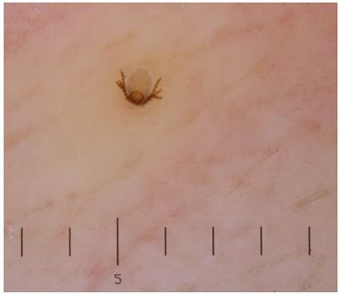
A 60-year old woman residing in Vermont presented to the dermatology clinic for a routine annual skin exam. She had no complaints. On physical exam, a pink papule was seen on the patient’s back, with a centrally embedded tick (Figure 1). The tick was removed and sent for identification, with the plan to give a single prophylactic dose of doxycycline if identified as Ixodes scapularis.

Laboratory Identification

The tick was examined and noted to be less than 1 mm in size, with six legs (Figure 2). This was identified as the larval stage of Ixodes scapularis.

ixo1

Figure 1. Photograph of Ixodes scapularis larva embedded in the patient’s back. Six legs are visible.

ixo2

Figure 2. Photograph of the tick larva received in the laboratory, demonstrating a light tan-brown color.


Discussion

I scapularis, also known as the blacklegged tick, deer tick, or bear tick, is most clinically significant for its ability to transmit the pathogens Borrelia burgdorferi, Babesia spp., and Anaplasma phagocytophilum. It has four separate life stages (egg, larva, nymph, and then adult), spanning approximately 2 years. Each of these stages feeds on different preferred host animals.

Eggs are deposited on the ground by blood-engorged females in the late spring, where they subsequently hatch into 6-legged larvae. Because they have not yet fed, larva forms generally do not carry or transmit B. burgdorferi or other tick-borne pathogens. Trans-ovarial transmission of Borrelia, Anaplasma, or Babesia from adult I. scapularis females to eggs of is not a significant mode of pathogen transmission; however, in a similar tick species, I. ricinus (prominent in Europe), trans-ovarial transmission of Babesia divergens does occur, and so infection may be transmitted by larvae. The I. scapularis larvae will take their first blood meal from small mammals and birds, and then when engorged fall to the ground and molt into nymphs.

The nymph forms, which have already taken a blood meal, can carry pathogens and in fact are more likely to transmit pathogens to humans than the adult form of the tick. This is because the nymph form is much smaller (<2mm in size) than the adult form, and therefore is likely to go undetected when it attaches to a host. The nymphs are dormant over the winter, and re-activate the following spring to take their second meal. By fall, nymph forms have molted into adult ticks, which prefer to feed on white-tailed deer. However, while these deer support the tick population, they are not a large reservoir for Lyme disease. Rather, it is the white-footed mice preferentially fed upon by larvae and nymph forms that act as the main reservoir for B. burgdorferi, B. microti, and A. phagocytophilum. The female adults of I. scapularis are red to orange and larger than males, around 1/8 of an inch long, with a dark brown to black dorsal shield. If females do not feed in the fall, they can remain dormant over the winter and may emerge if the weather gets temporarily warmer (so the onset of cold weather does not necessarily mean the risk of tick exposure is over). Male adults do not take blood meals, and so do not transmit blood-borne pathogens.

To be considered in the differential diagnosis is Dermacentor variabilis, or the American dog tick. This tick species is larger than Ixodes spp., and adult forms have a white-to-gray collar on their backs. D. variabilis have more rectangular-shaped head and mouth parts than the deer tick. Both nymph and larvae forms are yellow-brown in color before feeding, and then turn gray once engorged. It is extremely uncommon for nymph and larval forms of D. variabilis to feed on humans, in contrast to I. scapularis. D. variabilis does not transmit Lyme disease, though in endemic areas it may transmit Rickettsia rickettsii or Francisella tularensis.

Because the tick in the presented case was identified as an I. scapularis larva, the patient was not treated with antibiotics as there was an exceedingly low risk of pathogen transmission.


-Alison Krywanczyk, MD is a 3rd year anatomic and clinical pathology resident at the University of Vermont Medical Center.

Wojewoda-small
-Christi Wojewoda, MD, is the Director of Clinical Microbiology at the University of Vermont Medical Center and an Assistant Professor at the University of Vermont.

Toilet Paper Safety

As the years have passed, I have noticed many changes in the toilet paper dispensers in the healthcare setting. First there was the standard rolling style. This was great- you could get as much paper as you wanted, and the only issue was whether or not the roll was installed properly (I prefer “over”). The next style to come along was the bumpy roller. As it rolled out, the lop-sided holder would cause tissue to rip off before the user was ready. Then came covered paper holders that forced the user to reach under a sharp edge for paper access. The latest version I have seen completely covers the roll leaving a tiny access port that allows one piece of paper to be ripped off at a time.

As each dispenser style was replaced by a newer, more insidious model, I began to wonder why hospitals were being so cheap with the paper. Was it being stolen often? Was there a black market for toilet paper? Did the hospitals understand that each newer model forced staff to remain in the rest rooms for longer periods of time in order to get an adequate tissue supply? Surely this was affecting productivity in the work place. Clearly I had given this too much thought, and I let it bother me. I learned this year, however, that I was wrong about the topic for many years.  I found out that in hospital rooms with patients under contact precautions (such as patients who have contracted C. difficile) all of the open paper products must be discarded. In fact, it is a common practice to dispose of any open tissue when any patient room is cleaned. This latest dispenser designs prevents the wasting of paper and actually saves money. Once I received education about the issue, I had a better mindset about the tissue issue.

This is often true with laboratory safety, and providing the necessary education can truly improve safety compliance. There are many who have worked in the lab setting for years, and some have ignored safety regulations while others have followed them grudgingly. Often, the staff approach to lab safety can be improved with basic knowledge; information about the regulations, leadership expectations, and potential consequences of non-compliance.

I approached a lab manager about the need for his staff to utilize face protection when pouring chemicals. He said he was not aware of the need, and it would be an enormous change for the staff. We had a discussion about OSHA’s Chemical Hygiene standard and the Bloodborne Pathogen standard, both of which require face protection when handling open specimens and chemicals. Once he knew this and could also locate it in the safety policies, he immediately covered the information with his staff and compliance was improved. In this case, simple knowledge of the regulations was enough reason for the lab safety to be improved. Knowing the reason why is an important motivator for lab staff.

Lab leaders can make a strong impact on PPE compliance both by voicing expectations with staff and by being a good role model. If you lead lab safety, talk to new employees about what is expected, and regularly remind current staff about the safety policies that are to be followed. Every successful leader also has to be a positive role model. If you expect certain safety practices to be followed, you need to make sure you follow them when you are in the lab as well. A safety professional that walks through the lab in mesh sneakers is going to have a (pardon the pun) paper-thin positive impact on the overall culture.

Some long-term lab employees who regularly comply with safety regulations do so because they have learned an unfortunate lesson. Lab staff that has been the victim of an exposure or injury knows the consequences, and sometimes the cost has been very high. Exposures from an unknown source, for example, can result in treatments that cause illness and that will interfere with personal lives. An exposure that results in contracting an illness or a career-altering injury can be devastating. Our goal as lab safety professionals should be to get staff to comply with regulations proactively, rather than as a response to an incident. Teaching about potential consequences often can have an impact on safety behaviors. You may be surprised at how little laboratorians (and lab leaders) may think of the effects of poor safety conduct. Use real life incidents to tell stories and discuss other possible bad outcomes of non-compliance.

As the average age of laboratory professionals in the country continues to rise, we may be working with some folks who have had the same weak safety mindset for quite some time. They remember the days of eating, drinking and smoking in the lab, and they don’t understand why all of these rules are now in place. They’re healthy today, aren’t they? It’s time to change that way of thinking. It’s time to explain that while they may have practiced unsafe behaviors without incident, it just means they were lucky, not smart. Getting staff to think about the regulations, the expectations, and the consequences will help them to have a new and positive mindset about the lab safety issue.


Scungio 1

-Dan Scungio, MT(ASCP), SLS, CQA (ASQ) has over 25 years experience as a certified medical technologist. Today he is the Laboratory Safety Officer for Sentara Healthcare, a system of seven hospitals and over 20 laboratories and draw sites in the Tidewater area of Virginia. He is also known as Dan the Lab Safety Man, a lab safety consultant, educator, and trainer.

Choosing Wisely

Having recently completed the 2016 survey regarding topics to be considered in the updated ASCP Choosing Wisely campaign, there were several thoughts that came to mind.

First, I applaud the ASCP for taking the initiative to participate early on with Choosing Wisely and now to review and update these elements. As we all know, as the evidence and needs of our patients change, so must we. We should always strive to maintain the “latest and greatest” particularly given the continued growth of laboratory medicine. Identifying appropriate and necessary laboratory utilization shows our concern as individual practitioners and as a professional society. Our involvement clearly influences patient outcomes and we must remain diligent about putting forth this message.

Secondly, I will admit that there were numerous tests and the questions regarding their usefulness listed in the Survey that were completely unfamiliar to me. This highlighted how lab menus are expanding and many of these were subspecialty oriented, pointing out how intricate and complicated our scope of laboratory practice has become. With the multitude of tests available and their complexity, is it any wonder our clinical colleagues suffer the same, and I venture to say even MORE, angst than we face given this array of testing options? Therefore it is even more important, then, that we as a Society continue to be about the forefront of laboratory initiatives, educating ourselves and helping those healthcare providers that live outside “our walls.”

Of course, the Choosing Wisely campaign is also focused on patients and the information needed for solid patient-centered decision-making. It is thus incumbent upon us to participate in this national campaign, for who better than laboratory professionals to help navigate these waters on the behalf of patients?

The onus is upon us, then, to pursue this initiative and provide guidance regarding laboratory utilization, appropriate testing, testing intervals, adequate test menus and laboratory interpretation. The ASCP participation in the Choosing Wisely campaign allows us to solidly insert our expertise in this arena. Each of us, as members, is responsible for our participation, not just in this Survey, but on an ongoing daily basis as part of the patient care team.


Burns

-Dr. Burns was a private practice pathologist, and Medical Director for the Jewish Hospital Healthcare System in Louisville, KY. for 20 years. She has practiced both surgical and clinical pathology and has been an Assistant Clinical Professor at the University of Louisville. She is currently available for consulting in Patient Blood Management and Transfusion Medicine. You can reach her at cburnspbm@gmail.com.

Microbiology Case Study: A 21 Month Old Boy with Diaper Rash and Diarrhea

Case history

The patient was in his otherwise healthy state of being until 3 days prior when he developed non-bloody diarrhea. On the morning of presentation the stool had become bloody. The patient was afebrile, had some reduced intake of food, but drinking fine. Most notably, he periodically stops walking and bends over as if he is in pain. This happened 3-4 times the previous day and these episodes tended to last for about one minute, after which the boy would continue to play. He does not attend daycare and his immunizations are up to date.

Stool culture was sent and a predominant organism was an oxidase negative, lactose-fermenting, Gram-negative rod (Figure 1). The organism was non-sorbitol fermenting based on growth on Sorbitol-MacConkey agar, and grew as mauve colonies on E. coli 0157 screening agar. MUG testing was negative.

O157.png
Figure 1: Subculture of the disease causing organism on (A) MacConkey, (B) Sorbiol-MacConkey, (C) and E. coli O157 screening agars.

Discussion

The isolate was E. coli O157. Isolates of E. coli O157 commonly produce shiga toxins (sxt1 and sxt2) which are responsible for diarrhea, hemorrhagic colitis, and most famously hemolytic-uremic syndrome (HUS). Typical illness starts with non-bloody diarrhea which becomes bloody after 2-3 days due to onset of hemorrhagic colitis. Often severe abdominal pain and low grade fever are present as well. HUS is a serious complication of E. coli O157 infection which results in acute renal dysfunction. HUS most often occurs in children < 5 years of age, of which 15% of those with laboratory confirmation of E. coli O157 developing this complication, compared to 6% in the general population. It is possible for other E. coli to produce shiga toxins, with 1% of HUS is caused by non-E. coli O157 infection.

It is recommended that all patients with suspected HUS should have stool cultured on selective and differential media for detection of E. coli O157 and direct shiga toxin detection should be performed to identify non-E. coli O157 isolates that are producing toxin. E. coli 0157 isolates look exactly the same as non-E. coli O157 normal fecal flora on 5% sheep blood, chocolate, and MacConkey agars. All E. coli ferment lactose on MacConkey agar (Figure 1A). E. coli O157 can be differentiated from other E. coli strains by growth on Sorbitol-MacConkey (SMAC) agar; E. coli O157 is a non-sorbitol fermenter while most other E. coli will ferment sorbitol (Figure 1B). Chromagenic agar for E. coli O157 is another option to screen stool specimens for E. coli O157. E. coli O157 grow mauve colored colonies on this particular agar (BBL CHROMagar O157 , Becton Dickinson) (Figure 1C). A summary of this data can be found in Table 1.

Growth of organisms suspicious for E. coli O157 on any media requires confirmation prior to reporting. Biochemical confirmation tests include E. coli O157 antiserum or latex agglutination and 4-methylumbelliferyl-beta-D-glucuronide (MUG) testing. For latex agglutination or antisera testing, it is essential to test the isolate of interest with the E. coli O157-specific reagent as well as a non-specific control to exclude non-specific binding. Unlike most E. coli strains, E. coli-O157 does not express beta-glucuronidase and is therefore MUG test negative (Table 1).

Table 1. Characteristics of E. coli O157 in comparison to other E. coli strains

Test Non-E. coli O157 E. coli O157
Appearance on MacConkey agar Lactose fermenter Lactose fermenter
Appearance on Sorbitol-MacConky agar Sorbitol fermenter Non-sorbitol fermenter
MUG testing Positive Negative

For direct detection of shiga toxin, there are several commercially available immunoassays available for detection of shiga toxin protein. New on the market are multiplex gastrointestinal panels that can be used for molecular based detection of shiga toxin genes sxt-1 and sxt-2 among a host of other agents of gastrointestional disease.

E. coli O157 is spread via fecal oral route. It can be acquired directly from person to person or indirectly through food and water sources contaminated with fecal matter from infected humans and animals. Classic scenarios are undercooked ground beef, leafy greens, unpasteurized milk and juice, petting zoos, and contaminated drinking water. The incubation period prior to symptoms is 3-4 days (range 1-8 days).

Treatment for E. coli O157 is largely supportive consisting of fluids to prevent dehydration. The role of antibiotics is controversial with some studies suggesting antibiotics increase the risk of developing HUS while others found no association between the their use and increased HUS.

Following our patient’s stool culture result for E. coli O157, he was recalled to the Emergency Department for evaluation. He was still having diarrhea and vomiting, but it was reduced compared to the previous day. The patient was given fluids and sent home without antibiotic treatment and via phone conversation with his mother, his symptoms resolved a few days later.

-Erin McElvania TeKippe, PhD, D(ABMM), is the Director of Clinical Microbiology at Children’s Medical Center in Dallas Texas and an Assistant Professor of Pathology and Pediatrics at University of Texas Southwestern Medical Center.

Common Thrombotic Microangiopathies

Primary thrombotic microangiopathy (TMA) syndromes encompass diseases that present with thrombosis in small and medium sized blood vessels due to endothelial injury. They are specific disorders that require specific treatment. The initial assessment is focused on confirming that the patient has true microangiopathic hemolytic anemia (MAHA) with or without thrombocytopenia. If MAHA and thrombocytopenia are confirmed it is important to differentiate the primary etiologies, which include:

  1. Thrombotic thrombocytopenic purpura, or TTP, which occurs as a result of severe ADAMTS13 deficiency
  2. Atypical Hemolytic uremic syndrome, or aHUS, which occurs as a result of complement dysregulation
  3. Hemolytic uremic syndrome, or HUS, which occurs as a result of Shiga toxins

TTP results from a severe deficiency of ADAMTS13 (defined as activity <5-10%). ADAMTS13 (a disintegrin and metalloproteinase with a thrombospondin type 1 motif, member 13) is an enzyme that cleaves large von Willebrand multimers. TTP can be hereditary (called Upshaw-Shulman syndrome) but is usually acquired as a result of an inhibitory autoantibody toward ADAMTS13. The decision to treat TTP is usually clinical since ADAMTS13 assays are often not available and may take several days to return a result if sent to a reference laboratory. TTP typically has more systemic manifestations of organ injury compared to other primary TMA syndromes.

Atypical HUS (aHUS) occurs via a complement-mediated pathway most commonly due to gene mutations of complement factors. Patients with antibodies to complement proteins comprise a smaller proportion of aHUS patients. Penetrance of aHUS is low with only a fraction of gene mutation carriers developing the syndrome. In most cases, a complement activation trigger precedes the manifestation of aHUS.

Most cases of HUS are sporadic, resulting from Shiga toxins produced by Shigella dysenteriae and some serotypes of Escherichia coli (especially O157:H7 and O104:H4). Shiga toxins preferentially injure the renal system by binding to CD77 on kidney epithelial and mesangial cells and endothelial cells. This binding causes downstream ribosomal inactivation leading to programmed cell death (apoptosis). The sporadic form of HUS is associated with bloody diarrhea.

The rationale for focusing on the distinction between HUS, aHUS and TTP is due to the different treatment options for each disease. TTP requires urgent plasma exchange to prevent death, while HUS requires treatment of the infection with little benefit from plasma exchange, while aHUS requires treatment with specialized anti-complement medication. Plasma exchange has inherent risks, so determining the cause of TMA is crucial. While no single clinical feature can be used to determine whether TTP, aHUS, or HUS is responsible for a patient’s symptoms, distinguishing factors include:

  1. Patient age – any primary TMA syndromes may occur at any age; however, children typically present with HUS, aHUS, or hereditary TTP, while adults more commonly present with acquired TTP
  2. Kidney injury –the degree of injury is usually less in TTP than in HUS; aHUS affects the arterioles while HUS tends to affect the glomeruli
  3. Systemic symptoms – up to two-thirds of TTP patients will have some neurologic symptoms; overtly bloody diarrhea is more typical for HUS; aHUS primarily involves kidney injury
  4. A previous episode or family history of aHUS – indication for screening for complement dysregulation in aHUS


Want to learn more? Check out: George JN, Nester CM. Syndromes of thrombotic microangiopathy. N Engl J Med. 2014;371(7):654-66.


Rogers

-Thomas S. Rogers, DO is a third-year resident at the University of Vermont Medical Center, a clinical instructor at the University of Vermont College of Medicine, and the assistant medical director of the Blood Bank and Transfusion Medicine service.


Microbiology Case Study: A 73 Year Old Man with Altered Mental Status and Fever

Case History

A 73 year old man was brought to the emergency room with altered mental status and fever, which developed a few days following a 1-2 day illness characterized by myalgia and diarrhea. He was admitted to the hospital and blood cultures were drawn.

Laboratory Identification

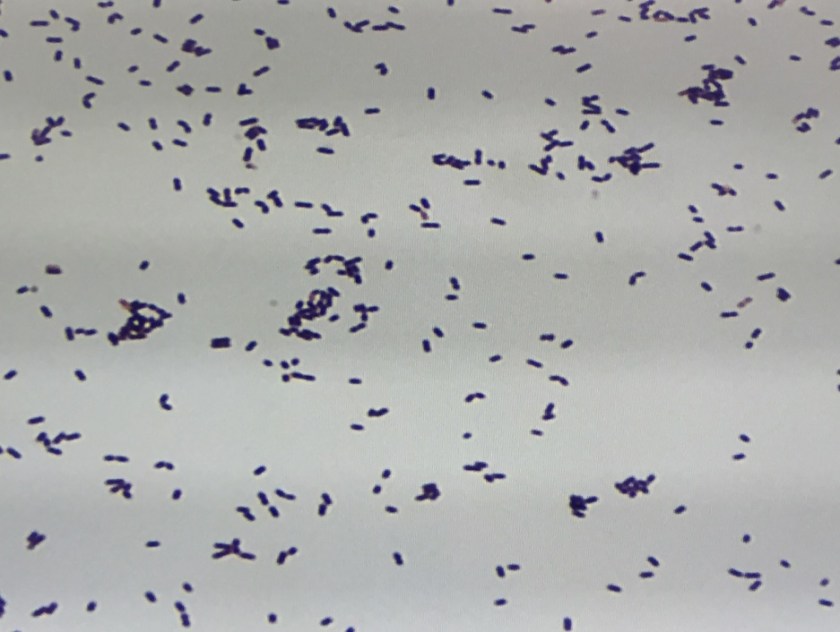
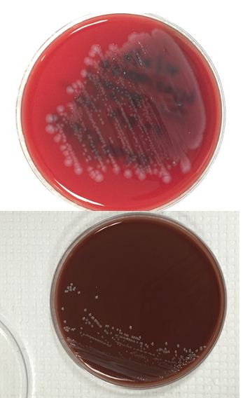
The bottles flagged positive after 12 hours and Gram stain showed small, Gram positive rods (Figure 1). Growth of white, smooth translucent colonies was seen on the blood and chocolate plates, with a small rim of beta-hemolysis on the blood plate (Figure 2). MALDI-TOF confirmed the identification as Listeria monocytogenes.

list2

Figure 1. Gram stain morphology of the colonies growing, demonstrating short Gram positive bacilli.

 list1

Figure 2. Smooth white colonies growing on the blood and chocolate plates, with a soft rim of beta-hemolysis visible on the blood plate.

Discussion

Listeria monocytogenes is prevalent throughout the environment, and can also colonize the human gastrointestinal tract. Humans are exposed by consumption of contaminated food, particularly soft cheeses, deli meats, and fruit. Listeria can grow at 4C which means it can multiply in refrigerated foods, making even low-level contamination a potential hazard. On gram stain, it is a short gram positive rod which may form chains. In some cases, the rods may be so short as to resemble chains of Streptococci, and with the soft surrounding beta hemolysis, could potentially be confused for Group B Streptococcus. However, Listeria is catalase positive, while Group B Strep is negative. Another characteristic feature of Listeria is the “tumbling motility” on wet prep at 20-25C, or “umbrella motility” in tube agar. Listeria also has the unique feature of manipulating the host cells’ intracellular actin framework, using it to facilitate direct cell-to-cell spread of the bacteria. The main virulence factor is the listeriolysin toxin, which is postulated to permit survival of the organism within macrophages via cytotoxic activity.

Listeria can cause a self-limited febrile gastroenteritis in previously healthy individuals, but typically only if they consume a large inoculum. However, in neonates, the elderly, or the immunosuppressed, it can invade and cause sepsis, meningitis, or meningoencephalitis. In pregnant women, Listeria can cross the placenta and lead to intrauterine fetal demise, premature labor, or neonatal meningitis, as well as the typically fatal condition granulomatosis infantiseptica in which the newborn develops widespread abscesses throughout multiple organ systems. Infection during pregnancy usually happens during the 3rd trimester, though the effects seem to be more severe with earlier infection.

 Listeria has been cultured from the stool of up to 3.4% of healthy, asymptomatic humans, and so there is little utility in stool cultures for Listeria except for epidemiologic purposes during an outbreak. Infections due to outbreaks of Listeria are far less common than sporadic infections, which comprise 95% of Listeria infections. Additionally, traditional stool cultures are poor at detecting Listeria and selective media is usually required. Blood and cerebrospinal fluid are the preferred sites of culture if there is suspicion for disseminated infection. Meningitis caused by Listeria is unique in that is can cause a lymphocyte-predominant CSF pleocytosis, which may result in confusion for viral meningitis. Additionally, gram stains of the CSF are only positive in approximately 1/3 of patients, so a high index of suspicion needs to be maintained while awaiting final culture results. While antibiotic treatment is not recommended for otherwise healthy patients with febrile gastroenteritis, it is recommended for those with disseminated infection or at high risk of dissemination (i.e. extremes of age, immunocompromised, or pregnant).


-Alison Krywanczyk, MD is a 3rd year anatomic and clinical pathology resident at the University of Vermont Medical Center.

Wojewoda-small

-Christi Wojewoda, MD, is the Director of Clinical Microbiology at the University of Vermont Medical Center and an Assistant Professor at the University of Vermont.