In the peak of the flu season we might see many people wearing masks in physician offices and hospitals. In the news today, as the 2019 Novel Coronavirus (SARS-CoV-2) continues to spread, we see many images of people wearing different types of respirators, some are N95 respirators and others are surgical masks. Not all experts agree on the efficacy of these pieces of personal protective equipment in the face of viruses, but if you’re going to use them, it is important to know how, when and why.
OSHA’s Respiratory Protection standard (1910.134) provides information about requirements for staff who may potentially be exposed to airborne pathogens. These requirements include specific instructions for choosing the proper respirator, for providing fit-testing, and for user training. The College of American Pathologists (CAP) also expects labs to determine the risk of airborne pathogen exposure for each employee, and they require labs to have a plan which outlines engineering and work practice controls that reduce exposure potential.
The purpose of a respirator is to protect the employee from contaminated or oxygen-deficient air. Therefore, two classes of respirators are common; air-purifying respirators which use filters to remove contaminants from the air you breathe, and atmosphere-supplying respirators which provide clean air from an uncontaminated source. These types of respirators can also be classified further as tight-fitting or loose-fitting. Tight-fitting respirators need a tight seal between the respirator and the face and/or neck of the user in order to work properly. For now, let’s focus on the air-purifying respirators which are in high demand these days as a potential 2019-nCoV pandemic looms.
In the laboratory, N95 respirators are probably the most commonly-used respirators, often used for protection against tuberculosis and other airborne pathogens. These respirators filter out 95% of airborne pathogens that are 0.3 microns or larger. While the exact size of the 2019-nCoV is not yet known, most coronaviruses are slightly larger than 0.1 microns. Does that mean a N95 respirator (recommended by the CDC) will not offer protection from the coronavirus? Not necessarily.
According to biosafety expert Sean Kaufman (www.saferbehaviors.com), the filter in the N95 respirator works three ways- through interception, impaction, and diffusion. Interception collects larger particles which are blocked by mask fibers, and impaction collects larger particles which have too much inertia to be moved around the filter fibers. Diffusion occurs as smaller particles are bombarded with larger air molecules and are pushed against filter fibers. Most of the bacteria or virus particles are removed from the airstream making the respirator quite useful and protective (HEPA filters on a Biological Safety Cabinet work in much the same way).
Employees who may need to wear a tight-fitting respirator as part of their job are required to have fit-testing every year. This is required by OSHA, and contracted employees (such as pathologists) should be fit-tested as well. Employees who may need such respirators would be those who work in microbiology labs, cytology techs who participate in patient procedures, and others. Labs should perform a risk assessment for each job category to determine the type and level of potential harmful airborne exposure.
Procedure masks, such as those handed out when people suspect they have the flu, are not technically considered respirators. Often, the person who is sick will wear these masks in order to prevent the spread of droplets when coughing or sneezing. They can protect others in the area, but they do not protect the user from harmful airborne pathogens or vapors.
Can these surgical masks be useful for the healthy public when a coronavirus is present? Sean Kaufman says “yes. If you wear a surgical mask in a potentially contaminated environment (on a commuter bus, for example),” Kaufman says, “it can keep you from touching your nose or mouth- two major routes of entry for viruses. Behaviorally speaking, these masks do offer some protection.”
Knowing when and why you use a respirator is vital, but knowing how to use it is important as well. Tight-fitting respirators should never be used without fit-testing to make sure the correct size is being used. Otherwise, the protection offered will be limited. Make sure your staff is properly trained and protected to work in environments where the air is not safe to breathe, and teach others about the usefulness of respirators when the flu and other viruses are lurking!
–Dan Scungio, MT(ASCP), SLS, CQA (ASQ) has over 25 years experience as a certified medical technologist. Today he is the Laboratory Safety Officer for Sentara Healthcare, a system of seven hospitals and over 20 laboratories and draw sites in the Tidewater area of Virginia. He is also known as Dan the Lab Safety Man, a lab safety consultant, educator, and trainer.
On February 4th, the FDA announced an Emergency Use Authorization for the CDC’s 2019 Novel Coronavirus real-time RT-PCR Diagnostic Panel. Here’s the press release:
Audience: Clinical Laboratory Professionals
Subject: Laboratory Update: Information about Emergency Use Authorization for 2019 Novel Coronavirus (2019-nCoV) Real-Time RT-PCR Diagnostic Panel
Level: Laboratory Update
This message is to ensure that clinical laboratories are aware that CDC has developed a new laboratory test kit called the CDC’s 2019 Novel Coronavirus (2019-nCoV) Real-Time RT-PCR Diagnostic Panel, for use in testing patient respiratory specimens for 2019-nCoV. On February 4, 2020, the Food and Drug Administration (FDA) issued an Emergency Use Authorization (EUA) to enable emergency use of the test kit in the United States. All EUA documents are available on the FDA website.
The test kit will be available for ordering today from the International Reagent Resource (IRR). Formerly, U.S. diagnostic testing for 2019-nCoV was only being conducted at CDC; however, the FDA EUA and distribution of the tests will allow 2019-nCoV testing to take place at laboratories designated by CDC. This includes U.S. state and local public health laboratories and Department of Defense (DoD) laboratories.
Clinical laboratories should NOT attempt viral isolation from specimens collected from 2019-nCoV persons under investigation (PUIs). For interim guidelines for collecting, handling, and testing clinical specimens from PUIs for 2019-nCoV, please see the CDC 2019 Novel Coronavirus website.
The FDA website lists current EUA assays, and also includes a link to terminated EUA assays. Each pathogen-specific EUA includes the device-specific Letter of Authorization, fact sheets, and manufacturer instructions/package inserts. These documents are updated when amendments are made (e.g., additional specimen types, extraction methods, procedural clarifications), so check the website routinely to ensure your laboratory staff members have the most up-to-date information.
Register for CDC Health Alert Network (HAN) notifications, including updates about the 2019 Novel Coronavirus. Enter your email address, search for HAN, and sign-up.
If you have any questions, please contact LOCS@cdc.gov.
Medical school councilors have good intentions in mind when they steer medical students who realize that direct patient care isn’t their strong suit into pathology. But I am different kind of pathologist – the one who sees (or talks to) patients every day. I am a member of unique subspecialty – Transfusion Medicine – which is the most patient-centric subspecialty of all pathology subspecialties. And, contrary to the popular wisdom, I like seeing patients.
Don’t get me wrong though, my heart and soul still live in the lab, deeply rooted in understanding test performance, troubleshooting and quality control. But direct patient care helps to put all the work I have done in the lab into a perspective.
One program that became especially dear to my heart is our chronic RBC exchange program for the kids and adults with sickle cell anemia who have high risk of developing serious complications from the disease, such as stroke, acute chest syndrome, and severe iron overload. As an apheresis physician I see these patients quite frequently due to the nature of the program – chronic RBC exchanges every 4 to 6 weeks. This also means that I quickly had to learn quite a lot not only about managing the exchanges, but also about patients’ success and failures, spend time explaining to parents the benefits of the program and engaging them to maintain compliance with rigorous schedule. The work is not immediately rewarding. All the adjustments I do to the plan of care show changes in lab values in a month or two at best. But it is not entirely about numbers. Another aspect that makes this program special is when you notice that the kids you treat are doing better at school, have less ED visits and overall live a more fulfilling life.
Sometimes the patient interaction is not as direct as in the case of the sickle cell RBC exchange program. For example, being part of the obstetric team that cares for the patient with severe hemolytic disease of fetus and newborn is also extremely rewarding. And the more challenging clinical question is the more rewarding it is in the end. Just this summer we had a patient who developed an antibody to very high frequency antigen that is present in 99.7% of the population and finding the right donor for intrauterine transfusion involved quite a few people in at least 3 cities. When all the pages, phone calls, emails, and personal conversations between me and residents, obstetricians, anesthesiologists, pediatricians, and blood suppliers result in a positive outcome for mom and baby – I feel elated. And who wouldn’t?! That is why I enjoy what I do!
-Aleh Bobr MD is currently the medical director of blood bank and tissue services at University of Nebraska Medical Center in Omaha, NE. He did his residency in Anatomic and Clinical pathology and Fellowship in Transfusion Medicine at Mayo Clinic Rochester, MN. Prior to that he did his post-doctoral research fellowship in Immunology with focus on dendritic cell biology at University of Minnesota and Yale University. He received his medical degree from Vitebsk State Medical University in Vitebsk, Belarus. Current interests include application of apheresis, platelet refractoriness.
I often have an argument or discussion with my spouse about facts versus opinions. Although both concepts represent information, my brain is mostly concerned with facts as a scientist and a problem solver. My spouse, having spent years in the hospitality, banking, and real estate business, is “all about the customer,” with success rooted largely in recognition of and alignment to their opinions. In my current role as CMO at ASCP where we have “concierge customer service” as one of our principles, I have adapted to listening, understanding, and operationalizing the opinions of a diverse group of individuals. However, when it comes to science, I feel that it is important to remain with facts until the point where science runs out of answers, and we have to guess about something. Where I run into trouble is that I have the opinion that people who do not understand the facts should not necessarily espouse their opinions. Someone once said an expert is someone who knows everything about a topic as well as everything that is wrong about a topic—referring to common misunderstandings that flow through our common knowledge. Opinions are like belly buttons—everyone has one, but some are cleaner than others.
When it comes to journalism, I am unfortunately a purest. I just want the facts. I was, therefore, a bit taken aback by a New York Times article discussing an innovative technology in neurosurgery for intraoperative consultations for brain tumors. My visceral negative response emerged from the surgeon discussed in the article owning part of the company that built the device for the study. My secondary concerns stem from the article discussing intraoperative consultations done by pathologists and, yet, not a single pathologist was interviewed for the article. But the biggest concern I had which made me delve deeper into the topic was the enormous number of inaccurate facts or complete untruths presented in the article. It is hard to say it wasn’t poor journalism but, as a scientist, I had to go to the source.
The scientific article in question was “Near real-time intraoperative brain tumor diagnosis using stimulated Raman histology and deep neural networks” published on Jan 6, 2020. The New York Times article, “A.I. Comes to the Operating Room” was published the same day. I read the article myself, and when it didn’t quite pass my sniff test, asked three of my colleagues who are experts in this area to also read the paper and the news report. Jane Brock, a breast pathologist who is truly an innovative thinker and dreams of the day when pathologists can study tissue immediately with confocal laser imaging and rapid molecular testing—part of her research—said of the technology, “this is a great paper and a great microscope.” She further mentioned how brain is ideal because it is homogenous, easily flattened, and amenable to artificial intelligence (AI) review because of the limited parameters needed to be evaluated for clinical decision making intraoperatively. “This is definitely the future of pathology—getting rid of frozen sections in favor of fresh tissue imaging,” Dr. Brock said. “It also means you can take tissue for research/molecular diagnostics, image it, and not waste it just by imagining it [on frozen section]. The time savings are huge.” It was exciting to hear Dr. Brock’s enthusiasm for the technology conceptually and how it could be a boon for pathologists’ ability to consult during surgery.
Dr. Rebecca Folkerth, a surgical neuropathologist for more than 20 years before she became the neuropathologist for the Office of the Medical Examiner of New York, had some concerns about the science in the paper and the maturity of the technology to replace a consultation with a pathologist today. “The questions asked [in the paper] were of necessity extremely basic, and hardly represent the real world as encountered in the operating room and neuropathology laboratory,” said Dr. Folkerth. She was concerned in the news article with the statement “Final pathologic diagnosis is increasingly driven by molecular rather than morphologic criteria.” “That is actually true for a minority of nervous system lesions, such that the ‘gold standard’ for diagnosis remains ‘macroscopic (gross) pathology’ [essentially imaging] and ‘cytologic and histoarchitectural features’ as well as clinical and laboratory findings. In other words,” concluded Dr. Folkerth, “the training and experience of a physician [pathologist] is what allows the synthesis of all data points to arrive at a comprehensive interpretation.” With regard to the immediate application to real world practice, Dr. Folkerth said, “It is telling that no ‘gliosis/treatment effect’ cases were analyzed [because] distinction of post-treatment changes from a neoplastic process in the brain is one of the most difficult encountered in clinical neuropathology.” Other cellular processes that occur in the brain were also not in the study. “A glaring omission of this paper,” Dr. Folkerth concluded, “were the consequences of the errors in ‘predicting diagnosis’. Were these ‘class A’ [leading to radical changes]?” In the article, the surgeon states that neuropathologists “hate frozen sections” which Dr. Folkerth says is simply not true. Perhaps the author should have interviewed an actual neuropathologist for this piece. Both Dr. Brock and Dr. Folkerth had concerns about the practicality of the technology where Dr. Brock felt it was currently “too expensive” (relative to current practice which provides more information across the spectrum of neuropathology) while Dr Folkerth was not clear who this process could work in smaller or decentralizes or underserved surgical settings (as the new article suggests) without a lower cost. Dr. Folkerth agreed with Dr. Brock, however, that this technology “may well represent a revolution in intraoperative decision-making and outcome.”
Lastly, I spoke with Dr. Jason Hornick, who in addition to be an internationally renowned surgical pathologist, has been in charge of quality of intraoperative consultations at one of the premiere hospitals in the US. The “frozen section [procedure] does not often take longer than 30 minutes, and is not often ‘far less accurate’ than in the study,” Dr. Hornick began. “The rates of significant discordance between intraoperative diagnosis and the final diagnosis in published surveys (for all of surgical pathology) are generally less that 1.5%.” Dr. Hornick (and I) agree that the surgeon quoted in the article is bashing the practice of pathology without any accuracy to his statements. He is not quoting facts and he is not quoting opinions. He is simply saying things that are wrong and not supported by data or years of experience. Dr. Hornick also mentioned that the cost of frozen sections are trivial compared with the cost of this technology and, as pointed out by Dr. Folkerth, the pathologists’ intraoperative consultation is much broader than just reviewing a slide for signs of tumor. Dr. Hornick said it best: “The expert consultation provided intraoperatively by the pathologist to the surgeon is not restricted to making an accurate diagnosis; pathologists are uniquely suited to integrating the patient’s clinical history, imaging, prior pathology, and surgical findings to assist the surgeon in making surgical decisions.” He concluded, “The intraoperative consultation is not a laboratory test; it is a consultative opinion by an expert physician who often understands the patient’s disease better than the surgeon.”
With these three experts’ views including their clarification of the facts and their opinions, it seems pretty clear, in my opinion, that this news article is presenting an inappropriate picture of the practice of pathology and making claims about this technology which are not, in fact, accurate or fair. What struck me, however, was the comment by the surgeon that when he was working with pathologists, he may only ask two questions because of the very long time for the frozen section but with this technology he can ask six or seven questions and get the answers really quickly. If this surgeon needed to know the answers to those question, that is, if they were mission critical to patient care, why didn’t the surgeon work with the pathology team and demand higher quality and faster turnaround time rather than investing time and money in a novel, expensive technology (from which he profits) which can only provide a fraction of the answers that a pathologist can provide? Unless the technology completely replaces a pathologist—which it doesn’t seem to be able to do—the pathologists and frozen section labs still have to be available. Thus, costs are increased, not decreased. The only parameter for increased value for the patient would appear to be time savings; however, most clinicians and pathologists would agree that the value to a patient of a two-minute AI read versus less than 20-minute intraoperative consultation is minimal compared with the cost difference.
But outside of all of this, as the entire field of pathology faces pressure from technologies—largely driven by non-pathologists—we have to realize that pathologists’ consultations, whether intraoperative or on permanents, are a tool of quality directly for the patient. If a surgeon is able to perform a surgery and discard tissue, use a system from which he/she profits, or depends solely on a computer algorithm informed by a couple of hundred cases, where is the quality check for the patient? How do we know that was tumor that was removed? Pathologists are paid to perform intraoperative consultations and ASCP works very hard to ensure that pathologists are not only fairly paid for their work but that their involvement occurs whenever it can improve quality and care for the patient. Dr. Folkerth alone has seen thousands of cases on intraoperative consultation for neuropathology and the collective knowledge of currently living neuropathologists would be millions of cases. If such knowledge were captured by an AI across the full spectrum of neuropathology, the technology would truly be remarkable. But you can’t have such integration of knowledge without involving pathologists.
-Dan Milner, MD, MSc, spent 10 years at Harvard where he taught pathology, microbiology, and infectious disease. He began working in Africa in 1997 as a medical student and has built an international reputation as an expert in cerebral malaria. In his current role as Chief Medical officer of ASCP, he leads all PEPFAR activities as well as the Partners for Cancer Diagnosis and Treatment in Africa Initiative.
An 80 year old female had a history of chronic iron deficiency anemia with unknown cause and comorbidities included hypothyroidism, congestive heart failure (CHF), severe aortic stenosis and COPD. The patient presented at the ED with initial presentation with increasing shortness of breast, NYHA class 3-4. She was admitted to the hospital for further treatment for CHF, as well hyperventilation, sleep apnea and COPD. Her serum iron and iron saturation were tested and results were 2 umol/L (reference range for iron: 10-29 umol/L) and 7% (reference range: 14-51%), respectively. Part of her investigations included a qualitative fecal test to screen for gastrointestinal bleeding. The immunochemical fecal occult blood test was performed using a CLIA waived Hema Screen SpecificTM POCT test (Immunostics, Inc, USA) in the hospital lab. Hema Screen Specific test is a qualitative, sandwich dye conjugated immunoassay that uses a combination of monoclonal and polyclonal antibodies to detect the globin component of hemoglobin in the fecal samples. The manufacture recommended using Hema Screen Specific test in routine physical examines, hospital monitoring of bleeding in patients and for screening for colorectal cancer or gastrointestinal bleeding for any source (statement from the product package insert).
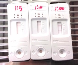
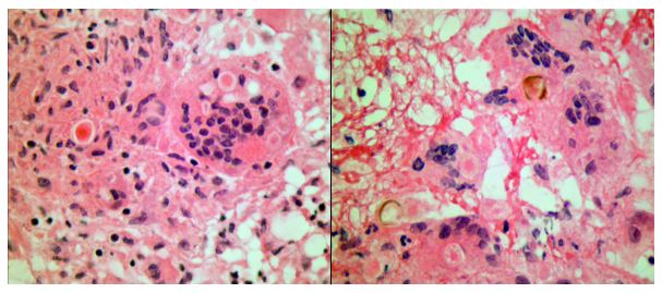
The specimen submitted to the lab was markedly red (Image 1), yet Hema screen test returned a negative result. Since this device is designed to detect occult blood in fecal samples, a prozone effect was suspected, as the stool appeared to contain overt hemorrhage. The specimen was reanalyzed with serial dilutions by a factor of 5, 10, and at 100 × dilution. The FIT result became clearly positive for blood (Image 2). The patient received a colonoscopy, which revealed internal hemorrhoids, severe diverticulosis in the left colon, as well as multiple angiodysplastic lesions. One such lesion was in the ascending colon and was actively bleeding at the time of colonoscopy. The others, which were not bleeding, were distributed in the proximal ascending colon, hepatic flexure, and proximal transverse colon. All angiodysplastic lesions were treated with argon plasma coagulation.
Image 1. Fecal specimen demonstrating overt hemorrhage.Image 2. Fecal immunochemical test performed on the patient sample submitted. Serial dilutions of fecal specimen were performed. At the dilution factor of 1:100, the result showed positive. Saline was used to dilute the fecal sample.
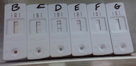
Moreover, we have tested the device with another bloody fecal sample during the initial evaluation. When an appropriate dilution factor was used, the prozone effect begins to lose its interference as show in Image 3.
Image 3. Fecal immunochemical test (FIT), showing control (C) and test (T) lines, applied to serial dilutions of fecal specimen (B ─ undiluted, C ─ 10 ×, D ─ 100 ×, E ─ 1 000 ×, F ─ 10 000 ×, G ─ 100 000 ×). At 10 × dilution, the prozone effect begins to lose its interference, and at 100 × the test is clearly positive. At dilutions higher than 1 000 ×, the concentration of blood becomes too low to return a positive result. (Image 3 provided by Dr. Andrew Lyon, PhD, DABCC, FCACB, Lab Medicine & Pathology, Saskatchewan Health Authority)
Discussion
The prozone effect (or Hook effect) has long been appreciated as a source of interference in immunoassays.1 It typically occurs in sandwich assays, of which the FIT test is an example.2 When the concentration of the analyte is excessively high, it oversaturates the capture and detection antibodies in favor of forming single antibody:analyte complexes, rather than sandwiches. This results in a false negative result where the assay is unable to detect the analyte. The solution to the prozone effect is serial dilution to lower the concentration of the analyte.
The FIT test is designed to detect microscopic amounts of blood, hence its function in screening for fecal occult blood. A number of hospital labs use this test in an acute care setting to screening bleeding in patients. However, its capacity is oversaturated in specimens containing overt hemorrhage, as in our patient. In these cases it is nevertheless important to prove that the red color of the specimen is truly due to blood, as bright red stool can be caused by a wide range of dietary factors. Some examples are red food coloring, beets, cranberries, and tomato juice.3 If these possibilities are not ruled out, the patient may become subject to the risks of unnecessary endoscopy. Serial dilution of the specimen is extremely useful in this type of situation.
References
Dasgupta A, Wahed A. Clinical Chemistry, Immunology and Laboratory Quality Control: A Comprehensive Review for Board Preparation, Certification and Clinical Practice. Amsterdam: Elsevier; 2014. 2.11.
Allison JE, Fraser CG, Halloran SP, Young GP. Population Screening for Colorectal Cancer Means Getting FIT: The Past, Present, and Future of Colorectal Cancer Screening Using the Fecal Immunochemical Test for Hemoglobin (FIT). Gut and Liver. 2014 Mar;8(2):117-30. https://doi.org/10.5009/gnl.2014.8.2.117
-Hao Li, MD is a currently a first year anatomical pathological resident at Western University, London ON, Canada. Prior to be a pathology resident, he was a neurosurgery resident at the University of Saskatchewan, Saskatoon SK, Canada. When he was at the University of Saskatchewan, he spent his third year primarily in neuropathology, with also some general anatomical pathology and clinical pathology. Through these experiences, he has come to realize that his passion and calling lay more in pathology than in surgery. He has successfully transferred into pathology, and started a new residency in anatomical pathology in July 2019. Having a background in the clinical neurosciences, he hopes to eventually pursue a fellowship in neuropathology, and possess the skill set to practice both anatomical pathology and neuropathology.
Welcome back everybody! Thank you for all the engagement on my last post, Up In Smoke¸ where I discussed the plenary publications surrounding the vaping crisis and EVALI as new pulmonary pathology entity. This month, let’s start 2020 off right. After the holiday break and going on some of my last pathology residency interviews, I’d like to reflect on this new year by taking a look at 20 exciting things on the horizon for those of us in pathology and laboratory medicine!
So, let’s take a look at 2020 with some 20/20 vision… (sorry, not sorry)
20. Big, big, big, big data
Image 1. Can’t mention databases without this invaluable website that has made me look somewhat semi-competent in many instances! Thanks Pathology Outlines (Source: um, pathologyoutlines.com)
Last year, Elsevier’s Clinical Solutions Director in China discussed three topics that would impact our profession in 2019—so let’s start there. These first three go hand-in-hand in prepping the stage for 2020. Up first: the never-ending explosion of biomedical information and the continuing tidal wave of health data we don’t even know what to do with just yet! It’s a very interesting estimate that, by 2020 (aka now!) the whole sum of medical knowledge will double every 73 days. How on earth are we to manage, when compared to 1950 it would have taken 50 years to double? Well, the argument in the linked Elsevier blogpost discusses how evidence-based inquiry databases will store and organize this knowledge for us: think UpToDate, or ExpertPath, or ImmunoQuery…some of you are nodding your heads in relief, great, I’ll move on.
19. Precision Medicine
Image 2. PD-L1, or programmed death ligand 1, is one of several new targets for cancer therapy that utilized cellular checkpoints in cell cycles alongside T cell and NK cell functional immunity to fight cancer a little more precisely than classic chemotherapy regimens. Look at you, all up to date, and stuff. (Source: AstraZeneca graphic, azimmuno-oncology.com, content)
The second topic last year’s Elsevier’s blogpost discussed was the growth and rapid development of highly specific, targeted, individualized treatment plans. The mainstay example is of course how oncology treatments are moving away from one-size-fits-all chemotherapies to individualized mutation-specific immunomodulating therapy. (We’re moving like melting glaciers but moving nonetheless.) I was definitely well equipped with my ASCP online CE credits as I found myself discussing testing patients during my heme/onc training for PDL-1 and other tailored targets. We’re just starting to ride this wave and it’s definitely growing fast.
18. AI in healthcare (part 1)
Image 3. Artificial Intelligence is getting really good at pattern recognition. Why did I choose this picture? Oh, because it’s a study of how China-based JF Healthcare, a Siemens off shoot start up AI group, designed an algorithm that beat radiologist at Stanford on precision, delivery, and accuracy. Woah. (Source: hitconsultant.net)
Yep. I went there—it’s exciting! But notice I’ll come around after some other topics to really get into the heart of AI in path. Basically, the last point in the blogpost discussed the way smart software has been growing in medicine; particularly with radiology and surgery, using advancements in robotics and detection software to predict and stratify clinical information for patient care. Within this context let me quote them directly for you, “…there remains some uncertainty around the role of AI and its true impact on pathology, it is important to recognize that AI-based technologies or machines will never replace pathologists. Instead, such innovations will play an assistive role, augmenting the decision-making capabilities of pathologists and helping them perform better and faster…” All my pathologist friends may now exhale. It’s going to be okay. We’ll talk more about this at #10.
17. New Tech, New Toys
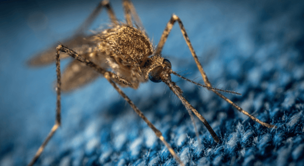
Image 4. You don’t have to go far to read about new tools and new tech. How’s this: a saliva-based rapid Malaria detection assay, courtesy of our friends at ThePathologist.com. New, rapid, accurate and deliverable diagnostics…now within spitting distance. Nailed it.
Pathologists are like the 007’s of the clinical team…at least when it comes to developing tech. There are so many new gadgets and tools we clinicians have available to us today. I delivered a recent TEDx talk where I discussed the “unrecognizable future” of medicine—and obviously now look for new and exciting ways to tell people I gave a TEDx talk. The important thing is that 73 days of doubling medical knowledge is happening so fast we don’t even know what we have available to us! Finger-print drug tests, smartphone facial capillary blood pressures, liquid biopsies, virtual MS-based immunohistochemical stains that never actually stain a single cell, cytology AI, deep data mining of free text pathology reports…it’s not a short list. It’s exciting, and we should all be sharing and collaborating to use these exciting tools together in creative ways for positive outcomes!
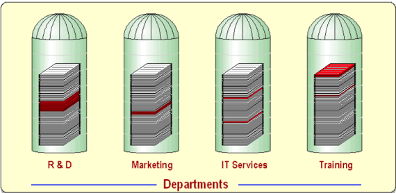
16. No More Silos
Image 5. Business and management have long discussed the importance of tearing down silos of knowledge in order to improve workflow and outcomes. It’s a growing conversation in healthcare and we’ve got our own isolated pockets of data that need to come out to the forefront, too. (Source: ERP Consulting, Estes Group Image, estesgrp.com)
Last year, I wrote a few pieces here on Lablogatory that mentioned High-Reliability Organizations (HROs) which require absolutely full sharing of responsibility as well as knowledge in order to solve problems and improve patient-care outcomes. There are many ways siloed thinking can harm the progress of any institution. It takes leadership, creative problem-solving, transparency, and teamwork. In the coming months of 2020, keep an eye out for pockets within your organizational environment that act as black holes or veils to keep pieces of critical data from the rest of the team. Encourage discussions between you and your peers, check biases about what you think might be important for one team vs. another, and try to share successes and failures as a group.
15. New Types of Colleagues
Image 6. We’re all different. And that’s ok. Each one of us is a brain, and an athlete, and a basket-case, a princess, and a criminal. Okay maybe not the last one, but we can all contribute in some important meaningful way. (Source: The Breakfast Club, 1985)
What I just mentioned about engaging in new conversations with folks you might not have worked with before—its not groundbreaking, its just good practice! In order to tear down #16’s silos, we’ve got to seek out and explore new ways to collaborate with colleagues outside our everyday scope. There will always be discussions about generational divides and differences that create culture strife in the workplace, or political/opinionated schisms that divide even the most cohesive of medical specialties. (I’m looking at you ACOG, ACP, ASCCP, and others: it’s Cervical Cancer Awareness Month, can we just agree on some guidelines already…) Soapbox over. But seriously, this isn’t a new concept. Feel like a lab half filled with boomers and millennials can’t make the cut? Well, the Harvard Business Review gave us great recommendations for this exact type of interpersonal growth exercise—in the NINETIES! The take home message: having an open culture and proactive leadership allows for fruitful exchange and growth!
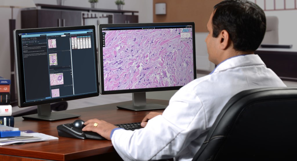
14. Digital Pathology!
Image 7. Bigger, I want these screens bigger! The desk of tomorrow’s anatomic pathologist might have less glass and more pixels, screens, and queued data with high-output servers that are stocked with smart software to sift out normal results so they can focus on really tough morphologies. Maybe even with augmented reality software, or other crazy stuff I can’t think of yet! (Source: Inspirata, digital pathology)
It’s coming. You can’t stop it. It’s exciting. I don’t care what you think. Well I actually do care, but don’t knock digi-path till it grows into whatever it’s going to become. The desk of the (anatomic) pathologist-of-the-future will look a lot different from today and that’s really cool. Once upon a time, a very long, long time ago—in the eighties maybe—radiologists still had films where we actually used radiation to change the exposure of images to be read over a light box. Classic scene, right? Doctor, the x-ray is ready! *THWIP* *CLICK* *BUZZ* and cue the contemplative stare on the wall light. Then, they went digital and get to hang out in the dark with four computer screens and coffee, and really comfy chairs. I mean what a form of progress, can’t deny.
13. MS Methodologies
Image 8. Okay, MSI crash-course time. All you really need to know is that this method allows for great specimen preservation on tiny samples, high resolution, the ability to combine with molecular testing, and fascinating implications for margin detection, mutation analyses, and more! (Source: https://blog.waters.com/molecular-visualization-ms-imaging-delivers-insights-for-cancer-research)
In my mailbox this month, is another excellent edition of The Pathologist and in it there’s a great article on Mass Spec imaging transitioning from a research tool to a clinical one. Woah. We’ve all talked about and praised MALDI-Tof methods for microbiologic assays and detection, but the expanse of mass spectrometry has developed rather quickly. Now, it’s looking for a niche in routine laboratory diagnostics outside of the old chemistry analyzer… It’s a new, non-destructive way of examining tissue and gleaning data from the smallest pieces of gross specimens. We’re onto something here, keep an eye on MSI.
12. Molecular—Need I say More?
Image 9. Move over International Space Station, the folks at Thermo Fischer Scientific want to share their take on the Next Generation of molecular testing. (Source: The Pathologist)
Same edition of The Pathologist, about 25 pages back: a discussion on the value of molecular Next Generation Sequencing. I’ve already bored half of you, wait! Come back. I agree with you, you can only call it Next-Gen so many times before a whole generation of laboratorians get bored of talking about new tumor markers or mutations. But what’s happening with NGS testing that you should know? Simply put, there are NGS analyzers that are faster, with smaller footprints, combined with smarter software that is making molecular more feasible for laboratories that used to shy away from the notion of including NGS or LDTs in their lab testing menus. This means more labs, running more molecular, for more specific populations, in real time that can collaborate with that many more new colleagues while breaking silos—well just look up at #18, 17, 16, and 15!
11. Global Health
Image 10. From Dr. Razzano’s post on Lablogatory
Dr. Dana Razzano recently interviewed me for her global health series, and we got the chance to talk about the important intersection of laboratory medicine and global public health. Getting involved in a community—especially for those of us in healthcare—often includes a survey of what kind of health challenges you face. For some it’s access to clean resources like water, for others it’s a complex system of reimbursement and billing issues that complicate delivery of care, or even more basic assessments reveal high rates of local infections with preventable illness. But you can’t tackle infrastructure change, political reform, or vaccine education single-handedly. Global health is an increasing part of our global world and, if we stay true to our professional values, we should be at the forefront.
10. AI in Healthcare (part 2)
Image 11. Drawing to represent AI from my TEDx talk, Unrecognizable Medicine 2019, TEDxAUCMED
Oh I told you I’d come back to this. Some folks are still apprehensive about AI—that’s ok—I am too, but only because I want to make sure it’s done right. Don’t expect any Skynet stuff, we’re not going that deep. So let me tell you some of the things I got to see on the residency interview trail that piqued my interests. At one hospital system, I saw plans for their anatomic pathology department to go fully digital with augmented AI software to help score mitoses and other morphologic traits by 2025. At another institution, I saw plans for data mining historical free text pathology reports to predict and stratify future specimens before they even got signed out! At a third system, I saw the utilization of smart software to predict clinical lab values for a patient’s personalized reference range…pre-analytically! This stuff is coming in hot so watch for it! What AI-related advancements are you seeing in your neck of the lab?
9. Patient Consultation
Image 12. Courtesy of SUNY Upstate Pathology Department via Twitter, a newly renovated pathology residency review room and patient consultation suite for the dedicated purpose of this invaluable interaction.
Another thing noteworthy of my residency trail are institutions which are championing the face-to-face consultative role of the clinical pathologist in patient care. We, at the end of the day, are consultants to all; physicians and patients alike. And many in our field are celebrating this role by pushing the envelope toward a progressive and effective future for pathology and laboratory medicine at large.
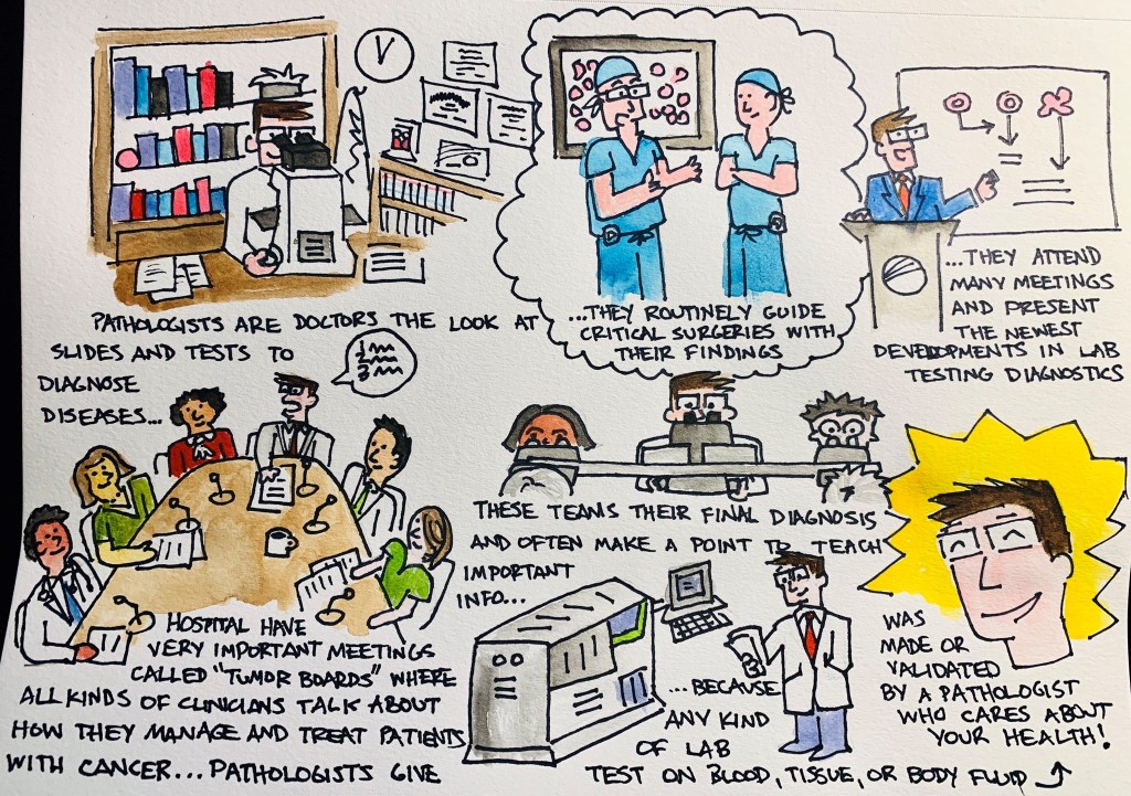
8. Graphic Medicine
Image 13. My very first #pathdoodle – What do Pathologists do?
Graphic medicine is fantastic! I wish I could have discovered this years ago. Nevertheless, in the last two years I have sought out and read numerous pieces which bridge a significant gap between clinician and patient. And if we in laboratory medicine plan to reach patients more directly, we shouldn’t be afraid to come up with creative solutions to communicate with them. My (current) approach, #PathDoodles which I post occasionally on social media, attempts to explain concepts in pathology to the everyday lay person. What will you do to reach your patients?
7. Wellness Programs
Image 14. Anonymous survey responses to Dr. Saint Martin’s program from trainees at Loyola University Medical Center. (Source: The Pathologist)
Another interesting thing I encountered on the residency interview trail was the way in which different programs addressed the concept of wellness. Some did, some did not, but everyone discussed it. Because, after all, it is important; not just for residents and physicians, but medical lab scientists, support staff, and …yes… even administrators. Work and work-life stresses and balance take a psychological and emotional toll, and in order to be happy and healthy we need ways to strengthen our mental health along the way. Last year, Dr. Marissa Saint Martin won a 2018 award from the American Association for Physician Leadership for her work in preparing residency resilience and promoting wellness through structured curricula at Loyola Health System and Mount Sinai in Miami. She’s been featured with ASCP numerous times as well as in The Pathologist. It’s promising to see such strong support for residency trainees; keep an eye out for other praise-worthy programs this year!
6. Social Media
Image 15. It takes a new kind of clinician to serve a new kind of patient. How will you rise to meet the challenges on the horizon that we can’t predict yet?
You caught me. I can’t make any predictions about the future of health care delivery, education, or collaboration without touting the importance of social media. As a member of the official ASCP Social Media Team (Go #ASCPSoMeTeam!), I’m a proud contributor not only to the content that we publish online, but to the message that unifies and spreads our #StrongerTogether mantra. Social media is fantastic tool to reach new colleagues, spread new ideas, and make new advancements in both our field and in reaching people where they are.
5. Moving Out of the Basement
Image 16. Is this closer to a representative image of your lab than you’d wish? Don’t worry, you’re not alone. Basement labs are so last season. (Source: Seret Studios, Bridge Basement, Brooklyn NYC)
One of the most memorable interviews I had this season was with a transfusion specialist who recounted to me his memory of training in pathology during his early days of residency. Green and eager, he told me how he remembered being taken on a tour of the facilities to be shown his desk/work space and upon reaching an underground level of the hospital building a sign read ‘BASEMENT’ with an adjacent one ‘PATHOLOGY’ …he paused to say it out loud, “What’s a basement-pathologist?” We bonded over the stifled stereotypes in our work and shared stories of great and terrible lab spaces we’ve seen. Some programs are renovating, some (for better) are moving on up… How are your facilities?
4. The Pipeline Problem
Image 17. The …scope of a pathologist has changed over time (see what I did there?). Sure, lots of us push glass and diagnose entities on tissue samples with complex investigations and work up, but many more of us ensure quality laboratory efficacy, develop new cutting-edge tests, manage donor centers and transfusion protocol, address infectious or public health crises, and more! I think the more we celebrate our profession, the more will join and contribute to our #StrongerTogether culture!
Well I mentioned stereotypes. I’ve talked about it before. You already know about shortages in pathology from physicians to lab scientists. And don’t get me started on pay—especially for lab workers and those in forensic path! But this is a topic I think we’re going to see a boon in media coverage in the coming years, starting now. Some of our awesome colleagues are talking about the “pipeline problem” from a myriad of angles: addressing medical student clerkships, exposure to laboratory workflow in school curriculum, advertising the infinite possibilities of careers within our profession…and more. CAP, ASCP, USCAP, and other professional societies have done amazing work in their due diligence to represent our field and advocate for the advancement of our work and image. A former CAP president once said, “Pathology is what a pathologist does,” and well, it’s a good start. Where do you see solutions to our pipeline problem?
3. PA’s and… CPA’s?
Image 18. Pathologist assistants work directly under the supervision of a pathologist in a similar way that physician assistants work with physicians. Clinical pathologist assistants help to determine the cause of disease through the examination of blood and other bodily fluids and tissues. (Source: UAB)
Now this, this is a cool concept and it’s the first time I’ve seen it. We all know and love our pathology assistants (PAs)—especially resident pathologists—as they make the training, education, and workload a better process for learning. They have awesome training and operate essentially as highly specialized clinicians in anatomic pathology. But what about the clinical, laboratory medicine side of things…? I recently saw a program advertised at the University of Alabama at Birmingham that trains clinical PAs for consultative and ancillary support roles to the clinical pathologist! Fantastic! Read more about it here! There are a growing number of DCLS (doctor of clinical laboratory science) professionals, now we’ve got a new pipeline to invite talented folks to work with us in a new way.
2. I’ll Have a Residency!
Image 19. Need I say more? What does the paper say?? Where am I going to end up? It’s a whirlwind of a season and a crazy system—once in a lifetime sort of adventure! Stay tuned, I’m sure I’ll have a post or ten about the next chapter of my career… (Image source: AAMC)
I have seen so many fantastic programs across the US these past few months, and so many decisions go into the way medical graduates rank programs to match into. To learn how the AAMC/NRMP matching system works, watch this video (maybe with some calming tea). We’ll see where I end up matching to, but I’m excited for the next chapter and to really dive into pathology even more!
1. You!
Obviously, the most important thing to keep an eye out for this year is you! Your goals, your ambition, your plans, your ideas and thoughts, and all the ways you contribute to our fantastic profession! I encourage you all to share, collaborate, and be creative with the ways in which we advance the future of laboratory medicine and continue to keep in mind that we do these things for our patients everywhere.
Thanks for reading, see you next time!
–Constantine E. Kanakis MD, MSc, MLS (ASCP)CM completed his BS at Loyola University Chicago and his MS at Rush University. He writes about experiences through medical school through the lens of a medical lab scientist with interests in hematopathology, molecular, bioethics, transfusion medicine, and graphic medicine. He is currently a 2020 AP/CP Residency Applicant and actively involved in public health and education, advocating for visibility and advancement of pathology and lab medicine. Follow him on Twitter @CEKanakisMD
A 60 year old male with a past medical history of ulcerative colitis requiring total proctocolectomy and immunomodulatory therapy followed by an anti-Tumor Necrosis Factor α blocker for the last two years and primary sclerosing cholangitis with subsequent decompensated cirrhosis that ultimately required an orthotopic liver transplant on tacrolimus and prednisone for immunosuppression presents 17 days post-transplant with worsening headache for two weeks with associated word finding difficulty and expressive aphasia.
Laboratory and Diagnostic Findings
Brain magnetic resonance imaging demonstrated, a “Heterogeneous, partially hemorrhagic and centrally necrotic mass within the posterior left temporal lobe…infectious etiologies such as pyogenic/non-pyogenic abscesses to include fungal organisms, are highest on the differential” (Image 1). At the time of admission, his complete blood count demonstrated a leukocytosis (16.48×10⁹ cells/L), anemia (hemoglobin of 7.8 g/dL, hematocrit of 24.8%) and a normal platelet count (367×109 cells/L). The automated differential showed 82% neutrophils, 10% lymphocytes, 6% monocytes, 1% eosinophils, and 1% basophils. A lumbar puncture was performed to obtain cerebral spinal fluid (CSF) and the analysis showed a glucose of 60 mg/dL, protein of 34 mg/dL, nucleated cell count of <1, and 6 red blood cells (completely normal CSF indices). Broad spectrum antimicrobials (Vancomycin, Piperacillin/Tazobactam, Metronidazole and Micafungin) were initiated. A 1,3-β-D-glucan test had a result of >500 pg/mL in both serum and CSF. Galactomannan, Histoplasma urine antigen, Cryptococcus antigen and other fungal testing were negative. Antifungal therapy was changed to voriconazole. Craniotomy was determined to be the best course of action and the patient was taken to surgery for debridement and pathologic evaluation.
Frozen section evaluation during the time of surgery showed granulomatous inflammation. Septate hyphae were observed on the fungal smear. Following surgery, amphotericin was added. Histologic evaluation of the tissue submitted from surgery showed pyogranulomatous inflammation with pigmented, spore-like structures present in multinucleated giant cells on hematoxylin and eosin (H&E) stain (Image 2). Grocott’s methenamine silver (GMS) stain also highlighted short segments of septate hyphae (Image 3).
Cultures from the surgical debridement grew a mould with central pigmentation (Image 4). Direct microscopic examination of the mould revealed thick-walled, oblong conidia with 3-5 cells, and uniformly pigmented hyphae (Image 5). A germ tube test showed germ tubes originating from both ends of the conidia consistent with Bipolaris species.
Image 1. T1-weighted (left) and T2-weighted (right) magnetic resonance imaging of the brain demonstrating a left temporal lobe mass. Image 2. Hematoxylin and Eosin stained photomicrographs showing pyogranulomatous inflammation with giant cell formation and circular structures within them (left) (40x objective magnification). The right shows gold-brown pigmented structures within granulomatous inflammation (40x objective magnification). Image 3. Grocott’s methenamine silver stain highlighting short segments of irregular septate hyphae in the brain debridement specimen (10x objective magnification).Image 4. Mature wooly brown-black colony on potato dextrose agar. Image 5. Photomicrograph of a lactophenol blue tape prep of the mature fungal colony. Pigmented hyphae and short 3-4 cell conidia are readily identified (40x objective magnification). This specimen also tested germ tube positive (not shown), indicating that this dematiaceous fungus is Bipolaris spp.
The patient’s mental status significantly improved following surgical debridement, 2 weeks of liposomal Amphotericin B, as well as long term treatment with voriconazole. The voriconazole was later switched to posaconazole due to concerns for fluoride toxicity. He completed a year of posaconazole with significant improvement of the abscess observed on imaging and resolution of headaches with no other visual problems. He continued to recover cognitive function with some residual difficulty with reading, comprehension and speech that eventually resolved.
Discussion
Phaeohyphomycosis refers to infections caused by dematiaceous fungi that exist in a variety of forms when seen in tissues and commonly involves skin, soft tissue and nasal sinuses.1 In rare cases, central nervous system (CNS) involvement has been reported. CNS phaeohyphomycosis is predominantly seen in immunosuppressed patients; however, cases involving immunocompetent individuals do exist.2 In one case series from Houston, Texas, five of seven cases of cerebral mycosis were caused by a dematiaceous mould.3 Interestingly, the patient presented in this case came to medical attention around the Dallas-Fort Worth area of Texas.
Cladophialophora bantiana is the most common dematiaceous fungus associated with CNS phaeohyphomycosis, but rare cases of Bipolaris species have been reported previously in literature.4-6
We report a case of CNS phaeohyphomycosis by Bipolaris species following orthotopic liver transplant with an excellent patient outcome. This case is unusual, in part, because the typical hospital course of a patient with phaeohyphomycosis is generally dismal.7 The stories of successful treatment often involve complete debridement of discrete lesions.7-8 In our case, the patient underwent surgical debridement and treatment initially with liposomal Amphotericin B and later transitioned to long term therapy with newer azole antifungals.
References
Revankar SG, Sutton DA, & Rinaldi MG, (2004). Primary Central Nervous System Phaeohyphomycosis: A Review of 101 cases. CID, 38, 206-2016
Filizzola MJ, Martinez F, & Rauf SJ, (2003). Phaeohyphomycosis of the central nervous system in immunocompetent hosts: report of a case and review of the literature. Int J Infec Dis, 7, 282-286
Raparia K, Powell SZ, Cernoch P, Takei H, (2010). Cerebral mycosis: 7-year retrospective series in a tertiary center. Neuropathology, Jun; 30(3): 218-223.
Frank T, Esquenazi Y, Nigo M, Wanger A, Portnoy B, & Shepard S, (2016). Disseminated Phaeohyphomycosis with Brain Abscess and Biliary Invasion Due to Bipolarisspp. In an Immunocompetent Patient. Annals of Clinical & Laboratory Science, 46(4).
McGinnis MR, Campbell G, Gourley WK, & Lucia HL, (1992). Phaeohyphomycosis Caused by Bipolaris spicifera, An Informative Case. Eur. J. Epidemiol, 8(3), 383-386
Rosow L, Jiang JX, Deuel T, Lechpammer M, Zamani AA, Milner DA, Folkerth R, Marty FM, & Kesari S, (2011). Cerebral phaeohyphomycosis caused by Bipolaris spiciferaafter heart transplantation. Transpl Infect Dis, 13, 419-423.
Gadgil N, Kupfermen M, Smitherman S, Fuller GN, Rao G, (2013). Curvularia brain abscess. J Clin Neurosci, Jan;20(1): 172-175.
-John Markantonis, DO is a second year Clinical Pathology resident at UT Southwestern in Dallas. He has interests in Medical Microbiology and Transfusion Medicine.
-Dominick Cavuoti, DO is a Professor at UT Southwestern in the Department of Pathology. He is multifaceted and splits his time as the Medical Director of the Parkland Hospital Clinical Microbiology Laboratory and Parkland Cytology attending among other administrative and educational activities.
-Clare McCormick-Baw, MD, PhD is an Assistant Professor of Clinical Microbiology at UT Southwestern in Dallas, Texas. She has a passion for teaching about laboratory medicine in general and the best uses of the microbiology lab in particular.
“I do not really understand what pathology is,” I said
during my first round of interviews at ASCP. “In fact, I have a website page in
front of me that describes it and I still do not really get it. I want to be
upfront about that before we go any further in my interview process,” I
followed. Needless to say, I got the job, but that experience really stuck with
me. As I learned more and more about pathology and laboratory medicine, I was
amazed that I had not known more about it. I had been to the doctor all my
life, I had received some serious diagnoses, and I thought I was pretty
well-versed in what my medical care entailed.
In the last few years that I have been with ASCP I have
become passionate about educating patients about the role the medical
laboratory plays in patient care. Without that understanding, patients will be
less empowered and less likely to advocate for themselves. Their family doctors
might order tests that they do not want or not order ones they that do. They
might not understand certain results, which means that they are less likely to
take an active role in their care. The more we education patients and their
caregivers about pathology and laboratory medicine, the higher quality health
care we create. Educated patients are empowered patients and it is imperative
that education includes the laboratory.
Through directing the ASCP Patient Champions program, I have
been fortunate to meet incredible patients, all who have some understanding of
the role the laboratory played and plays in their care. Hearing them say that
without the laboratory, they would only be a memory, is incredibly powerful and
humbling. The active role these patients play in their care has allowed them to
be more resourceful and more hopeful. For some of them, seeing their own slides
has been a cathartic experience because they could suddenly see the enemy they
were fighting. Others are now educating new patients about their lab tests and taking
time from their own busy schedules to volunteer at hospitals and clinics.
It can also be an inspirational experience for laboratory
professionals and pathologists to hear how they impacted a patient’s life. I
have personally shed many tears when interviewing patients so I can only
imagine what it is like to hear from someone whose life you have impacted, let
alone meet them in person. It can also really help patients to have their
diagnosis be explained by someone working in the lab and to understand why
their blood is drawn or why a biopsy is needed.
This new series on Lablogatory called Patient Advocacy, will explore the topic of patient advocacy from laboratory professional, pathologist, and patient perspectives. Each month, you will hear how patient interactions have impacted lives and what we can do to make more people aware of the crucial role the medical laboratory plays in patient care. You are all changing and saving lives every day. Let’s learn together how we can increase our patient advocacy to help them even more.
-Lotte Mulder, EdM, is the Senior Manager of Organizational
Leadership and Patient Engagement at ASCP. She earned her Masters of
Education from the Harvard Graduate School of Education in 2013, where
she focused on Leadership and Group Development. After she graduated,
Lotte started her own consulting company focused on establishing
leadership practices in organizations, creating effective organizational
structures, and interpersonal coaching. She has worked in Africa, Latin
America, Asia, and the U.S. on increasing leadership skills in young
adults through cultural immersion, service learning and refugee issues,
and cross-cultural interpretation. She is currently working toward a PhD
in Organizational Leadership.
One of the challenges of providing healthcare to patients of any type is “staying current” or “keeping up with the literature.” This can be especially challenging in the diagnostics laboratory where novel or unique approaches to a given test or test method or disease may show early promise but have no clinical utility, be too expensive, or not actually significantly change work-flow and/or patient value to justify implementation. On the other hand, sometimes a technology or test which is in development or approval can be so anticipated that clinicians and laboratorians are frustrated that it is not yet available.
In global health, there is a different problem that is encountered every day. There are technologies and tests that are approved, have documented clinical utility, and add great value to patients but they are simply not available because of supply chain, cost, administration, or geography. In such situations, the practitioners in these settings face extreme frustration—especially with stock-outs—and can become jaded and non-dependent on laboratory testing as part of care. This latter issue is a major challenge in cancer care where cancer diagnoses are required before treatment can begin; yet, in a large number of countries, access to cancer diagnostics routinely is not available. It is to that end that ASCP along with a whole host of NGO, industry, academic, and government partners are making great efforts to improve cancer care in each part of the continuum.
In this environment, however, disruptive innovations are, in fact, much easier to recognize as forthcoming. In the early 2000’s when I was working and traveling in Malawi, our project had a landline in the hospital to call the landline at the doctor’s house for issues overnight with patients. This required 24-hour nurses to be physically in the ward, tied to the phone and the patients. Landlines were expensive to install, had a very long waiting list to be installed, and, for the most part, the majority of the population in the country had never had a phone line in their dwelling. By the mid-2000’s, our project had one or more cellphones (as did the nurses) and communications through texting were nearly constant (especially since it was less expensive than making a phone call). By 2010, cell phones were ubiquitous in Malawi (and almost everywhere else in Africa) and there was no demand for landlines. Although this is a commonly used example, consider the adoption of cellular telephones and now smartphones in the US compared with Africa. There was push back, denial, avoidance, and even refusal to use them because there was an existing, well established system of landline communication. If you want to install cable television and internet in your home as late as 2016, you were often required to bundle with a landline. The point is that the adoption pattern was significantly different because there was a pre-existing competitor with the new technology although—clearly—the new technology was superior.
Now consider a woman of 35 years who has a breast mass on mammogram in downtown Boston today. She will likely have an imaging study with immediate ultrasound and fine needle aspiration and/or core biopsy subsequent. A pathological diagnosis will be issued within 3 to 4 business days (or sooner) which includes a histological diagnosis along with hormone receptor status and Her2 staining. She will see a clinician likely within a week for a positive cancer diagnosis and a treatment plan will be decided upon and executed. If we consider a similar woman in downtown Nairobi, Kampala, or Lagos, they may, in fact, have a similar experience because of the recent efforts globally to improve cancer awareness, diagnosis, and treatment. There may be some delays (reports may take several weeks), potential stock-outs, etc. but, in these major cities, the services might exist. They are likely, however, provided in private clinics, will cost a premium, and may or may not have any guarantees about quality.
The reality, however, is that the vast majority of women in the US or Europe who present with breast cancer do so at a very early stage because of active screening programs which include mammography. The vast majority of women in low- and middle-income countries (LMICs) present with later staged disease because of lack of screening. The latter group of women, however, often live in rural conditions and/or poverty conditions such that seeking care for a breast mass (of any size) will require them to spend time and money to travel to one of the major cities and attempt to access services. With this situation, many of these cancers are detected by the health system at a late stage where curative therapy windows have been missed.
Onto these observations let’s now overlay access to a test for a breast mass that can be performed on a fine needle aspiration biopsy and resulted in ~4 hours which will provide a diagnosis of cancer (or benign) along with prognostic features directing treatment. If we consider the woman in Boston, we may see such a test providing an incremental improvement in care because billing systems, litigation fears, compliance requirements, or accreditation standards still include routine histology and immunohistochemistry to be performed on a tissue biopsy. To some degree, the test may be rejected because it is adding a cost over the standard costs without adding value (other than speed) to the results. However, for the woman in the rural village who likely has access to a community health worker, access to such a test could mean that she starts oral therapy the same day she has the health visit without ever having to leave her village. We have now removed the journey to a clinic that can performed a biopsy, the costs associated with that travel, the time lost while traveling and waiting for a result, and removed the risk that this is not breast cancer—which would mean all the time and money were wasted. For this woman, enormous value is created for her with a test that is performed same day with immediate results.
This concept of point-of-care (POC) cancer diagnostics would arguable meet resistance in the US or European system because of competition with existing systems and other issues as mentioned previously. In an LMIC setting, as there may be no competition, such an innovation would sweep the system and become standard of care—almost regardless of cost. This last bit is very important because traditional systems for performing histology and IHC are complex, costly, and require multiple highly trained individuals to get a quality result. If that process costs $75 to $100 US dollars (to the health system) to provide and, for the individual patient, $10s to $100s of dollar for the travel, lodging, and lost wages, the cost of such a test could, in a stable, high-income country (HIC) market, fetch a hefty price. However, if such a test is priced at $25 to $50 USD (half the cost of the current system excluding the travel), the immediate replacement of the old system with this new system for the given indication must and will occur. This uptake is amplified in an LMIC when the POC test moves to the patient in a geographically distributed process. Breast cancer is an obvious target for such an approach because the tumors are easily accessible, the disease is quite common globally, and the primary therapies are very inexpensive. Could such a test have an impact in an LMICs for bone marrow-based, lung, bladder, colon, prostate, liver, kidney, or soft tissue tumors? The answer to that question lies in the availability of therapy, incidence of disease, and access to radiological equipment rather than availability of the actual POC device. That is, once you have a POC test for one cancer, creating a subsequent POC test for another cancer is a surmountable technical hurdle. But will such a test be able to have an impact because of the alignment of the other factors? It is likely that as you are reading this sentence, you have thought of a few yourself but there are certain cancers where you are likely thinking, “not possible”.
For breast cancer, two such POC approaches are coming down the pipeline. The first is the Cepheid GeneXpert Breast STRAT4 assay which measures quantitative RNA (qRNA) for ESR1, PGR, ERBB2, and MKi67. These four assays are surrogates for standard immunohistochemical staining for ER, PR, Her2, and Ki-67, respectively. In a series of published and in press feasibility and validation studies, the qRNA assay is essentially equivalent to IHC. There are nearly a dozen studies of this new testing cartridge using formalin-fixed, paraffin embedded (FFPE) tissue throughout Africa where the test is being compared to standard IHC. However, in at least one site, the test is being performed directly on FNA material. The second test is from the laboratory of Dr. Sara Sukumar at Johns Hopkins which uses a set of DNA methylation markers that can separate benign from malignant disease on FNA using only 10 markers. By combining these two approaches (benign vs. malignant followed by STRAT4 for positive tumors), a diagnosis of malignant breast disease with prognostic factors for treatment could be obtained in less than 4 hours.
Let’s jump forward to the point in time when both of these POCs are available (or, in fact, any POC for cancer is available). How would they change the approach to breast or other cancer in an LMIC? Because both tests require only an FNA of a mass and because tumors of the breast and other organs today are often late staged, community health workers could be trained to evaluate patients with masses, perform the sampling, and run the test in a remote village. Regardless of stage, starting a breast cancer patient on estrogen receptor antagonists can provide palliative relief or pre-surgical treatment. As a population down stages—which occurs as community health workers begin routine screening—the testing can triage benign and malignant disease at a fraction of the cost for both the system and the patient. Based on population epidemiology, nearly exact costs for these services can be predicted for a population and stock outs can be avoided. Corollary note: Only for those cancers for which you HAVE a POC.
How would these tests change the approach to breast cancer in an HIC? There would likely be resistance at many levels but, eventually, the relatively low cost and the increased patient value would allow the tests to replace or displace standard diagnostics. Without complete replacement, there could, at a minimum, be multimodality redundancy which increases quality. However, the tests would find purchase within the system because in some settings their cost and added value would make any other choice impossible.
For both settings, we can now add other market entrants, other tests for other cancers, and a generalize increased in cancer awareness in the community, all of which would increase demand, improve morbidity and mortality, but decrease costs. Such a situation would be highly valued by the patients and, therefore, is the most important eventuality as this disruption ensues. Recognizing forthcoming change is sometimes hard and sometimes easy; however, accepting and embracing forthcoming change in healthcare can lead to best outcomes for our patients—the central mission of ASCP.
Dr. Milner has no financial disclosures regarding this blog post and has received no fiscal or in-kind support from any entity, named or otherwise, that involves this blog post.
References
Wu NC, Wong W, Ho KE, Chu VC, Rizo A, Davenport S, Kelly D, Makar R, Jassem J, Duchnowska R, Biernat W, Radecka B, Fujita T, Klein JL, Stonecypher M, Ohta S, Juhl H, Weidler JM, Bates M, Press MF. Comparison of central laboratory assessments of ER, PR, HER2, and Ki67 by IHC/FISH and the corresponding mRNAs (ESR1, PGR, ERBB2, and MKi67) by RT-qPCR on an automated, broadly deployed diagnostic platform. Breast Cancer Res Treat. 2018 Nov;172(2):327-338.
Wasserman BE, Carvajal-Hausdorf DE, Ho K, Wong W, Wu N, Chu VC, Lai EW, Weidler JM, Bates M, Neumeister V, Rimm DL. High concordance of a closed-system, RT-qPCR breast cancer assay for HER2 mRNA, compared to clinically determined immunohistochemistry, fluorescence in situ hybridization, and quantitative immunofluorescence. Lab Invest. 2017 Dec;97(12):1521-1526.
Downs BM, Mercado-Rodriguez C, Cimino-Mathews A, Chen C, Yuan JP, Van Den Berg E, Cope LM, Schmitt F, Tse GM, Ali SZ, Meir-Levi D, Sood R, Li J, Richardson AL, Mosunjac MB, Rizzo M, Tulac S, Kocmond KJ, de Guzman T, Lai EW, Rhees B, Bates M, Wolff AC, Gabrielson E, Harvey SC, Umbricht CB, Visvanathan K, Fackler MJ, Sukumar S. DNA Methylation Markers for Breast Cancer Detection in the Developing World. Clin Cancer Res. 2019 Nov 1;25(21):6357-6367.
-Dan Milner, MD, MSc, spent 10 years at Harvard where he taught pathology, microbiology, and infectious disease. He began working in Africa in 1997 as a medical student and has built an international reputation as an expert in cerebral malaria. In his current role as Chief Medical officer of ASCP, he leads all PEPFAR activities as well as the Partners for Cancer Diagnosis and Treatment in Africa Initiative.
In the Immunohistochemical stain lab, Rory made up his special stains under the chemical fume hood. One of the reagents he used was hydrochloric acid. At the end of each month there was usually a little bit of acid that needed to be disposed of as waste. He poured the waste acid into a glass jar and labeled the jar as “waste HCl.” He then carried the jar through the door to the room next door where there was an acid storage cabinet. That was where the contracted chemical waste vendor picked up other wastes from the lab.
Lydia was working the night shift in blood bank when she was changing the waste container on the automated type and screen analyzer. She splashed some waste into her eye when pulling the container out of the analyzer. She rubbed some water from the restroom sink in her eyes and decided not to report the incident as she was already in trouble with the supervisor for her continued absences.
I often talk to Lab Safety Professionals about using their “Safety Eyes” while performing their duties. It’s a latent ability we all have and can develop with some practice. With it, one can walk into a laboratory and quickly see safety issues and even make a swift assessment of the overall safety culture. Much of what can be seen using that super-power belongs to the lab’s physical environment- that which lies on the surface and should be visible to all. But sometimes there are deeper issues, those that may be more hidden. With practice, one might easily spot incorrect use of PPE, unlabeled chemicals or trip hazards. But how do you spot those other safety issues that can be just as dangerous- or even more so? How can your Safety Eyes ability be honed into something more powerful….like X-ray vision?
In the first scenario above, you may see nothing wrong, especially if you’ve performed that process yourself for years. One week later the EPA inspector came in for a laboratory waste audit, and they cited the lab for moving waste from the point of its generation to another area which was not designated as a Central Accumulation Area (CAA). Hazardous (chemical) waste cannot be moved to another location outside the line of sight of its generation point unless that other area is treated a CAA.
In the second scenario Lydia woke up the next day because her eye began to burn. She went to the emergency room and told her story. Because she missed the window of opportunity for proper treatment of an unknown source exposure to biohazards, she had to undergo long-term treatments which involved strong medications which have unpleasant side effects. She also had to be tested regularly for Hepatitis and HIV.
Some people you may know in the lab have been performing unsafe acts for years with little or no known consequences. Have they been doing the right thing or have they been lucky? What will it take to correct those unsafe actions? A fine? An exposure or injury? Hopefully not. Sometimes the reason unsafe acts occur is that staff is unaware of the regulations or the potential consequences. Influencing others’ safety behaviors is another more subtle super-power of the Lab Safety Professional, but it can be both important and useful.
As a safety professional, make sure you develop your basic super powers- your Influence and your Safety Eyes- but also be sure to augment what you already know how to use. Learn to use some X-ray Vision. Look more deeply for those processes and actions that may have been in place for years. It is not too late to make a change and prevent an incident that was years in the making.
–Dan Scungio, MT(ASCP), SLS, CQA (ASQ) has over 25 years experience as a certified medical technologist. Today he is the Laboratory Safety Officer for Sentara Healthcare, a system of seven hospitals and over 20 laboratories and draw sites in the Tidewater area of Virginia. He is also known as Dan the Lab Safety Man, a lab safety consultant, educator, and trainer.