Microbiology Case Study: Not An Ordinary Sore Throat, but One Accompanied by Headache 

Case History

An 18 year old healthy female presented to the emergency department of a tertiary care hospital in Minnesota for headache, vomiting, and sore throat. She did not have any significant past medical history. Due to meningitis concerns, lumbar puncture and head computed tomography (CT) imaging were performed. The CT scan showed an accumulation of fluid in the posterior right frontal sinus with scattered mucosal thickening. However, her cerebrospinal fluid (CSF) profile was insignificant, with normal protein and glucose levels. CSF culture was ordered, and two sets of blood cultures were drawn. 

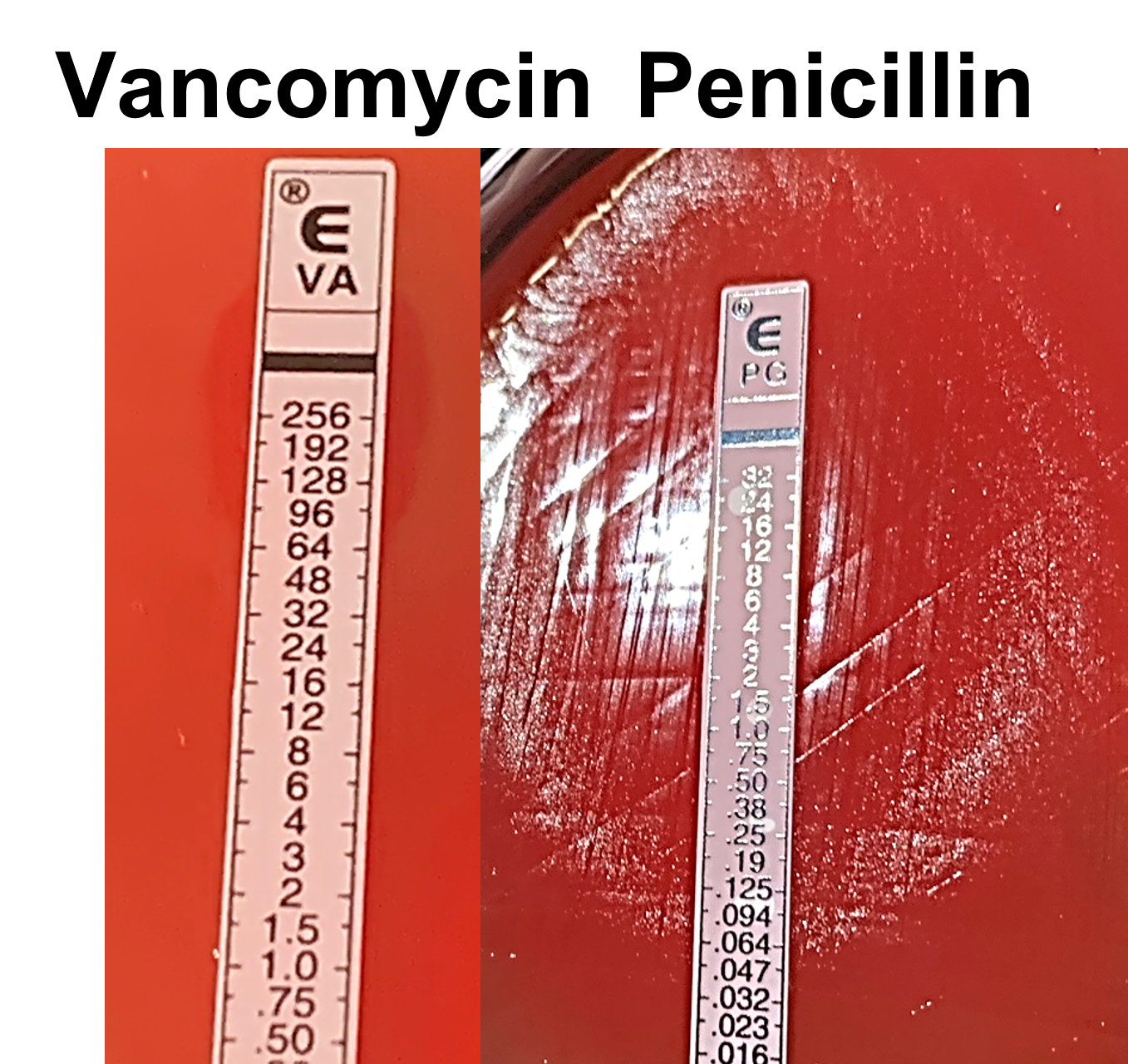
Based on the examination and presenting symptoms, pharyngitis was suspected, and she was discharged with Amoxicillin (500mg Q6H for five days). However, her strep throat screening returned negative. Her blood culture was negative. CSF culture was also negative. Cryptococcal antigen and Enterovirus PCR were performed; however, both results were negative.

She returned to the ED two days later for a worsening headache and newly developed photophobia. Additional history revealed that she went swimming in a lake two weeks prior to her first presentation at the ED. Her CSF was sent for the Ova and Parasite (O&P) exam for suspicious parasitic meningitis. The CSF O&P test was negative. CSF PCR for amoeba was also performed at a reference laboratory, and the results came back positive with Balamuthia mandrillaris. The patient was then given flucytosine, fluconazole, and azithromycin. 

Figure 1. Photo Credit CDC: Balamuthia – free living parasite observed under light microscopy.

Discussion

Balamuthia mandrillaris belongs to a group of free-living amoebae, including Acanthamoeba species and Naegleriafowleri, that cause fatal encephalitis.1 Balamuthia mandrillaris is the only known species of the genus Balamuthia that causes infections in humans. Encephalitis caused by B. mandrillaris is known as granulomatous amoebic encephalitis (GAE). GAE is characterized as a subacute to a chronic infection that can last several months to years.2 GAE differs from primary amoebic meningoencephalitis (PAM) caused by Naegleria fowleri, which typically causes an acute onset lasting a few days. 

While ecological niches of B. mandrillaris are not well understood, they have been reported to be isolated from dust, soil, and water.r Both trophozoite and cyst forms can enter the body through the nasal passage or ulcerated/broken skin; however, the trophozoite stage causes associated disease manifestation and represents a diagnostic stage.2 

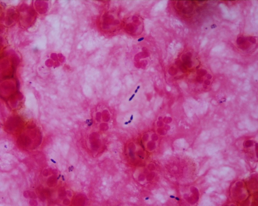
Brain-eating amoebas are traditionally difficult to diagnose. Hematology and chemistry profiles of CSF of affected individuals are generally unremarkable, although, sometimes, increased monocytes and lymphocytes, along with increased protein levels, are seen in some cases of GAE.3 

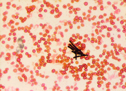
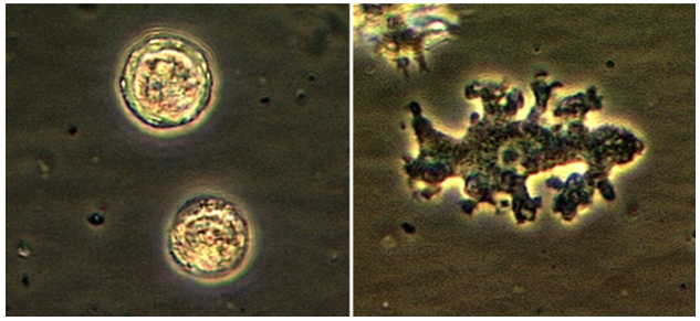
The most common method of laboratory diagnosis of B. mandrillaris is a microscopic examination of CSF wet mount (Figure 1) or via immunohistochemical staining of CSF or brain biopsy.1 With advancements in technology, species-specific nucleic acid amplification tests (NAAT) can be performed to diagnose B. mandrillaris infection accurately. However, there is no commercially available NAAT for the free-living amoeba. Only very few laboratories, such as State departments of health laboratories, Centers for Disease Control (CDC) and Prevention, or commercial reference laboratories, develop these tests as a laboratory-developed test (LDT). Histological assessment of biopsies from brain lesions may reveal tumor-like appearance or perivascular monocytic necrosis of affected areas.1 While there have been significant technological advancements, the prognosis stays at less than a 5% survival rate,6 with only roughly 25% of cases diagnosed antemortem. One possible reason for delayed laboratory diagnosis is the challenges in performing the microscopic examination in clinical microbiology laboratories since it requires expertise for accurate identification of the organism. Additionally, most clinical microbiology laboratories do not readily have an in-house LDT for free-living amoeba NAAT. Therefore, the turnaround time for diagnosing B. mandrillaris or any free-living amoeba is typically longer when specimens have to be sent out to reference laboratories. 

Diagnosis of B. mandrillaris encephalitis solely based on clinical symptoms is often challenging due to similar presentation in other causes of infectious encephalitis. B. mandrillaris can affect both immunocompetent and immunocompromised individuals.1,3,4 The first B. mandrillaris case was reported in a deceased baboon in the San Diego Zoo in 1986.1  The majority of patients were diagnosed postmortem.1 While most B. mandrillaris infections are actively acquired through nasal passages or skin penetration, rare post-mortem cases of passive transfer of the organism from organ transplantation have been reported.6 With technological advancement, there have been successes in pre-mortem diagnoses in recent years.1,4 According to the known cases, individuals of Latin American origin are more likely to contract the disease; it is unknown if it is due to increased exposure or a genetic predisposition.1  Similar to other free-living amoebae, B. mandrillaris can be generally found in warmer climates or tropical regions. Of approximately two hundred cases reported worldwide, about 34 were reported in Latin America, from Mexico to Brazil, while some were from Japan, New Zealand, England, and other European countries. The Southwestern United States also contributes 30 cases, mostly in Arizona, Texas, and California.1  In the United States, there have only been 109 cases directly reported to CDC from 1974 to 2016.2,7  We believe that this is the first case of Balamuthia reported in Minnesota. The number of exact cases would be difficult to be determined due to misdiagnosis and rare occurrence of the disease or cases not reported to CDC or the state department of health. 

While investigational drugs for B. mandrillaris GAE are in development, combination therapy of flucytosine, fluconazole, pentamidine, and azithromycin or clarithromycin has shown successes.2 Our patient was successfully treated with flucytosine, fluconazole, and azithromycin. 

References

  1. Matin A, Siddiqui R, Jayasekera S, Khan NA. Increasing importance of Balamuthia mandrillaris. Clin Microbiol Rev. 2008 Jul;21(3):435-48. doi: 10.1128/CMR.00056-07. PMID: 18625680; PMCID: PMC2493082. 
  2. Centers for Disease Control and Prevention. (2019, August 23). CDC – Dpdx – free Living Amebic Infections. Centers for Disease Control and Prevention. https://www.cdc.gov/dpdx/freelivingamebic/index.html.
  3. Kofman A, Guarner J. Free Living Amoebic Infections: Review. J Clin Microbiol. 2021 Jun 16:JCM0022821. doi: 10.1128/JCM.00228-21. Epub ahead of print. PMID: 34133896.
  4. Pietrucha-Dilanchian, P., Chan, J. C., Castellano-Sanchez, A., Hirzel, A., Laowansiri, P., Tuda, C., Visvesvara, G. S., Qvarnstrom, Y., & Ratzan, K. R. (2011). Balamuthia mandrillaris And Acanthamoeba Amebic Encephalitis With Neurotoxoplasmosis Coinfection in a patient with Advanced HIV Infection. Journal of Clinical Microbiology, 50(3), 1128–1131.
  5. Ong TYY, Khan NA, Siddiqui R. 2017. Brain-eating amoebae: predilection sites in the brain and disease outcome. J Clin Microbiol 55:1989 –1997. https://doi.org/10.1128/JCM. 02300-16.
  6. Centers for Disease Control and Prevention. 2011. Balamuthia mandrillaris transmitted through organ transplantation—Mississippi, 2009. Am J Trans-plant 11:173–176. https://doi.org/10.1111/j.1600-6143.2010.03395_1.x.
  7. Jennifer R Cope, Janet Landa, Hannah Nethercut, Sarah A Collier, Carol Glaser, Melanie Moser, Raghuveer Puttagunta, Jonathan S Yoder, Ibne K Ali, Sharon L Roy, The Epidemiology and Clinical Features of Balamuthia mandrillaris Disease in the United States, 1974–2016, Clinical Infectious Diseases, Volume 68, Issue 11, 1 June 2019, Pages 1815–1822, https://doi.org/10.1093/cid/ciy813

-Alejandro Soto, MLS (ASCP)CM is a junior medical technologist who is passionate about clinical microbiology.

-Phyu M. Thwe, Ph.D., D(ABMM), MLS(ASCP)CM is Microbiology Technical Director at Allina Health Laboratory in Minneapolis, MN. She completed her CPEP microbiology fellowship at the University of Texas Medical Branch in Galveston, TX. Her interest includes appropriate test utilization and extra-pulmonary tuberculosis.

Peritoneal Problems

A 74 year old male patient with an extensive cardiac history initially presented to the ER with black stool, warranting a CT scan, upper endoscopy, and colonoscopy, identifying a large, obstructive mass in the colon, smaller, yet unresectable polyps, and subcentimeter liver lesions and lung nodules. The colonic mass was biopsied, consistent with adenocarcinoma; however, the liver lesions were too small to characterize. One month after the onset of symptoms, a right hemicolectomy was performed, and the pathology was signed out as moderately differentiated adenocarcinoma, microsatellite stable, with evidence of lymphovascular and perineural invasion, placing the patient’s stage at IIA (pT3, pN0, cM0). Through shared decision-making, the medical oncologist and patient elected for surveillance due to multiple comorbidities. Forgoing adjuvant therapy, the patient was discharged to physical therapy/rehabilitation. The patient returned for imaging 4 months after his hemicolectomy, demonstrating an enlargement in one of the liver lesions, but then, the patient was lost to follow-up for 20 months.


The patient reestablished care and surveillance imaging, which demonstrated a hypodense liver lesion (in a background of poorly visualized subcentimeter liver lesions), a nonocclusive thrombus in the right portal vein, a heterogenous enhancement of the left portal vein (suggestive of an underlying tumor thrombus), and an 8 cm heterogenous right adrenal mass. Based on the most recent CT scan, the differential diagnoses of the adrenal mass include metastatic disease or a primary adrenal lesion including adrenal cortical carcinoma or pheochromocytoma (for which biochemical analysis should be performed before attempting a biopsy). Extensive peritoneal lymphadenopathy was visualized as well. The area of the right hemicolectomy, however, did not show evidence of recurrence. After biochemical evaluation for metanephrines ruled out a pheochromocytoma, the patient underwent a CT scan-guided adrenal FNA and core biopsy.

The Diff-Quik smear assessed at the time of biopsy revealed a highly cellular specimen, some cells with bare nuclei, enlarged nuclei, and some pseudoglandular structures.

Images 1-2: Adrenal Gland, Right, Fine Needle Aspiration. 1-2: DQ-stained smears

Telepathology confirmed an adequate sample of tumor cells present, and core biopsies were obtained.

The following morning, the pap-stained smears and H&E cell block sections were screened. The cells appeared polygonal with a high N/C ratio and prominent macronucleoli. Cell arrangements formed thickened trabeculae. However, the cytoplasm is more granular than the lipid-rich cytoplasm seen in an adrenal cortical carcinoma. The H&E cell block sections depicted a beautiful trabecular pattern with endothelial cells wrapping the periphery.

Images 3-6: Adrenal Gland, Right, Fine Needle Aspiration. 3-4: Pap-stained smear; 5-6: H&E Cell Block sections.

The preliminary morphology was interpreted as carcinoma, and both cytotechnologist (or cytologist, as we now prefer to be called) and pathologist suggesting features of adrenal cortical carcinoma; however, the IHC markers proved otherwise!

Images 7-9: Adrenal Gland, Right, Fine Needle Aspiration, IHC Cell Block Sections. 7:HepPar1+; 8: Arginase+; 9: pCEA (canalicular pattern)+.

Other differential diagnoses considered renal cell carcinoma and pheochromocytoma (to be safe). The IHC profile ruled out adrenal cortical carcinoma as the cells of interest were negative for inhibin, calretinin, and Melan A. Negative PAX-8, EMA, AE1/AE3, and vimentin staining ruled out renal cell carcinoma, and negative chromogranin, synaptophysin, GATA-3, vimentin, and S100 staining enabled us to safely say that a pheochromocytoma was out of the equation as well. Positive staining for HepPar1, arginase, pCEA (canalicular pattern), and CAM5.2 supported the unlikely diagnosis of metastatic hepatocellular carcinoma (HCC).

This diagnosis placed the patient at Stage IV HCC. It came to light that the patient has a remote history of hepatitis and a high-risk history of drinking, contributing to a poor prognosis. Due to the patient’s condition, they held off on HCV antiviral therapy and decided to observing viral load through regular blood work. The patient and clinician discussed the risks and benefits along with alternatives of systemic therapy, as his multiple comorbidities still pose a significant risk. Immunotherapy was determined to be the best option to delay the progression of his cancer and maintain quality of life.

-Taryn Waraksa, MS, SCT(ASCP)CM, CT(IAC), has worked as a cytotechnologist at Fox Chase Cancer Center, in Philadelphia, Pennsylvania, since earning her master’s degree from Thomas Jefferson University in 2014. She is an ASCP board-certified Specialist in Cytotechnology with an additional certification by the International Academy of Cytology (IAC). She is also a 2020 ASCP 40 Under Forty Honoree.

Validations/Verifications of Alternative Anticoagulants for Platelet Clumping

Platelet clumping can cause a falsely lowered platelet count on hematology instruments and can be difficult to resolve. With thrombocytopenia, physicians need an accurate count to diagnose, treat, or monitor patients. Clumping can be due to pre-analytic issues with specimen handling, can be caused by medications, or may be an in vitro phenomenon caused by anticoagulants. The clumping makes precise counting impossible and even estimates can be very tricky. If there are clumps, and recollection of the sample still yields platelet clumping, then many labs will have an alternate tube drawn or an alternative method to help resolve clumping.

Many of us have heard of using sodium citrate tubes for patients who have clumped platelets in EDTA. So, if you are having platelet clumping headaches, you can just order some sodium citrate tubes and start using those on your hematology analyzers, right? Not so fast. There are many published references of the use of sodium citrate tubes to resolve EDTA induced thrombocytopenia but we still see samples in which the clumping is not resolved with the sodium citrate tube. Published studies have shown that several other alternate methods have been helpful in resolving platelet clumping issues. These include drawing specimens in CTAD, ACD, or ‘ThromboExact’1 tubes, or adding amikacin or kanamycin to the EDTA after the specimen is drawn.

So, why can’t we just order one of these other tubes and start reporting results? Hematology analyzers are only FDA approved for EDTA tubes. Before you can use any modified method, and before you can report any patient results, your laboratory must do validation or verification studies to prove that the method produces valid results.

A validation provides objective evidence that a test performs as intended. A validation uses a defined process and is used when setting up and implementing a new test. One example is a laboratory developed test (LDT), which is a test performed by the clinical laboratory in which the test was developed. A LDT can be one that is neither FDA-cleared nor FDA-approved or can be one that is FDA cleared/approved but has been modified by the performing laboratory. The use of sample types or the use of collection devices not listed in manufacturer instructions constitute modifications, by this definition. In a validation, accuracy should be tested with at least 40 samples across the analytical measurement range (AMR). Correlations are then performed. Precision should be tested over approximately 20 days. A verification, on the other hand, uses an abbreviated process and is used when setting up and implementing new tests that are cleared or approved by FDA. Before reporting patient results, the laboratory must demonstrate that a test performs in agreement with prior claims and must demonstrate performance specifications are comparable to the manufacturer’s specifications. Verification therefore is a confirmation that a test method meets specified requirements and would be applied to a method which has already been validated. For a verification, a smaller sample size may be used, and precisions tested over 5 or more days.

Table 1. Validations vs. Verifications

So, which would you do if you wanted to use an alternate method for reporting platelet counts? Hematology analyzers are only FDA approved for platelet counts on EDTA, but the by which the sample is analyzed does not change with an alternate tube, so it may be possible to do a limited validation or verification with a smaller sample size. A laboratory needs to prove correlation, accuracy, and precision. Follow your laboratory SOPs for validation/verification and consult with your accrediting agencies, if necessary. A plan needs to be written and signed off by laboratory director. Choose the alternative method you wish to investigate and run correlations for platelet counts on EDTA and the alternate anticoagulant. If your instrument has more than one platelet mode, it is important to run samples in the mode which you would normally use for thrombocytopenia or flagged platelet counts. Apply any dilutional factors and calculate correlations. This data will be Included in your report, which, along with a procedure needs to be signed by the laboratory director.

The most important thing is to write a plan and a follow-up report according to your SOPs and to make sure any requirements of accrediting agencies are included. There can be some differences in interpretation of standards, so it is the laboratory’s responsibility to make sure what you have done meets the standards that apply to your lab.

The use of alternate tubes for platelet counts has been well reviewed in literature. Sodium citrate tubes are the most common, likely because they are the easiest to use and the most cost effective. Remember though that sodium citrate and other methods cannot resolve all case s of pseudothrombocytopenia. There are several special notes to consider. Counts from sodium citrate tubes are known to be stable for approximately 3 hours, after which counts decrease. As well, it has been shown in literature that sodium citrate tubes do show a negative bias. It has been reported that the 10% dilutional factor may be too low. Some studies have been done to determine dilution factors that correlate more closely with EDTA tubes, and researchers have suggested factor of 17%-25%. If your laboratory wishes to determine its own dilutional factor for sodium citrate or other tubes, this will also have to be included in your platelet studies. Lastly, CBCs are calibrated for EDTA, so only the platelet count should be reported from an alternative anticoagulant.

The end of another busy and challenging year is upon us, and at this time of year we can find ourselves rushed to finish ‘end of year’ tasks such as competencies and continuing education requirements. and a response to Sysmex’s recent webinar “Those Sticky, Tricky Platelets – Solving the Puzzle of Platelet Clumping” (Oct.20,2021). After the webinar I had many questions from techs asking, “Do we need to validate our alternative method?” and “How do we go about doing that?” The webinar discusses pseudothrombocytopenia and its causes in more detail than my earlier blog from Oct 2019, “Hematology Case Study: The Story of the Platelet Clump: EDTA-Induced Thrombocytopenia”. The webinar can be found at https://webinars.sysmex.com/webinars/11ae743e-ac99-47e7-acb7-2b24cedc1a1a and is available for CEU, free of charge.

References

  1. Baccini V, Geneviève F, Jacqmin H, et al. Platelet Counting: Ugly Traps and Good Advice. Proposals from the French-Speaking Cellular Hematology Group (GFHC). J Clin Med. 2020;9(3):808. Published 2020 Mar 16. doi:10.3390/jcm9030808
  2. Bizzaro N. (2013): Pseudothrombocytopenia. In: Platelets, Vol. 3, ed Bizzaro N, Elsevier, Amsterdam, pp. 989–997 
  3. Chae H, Kim M, Lim J, Oh EJ, Kim Y, Han K: Novel method to dissociate platelet clumps in EDTA-dependent pseudothrombocytopenia based on the pathophysiological mechanism. Clin Chem Lab Med 50, 1387–1391 (2012)
  4. Socha, Becky. Calibration and Calibration Verification: Who, What, Where, When, Why, How & Did I Pass or Fail?. AMT 81st Educational Program and annual meeting, 2019
  5. Zhou X, Wu X, Deng W, Li J, Luo W: Amikacin can be added to blood to reduce the fall in platelet count. Am J Clin Pathol 136, 646–652 (2011)
  6. https://www.cms.gov/Regulations-and-Guidance/Legislation/CLIA/downloads/6065bk.pdf
  7. https://www.cap.org/laboratory-improvement/proficiency-testing/calibration-verification-linearity
  8. https://www.westgard.com/cal-verification-criteria.htm
  9. https://labmedicineblog.com/2019/10/29/ hematology-case-study-the-story-of-the-platelet- clump-edta-induced-thrombocytopenia/
Socha-small

-Becky Socha, MS, MLS(ASCP)CMBBCM graduated from Merrimack College in N. Andover, Massachusetts with a BS in Medical Technology and completed her MS in Clinical Laboratory Sciences at the University of Massachusetts, Lowell. She has worked as a Medical Technologist for over 40 years and has taught as an adjunct faculty member at Merrimack College, UMass Lowell and Stevenson University for over 20 years.  She has worked in all areas of the clinical laboratory, but has a special interest in Hematology and Blood Banking. She currently works at Mercy Medical Center in Baltimore, Md. When she’s not busy being a mad scientist, she can be found outside riding her bicycle.

Microbiology Case Study: An Adult Patient with a Tender Mass and Rash

Case History

An adult patient with no significant past medical history presents with a tender right inguinal mass and rash over the right buttock measuring 5×7 cm. A skin punch biopsy was performed on the gluteal rash and sent to histopathology for analysis. Histology (Image 1) revealed an intradermal acantholytic vesicular dermatitis and associated folliculitis. Chronic inflammatory infiltrates surrounded neurovascular bundles as well as adnexal structures. Multinucleated Tzank cells were identified with the characteristic multinucleation, margination, and molding. Scattered eosinophilic Cowdry A inclusions were seen. Stains for bacteria and acid-fast bacilli (AFB) were not performed. A periodic acid-Schiff (PAS) stain (Image 2) demonstrated the absence of fungal elements.

Image 1. A hematoxylin and eosin (H&E) slide reveals a chronic inflammatory infiltrate surrounding (A) neurovascular bundles and (B) adnexal structures. (C) Tzank cells and (D) Cowdry A inclusions are also seen.
Image 2. A PAS stained slide of the same region as Image 1. (A) highlights a chronic inflammatory infiltrate where no fungal hyphae are seen.

Histopathology demonstrated “folliculitis suspicious for herpetic dermatitis.” PCR molecular testing for herpes simplex virus (HSV) and varicella zoster virus (VZV) were ordered on the punch biopsy. HSV was not detected; however, VZV was detected by PCR (Image 3, Image 4).

Image 3. The Simplexa VZV Direct Assay (Diasorin) targets a portion of the VZV DNA polymerase. The PCR amplification curve reveals the presence of VZV DNA (green) as well as that of the internal control (purple).
Image 4. A separate PCR assay targeting TP53 was performed to assess DNA quality of the fixed tissue. The presence of TP53 amplification in both the IC, the patient sample (Sample), as well as other samples on the same run (unlabeled) demonstrates the DNA quality is adequate. The absence of amplification of the NTC demonstrates a lack of nucleic acid contamination.

Discussion

Varicella zoster virus (VZV) is an enveloped double-stranded DNA virus belonging to the herpesviridae family.5 Transmission during primary infection occurs via inhalation of aerosolized respiratory secretions or lesional secretions, and to a lesser extent, via direct contact with lesional secretions. Transmission during secondary infection occurs mainly via physical contact with the secretions of herpetic lesions or the lesions themselves. The window for primary infection of transmissibility is 1-2 days before the onset of the rash lasting until either all lesions have crusted over or 24 hours have passed without the formation of new lesions, whereas secondary infections are only contagious during the presence of active lesions.6 Primary infection causes chicken pox, which is characterized by a vesicular rash, fever, and malaise. After primary infection, VZV resides in the dorsal root ganglia and trigeminal ganglia. VZV may reactivate, possibly as a result of stress or some other immunosuppressive state, as a painful vesicular rash known as shingles or herpes zoster. The rash is limited to the dermatome innervated by the ganglion from which the virus reactivated. Severe cases of shingles may result in meningitis, myelitis, as well as encephalitis, and can be fatal.1 Though the lesions of herpes zoster (secondary VZV infection) are infectious, they are significantly less so than those of varicella (primary VZV infection).6

The histology of VZV infection is characterized by intradermal and sub-epidermal vesicles with associated acantholysis, necrosis, and spongiosis. Tzanck cells demonstrate the characteristic “3 Ms” of multi-nucleation, marginated chromatin, and nuclear molding. The dermis is notable for perivascular, periadnexal, and perineural lymphocytic infiltrates. Folliculitis and syringitis may be present along with small vessel necrotizing vasculitis. Late stage lesions are notable for encrusted ulcers. Though there is significant histologic overlap between VZV infection as those caused by others in the herpes family, VZV histology tends to demonstrate a more substantial follicular involvement.2 Besides other herpes viruses, the differential diagnosis includes erythema multiforme, coxsackievirus, ecthyma contagiosum, pemphigus vulgaris and paravaccinia infection.3

While molecular methodologies are now the gold standard for diagnosis, a number of modalities including immunohistochemistry, immunofluorescence, in-situ hybridization, and serology can be used to aid in diagnosis.3 In the aforementioned case, diagnosis was made using a real time polymerase chain reaction (RT-PCR) assay (Simplexa VZV Direct Assay, Image 3) using previously extracted DNA. Forward and reverse primers target a well conserved portion of the VZV DNA polymerase. In between synthesis cycles, fluorescent probes anneal to the target sequence, separating the fluorophore from the quencher, thus generating a fluorescent signal. Amplification is measure by the cycle threshold (Ct), the number of PCR cycles needed for the fluorescent signal to exceed the background. An internal positive control (IC) is spiked in to assure negative results are not the result the presence of PCR inhibitors. To assess the quality of DNA present, a separate PCR was also performed on TP53, which amplifies if sufficiently high quality DNA is present, irrespective of the presence of VZV DNA (Image 4). A negative control (no template control, NTC) should be run to interrogate the presence of nucleic acid contamination.4

Treatment, if warranted, should be administered as soon as possible. Antiviral options include acyclovir, valacyclovir, or famcyclovir. Central nervous system, ocular, or renal VZV cases are considered emergencies and are typically treated with intravenous acyclovir.6 While resistance is rare, at least three mechanisms of resistance have been shown to endow VZV resistance to the aforementioned drugs: reduced or absent thymidine kinase, altered thymidine kinase activity leading to decreased phosphorylation of the drug, or decreased affinity of VZV DNA polymerase for acyclovir triphosphate.5, 8 If an infection with a resistant strain is identified or suspected, foscarnet is often used in place of acyclovir. Unlike the nucleoside analogs, this pyrophosphate analog does not rely on phosphorylation for the activation of its anti-VZV DNA polymerase activity.7 Historically plaque reduction assays were used, but this method is both labor intensive, low yield, and slow. Thus, molecular testing interrogating mutations in the DNA polymerase or thymidine kinase genes have increased in popularity.8

Two live attenuated vaccines are available, either in isolation or in combination with the measles mumps, and rubella vaccines (MMRV), in a 2 dose series to prevent primary infection. Since the VZV vaccine contains live virus, it should not be administered to pregnant women or the severely immunocompromised. Vaccine administration has been found to be 90% effective in preventing primary infection and 99% effective at preventing severe or complicated disease.7 Additionally, there is a recombinant vaccine consisting of the VZV glycophorin E protein in addition to an adjuvant that is used to prevent shingles. This formulation is recommended for adults over the age of 60 in prevention of secondary infections as well as to immunocompromised individuals at higher risk from exposure to the live attenuated vaccine.9

References

  1. Depledge DP, Sadaoka T, Ouwendijk WJD. Molecular Aspects of Varicella-Zoster Virus Latency. Viruses. 2018;10(7):349. Published 2018 Jun 28. doi:10.3390/v10070349
  2. Busam, K. J. Dermatopathology. 2nd Edition. Published 2014.
  3. Hall, B. Diagnostic pathology: Nonneoplastic Dermatopathology. 3rd Edition. Published 2021.
  4. Simplexa™ VZV Swab Direct REF MOL3655. 2021
  5. Sauerbrei A. Diagnosis, antiviral therapy, and prophylaxis of varicella-zoster virus infections. Eur J Clin Microbiol Infect Dis. 2016;35(5):723-734. doi:10.1007/s10096-016-2605-0
  6. https://www.cdc.gov/chickenpox/about/transmission.html
  7. https://www.cdc.gov/vaccines/vpd/varicella/hcp/index.html
  8. Piret J, Boivin G. Antiviral resistance in herpes simplex virus and varicella-zoster virus infections: diagnosis and management. Curr Opin Infect Dis. 2016;29(6):654-662. doi:10.1097/QCO.0000000000000288
  9. https://www.cdc.gov/vaccines/hcp/vis/vis-statements/shingles-recombinant.html

-Jeremy Adler, MD is a Molecular Genetic Pathology fellow at the University of Chicago Medicine and NorthShore University HealthSystem. He completed his MD at SUNY Stony Brook and his AP/CP residency at the Pennsylvania Hospital of the University of Pennsylvania Health System.

-Paige M.K. Larkin, PhD, D(ABMM), M(ASCP)CM is the Director of Molecular Microbiology and Associate Director of Clinical Microbiology at NorthShore University HealthSystem in Evanston, IL. Her interests include mycology, mycobacteriology, point-of-care testing, and molecular diagnostics, especially next generation sequencing.

Safety in the Moment

Often I am asked how one who is responsible for laboratory safety (yet has other duties as well) can get the job done well. In today’s labs there is tight staffing, tight budgeting, and a score of regulatory duties that must be accomplished, and not all of these things revolve around safety. Many who oversee the lab safety program also must run the point of care program, the lab quality program, or even manage all of the day to day operations of the department. It’s a great deal to juggle, but there are methods you can use to make sure that laboratory safety doesn’t take a back seat.

One way to incorporate safety into your multiple roles each day is to start every meeting or huddle with a safety moment or story. Ask for a team member to discuss a safety story they witnessed or in which they were involved. Placing safety first lets the team members know it has priority, and relating an issue or incident has benefits as well. The safety moment may be as brief as reporting on how an employee provided PPE to a vendor that came into the department. That is a safety success worth mentioning, and there are doubtless others that can be mentioned. These safety stories may also be those that do not necessarily illustrate a success. Telling people about an incident and asking how it could have been avoided is a fast yet educational plus for your safety culture. Reviewing safety incidents is also beneficial so that others know what happened and they can be thinking of how to avoid the same thing from happening to others or themselves. Talking about safety in these ways takes little time, but if safety is incorporated into the language of the department, the culture will remain improved, and it is easy to fit this habit into your schedule.

Acting as a consistent role model is another way to incorporate safety into your multiple roles. Make sure you wear the correct clothing and shoes. If you walk in and out of the department, you should dress the part. Open-toed shoes or mesh sneakers should not be worn. Wear PPE when performing any work in the lab, including huddles or team meetings. It doesn’t take any extra time to model the safety behaviors you expect from the staff, and doing this shows the staff where safety stands in the department.

A third way to insert safety into your busy day is to make sure you are able to quickly spot safety issues and address them immediately. Developing your “Safety Eyes” is a vital tool – learn how to notice safety problems as you work in the lab. Train yourself to be able to do this by looking for one thing each week. For instance, look for PPE and dress code issues on week one. Purposely notice what people are wearing on their feet, look for proper PPR like lab coats and gloves. Check to see that they are worn properly. If you do this for one week, you will become much better at noticing issues with just a glance. The next week look for proper chemical labels, then fire safety issues, etc. Once your Safety Eyes are enabled, you will be able to easily see issues and manage to rectify them while performing your other lab duties.

No matter your role in the laboratory, part of the job involves talking to other people. Make safety a part of those conversations when the opportunity arises. You might speak to your lead technologist about an instrument installation. Ask about new reagents that might need to be added to the chemical inventory.  Find out if there will be new waste streams generated. Was a risk assessment performed to look for other possible dangers?

Incorporating safety into your already busy day might seem like an impossibility, but it can be done. It is important that it is done. You are managing different parts of the lab, but if people are getting injured and exposed because there is no focus on safety, there won’t be much left to manage! Try these few ways to blend safety into your schedule- add one at a time and see how it works. In time you will notice that these small tasks make a big improvement on your lab safety culture.

Dan Scungio, MT(ASCP), SLS, CQA (ASQ) has over 25 years experience as a certified medical technologist. Today he is the Laboratory Safety Officer for Sentara Healthcare, a system of seven hospitals and over 20 laboratories and draw sites in the Tidewater area of Virginia. He is also known as Dan the Lab Safety Man, a lab safety consultant, educator, and trainer.

Microbiology Case Study: A Female with Diabetes and Renal Disease

Case history

A middle-aged female with a past medical history of diabetes and end stage renal disease resulting in kidney transplant presented for evaluation of right hip and knee pain for the previous two months. An MRI of the hip revealed a large effusion with evidence of septic arthritis, myositis in the surrounding muscle, and osteomyelitis of the hip. Blood cultures remained negative for the duration of her presentation. The patient underwent a joint aspiration, and synovial fluid was sent to the microbiology laboratory for culture. Due to subsequent culture positivity and the extent of the involvement of the surrounding anatomy, the patient was started on ceftriaxone and underwent a total joint replacement. Her symptoms improved post-procedure, and post-operative vertebral MRI and TTE revealed no evidence of osteomyelitis or endocarditis. The patient was discharged on post-operative day six with continued IV ceftriaxone for an additional 5 weeks.

Laboratory identification

The synovial fluid received in the microbiology laboratory was plated onto blood, chocolate, and MacConkey agars. No organisms were visible on direct Gram stain, but the culture revealed scant growth of alpha-hemolytic colonies on blood and chocolate plates. These colonies were comprised of faintly staining gram positive rods (Image 1). The organism was catalase negative. Given the characteristic appearance by Gram stain, the organism was inoculated to a triple sugar iron (TSI) slant where it demonstrated H2S production. A definitive identification of Erysipelothrix rhusiopathiae was achieved by MALDI-TOF MS.

Image 1. Synovial fluid culture sent to the microbiology laboratory. E. rhusiopathiae colonies growing on Sheep’s blood agar are denoted by black arrowheads. Characteristic Gram stain of the E. rhusiopathiae colonies from the plate revealing poorly staining gram positive rods. TSI slant from the colonies demonstrating H2S production.

Discussion

Erysipelothrix rhusiopathiae is a facultatively aerobic, non-spore forming, gram positive pathogen that is a resident of the digestive and respiratory tracts of mammals, bird, fish, and pigs.1 It is the etiological agent of Swine Erysipelas, causing either an acute septicemia, cutaneous disease, endocarditis, or chronic arthritis in pigs. Human infections with E. rhusiopathiae are usually due to exposure to infected animals or contaminated animal products or environments. Certain occupations with frequent animal exposure are at increased risk for infection (including fishermen, veterinarians, farmers, and butchers). Infection requires entry into the skin through cutaneous abrasions, which can be caused by sharp hooks, fish scales, teeth, and other occupational tools or hazards that damage epithelial barriers.1,2

Human E. rhusiopathiae infection can manifest as three distinct forms. An acute, localized cellulitis named eryspieloid (not to be confused with streptococcal erysipelas) is the most common manifestation. This usually impacts the hands, fingers, or other parts of the upper extremities that have contact with animals or animal products.3 A generalized cutaneous form more often associated with systemic symptoms including fever, joint aches, lymphadenitis, lymphadenopathy, and arthritis can also occur. Finally, septicemia frequently associated with endocarditis is a third manifestation. E. rhusiopathiae endocarditis is often subacute, with a tropism for native valves (particularly the aortic valve). Due to its indolent nature, this presentation often requires valve replacement at the time of diagnosis and is associated with increased mortality.1,4 While cases of non-severe eryspieloid may self-resolve, ampicillin or penicillin are the treatments of choice for cutaneous and systemic infections. Cephalosporins and fluoroquinolones are also efficient alternative agents.3 Importantly, the organism is intrinsically resistant to vancomycin, thus accurate and timely identification is critical to ensure appropriate intervention (Image 2). Susceptibility testing is generally not performed but may be useful in the setting of penicillin allergy.

Image 2. E. rhusiopathiae is intrinsically resistant to vancomycin. E. rhusiopathiae exhibits elevated MICs to vancomycin. Penicillin is the treatment of choice.

Laboratory identification of E. rhusiopathiae can be challenging.  Erysiepelothrix can easily decolorize during gram staining and can be mistaken as gram negative due to lack of stain retention. Additionally, the cells can exhibit variable morphologies including pairs, chains, and filaments. Colonies can also exhibit variable morphotypes when grown on routine media, including both rough and smooth forms.2An environmental exposure to animals was investigated in this patient’s case to possibly serve as the source of infection. While a direct link cannot be definitively proven, it was revealed that the patient owned a large fish tank which she regularly cleaned which could have been a potential source of infection. 

References

  1. Wang Q, Chang BJ, Riley TV. 2010. Erysipelothrix rhusiopathiae. Veterinary Microbiology 140:405-417.
  2. Clark AE. 2015. The Occupational Opportunist: an Update on Erysipelothrix rhusiopathiae Infection, Disease Pathogenesis, and Microbiology. Clinical Microbiology Newsletter 37:143-151.
  3. Veraldi S, Girgenti V, Dassoni F, Gianotti R. 2009. Erysipeloid: a review. Clinical and Experimental Dermatology 34:859-862.
  4. Brooke CJ, Riley TV. 1999. Erysipelothrix rhusiopathiae: bacteriology, epidemiology and clinical manifestations of an occupational pathogen. Journal of Medical Microbiology 48:789-799.

-Timothy J. Kirtek, M.D., originally from Grand Blanc, Michigan, graduated from American University of the Caribbean School of Medicine located on the island of Sint Maarten. There, he conducted research on tropical arboviruses including Dengue, Chikungunya, and Zika viruses. He then returned to Michigan to complete his clinical training and, upon graduation from medical school, moved to Dallas, Texas where he is currently an Anatomic and Clinical Pathology resident physician at UT Southwestern.

-Andrew Clark, PhD, D(ABMM) is an Assistant Professor at UT Southwestern Medical Center in the Department of Pathology, and Associate Director of the Clements University Hospital microbiology laboratory. He completed a CPEP-accredited postdoctoral fellowship in Medical and Public Health Microbiology at National Institutes of Health, and is interested in antimicrobial susceptibility and anaerobe pathophysiology.

Where have all the Techs Gone?

Electronic media is replete with articles and editorials of employers lamenting the shortage of workers. Signs offering hiring bonuses hang outside of restaurants, stores, and other retail outlets all across the country.

The inability to find workers has forced employers to take another look at their business model and reevaluate whether the model is still viable in its current form. The power balance in the employer/ employee dynamic has shifted. Employers accustomed to having their choice of applicants now find themselves scrambling to find workers.

No schools, No students

The healthcare industry, including the medical laboratory, is not exempt from the shortage despite healthcare experts and administrators knowing that the trending laboratory employee shortage was inevitable years ago.

Laboratory school administrators and managers have been sounding the alarm about the lack of community college and university medical technology program applications. Many academic medical technology programs are shuttered due to a lack of students.  The decrease in the number of students going into the laboratory field and the normal attrition rate of older workers retiring or moving on to higher-paying occupations has led to a high vacancy rate and a loss of expertise.

Burnout

The pandemic has added more pressure on a cohort of employees experiencing the stress of a new and unknown danger. These allied health professionals were (and are) the front-line response to a disease threatening everyone, regardless of economic or social demographics. Lab worker burnout has become a documented phenomenon

We call them heroes, but in reality, these are the same people working every day (pandemic or not), serving patients and delivering quality test results. Labs across the nation are filled with these everyday people. But just like everyone, laboratory workers have families, feelings, and needs they are trying to meet while being asked to give a little more. Many have little left to give and are now leaving the field to pursue other less stressful occupations or to simply enjoy the life they have worked so hard to build.

Start recruiting early

How can healthcare organizations stem the tide of those choosing to leave the lab and simultaneously attract young fresh minds to the unglamorous and less financially rewarding but necessary field of laboratory testing?

Presentations to elementary school children are a great way to introduce the next generation to the laboratory field. What child doesn’t like looking into a microscope to see their own red and white blood cells? Roadshows put on in junior high and high schools are a great way to kindle interest in healthcare just when students are beginning to ponder the question of what they want as a career.

Educational Aid

The cost of college continues to rise. Scholarships are often garnered by high-performing “A” students. But there is a pool of “B” students that could also benefit from financial assistance and would be just as welcomed into clinical laboratories. Broadening and diversifying the qualifications to receive a scholarship and financial aid could conceivably add to the pool of potential laboratory workers. Another unique idea is to allow laboratory workers’ dependents access to unused employee educational benefits.

Wellness in the Lab

Resources should also be dedicated to retaining technicians and technologists who are considering leaving the laboratory field.  The level of compensation is meaningful, but studies have shown that employees often leave the job for more esoteric reasons. Reducing stress, supporting a culture of wellness, inclusiveness, and belonging can differentiate one workplace from another. The theme of workplace wellness was extensively discussed at this year’s ASCP 2021 annual meeting in Boston.

The Need is Real

The pandemic has highlighted the importance of the laboratory to the health of the nation. The medical laboratory should use this moment in the spotlight to advocate for more resources and emphasize the necessity for more laboratory programs and students to meet the future testing needs of the nation.

Of course, many lab managers are wondering what to do today to stem the slow leak of personnel. Providing mental health support and financial incentives do work to keep these knowledgeable workers in the lab. Managers realize that laboratory science is a demanding high acuity job with little or no margin for error. To maintain quality, the healthcare industry will need to change its perceptions about the laboratory and address the lack of technicians and technologists with the same interest and retention resources given to nurses and doctors.

-Darryl Elzie, PsyD, MHA, MT(ASCP), CQA(ASQ), has been an ASCP Medical Technologist for over 30 years and has been performing CAP inspections for 15+ years. Dr. Elzie provides laboratory quality oversight for four hospitals, one ambulatory care center, and supports laboratory quality initiatives throughout the Sentara Healthcare system.

Disruption in Cancer Care: Good or Bad … What’s Next?

The concept of disruption often has negative connotations. Everyone on the planet can understand the phrase, “COVID-19 has disrupted our lives” without explanation. Although this disruption has been global, the disruption and ensuing impact this has had on non-COVID-19 related healthcare and, specifically, oncology, have been dramatic.

Surgeries, chemotherapy and other medical treatments were canceled or delayed by months, and volumes of testing across the cancer landscape dropped to minimums. Existing infrastructure furthered the deployment of telehealth consultations and, eventually, clinics were reopened; however, there is no question that many people with cancer face being diagnosed at a more advanced stage of disease, with worse outcomes.

On 25-26 October, the World Cancer Leaders’ Summit, organized by the Union for International Cancer Control and hosted by the American Society for Clinical Pathology, brought together more than 600 leaders from some 100 countries. One of the major topics of discussion was, “What do we do for oncology after COVID-19?”

In addition to examining heart-wrenching data on disruptions to cancer services, there were also positive discussions about what we have learned from this pandemic, how we have adapted, and what novel approaches we should keep that could create optimal, more efficient, or more impactful cancer care.

The positive side of disruption

When applied to innovative technologies or ways of thinking, “disruption” can be positive, particularly when we consider the many advancements happening so quickly with treatments, including immunotherapies like check-point inhibitors, mRNA cancer vaccines, CAR-T therapy, epigenetic therapies, that the different members of the cancer community are often running to catch up.

Some of these advances are simply operational efficiency (i.e., getting more output from the system by improving the inputs and the usage) while many are transformative innovations (i.e., immunotherapy for lung cancer and melanoma). And some advances are considered “disruptive” because they are not just a new way of doing something better but allow an entirely new approach that previously wasn’t available and that radically improves prevention, diagnosis, treatment or supportive care.

A disruptive revolution in cancer detection

In oncology, a true disruptive innovation is taking place with universal cancer screening (UCS) or multi-cancer early detection (MCED). The earlier a cancer is detected and the patient can start treatment, the higher the chance of survival. The current paradigm for cancer care is suspicion of cancer leads to diagnosis, which leads to treatment. Suspicion rests in either the results of a screening test or when a person shows symptoms, and diagnosis involves a biopsy that must be analyzed.

Primary care doctors and not just oncologists will be able to use UCS and MCED testing platforms. Tests will be performed on a timescale (e.g. annually, every five years) relevant to the person’s age, medical and family history as well as the type of cancer being detected for, rather than wait for a patient to present with symptoms. Furthermore, these platforms will be able to detect 20 to 50 or more cancers from a single sample and for myriad cancer stages, including precursor or pre-invasive cancer, and there is no need for a separate diagnostics phase: the result itself would dictate a treatment because the UCS/MCED platforms not only detect the cancer but can, in theory, give an origin and medical response parameters.

Whereas the current paradigm involves primary care, oncology, surgery, radiology, pathology, nursing, etc., this new paradigm would only involve primary care and an insurance provider.

Innovating, Creating and Breaking Down Barriers

The transition from traditional oncology to such novel platforms – as with all disruptive technologies – will not be smooth as we are talking about entire businesses and careers connected to traditional oncology possibly become obsolete. People with cancer, however, are expected to have shorter, more efficient journeys, likely with better outcomes and at a lower cost.

In LMICs, where oncology care systems are not nearly as developed as in HICs and where governments, unlike the US, are generally assumed or expected to pay for cancer services, UCS/MCED will require fewer dollars and provide better results than investing in the infrastructure required to create traditional cancer care systems. If this theoretical framework (UCS/MCED for cancer) does demonstrate the value in promises, it would set the stage for similar paradigms in other non-communicable diseases for which infrastructure and resources in LMICs are often lacking.

UCS/MCED was a hot topic at the WCLS. The leaders that were involved in the meeting sit on either side of a fence with regards to this innovation. There are those that support this technology’s development as quickly as possible, anticipating better patient outcomes, more efficient systems, less healthcare spending and more revenue. There are also opponents to this innovation, who throw up barriers resulting from fear of losses (revenue, employment, testing volume, referral networks, etc.).

The barriers they present, however, are important only if they are true barriers and not just perceived barriers. Why? True barriers are likely to require the engagement of the traditional oncology system to overcome; yet the act of overcoming those barriers may herald the disruptive innovation they fear. When an existing system must participate in its own creative destruction, can such a disruptive innovation take place?

No doubt the participants of the WCLS will continue to ask this question and let’s hope they find some answers for the sake of our patients.

milner-small


-Dan Milner, MD, MSc, spent 10 years at Harvard where he taught pathology, microbiology, and infectious disease. He began working in Africa in 1997 as a medical student and has built an international reputation as an expert in cerebral malaria. In his current role as Chief Medical officer of ASCP, he leads all PEPFAR activities as well as the Partners for Cancer Diagnosis and Treatment in Africa Initiative.

Microbiology Case Study: A Young Adult in Septic Shock

A 23 year old female with a previous medical history of endocarditis, hepatitis C, IV drug use, and aortic insufficiency status post emergent aortic valve replacement, presented to the ER in septic shock. After one week of hospitalization, she left against medical advice, and did not complete her prescribed course of antimicrobials.

One month later, she returned to the ER with tachypnea, lactic acidosis, and altered mental status, secondary to septic shock and she was admitted to the ICU. She was started on broad spectrum antibiotics based on the cultures from her previous hospitalization. Within one day, blood cultures from her central line were positive for growth of Serratia marcescens. Echocardiogram demonstrated prosthetic valve endocarditis with severe aortic regurgitation. Previous imaging had shown scattered septic emboli throughout her viscera, extremities, and now, MRI/MRA revealed cerebral lesions as well.

Ten and twelve days into her current hospitalization, blood and heart valve tissue cultures (respectively) were both positive for growth of the below-pictured organism. What is this causative organism?

Image 1. Central line blood culture.
Image 2. Heart valve tissue culture.

MALDI-ToF-MS identified the yeast from the blood culture and heart valve as Trichosporon asahii. It is a yeast-like basidiomycete. It is commonly found in soil, but is also a normal colonizer of mucous membranes of the GI and respiratory epithelium, and skin. It may also infect hair shafts and is the causative agent in “white piedra”. It is involved in several opportunistic infections in the immunosuppressed. Of all Trichosporon species, T. asahii is the most common cause of disseminated infection, especially in those with hematologic malignancies (leukemia, multiple myeloma, aplastic anemia, lymphoma), solid tumors, AIDS, and solid tumors. In immunocompetent patients, Trichosporon may cause infections including endophthalmitis following cataract surgery, endocarditis, following prosthetic heart valve replacement (as seen in this patient), and peritonitis in IV drug abusers or those receiving continuous ambulatory peritoneal dialysis (CAPD).

Trichosporon colonies are powdery, cream-colored, and with age, may develop surface wrinkles. On cornmeal Tween 80, yeast can either grow alone or in short chains. True and pseudohyphae may be seen. Barrel-shaped arthroconidia are typically present. Variable growth is seen on media containing cycloheximide. It may also cause Cryptococcal antigen agglutination tests to be falsely positive.

Diagnosis is typically via blood culture.

Combination therapy with amphotericin B and an -azole drugs seems to be the most successful treatment option.

Resources

  1. Brandt, ME, Lockhart, SR. Recent developments with Candida and other opportunistic yeasts. Curr Fungal Infect Rep. 6(3); 170-177. 2012.
  2. Dimorphic Systemic Mycoses | Mycology | University of Adelaide Accessed 10/22/21.
  3. Love, G. Mycology Benchtop Reference Guide. College of American Pathologists. P20. 2013.
  4. Maves, RC. Trichosporon Infections. Emedicine.medscape.com. Updated Feb 12, 2018. Accessed 10/18/21.
  5. Procop, GW, Church, DL, Hall, GS et al. Koneman’s Color Atlas and Textbook of Diagnostic Microbiology. 7th Edition. P 1366-1369. Wolter’s Kluwer Health. 2017.
  6. Ramos, JM, Cuenca-Estrella, M, Gutierrez, F, et al. Clinical case of endocarditis due to Trichosporon inkin and antifungal susceptibility profile of the organism. J Clin Microbiology. 42(5):2341-4. 2004.
  7. Trichosporon | Mycology | University of Adelaide Accessed 10/22/21.

-Jenny Pfeiffer, MD is a 1st year Anatomic and Clinical Pathology resident at the University of Vermont Medical Center.

-Christi Wojewoda, MD, is the Director of Clinical Microbiology at the University of Vermont Medical Center and an Associate Professor at the University of Vermont.

Microbiology Case Study: A 62 Year Old Male with Altered Mental Status

Case Description

A 62 year old male with unknown past medical history was dropped off at the emergency department by EMS after being found altered with concern for IV drug use. On presentation he was febrile to 104.5o F, tachycardic, and although he was initially responsive, his mental status deteriorated. Labs were drawn and broad-spectrum antibiotic coverage with vancomycin, cefepime, and metronidazole was initiated in the ED. He then had a tonic-clonic seizure event and was given intravenous levetiracetam. A CT brain showed a right inferior temporal lobe lesion, initially interpreted as likely glioblastoma multiforme, causing subfalcine and uncal herniation. MRI revealed a ring-enhancing mass measuring 3 cm x 3 cm x 3 cm in the right temporal lobe with significant surrounding edema. CT of the temporal bones also revealed right mastoiditis (Figures 1 and 2).

Figure 1. Coronal T1 post-contrast MRI demonstrating the ring-enhancing mass in the right temporal lobe (arrow).
Figure 2. CT of temporal bones with IV contrast demonstrating opacification of the right mastoid air cells and abnormal soft tissue within the epitympanum (arrow).

Neurosurgery evacuated 17 mL of fluid from the mass and a ventricular drain was placed. Gram stain of the evacuated fluid identified many white blood cells and few gram-positive cocci in pairs, chains and clusters (Figure 3). Postoperatively, the patient was mechanically ventilated and medicines were used to support his blood pressure in the ICU. Broad-spectrum antibiotics were continued for CNS penetration and activity against possible oral flora.

Figure 3. Representative Gram stain of the pus drained from the abscess. Multiple couplets of lancet-shaped gram positive organisms identified. Slight halo around the bacteria suggests the presence of a capsule.

An aerobic culture of drained contents from the brain ultimately grew characteristic alpha-hemolytic colonies with central umbilication (Figure 4) which were subsequently identified by MALDI-TOF and optochin disk as Streptococcus pneumoniae. Admission blood cultures also grew Streptococcus pneumoniae with characteristic “bullet-” or “lancet-shaped” gram-positive cocci in pairs on Gram stain. Fungal and acid-fast bacillus cultures had no growth. Following susceptibility testing; antibiotic coverage was narrowed to IV Penicillin G.

Figure 4: Representative, archival image of a blood agar plate with alpha-hemolytic, centrally umbilicated colonies and optochin susceptibility (P disk).

The patient remained unresponsive and required continued intensive medical support. Although blood cultures were sterilized, he continued to have fevers and persistent leukocytosis. Gram stain of ventricular drainage re-demonstrated gram positive diplococci. The patient was transitioned to comfort care and expired on day 5 of hospitalization, the cause of death was sepsis.

Discussion

This case of a brain abscess demonstrates an unusual intracranial complication of Streptococcus pneumoniae. S. pneumoniae (or pneumococcus) is a commensal of the upper respiratory tract (URT) and important opportunistic pathogen. Up to 65% of children and less than 10% of adults are colonized by S. pneumoniae. Dissemination of S. pneumoniae beyond its niche in the nasal mucosa leads to a spectrum of disease including lobar pneumonia, meningitis, sepsis, sinus infections and middle ear infections.1 Local dissemination of S. pneumoniae to the central nervous system (CNS) is the most common intracranial complication of otitis media and mastoiditis. These patients can present with fulminant “otogenic” meningitis. About a third of these cases require myringotomy or mastoidectomy.2 Focal parenchymal brain infection by pneumococcus, however, is uncommon.

This patient presented with signs of mass effect due to a large temporal lobe abscess warranting emergent neurosurgery. Broadly, focal parenchymal brain infections arise either by hematogenous dissemination of organisms or contiguous spread from an adjacent infection. The age, immune status, and any underlying disease present in the patient help predict the pathogen. Brain imaging is also helpful. Hematogenous spread, usually from endocarditis, tends to produce multiple lesions at the grey-white matter junction,3 while direct seeding causes solitary lesions.4,5 In this older patient with a relatively intact immune system and a possible history of intravenous drug use, hematogenous spread of bacteria was considered. However, a large single lesion in the temporal lobe with a plausible adjacent nidus (opacified mastoid air cells) is most consistent with contiguous spread.

A wide range of organisms should be considered when evaluating brain abscesses, though S. pneumoniae is a relatively rare culprit. A meta-analysis of 9,699 patients with brain abscesses found that S. pneumoniae was isolated from only 2.4%.6 The most common organisms were streptococci of the viridans group (34%) and Staphylococcus spp., most commonly S. aureus (18%). Even among patients with otogenic intracranial abscesses, S. pneumoniae is rarely implicated. Interestingly, the pathogen most frequently isolated from otogenic brain abscesses is Proteus mirabilis. 7,8

Once S. pneumoniae was identified, susceptibility testing was required to rule out acquired resistance to beta lactam and cephalosporin antibiotics, which is mediated by altered penicillin-binding proteins (PBPs).9 A more stringent susceptibility minimal inhibitory concentration (MIC) breakpoint applies to S. pneumoniae meningitis than other infections to account for drug distribution into the CNS.10 The hospital antibiogram reports that 97% of S. pneumoniae isolates are susceptible to Penicillin at MICs acceptable for treating non-meningitis infection but only 53% are susceptible at MICs for meningitis. Furthermore, 3.3% of all strains reported in the United States between 2001 and 2005 were also significantly resistant to ceftriaxone.11 This patient was covered by broad spectrum antibiotics until susceptibility testing demonstrated sensitivity to both penicillin and ceftriaxone.

References

1          Weiser, J. N., Ferreira, D. M. & Paton, J. C. Streptococcus pneumoniae: transmission, colonization and invasion. Nat Rev Microbiol 16, 355-367, doi:10.1038/s41579-018-0001-8 (2018).

2          Kaplan, D. M., Gluck, O., Kraus, M., Slovik, Y. & Juwad, H. Acute bacterial meningitis caused by acute otitis media in adults: A series of 12 patients. Ear Nose Throat J 96, 20-28 (2017).

3          Bakshi, R. et al. Cranial magnetic resonance imaging findings in bacterial endocarditis: the neuroimaging spectrum of septic brain embolization demonstrated in twelve patients. J Neuroimaging 9, 78-84, doi:10.1111/jon19999278 (1999).

4          Brouwer, M. C., Tunkel, A. R., McKhann, G. M., 2nd & van de Beek, D. Brain abscess. N Engl J Med 371, 447-456, doi:10.1056/NEJMra1301635 (2014).

5          Miller, J. M. et al. A Guide to Utilization of the Microbiology Laboratory for Diagnosis of Infectious Diseases: 2018 Update by the Infectious Diseases Society of America and the American Society for Microbiology. Clin Infect Dis 67, e1-e94, doi:10.1093/cid/ciy381 (2018).

6          Brouwer, M. C., Coutinho, J. M. & van de Beek, D. Clinical characteristics and outcome of brain abscess: systematic review and meta-analysis. Neurology 82, 806-813, doi:10.1212/WNL.0000000000000172 (2014).

7          Duarte, M. J. et al. Otogenic brain abscesses: A systematic review. Laryngoscope Investig Otolaryngol 3, 198-208, doi:10.1002/lio2.150 (2018).

8          Kangsanarak, J., Fooanant, S., Ruckphaopunt, K., Navacharoen, N. & Teotrakul, S. Extracranial and intracranial complications of suppurative otitis media. Report of 102 cases. J Laryngol Otol 107, 999-1004, doi:10.1017/s0022215100125095 (1993).

9          Chen, L. F., Chopra, T. & Kaye, K. S. Pathogens resistant to antibacterial agents. Infect Dis Clin North Am 23, 817-845, vii, doi:10.1016/j.idc.2009.06.002 (2009).

10        Weinstein, M. P., Klugman, K. P. & Jones, R. N. Rationale for revised penicillin susceptibility breakpoints versus Streptococcus pneumoniae: coping with antimicrobial susceptibility in an era of resistance. Clin Infect Dis 48, 1596-1600, doi:10.1086/598975 (2009).

11        Sahm, D. F. et al. Tracking resistance among bacterial respiratory tract pathogens: summary of findings of the TRUST Surveillance Initiative, 2001-2005. Postgrad Med 120, 8-15, doi:10.3810/pgm.2008.09.suppl52.279 (2008).

Miles Black, Ph.D. is a fourth-year medical student in the Medical Scientist Training Program at UT Southwestern Medical Center. His background is in enzyme biochemistry and Legionella pathogenesis.

Denver Niles, MD is the Medical Microbiology fellow at UT Southwestern Medical Center. Prior to his Medical Microbiology fellowship, he completed pediatric infectious disease training at Baylor College of Medicine/Texas Children’s Hospital.

Dominick Cavuoti, D.O. is a professor of Pathology at UT Southwestern Medical Center who specializes in Medical Microbiology, ID Pathology and Cytology.

-Clare McCormick-Baw, MD, PhD is an Assistant Professor of Clinical Microbiology at UT Southwestern in Dallas, Texas. She has a passion for teaching about laboratory medicine in general and the best uses of the microbiology lab in particular.