Be Safe…Be Very Safe

In the 1986 remake of the horror film “The Fly,” the character played by actress Geena Davis has a great line. When she warns another person about the extremely unsafe behaviors of the title character, she says, “Be afraid…be very afraid!” Clearly this woman in the story understands the dangers involved in hanging out with a man whose mind is slowly being dominated by a killer creature. As a lab safety professional, one of my greatest wishes is that laboratorians would understand the danger they can be in when they permit unsafe behaviors in those around them.

Coaching fellow lab workers about safer behaviors is perhaps one of the most powerful and important tools we have to improve the overall safety culture, but it is also one of the most difficult tools to use. There are a variety of reasons we don’t do well with speaking up when we notice unsafe activities. Some laboratorians are introverts, and saying something that could be perceived as forward or direct just isn’t natural for them. There are those who do not want to correct co-workers or friends since doing so might somehow damage the relationship. Others don’t say anything because doing so in the past had no noticeable results.

The damage done by not coaching others for safety is terrible, and unfortunately, it’s easy to do. Repairing this damage, on the other hand, can be a slow and difficult process. Albert Einstein said, “The world is not a dangerous place because of those who do harm, but because of those who look on and do nothing.” That means that when we see unsafe behaviors, we have a responsibility to do take action against them. Otherwise when we do nothing, we are essentially giving permission for those dangerous behaviors to go on. That will only lead to a worsening lab safety culture, and eventually there will be increasing amounts of injuries and exposures.

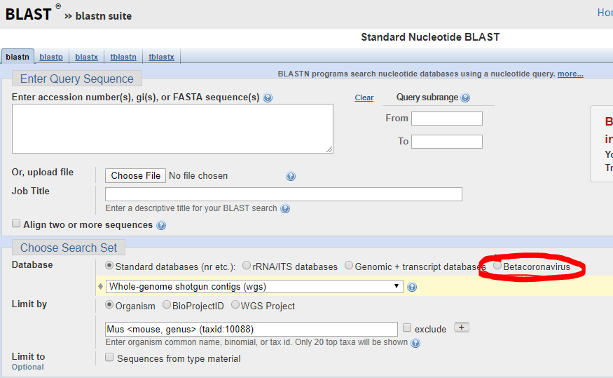
With the rapid spread of COVID-19 in the Unites States, the number of questions that have arisen about lab safety has climbed exponentially. I am excited about any uptick in interest in laboratory safety issues, but I wish it didn’t take a world-wide pandemic to cause it. The Centers for Disease Control (CDC) has offered very good lab safety instruction for the processing and testing of COVID-19 specimens          (https://www.cdc.gov/coronavirus/2019-ncov/lab/index.html), and virus testing is being performed in more labs each day.

It is vital for laboratorians to remember this. While the coronavirus is not to be taken lightly, the patient specimens we handle every day contain biohazards that are far more dangerous to us than COVID-19. Hepatitis, HIV, select agents, and many other pathogens reside in the blood and body fluids processed and tested in laboratories across the country, and many of the illnesses these agents can cause are very hazardous to human health. I hope we remember that when the hype about this latest virus passes.

Use Standard Precautions when working in the laboratory. Wear lab coats, gloves, and face shields. When you see a co-worker who is not properly attired, offer them the PPE they need. If you see an unsafe practice like eating, drinking, or using cell phones in the department, end it quickly. That is how infections occur, and that is how they spread into the community. Remember, unsafe behaviors can have a direct affect on the safety of the entire team. The sooner we can help everyone to understand that, the better we will all be at coaching others. In the original 1958 version of “The Fly,” the title character is caught in a spider’s web. His famous (and often imitated) last words were, “Help me! Help meeeee!” The scientist practiced unsafe behaviors until it was too late to turn back. Don’t let that be the case for anyone in your laboratory!

Dan Scungio, MT(ASCP), SLS, CQA (ASQ) has over 25 years experience as a certified medical technologist. Today he is the Laboratory Safety Officer for Sentara Healthcare, a system of seven hospitals and over 20 laboratories and draw sites in the Tidewater area of Virginia. He is also known as Dan the Lab Safety Man, a lab safety consultant, educator, and trainer.

Microbiology Case Study: 41 Year Old Male with Complaints of Abdominal Pain for One Month

Case History

A 41 year old African male presented to the ED with complaints of abdominal pain, weight loss, and decreased appetite over one month. He immigrated from Zimbabwe 10 years prior, and most recently visited 4 months ago. His past medical history is non-contributory. He was found to have microcytic anemia (Hgb 9.3 g/dL, MCV 77.0 fL), pneumonia, and focal small bowel dilation with thickening and inflammatory changes on abdominal CT.

Endoscopy revealed a large, villous, infiltrative mass in the third portion of the duodenum. On histologic examination of the duodenum biopsy, viable Schistosoma eggs were seen within the vessels in the lamina propria and associated with adenocarcinoma (Images 1 and 2). Schistosoma mansoni eggs were identified on stool ova and parasite exam (Image 3), and serology was positive for Schistosoma IgG antibody.

Image 1. 10x objective magnification of a hematoxylin and eosin stained histology slide of the duodenal biopsy with associated adenocarcinoma and multiple eggs can be visualized.
Image 2. 40x objective magnification of a hematoxylin and eosin stained slide from the duodenal biopsy of a man from Africa who presents with abdominal pain who is found to have a mass on endoscopy. Seen is a viable egg with intact miracidium and no evidence of calcification.
Image 3. 100x oil immersion objective magnification of an iodine prep from a concentrated formalin-fixed stool specimen demonstrating a Schistosoma mansoni egg with its characteristic large lateral spine.

Discussion

Although this is not a common infection to encounter in the US, prevalence in endemic areas ranges from 30-100%. Infection occurs through contact with water contaminated with human feces; common circumstances are irrigation ditches as well as bathing and washing water.1

Schistosomiasis may be an indolent infection in many immunocompetent hosts. Chronic infection requires a shift from inflammatory TH1 response to a modulatory TH2 response.2 There is also evidence that Schistosoma infection may downregulate the immune response by inducing M2 differentiation of macrophages.3 These anti-inflammatory macrophages have been associated with a microenvironment favorable to malignancy. It has also been shown that S. mansoni is a risk factor for hepatocellular carcinoma (HCC) and colonic adenocarcinoma, possibly by altering p53 activation, initiation of chronic granulomatous response that blocks venules, increasing cell turnover, and promotion of ROS and RNOS production.4 Anti-idiotype antibodies produced in chronic infection may also down-regulate both specific and non-specific immune responses.5

This case is an unusual presentation in the US in that viable ova are typically not seen, and S. mansoni is more likely to involve the distal colon and liver rather than the small bowel. There is a known association between S. mansoni infection and HCC, and there are sporadic reports of association with tumors of the prostate, ovary, uterus, and cervix.5,6 To our knowledge however, there are no other reported cases of duodenal adenocarcinoma with concurrent schistosomiasis.

References

  1. Chai J and Jung B. Epidemiology of Trematode Infections: An Update. 2019. Adv Exp Med Biol. 1154:359-409.
  2. Barsoum RS, et al. Human Schistosomiasis: Clinical Perspective: Review. 2013. Journal of Advanced Research. 4:433-44.
  3. Hussaarts L, et al. Chronic helminth infection and helminth-derived antigens promote adipose tissue M2 macrophages and improve insulin sensitivity in obese mice. 2015. FASEB J. 29(7):3027-39.
  4. El-Tonsy MM, et al. Schistosoma mansoni infection: Is it a risk factor for development of hepatocellular carcinoma? 2013. Acta Trop. 128(3):542-7.
  5. Palumbo E. Association Between Schistosomiasis and Cancer. 2007. Infectious Diseases in Clinical Practice. 15(3):145-8.
  6. Peterson MR and Weidner N. Gastrointestinal neoplasia associated with bowel parasitosis: real or imaginary? 2011. J Trop Med. 2011:234254.

-Daniel Welder, MD is a second year Clinical Pathology resident at UT Southwestern Medical Center in Dallas, Texas. He has interests in Hematopathology, Transfusion Medicine and dabbles in Microbiology.

-Dominick Cavuoti, DO is a Professor at UT Southwestern in the Department of Pathology. He is multifaceted and splits his time as the Medical Director of the Parkland Hospital Clinical Microbiology Laboratory and Parkland Cytology attending among other administrative and educational activities.

-Clare McCormick-Baw, MD, PhD is an Assistant Professor of Clinical Microbiology at UT Southwestern in Dallas, Texas. She has a passion for teaching about laboratory medicine in general and the best uses of the microbiology lab in particular.

An Asymptomatic 52 Year Old Female with a Surprise Finding on Colonoscopy

Case Presentation

A 52-year-old female with no significant past medical history is seen for a routine annual examination and is scheduled for a colonoscopy due to her age being over 50 years. The colonoscopy was performed and an isolated single worm was found within the cecum (Images 1-2). The worm was removed with cold forceps and subsequently placed in paraffin and sectioned (Images 3-5).

Image 1. The worm is depicted within the cecum attached to the mucosal wall by its anterior end.
Image 2. The worm is captured using cold forceps.
Image 3. Hematoxylin and eosin stained section of the worm.
Image 4. Higher power magnification, showing eggs with distinctive characteristic bilateral polar plugs and barrel shape.
Image 5. Higher power magnification, showing eggs with distinctive characteristic bilateral polar plugs and barrel shape.

Discussion

The worm was identified as Trichuris trichiura. The common name for this organism is the whipworm. It belongs to the Nematode classification of parasites, which are commonly referred to as roundworms. Adults measure up to 5 cm in length and have a tapered or whip-like anterior end. The eggs measure 50 x 25 µm, and have brownish thick shells on stool smear. The eggs also have a barrel shape and distinctive protruding polar plugs at each end. These morphologic characteristics of the egg are diagnostic of Trichuris trichiura. The lack of a tissue migration phase and a relative lack of symptoms characterize whipworm infection, with only those with a heavy parasite burden becoming symptomatic. If these symptoms do arise, they are usually mild, ranging from loose stools with minimal blood loss and nocturnal stools, to iron deficiency anemia and vitamin deficiency. As parasite burden increases, however, symptoms can progress to dysentery, colitis, or rectal prolapse. Prolapse is more frequent in the Pediatric population, but has been described in adults as well.

Trichuris trichiura has one of the simplest of the Nematode life cycles. Eggs are unintentionally ingested, hatching in the small intestine by way of exploitation of signaling molecules from the intestinal microbiome. The larvae then burrow through the villi and continue maturing in the wall of the small intestine. They then return to the intestinal lumen, migrating to the cecum and subsequently into the large intestine, where they finish the process of maturation. Finally, the worm uses its anterior end to anchor into the bowel mucosa, where it feeds on tissue secretions and uses its posterior end for reproduction and laying eggs. Female worms can live from 1-5 years and can lay up to 20,000 eggs per day.

Whipworm infection is principally a problem in tropical Asia and, to a lesser degree, in Africa and South America. Children are most commonly infected, and can experience failure to thrive as well as cognitive and developmental defects. Transmission is by the fecal-oral route, explaining the large incidence of infection in children from developing countries, as they are far more likely to be in physical contact with soil and environmental contaminants, with subsequent placement of their fingers in their mouths. The fecal-oral route can also be facilitated by improper washing and cooking of fruits and vegetables, as well as overall poor hygiene, no matter what the geographical location. In the United States, whipworm infection is exceedingly rare. When it does happen, it is most commonly seen in the rural Southeast. Although it is rare, the incidence of infection is reported to be as high as 2.2 million individuals within the United States, with 1-2 billion cases worldwide.

Studies often reveal eosinophilia in nematode infections from ongoing tissue invasion. However, the lack of a tissue migration phase in Trichuris life cycles makes this a rare laboratory finding. Other studies such as anemia can give an indication to the presence of the worm. Characteristic egg morphology on stool smear remains the cheapest and easiest way to diagnose infection, but polymerase chain reaction using new sequencing techniques are now available in some laboratories to detect the presence of Trichuris with great sensitivity and specificity. The parasite burden can be quantified per gram of stool by the Kato-Katz technique. This procedure filters stool through mesh, with the filtered sample being placed within a template on a glass slide. The template is then removed and the remaining fecal material is removed with a piece of cellophane soaked in glycerol, leaving only eggs on the slide.

Discovery of T. trichiura in our patient was an unexpected finding, as our patient had no symptoms.  Asymptomatic detection of T. trichiura has been described in the past, so this finding is not unique. The medication of choice is mebendazole, showing a cure rate of 40-75%. The drug works well by inhibiting glucose uptake from the gastrointestinal tract of the helminth. However, this drug is very expensive, and as a result is difficult to obtain. The patient is currently receiving an alternative drug called albendazole as outpatient therapy and will be switched to mebendazole as soon as resources become available should the need remain. The patient is following up with her primary care physician and is expected to make a full recovery.

References

  1. Donkor, Kwame; Lundberg, Scott;
    https://emedicine.medscape.com/article/788570-overview. Trichuris trichiura (Whipworm) Infection (Trichuriasis).
  2. Sunkara T, Sharma SR, Ofosu A. Trichuris trichiura-An Unwelcome Surprise during Colonoscopy. Am J Trop Med Hyg. 2018 Sep;99(3):555-556. doi: 10.4269/ajtmh.18-0209. PubMed PMID: 30187847; PubMed Central PMCID: PMC6169157.

-Cory Gray, MD is a second year resident in anatomic and clinical pathology at the University of Chicago (NorthShore). His interests include hematopathology and molecular and genetic pathology, as well as medical microbiology.

-Erin McElvania, PhD, D(ABMM), is the Director of Clinical Microbiology NorthShore University Health System in Evanston, Illinois. Follow Dr. McElvania on twitter @E-McElvania. 

Is It Safe to Process Histopathology Samples from Suspected Cases of COVID19?

According to this recently-published study in the Journal of Histotechnology, ” … experts are confident that 70% ethanol and 0.1% sodium hypochlorite should inactivate the virus. Formalin fixation and heating samples to 56oC, as used in routine tissue processing, were found to inactivate several coronaviruses and it is believed that 2019-nCoV would be similarly affected.”

Read the study: https://doi.org/10.1080/01478885.2020.1734718

How to Validate a COVID-19 Assay

The FDA is now democratizing the testing of the novel coronavirus: SARS-CoV-2 (the virus which causes the COVID-19 disease syndrome—I will call it COVID-19 from here on as that is the colloquial name most people know) by allowing high complexity testing labs across the United States. This move will permit more labs to test for COVID-19. A previous post by contributor Constantine Kanakis describes the biology of the virus, so I will not repeat that material. Instead, I will focus on some considerations in validating a Lab Developed Test (LDT) COVID-19 molecular assay.

The president of AACC, Carmen Wiley, said there are 11,000 high complexity testing labs in the US, which could qualify for performing this testing. However, not all of these labs have molecular and virology expertise, so others have placed the number of labs with qualified staff and instrumentation at 400.

Published Assays and Targets: As an overview, the figure below (Figure 1) summarizes some published COVID-19 assays. As you can see, the major strategy involves using the TaqMan probe strategy where a short probe is degraded by Taq polymerase releasing a fluorescent molecule (green ball) from a quencher molecule (blue ball). The TaqMan approach allows for quick performance of the assay and easy interpretation. One lab from Japan is using nested PCR amplification and sequencing of the Orf1a and S genes as well.

Figure 1. The COVID-19 genetic structure is abbreviated above with the different genes targeted displayed. The names of institutions that have published their assay procedure along with the TaqMan reagents that were reportedly used with each assay are shown above. Primers are represented by small arrows with a TaqMan probe in the middle represented by a black line with green and blue circles indicative of the fluorescent molecule and its quencher. The double set of arrows for the Japanese assay represents a nested PCR strategy.

In silico Cross-reactivity:

The FDA guidance allows cross-reactivity to be minimally assessed in silico by demonstrating “greater than 80% homology between primer/probes and any sequence present in the targeted microorganism.” The primer locations can be found in the publication of each protocol (except Thermo) and can be confirmed by checking the NCBI Blast site and they actually have a selection for beta-cornavirus (Figure 2) now that allows you to search for your primer’s reactivity across other related viruses- Very helpful!

Figure 2. Select Betacornavirus before entering your primer/probe sequence to confirm cross-reactivity.

Primer/Probe Design:

The N region is the most popular site to probe and is included in most kits once and the CDC kit three times. It was the reagent set for N3 in the CDC kit that was having difficulties, so you may decide to not include that component in your LDT. If you want to see how the different available primer sets align on the N gene sequence you can see below for the primers labeled based on their source. Many are overlapping, perhaps because many people thought the same site was a good target (Figure 3).

Figure 3. N-gene of COVID-19 along with labeled primers from some published assays. The information on the source of the sequence is shown on the bottom right with the link.

Commercially Available Assays:

An important part of validating your COVID-19 assay is to do so quickly. Thus commercially available kits would be helpful, however there are only two commercially available sources at this time: IDT and Thermo. IDT is producing a kit with the CDC design. Thermo produced their kit over the last few months and does not have any published validation information that I could find. Also Thermo when I checked just now for the catalog number, it says this product is unavailable… not sure what that means, but maybe you can try contacting them. Both IDT and Thermo list control plasmid reagents for their assays.

Controls for the Assay:

The wording of the FDA announcement was interesting in that it 1) did not require clinical samples, but allows “contrived clinical specimens.” “Contrived reactive specimens can be created by spiking RNA or inactivated virus into leftover clinical specimens.” A major difficulty is the access to actual COVID-19 RNA or inactivated virus. I noticed that the guidance didn’t say that the assay MUST use RNA. Thus most labs would have access to plasmid DNA, which could potentially be used.

Given the limited availability of RNA for validation use, a lab may consider performing much of the assay optimization with COVID-19 Plasmid DNA while waiting for access to RNA. I would like to be sure my assay could extract, amplify and detect RNA as part of the clinical validation.

Asuragen can produce Armored RNA, with synthetic RNA packaged inside of a viral capsid, which would be a useful control for extraction, amplification and detection. However, we heard this will not be available for another month.

Tom Stenzel (director of the Office of In Vitro Diagnostics and Radiological Health at the FDA’s Center for Devices and Radiological Health (CDRH)) said FDA, BARDA, and the CDC will prioritize and coordinate shipments of viral materials to labs when they are ready to validate tests according to a webinar with labs on Monday. Currently, the FDA is directing inquiries to BEI, which is reportedly prioritizing requests to send out samples in 12-72 hours.

Lastly, one could try to use in vitro synthesized RNA sequences surrounding your primer targets as a control for now and may have better luck in getting the product soon. This is the control that is being shipped with the CDC kits to public labs.

Limit of Detection is an unknown for what is likely to be clinically relevant as we don’t know what the levels look like in people with early vs. late vs. severe vs. mild disease. The FDA just says you should be able to detect 95% of samples (19 of 20) that are x1-x2 the limit of detection.

FDA Notification:

This is the final and important step. Once you go live, you must notify the FDA with an Emergency Use Assay (EUA) form within 15 days. Reviewing the form, there doesn’t appear to have complex explanations or overdue requirements for reporting, which wouldn’t be found in a standard lab validation document.

Final Thoughts/Future commercial solutions:

This information is the best of what I know right now based on current information- this is not a complete guide and the FDA guidance should be read closely for all compliance details. Information is changing quickly and is likely to change more if the number of COVID-19 cases in the United States increases. Cepheid, Luminex, and BioFire are reportedly working on assays that will be out in several months and would be easy to use for many labs that already have one or both of these systems-however it may require a full validation for an LDT, but I’m not sure as it is an EUA-further clarification on this point is needed. Although there are several commercial solutions available, we don’t know how demand could impact supply from each company. Fortunately, some large reference labs like LabCorp and Quest are looking to develop a COVID19 test. Good luck, stay safe, and feel free to contact me with any questions in the comments below so that everyone can benefit from the discussion!

References

In lieu of a list of references, I’ve included web links for the most current and direct sources of information.

-Jeff SoRelle, MD is a Chief Resident of Pathology at the University of Texas Southwestern Medical Center in Dallas, TX. His clinical research interests include understanding how the lab intersects with transgender healthcare and improving genetic variant interpretation.

FDA Guidance to Develop Novel Molecular Diagnostic Tests for SARS-CoV-2

A laboratory advisory from the CDC:

The Food and Drug Administration (FDA) issued new guidance on February 29, 2020, for laboratories to be able to develop novel coronavirus (COVID-19) molecular diagnostics tests and begin use prior to obtaining Emergency Use Authorization (EUA). This permits laboratories that are CLIA certified and meet requirements to perform high complexity testing to start offering severe acute respiratory syndrome coronavirus 2 (SARS-CoV-2) molecular diagnostic testing after validation is completed as outlined in the guidance. Laboratories should submit an EUA request to the FDA within fifteen business days after validation. FDA will be hosting a webinar to provide more information on March 2, 2020, at 3 pm ET.

Clinical laboratories should contact their state health departments for guidance if they have a suspected COVID-19 case specimen. Clinical laboratories should NOT attempt viral isolation from specimens collected from COVID-19 persons under investigation (PUIs). For interim guidelines for collecting, handling, and testing clinical specimens from PUIs for COVID-19, please see the CDC Coronavirus Disease 2019 (COVID-19) website.

Microbiology Case Study: A 52 Year Old with a Liver Abscess

Clinical history

A 52 year old patient with a history of recent travel to India presented to interventional radiology from an outside hospital for aspiration of a liver abscess, and was subsequently returned to the outside hospital. The patient had spent 2 months in India before returning to the US, and about 1 month later developed right upper quadrant pain. Abdominal CT showed 2 cystic masses measuring 2-4cm. Aspiration of the cysts yielded 0.5mL of bloody fluid, which was sent for bacterial culture and smear. Infectious disease prescribed antimicrobial treatment consisted of ceftriaxone and metronidazole, followed by paromomycin and levofloxacin.

Laboratory findings

A gram smear of the patient’s liver mass aspirate showed few neutrophils and no bacteria. Culture of the aspirate showed no growth at 5 days. Multiple sets of blood cultures collected at the outside hospital all showed no growth at 5 days.

Image 1. Entamoeba coli trophozoite seen in the patient’s stool ova and parasite exam, demonstrating an eccentric karyosome and coarse, irregular peripheral chromatin.

A single ova and parasite exam of the patient’s stool was sent and showed few Entamoeba coli trophozoites. A sample of the patient’s blood was sent to the Mayo reference lab for serum Entamoeba histolytica antibody testing, which came back positive. Stool was sent for Entamoeba histolytica antigen testing which was negative.

Discussion

Entamoeba coli is a non-pathogenic protozoan that can exist as a commensal organism in the human gastrointestinal tract. This organism has not been established to have any disease causing effect per se, but its presence may indicate exposure to water sources that could contain parasitic organisms. (3)

Entamoeba histolytica, by contrast, is a parasitic protozoal pathogen. Most infections are asymptomatic, but they can manifest as amebic dysentery or extraintestinal disease. The most common extraintestinal manifestation is amebic liver abscesses.1

Intestinal amebiasis occurs via ingestion of amebic cysts, typically through contaminated food or water, but also through other forms of fecal-oral contact. Infections are seen most commonly in areas with poor sanitation, but can be found in developed countries in patients who have migrated from or traveled to endemic areas.2

Once the amebic cysts pass into the small intestine, they form trophozoites, which are able to penetrate the mucous barrier of the gut and destroy intestinal epithelial cells. This leads to blood and mucus in the stool. (2) Once the amebae penetrate the gut wall, they are able to reach the blood and ascend through the portal system to the liver and form amebic liver abscesses.3

Clinical presentation of these abscesses typically includes right upper quadrant pain and fever in a patient with a history of travel to an endemic area. Serologic testing is used for confirmation if clinical presentation and imaging are suggestive, but this cannot distinguish between current infection and prior exposure, and up to 35 percent of uninfected inhabitants of endemic areas show positive serology.3 Stool microscopy may be the initial, and indeed only test available in some areas, but cannot differentiate E. histolytica from non-pathogenic E. dispar and E. moshkovskii strains.2

Image 2. E. histolytica trophozoite with ingested red blood cell, visible as a dark inclusion, as well as demonstrating a central karysome and fine, uniformly distributed chromatin. (CDC: https://www.cdc.gov/dpdx/amebiasis/index.html) Erythrophagocytosis is suggestive of E. histolytica.

Empiric treatment in the setting of consistent epidemiology, clinical picture, and radiology consists of metronidazole or tinidazole for tissue clearance followed by paromomycin, diiodohydroxyquin, or diloxanide furoate for intraluminal clearance.

Infectious diseases was taking care of this patient and decided her clinical syndrome is probably extraintestinal Entamoeba histolytica amoebiasis based on the results of the CT findings and the antibody in the right clinical setting.  Although her stool ova and parasite only showed Entamoeba coli, she clearly has been exposed to contaminated food or water.  In addition, the Entamoeba histolytica stool antigen was negative, but this can be an insensitive test.

References

  1. Leder, Karin, and Peter F. Weller. “Extraintestinal Entamoeba histolytica amebiasis.” UpToDate, Wolters Kluwer, 27 Jan. 2020, http://www.uptodate.com/contents/extraintestinal-entamoeba-histolytica-amebiasis?search=entamoeba%20histolyticatreatment&topicRef=5727&source=see_link. Accessed 4 Feb. 2020.  
  2. Leder, Karin, and Peter F. Weller. “Intestinal Entamoeba histolytica amebiasis.” UpToDate, Wolters Kluwer, 27 Jan. 2020, http://www.uptodate.com/contents/intestinal-entamoeba-histolytica-amebiasis?search=entamoeba%20histolyticatreatment&source=search_result&selectedTitle=1~46&usage_type=default&display_rank. Accessed 4 Feb. 2020.
  3. Weller, Peter F. “Nonpathogenic enteric protozoa.” UpToDate, Wolters Kluwer, 25 July 2019, http://www.uptodate.com/contents/nonpathogenic-enteric-protozoa?search=entamoeba%20coli%20treatment&source=search_result&selectedTitle=1~6&usage_type=default&display_rank=1. Accessed 4 Feb. 2020.

-Tom Koster, DO is a 1st year Anatomic and Clinical Pathology Resident at the University of Vermont Medical Center.

-Christi Wojewoda, MD, is the Director of Clinical Microbiology at the University of Vermont Medical Center and an Associate Professor at the University of Vermont.

The Forensic Pathologist as Patient Advocate

Patient advocates are simply people who care about patients as fellow human beings enough to act on that care. Forensic pathology fascinates many people, but hardly anyone realizes how strongly forensic pathologists advocate for patients.

Forensic pathologists have the responsibility of identifying human remains and determining the cause and manner of death for individuals that die suddenly and unexpectedly. Most often, we accomplish this mission by performing an autopsy. Death makes many people uncomfortable, and we’re accustomed to grim jokes about their work when meeting someone. Typically these comments carry an undertone that because decedents cannot talk, we don’t need interpersonal skills. Not only is this untrue, comments such as these provide an educational opportunity.

It is true that our patients have already died, but the relatives of our patients are very much alive. Those relatives have needs that we work to provide and questions we strive to answer. The most common question relatives have is “Why did my loved one die?” which is precisely what the pathologist is working to determine. We regularly talk with relatives of decedents that we’ve examined. We can tell family members why death occurred, including any implications that the death has for remaining members of the family. We can also help families begin to work through the social and bureaucratic requirements that death brings for those still living, such as the need to make arrangements for the disposition of the body and the need for a death certificate. (For example, after a person dies, that person’s financial accounts are frozen until a death certificate becomes available to unlock the accounts.)

Forensic pathologists work to develop a good relationship with the decedent’s relatives. Because anger and bargaining are part of grieving, conversations with relatives sometimes begin as though the relative and the pathologist are adversaries, but with time and compassion, the relationship usually transforms into a more appropriate professional relationship. A particularly important aspect of family interactions is listening to a grieving relative, because listening with care helps someone who is grieving. Attempting to build a good relationship with the decedent’s relatives does not mean that the pathologist is a blind advocate for the family. We won’t change the cause of death so that the family can reap more financial benefit from the death, for example. Lies will not help someone pass through the process of grieving in a healthy way – truth, time, and patient, loving care are the necessary therapeutic measures.

In the case of homicides, forensic pathologists advocate for the decedent by calling the death what it is and then testifying to the medical facts of that death when a suspect is tried in court. The pathologist testifies to the medical aspects of what caused death without trying to ensure that the suspect is either convicted or acquitted. Trying to sway the jury’s verdict is the work of attorneys; presenting the medical facts of why and how the decedent died is the work of the pathologist.

Forensic pathologists advocate for public health by providing an accurate cause of death. Death certificate data provide an essential component for assessing public health, and those data are an important determinant for allocation of medical research funds and for interventions to improve public health.

Like other pathologists, forensic pathologists typically do their work quietly in the background, advocating for their unique patients in their own special way. People give little thought to professional interactions with a forensic pathologist until forced to do so; in that difficult time we try to serve as best we can.

-Gregory G. Davis, MD, FASCP graduated from Vanderbilt Medical School and trained in pathology at Vanderbilt University Medical Center, Nashville, TN, followed by a fellowship in forensic pathology at the San Diego County Medical Examiner Office in San Diego, CA. Dr. Davis then joined the faculty at the University of Alabama at Birmingham, where he currently serves as a Professor and as Director of the Forensic Division of the Department of Pathology. Dr. Davis also serves as Chief Coroner/Medical Examiner for Jefferson County, Alabama, the county in which Birmingham is located. Dr. Davis has earned a Master of Science in Public Health from the UAB School of Public Health. His research interest is the application of epidemiology to the study and practice of forensic pathology, especially drug abuse. He has published 74 peer-reviewed manuscripts, including serving as lead author on the 2013 opioid position paper of the National Association of Medical Examiners. He is currently working as chair of a panel revising and updating the NAME opioid position paper for expected publication in 2020. He serves on the editorial boards of the Journal of Forensic Sciences and Forensic Science, Medicine, and Pathology. Dr. Davis is a Fellow At-Large Director on the Board of Directors of the American Society for Clinical Pathology.