Hematopathology Case Study: What’s in Those Histiocytes?

Case history

A 50 year old female with a past medical history significant for Sjogren’s syndrome and ventricular tachycardia s/p ICD placement presented for a routine chest X-ray in which a 1.8 cm spiculated left upper lobe lung mass was identified. A subsequent PET scan revealed FDG avidity. Other Imaging revealed no lymphadenopathy. The patient is a non-smoker and has no other comorbidities. A core needle biopsy with fiducial placement was performed.

Diagnosis

histio1
H&E 10x
histio2
H&E, 20x
histio3
H&E, 50x
histio4
CD3
histio5
CD20
histio6
CD79a
histio7
IgG
histio8
CD68
histio9
CD138
histio10
Kappa ISH
histio11
Lambda ISH

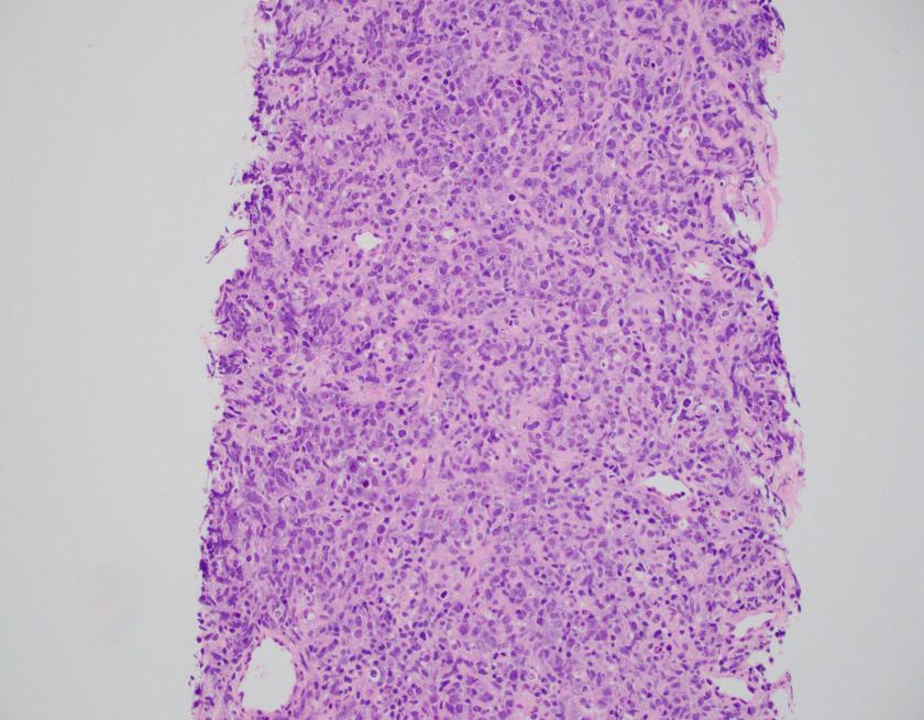
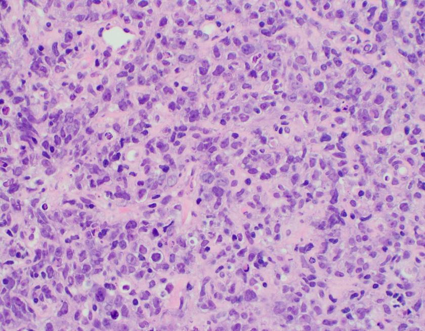
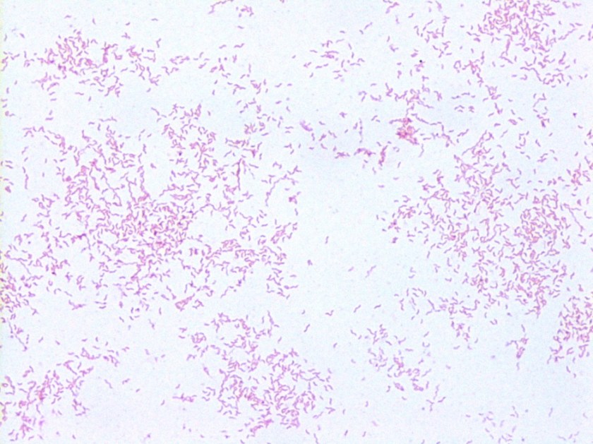
Sections of lung core biopsy material show numerous histiocytes containing eosinophilic intracytoplasmic globular inclusions. An admixed population of plasma cells are seen which are present in aggregates along with mature appearing lymphocytes. The plasma cells also demonstrate globular inclusions within their cytoplasm.

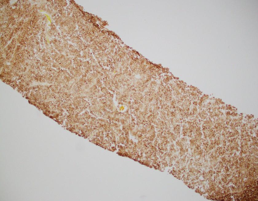
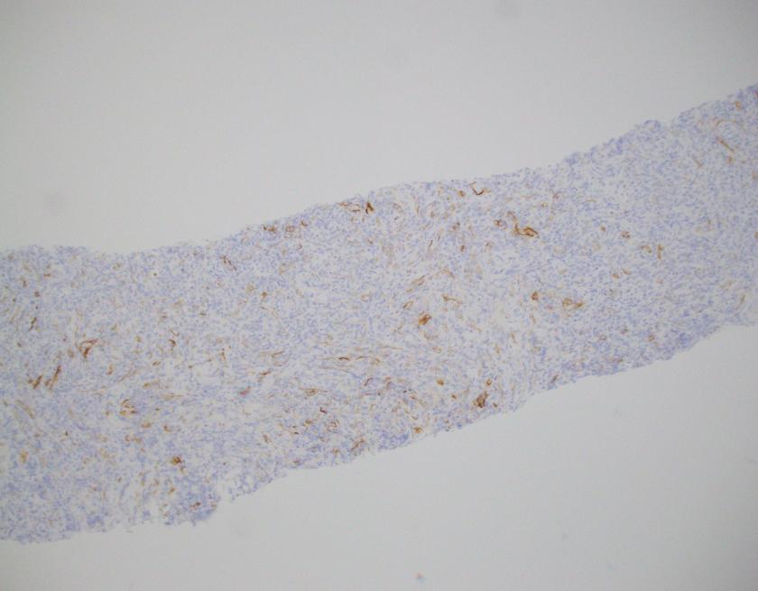
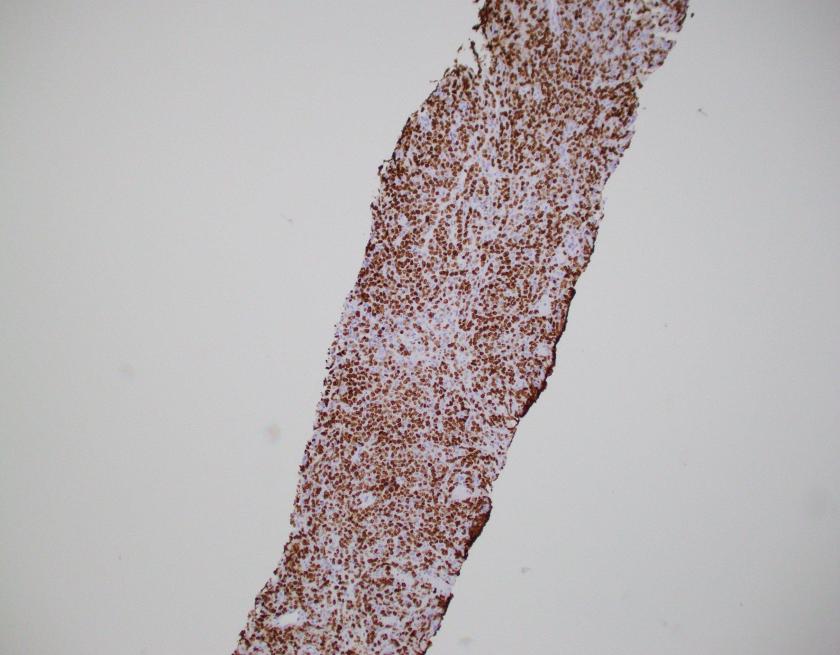
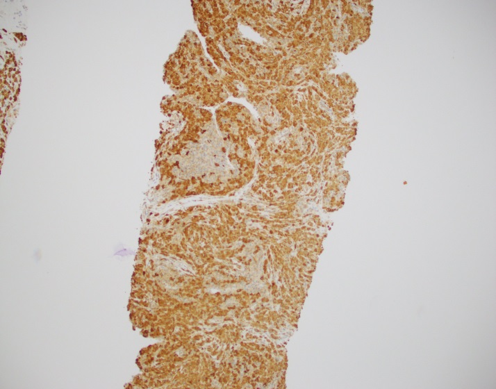
By immunohistochemistry, CD3 highlights scattered mature T-cells while CD20 highlights B-cells present in focal aggregates. Numerous plasma cells are present and are positive for CD138, CD79a, BCL2, and MUM1. By in situ hybridization, plasma cells are greatly kappa predominant. IgG is positive in the majority of the plasma cells with only rare cells staining for IgA and IgM. CD68 is positive in the numerous histiocytes.

IGH gene rearrangement studies by PCR demonstrated was positive, indicating a clonal population.

Overall, the findings are consistent with a crystal-storing histiocytosis with an associated plasma cell neoplasm or low-grade B-cell lymphoproliferative disorder.

Following the diagnosis, the patient received stereotactic body radiation therapy given the localized findings.

Discussion

In this case, the findings are morphologically consistent with crystal-storing histiocytosis (CSH), which is a rare lesion that is the result of intralysosomal accumulation of immunoglobulin. The immunoglobulin is stored as crystalline structures within histiocytes that occupy the vast majority of a mass forming lesion. Multiple sites can be involved, which include bone marrow, lymph nodes, liver, spleen, gastrointestinal tract, and kidney. Most often, the lesion is confined to a single site but occasional generalized forms with multiple organ involvement have been described. CSH is also often associated with B-cell lymphoproliferative disorders or plasma cell dyscrasias, but rarely are the result of chronic inflammatory conditions.

The assessment of CSH requires excellent staining to identify the quality of the histiocytes. As mentioned, CSH will show intracytoplasmic inclusions that are eosinophilic in nature. Mimickers of CSH include mycobacterial and fungal infections, mycobacterial spindle cell pseudotumor, malakoplakia, HLH, storage disorders such as Gaucher’s, as well as histiocytic lesions such as xanthogranuloma, Langerhans cell histiocytiosis, fibrous histiocytoma, Rosai Dorfman disease and rarely other eosinophilic tumors such as rhabdomyoma, granular cell tumor, and oncocytic neoplasms.1

A thorough review of the literature as well as a clinicopathologic study by Kanagal-Shamanna R et al revealed that the localized type of CSH was the dominant presentation in which over 90% of cases showed isolated masses. Per previous reviews, localized lesions were often found in the head and neck as well as lung.2 A study group in which 13 cases that showed CSH, 12 demonstrated an underlying lymphoma or plasmacytic neoplasm. Interestingly, in 5 of the cases, the histiocytic infiltrate was so prominent and dense that it obscured the underlying neoplasm. In these particular cases, immunohistochemistry and PCR were of great importance.

Although the majority of cases of CSH are the result of an underlying lymphoproliferative disorder or plasma cell neoplasm, rare cases of report inflammatory processes have been described, particularly in the setting of an immune mediated process such as rheumatoid arthritis or Crohn disease.

Overall, although a rare entity, it is important to be aware of CSH and its mimickers as this can be an elusive diagnosis to make, especially when the histiocytic infiltrate is dense.

References

  1. Kanagal-Shamanna R, et al. “Crystal-Storing Histiocytosis: A Clinicopathologic Study of 13 Cases,” Histopathology. 2016 March; 68(4): 482-491.
  2. Dogan S, Barnes L, Cruz-Vetrano WP “Crystal-storing histiocytosis: a report of a case, review of the literature (80 cases) and a proposed classification,” Head Neck Pathol. 2012; 6:11-120.


PhillipBlogPic-small

-Phillip Michaels, MD is a board certified anatomic and clinical pathologist who is a current hematopathology fellow at Beth Israel Deaconess Medical Center, Harvard Medical School, Boston, MA. His research interests include molecular profiling of diffuse large B-cell lymphoma as well as pathology resident education, especially in hematopathology and molecular genetic pathology.

Microbiology Case Study: A 7 Month Old Female with Fever and Seizure-Like Episodes

Case History

A 7 month old female presented to the emergency department (ED) due to fever and seizure-like episodes. Her mother reported the child had been persistently febrile for 5 days (Tmax 103.9°F) with rhinorrhea, fussiness and decreased oral intake. The patient experienced 3 seizure-like episodes on the day of admission, which the mother described as periods of “shaking” with eyes rolling back. The child was unresponsive during these episodes, which lasted 1 to 2 minutes each. The child had been taken to her pediatrician the day prior to presentation to the ED where she was given a shot of ceftriaxone for presumed otitis media. The child received a chest x-ray, influenza testing, and blood and urine cultures were collected. She also had a lumbar puncture performed and the cerebral spinal fluid (CSF) was sent for chemistries, bacterial culture and polymerase chain reaction (PCR) testing for meningitis/encephalitis pathogens. She was started on IV ceftriaxone.

Laboratory Testing

The child’s white blood cell count from peripheral blood was 7.1 TH/cm2 and chest x-ray, urinalysis and flu testing were unremarkable. The CSF was clear and colorless with 7 WBC/cm2, glucose of 57 mg/dL and protein of 21 mg/dL. The cytospin Gram stain identified no organisms. The meningitis/encephalitis panel detected the presence of human herpesvirus 6 (HHV-6).

Discussion

Human herpesvirus 6 is a member of the Herpesviridae family and was the sixth herpes virus identified. Structurally, HHV-6 possesses a double stranded DNA genome and is enveloped. Clinically, it is the etiologic agent of roseola infantum (exanthum subitum) in infants and toddlers. Primary infection occurs in early childhood and those infected can be asymptomatic or have a non-specific febrile illness while only the minority present with the characteristic red macular rash prominent on the trunk and extremities, lymphadenopathy and high fevers. HHV-6 is highly neurotropic and as such causes viral encephalitis with 5-15% of children experiencing febrile seizures as a result of this illness. HHV-6 is highly prevalent with a greater than ninety percent seropervalence rate. HHV-6 establishes latency in T lymphocytes and can reactivate & cause disease, especially in immunocompromised patients such as those recipients of stem cell or solid organ transplants.

Traditional laboratory methods of identification for HHV-6 were challenging as viral culture, while once the gold standard for active disease, is not practical for most labs and is no longer used in routine diagnostics. PCR from serum, plasma or CSF has become the preferred test as there are now FDA-cleared, commercial platforms that are easy to use, allow for rapid turnaround time and in the case of multiplex PCR panels, the ability to target multiple pathogens from one test. Serology, while helpful in the diagnosis of primary infections, may not be provide conclusive results in a timely manner and is of limited utility in reactivation. Other less commonly used methods include immunohistochemistry, in situ hybridization and electron microscopy.

The prognosis for patients infected with HHV-6 is generally good with self-limited illness not requiring treatment. Rarely, multi-organ involvement can occur and HHV-6 infection in immunosuppressed patients can be a major cause of morbidity and mortality. There is no antiviral therapy licensed for the treatment of reactivated disease in this setting, but approaches using ganciclovir and valganciclovir have been proposed.

In the case of our patient, her blood, urine and CSF cultures were negative and her antibiotics were stopped after cultures were no growth at 24 hours. She required no treatment other than supportive care with acetaminophen for fever control. Prior to discharge, she developed a fine rash on her face, the back of her neck and trunk that was characteristic of an HHV-6 rash. This case demonstrates the utility of multiplex PCR testing in providing rapid identification of pathogenic organisms allowing for real time diagnosis and the limiting of unnecessary treatment.


ET

-Eric Tillotson, MD, is a second year Anatomic and Clinical Pathology resident at the University of Mississippi Medical Center.

Stempak

-Lisa Stempak, MD, is an Assistant Professor of Pathology at the University of Mississippi Medical Center in Jackson, MS. She is certified by the American Board of Pathology in Anatomic and Clinical Pathology as well as Medical Microbiology. She is the director of the Microbiology and Serology Laboratories. Her interests include infectious disease histology, process and quality improvement and resident education.


Microbiology Case Study: A 56 Year Old Woman with Purulent Vaginal Discharge and Dysarthria

Case History

The lab received a purulent fluid from a frontal subdural empyema in a 56 year old woman with a several month history of sporadic bloody to purulent vaginal discharge and a two week history of severe headache, facial weakness, and recent dysarthria causing her to seek treatment. Pelvic exam and CT of the abdomen revealed a fungating cervical mass, later found to be adenocarcinoma, with possible fluid collection within the uterine cavity.

Lab Identification

Initial gram stain of the subdural fluid showed moderate neutrophils and no bacteria. The aerobic plate showed no growth at 48 hours. The thioglycollate broth grew “puffballs” containing long gram negative bacilli with tapered ends.


fusnuc1
Anaerobic blood agar plate on day 9. Note: large white colonies are contamination.
fusnuc2
Gram stain from anaerobic plate on day 9.

The anaerobic plate grew few grey colonies composed of similarly-shaped gram negative bacilli. MALDI-TOF mass spectrometer identified both colony morphologies as Fusobacterium nucleatum.

Discussion

F. nucleatum is a gram negative, spore non-forming, asacchrolytic, slow-growing, obligate anaerobe that can fluoresce chartreuse under UV light. It is one of 14 species within the Fusobacterium genus and is further classified into 5 subspecies (nucleatum, animalis, fusiforme, vincentii, and polymorphum) which have different pathogenic profiles. It is commonly associated with the oral cavity, though it is unclear if it is ever a constituent of usual flora. Its main virulence factors are invasion of epithelial and endothelial cells, induction of host immune response, and adhesion to tissue through a variety of adhesins. The FadA adhesin interacts with the ubiquitous cell junctional cadherins leading to increased permeability of the epithelial and endothelial barrier. This interaction may be why F. nucleatum infections can be found in such diverse locations within the body and are often part of a polymicrobial infection [1, 2].

F. nucleatum is a constituent of oral plaque and is strongly associated with gingivitis and periodontitis. It is present in increased quantity with increasing severity of disease and in patients with a history of smoking or uncontrolled diabetes mellitus. It is a known pathogen in infections and abscesses of the head and neck, brain, lung, abdomen, blood, and pleura. It is also commonly found in placental and fetal tissues and strongly associated with a variety of obstetrical conditions including preterm birth, chorioamnionitis, and neonatal sepsis. It has been implicated in at least one case as the causative agent of stillbirth. Its prevalence in cord blood in cases of neonatal sepsis is equal to or greater than that of E. coli and Group B Strep. There is also a known association between F. nucleatum and colorectal cancer and IBD, with current research investigating whether the bacteria could play a role in pathogenesis. [1].

F. nucleatum is generally responsive to treatment with a range of antimicrobials, though there are reports of strains resistant to clindamycin and beta-lactamase-based resistance to ampicillin [2, 3].

The infectious disease clinician covering the present case suggested that the patient may have been transiently bacteremic due to her fungating gynecologic malignancy and suffered a minor head trauma causing a small subdural hemorrhage that seeded the bacteria in a sufficiently protected anaerobic environment.


References

  1. Han TW. Fusobacterium nucleatum: a commensal-turned pathogen. Curr Opin Microbiol. (2015) 23:141-147.
  2. Denes E, Barraud O. Fusobacterium nucleatum infections: clinical spectrum and bacteriological features of 78 cases. Infection (2016) 44:475-481.
  3. Veloo ACM, Seme K, Raanges E, Rurenga P, Singadji Z, Wekema-Mulder G, van Winkelhoff AJ. Antibiotic susceptibility profiles of oral pathogens. Intl J Antimicrob Agents. (2012) 40:450-454.


-Taylor Goller is a Pathology Student Fellow at University of Vermont Medical Center.

Wojewoda-small

-Christi Wojewoda, MD, is the Director of Clinical Microbiology at the University of Vermont Medical Center and an Associate Professor at the University of Vermont.


Microbiology Case Study: A 16 Year Old with Rhinosinusitis

Case

A 16-year-old male presented with recurrent sinusitis and rhinitis. He had a history of left sinus surgery two years ago, and at that time pathologic examination of the tissue demonstrated eosinophilia and fungal culture grew Curvularia, consistent allergic fungal sinusitis. The patient was doing well without allergy or immunotherapy management until three months ago when he could not breathe out of the right nostril and began snoring loudly. He underwent bilateral endoscopic frontal sinusotomy with tissue removal of the ethmoid and sphenoid sinuses. Tissue was sent to the laboratory for fungal culture. After five days, fungal cultures grew mold on inhibitory mold agar with gentamicin. The surface was a gray speckled color (Image 1C). The reverse color of the mold colony was dark brown to black. The microscopic appearance can be seen in Image 1A-B.

Bipolaris figure
Image 1. A) Ellipsoid conidia with the common number of 3-5 septations stained with lactophenol cotton blue counterstain, B) a cluster of conidia surrounded by less lactophenol cotton blue stain better demonstrating brown melanin pigment in the cell wall, and C) a dark gray speckled fungal colony.


Discussion

These features are consistent with the identification of Bipolaris. Microscopic examination using lactophenol cotton blue tape prep demonstrated oblong conidia characteristic of Bipolaris (Image 1 A-B). The conidia are ellipsoidal with pale brown pseudoseptations that contain three to five septa. Four septa are the most common. Bipolaris is a dematiaceous fungus, meaning the cell walls contain dark brown melanin pigment. This can be seen by microscopic observation of the fungal cell wall which contains pigment (Image 1B) and is also demonstrated by the dark reverse color of the fungal colony.

To distinguish Bipolaris from Drechslera and Exserohilum, the Germ tube test can be utilized. When conidia are incubated with a drop of water on a glass slide for 8-24 hours, they will begin to form Germ tubes. Bipolaris species germinate from both poles of the oblong conidium at a 180 degree angles (hence the name “Bipolaris”), whereas Exserohilum germinate from just one pole at a 180 degree angle and Dreschslera species germinate at a 90 degree angle from the central cells of the conidium. Dreschslera can be confused for Bipolaris based on colony appearance and microscopic appearance, but unlike Bipolaris, Dreschslera is not associated with human disease.1

Pathogenic strains of Bipolaris include Bipolaris australiensis, Bipolaris hawaiiensis, Bipolaris maydis, Bipolaris melanidis, Bipolaris speicifera, and Bipolaris sorokiniana.2 The most common cause of infection is Bipolaris spifcifera. Bipolaris species are the most common cause of fungal sinusitis in immunocompetent individuals which often presents as allergic rhinitis. Allergic rhinitis could also be a risk factor for acquiring Bipolaris. Treatment often consists of prompt surgical excision to prevent expansion, superficial deformity and dissemination. If fungal chemotherapy is pursued, itraconazole and amphoterin B have been reported as effective agents.3

Bipolaris is one of the most common causes of allergic fungal sinusitis, typified by nasal polyps and mucus plugs consisting of eosinophils, fungal hyphae and Charcot-Leyden crystals. It is a type 1 and 3 hypersensitivity reaction mediated process due to high levels of mold-specific IgE.4 Skin prick testing is also positive in patients with allergic fungal rhinosinusitis (AFRS) which further indicates that the pathophysiology is an immunologic versus infectious process.4 While the exact process of fungal allergic sensitization has not been codified, chitin, a structural fungal protein has been shown to elicit a Th2 immune response.5 It will be interesting to see how this research evolves so that we might one day see why fungi can cause both erosive infections and allergies within human patients.


References

  1. Fothergill AW. Identification of Dematiaceous Fungi and Their Role in Human Disease. Clin Infect Dis. 1996; 22 (S2): S179-84.
  2. Shafili SM, Donate G, Mannari RJ, Payne WG, Robson MC. Diagnostic Dilemmas: Cutaneous Fungal Bipolaris Infection. Wounds. 2006; 18(1):19-24.
  3. Saenz RE, Brown WD, Sanders CV. Allergic Bronchopulmonary Disease Caused by Bipolaris hawaiiensisPresenting as a Necrotizing Pneumonia: Case Report and Review of Literature. The American Journal of Medical Sciences. 2001; 321(3):209-12.
  4. Manning SC, Holman M. Further evidence for allergic pathophysiology in allergic fungal sinusitis. Laryngoscope. 1998;108(10):1485–1496.
  5. Reese TA, Liang HETager AMLuster ADVan Rooijen NVoehringer DLocksley RM. Chitin induces accumulation in tissue of innate immune cells associated with allergy. Nature 2007; 3;447(7140):92-6.


 JS

-Jeffrey SoRelle, MD, is a 1st year Clinical Pathology Resident at UT Southwestern Medical Center.

Erin McElvania TeKippe, PhD, D(ABMM), is the Director of Clinical Microbiology at Children’s Medical Center in Dallas Texas and an Assistant Professor of Pathology and Pediatrics at University of Texas Southwestern Medical Center.

Microbiology Case Study: Elderly Woman with Signs of Shock

Case History

The lab received two sets of blood cultures drawn at the time of admission from an elderly woman presenting with escalating signs of shock in the setting of suspected ischemic bowel. An exploratory laparotomy led to resection of a gangrenous segment of large bowel without signs of perforation or abscess. The woman’s medical history in the previous three months included two episodes of GI bleed, including one associated with signs of ischemic bowel, treated conservatively, and a hospitalization for health care acquired pneumonia treated with levofloxacin and steroids.

Lab Identification

Bacterial growth was detected in two bottles from one site. The anaerobic bottle demonstrated gram negative bacilli. The aerobic bottle demonstrated gram positive cocci that were identified by the Verigene microarray as non-VRE Enterococcus faecium. However the Verigene system could not identify the gram negative rod.

The gram negative rod grew on blood, chocolate, and McConkey agar. It was a lactose non-fermentor. Additionally, the gram negative colonies were indole positive and oxidase negative.

escfer1

Image 1. Gram stain of anaerobic blood culture bottle.

escfer2

escfer3
Image 2. Blood and MacConkey agars with growth of the organism.


These colonies were identified by the MALDI-TOF mass spectrometer as Escherichia fergusonii.

Discussion

Originally described as a unique species in 1985, Escherichia fergusonii is the closest relative of the much more common and well-known Escherichia coli.1 It is differentiated from E. coli by being lactose and sorbitol negative but adonitol, amygdalin, and cellobiose fermentation positive. Due to its molecular and structural similarities to E. coli, platforms such as the MALDI-TOF or Vitek 2 may have difficulty discriminating between the two species.2, 3 It has been identified as normal intestinal flora of warm blooded animals, though not specifically humans, and has been implicated in a salmonellosis-like disease in sheep and cattle.3

Descriptions in the literature regarding the clinical relevance of E. fergusonii to human disease are limited. There have been reports of E. fegusonii acquiring known virulence factors from E. coli and Salmonella, however, the prevalence and significance of such transformation is not well understood. In one report, E. fergusonii was found to have acquired the E. coli O157 antigen, but did not acquire the associated virulence factors or Shiga toxin.4 It has been identified as the pathogenic agent in cases of urinary tract infection, wound infection, diarrhea, bacteremia, cholangiosepsis, pleuritis, and endopthalmitis. These reports are geographically widespread with E. fergusonii identified in patients from the US, Nicaragua, Italy, India, and Taiwan. There is also variable information on antimicrobial susceptibility. The original description by Farmer et al. reported that all strains were susceptible to colistin, gentamicin, and chloramphenicol, and resistant to penicillin.1 An analysis of 600 specimens from eastern India revealed high rates of resistance to gentamicin and chloramphenicol and moderate resistance to ampicillin.5 An analysis of urinary tract infection specimens from Nicaragua reported resistance to nitrofuratoin, ciprofloxacin, ceftriaxone, and amoxicillin-calvulanate.6

Interestingly, E. fergusonii has been identified as the most quickly evolved member of the Escherichia genus.7 Though it is unclear whether that rapidity of differentiation translates to an ability to acquire clinical relevant features, it can at least be concluded E. fergusonii is a widely distributed and possibly under-identified gram negative facultative bacillus with the potential to acquire virulence factors and antibiotic resistance that could make it a significant pathogenic organism in humans.

References

  1. Farmer JJ, Fanning GR, Davis BR, O’Hara C, Riddle C, Hickman-Brenner FW, Asbury MA, Lowery VA, Brenner DJ. Escherichia fergusonii and Enterobacter taylorae, two new species of Enterobacteriaceae from clinical specimens. J. Clin. Microbiol., 21 (1985): 77–81.
  2. Crawford-Miksza LK, Himathongkam S, Dodd ML, Badoiu AS, Badoiu OM, Guthertz LS. Misidentification of a variant biotype of Escherichia coli O157:H7 as Escherichia fergusonii by Vitek 2 compact. J. Clin. Microbiol., 47 (2009): 872–873.
  1. Gastra W, Kusters JG, van Duijkern E, Lipman LJA. Escherichia fergusonii. Vet. Microbiol., 172 (2014): 7-12.
  1. Fegan N, Barlow RS, Gobius KS. Escherichia coli O157 somatic antigen is present in an isolate of E. fergusonii. Curr. Microbiol., 52 (2006): 482–486.
  1. Mahapatra A, Mahapatra S, Mahapatra A. Escherichia fergusonii: an emerging pathogen in South Orissa. Indian J. Med. Microbiol., 23 (2005): 204–208.
  1. Bours PHA, Polak R, Hoepelman AIM, Delgado E, Jarquin A, Matute AJ. Increasing resistance in community-acquired urinary tract infections in Latin America, five years after the implementation of national therapeutic guidelines. Int. J. Infect. Dis., 14 (2010): e770–e774.
  2. Walk ST, Alm EW, Gordon DM, Ram JL, Toranzos GA, Tiedje JM, Whittam TS. Cryptic lineages of the genus Escherichia. Appl. Environ. Microbiol., 75 (2009): 6534–6544.


-Taylor Goller is a Pathology Student Fellow at University of Vermont Medical Center.

Wojewoda-small

-Christi Wojewoda, MD, is the Director of Clinical Microbiology at the University of Vermont Medical Center and an Assistant Professor at the University of Vermont.

Molecular Perspectives of Diffuse Large B-cell Lymphoma

Case

A 100 year old female was seen for follow-up for her hypertension, mild renal impairment, and fatigue. The patient also stated a three week duration of pain in the area of the right upper quadrant that radiates to her back. No other symptoms or concerns were expressed.

An abdominal CT was performed which showed a 6.6 x 2.1 cm soft tissue mass in the right posterior chest wall that also encases the 11th rib. Given the concern for a malignant process, a core needle biopsy was obtained for histology only.

b-cell1
H&E, 20x
b-cell2
H&E, 50x
b-cell3
CD20
b-cell4
CD10
b-cell5
BCL6
b-cell6
MUM1
b-cell7
Ki-67

The H&E stained sections show a diffuse infiltration of atypical lymphoid cells that are large in size with irregular nuclear contours, vesicular chromatin, and some with prominent nucleoli. Frequent apoptotic bodies and mitotic figures were seen. By immunohistochemistry, CD20 highlights the infiltrating cells, which are positive for BCL2, BCL6, and MUM1 (major subset). CD10 is negative within the atypical lymphoid population. CD3 highlights background T-cells. Ki-67 proliferation index is approximately 70%. EBER ISH is negative.

Overall, the findings are consistent with diffuse large B-cell lymphoma, NOS with a non-GCB phenotype by the Hans algorithm.

Discussion

Diffuse large B-cell lymphoma (DLBCL) is the most common B-cell lymphoma in adults comprising 30%-40% of new adult lymphomas. Approximately 50% of patients will be cured, even in advanced cases; however, those that fail conventional therapy ultimately succumb to their illness.1 Up to 30% of patients have refractoriness or relapse after initial therapy with rituximab based regimens, particulary R-CHOP (ritixumab, cyclophosphamide, doxorubicin, vincristine, and prednisone).

In the era of new molecular techniques and in the context of the heterogeneous nature of DLBCL, it has become important to accurately assess cell of origin (COO) as this has prognostic implications. With the seminal paper from Alizadeh and colleagues, gene expression profiling (GEP) by a microarray platform produced the concept of germinal center (GCB) versus activated B-cell (ABC) types of DLBCL.2 In the context of prognosis and R-CHOP therapy, the GCB type has a 3 year PFS of 75% as opposed to the ABC type that has a 3 year PFS of 40% (P<.001).3 Although GEP analysis is considered the ideal modality for determining COO, however, given the constraints of most modern hematopathology practices, surrogate immunohistochemical algorithms were developed to aid in COO determination. Of the multiple algorithms, the Hans algorithm is the most widely used and accepted for IHC determination of COO.

b-cell8
Adapted from Hans et al., Blood, 2004

The COO determination has revealed multiple genetic alterations that are shared between the GCB and ABC phenotype while distinct changes have been identified in each type. Molecular mechanisms at play include, but are not limited to, histone modification, blocks to terminal differentiation, cell cycle activation, PI3K/AKT signaling activation, mTOR pathway activation, as well as a multitude of other signaling cascades. A common shared dysregulated pathway between GCB and ABC types include mutations in CREBBP and EP300, which is in approximately 30% of DLBCL cases and slightly enriched in the GCB group. Mutations/deletions in these genes result in inactivation and alter histone modification subsequently thought to contribute to acetylation of BCL6, which is a key regulatory protein in lymphomagenesis. Up to 33% of DLBCL have mutations in MLL2, which has a broad effect on chromatin regulation and epigenomic alteration. Approximately 35% of DLBCL cases with up to two- to three-fold increase in ABC type cases have genetic alterations in BCL6, particularly chromosomal rearrangements and mutations in the 5’ sequence. Pasqualucci et al also described other factors that lead to BCL6 inactivation, including mutations in MEF2B and FBXO11.4

ABC type DLBCL often displays canonical pathway activation of NF-ƙB signaling, which ultimately promotes survival, proliferation, and inhibition of apoptosis. This potentially is a result of alterations in the CBM signalosome (CARD11, BCL10, and MALT1) with up to 10% of ABC-DLBCL cases having a mutation in CARD11. Another modality of ABC activation is through the B-cell receptor signaling pathway in which 20% of cases harbor a CD79A or CD79B mutation.  Interestingly enough, recurring mutations in MYD88 occur in ~30% of ABC-DLBCLs, which results in upregulation of NF-kB and Janus kinase-signal transducers. Other important genetic alterations include involvement by signaling pathways of spleen tyrosine kinase (SYK), PI3K, Bruton tyrosine kinase (BTK), and protein kinase C-β (PKC-β).

GCB type DLBCL often expresses CD10, LMO2, and BCL6 and has a less understood and distinct pathway when compared to ABC-DLBCL. The most common alterations include t(14;18) IGH-BCL2 (30-40%), C-REL amplification (30%), EZH2 (20%) and PTEN mutations (10%). These changes are almost never seen in ABC-DLBCL.

b-cell9.png
Adapted from Pasqualucci et al., Semin Hematol, April 2015

Although the findings in GCB and ABC type DLBCL are described, they are not absolute and multiple studies done by whole exome sequencing (WES) and whole genome sequencing (WGS) have elucidate further complexities and genetic changes. In 2015, data from Novak and colleagues revealed CNAs and mutations that were associated EFS, which also underscored the important 24 month milestone for survival.5 Morin et al in 2013 described 41 novel genes in DLBCL which demonstrated just how complex and heterogeneous DLBCL truly is (see figure below).6

b-cell10.png
Adapted from Morin et al., Blood, 2013.

As common as DLBCL is, there is much to be understood not only for lymphomagenesis, but for correct classification and risk stratification. Many targeted therapies have been designed and are in trials at the moment, but given the nature of DLBCL and its heterogeneity, more work on the molecular front is needed. Modalities for assessing COO are currently on the market but are not widely used. Perhaps COO determination by IHC may be an antiquated method, but it is currently the standard by which most pathologists practice. Overall, DLBCL in all its forms is not a uniform entity that can easily be defeated, but requires thought and diligence in achieving a cure.


  1. Lohr, JG et al. “Discovery and prioritization of somatic mutations in diffuse large B-cell lymphoma (DLBCL) by whole-exome sequencing,” Proc Natl Acad Sci USA. 2012; 109(10): 3879-3884
  2. Alizadeh AA, et al. “Distinct types of diffuse large B-cell lymphoma identified by gene expression profiling,” Nature 2000, 403:503-11
  3. Sehn, L and Gascoyne, R “Diffuse large B-cell lymphoma: optimizing outcome in the context of clinical and biologic heterogeneity,” Blood. 2015;125(1):22-32
  4. Pasqualucci, L and Dalla-Favera, Riccardo, “The Genetic Landscape of Diffuse Large B Cell Lymphoma,” Semin Hematol. 2015 April; 52(2): 67-76
  5. Novak, AJ et al. “Whole-exome analysis reveals novel somatic genomic alterations associated with outcome in immunochemotherapy-treated diffuse large B-cell lymphoma,” Blood Cancer Journal (2015) 5
  6. Morin, R et al. “Mutational and structural analysis of diffuse large B-cell lymphoma using whole-genome sequencing,” 2013;122(7):1256-1265


PhillipBlogPic-small

-Phillip Michaels, MD is a board certified anatomic and clinical pathologist who is a current hematopathology fellow at Beth Israel Deaconess Medical Center, Harvard Medical School, Boston, MA. His research interests include molecular profiling of diffuse large B-cell lymphoma as well as pathology resident education, especially in hematopathology and molecular genetic pathology.

Microbiology Case Study: A 34 Year Old Female with Nausea, Vomiting, Diarrhea and Tender Extremities

Case History

A 34 year old female presented to the emergency department with a chief complaint of nausea, vomiting and diarrhea as well as tenderness in her extremities. These symptoms had been present for the previous 4 days with multiple episodes of diarrhea, associated low grade fevers & chills and she had poor oral intake as a result. Her past medical history was significant for human immunodeficiency virus (HIV) and chronic kidney disease. She has not be compliant with her anti-retroviral therapy and infectious disease prophylactic medications. Her vitals were within normal range and her physical exam elicited tenderness to palpation of her extremities and torso. No rashes and no erythema are seen. Routine laboratory tests as well as infectious disease work up, which included blood, stool & urine cultures, C. difficile and ova & parasite exam, were ordered. Notable findings included a slightly elevated white blood count (11.3 TH/cm2), creatinine of 7.1 mg/dL, HIV RNA viral load of 671 VC/mL and an absolute CD4 count of 7 cells/cm2. Two days after collection, her blood cultures were signaled as positive by the automated instrument.

Laboratory Identification

ngono1
Image 1. Gram stain from the blood culture bottles showed Gram negative cocci arranged in pairs (1000x oil immersion).
ngono2.png
Image 2. Small, whitish glistening colonies grew on blood and chocolate agars after 48 hours incubation in a 35°C incubator with 5% CO2.

The pathogen of interest grew from two sets of blood cultures and the direct Gram stain showed Gram negative cocci arranged in pairs (Image 1). After 48 hours incubation, small, whitish colonies were observed on blood and chocolate agars. No growth was seen on the MacConkey plate (Image 2). The isolate was positive for both catalase and oxidase. It was identified as Neisseria gonorrhoeae by both MALDI-TOF MS and a Vitek NH card.

Discussion

N. gonorrhoeae is the second most common sexually transmitted infection (STI) in the United States, only surpassed by Chlamydia trachomatis and they are often acquired together as a co-infection. Uncomplicated infections with N. gonorrhoeae typically present as acute urethritis with discharge. Asymptomatic infection occurs in 10% of males and upwards of 50% of females. As a result, females are at risk for the development of ascending infections and pelvic inflammatory disease leading to further reproductive issues. Disseminated gonococcal infection is uncommon (less than 1% of all gonococcal infections) but can occur and manifests as purulent arthritis with or without an accompanying dermatitis. In the case of our patient, her tenderness to palpation of the extremities could be a symptom of this disseminated septic arthritis.

In the laboratory, N. gonorrhoeae can be fastidious and requires special media such as chocolate, Martin-Lewis, modified Thayer-Martin or New York City agars as well as an enhanced CO2 environment in order to grow. The Gram stain of N. gonorrhoeae is described as Gram negative cocci with adjacent flattened sides and helpful biochemicals include catalase and oxidase (both positive).  Traditionally, in order to further speciate members of the Neisseria genus, sugar fermentation was necessary. N. gonorrhoeae only ferments glucose, while another notable member, N. meningitides, ferments both glucose and maltose.  Additionally, N. lactamica ferments glucose, maltose and lactose. Currently, commonly used methods of identification include API NH strips and automated instruments such as Vitek and MALDI-TOF MS.

Susceptibility testing for N. gonorrhoeae is usually limited to testing for beta-lactamase activity, although CLSI guidelines are available if deemed necessary. Current therapeutic guidelines recommend empiric treatment of uncomplicated infections with intramuscular ceftriaxone and oral azithromycin.


ka

-Kristen Adams, MD, is a fourth year Anatomic and Clinical Pathology resident at the University of Mississippi Medical Center. 

Stempak

-Lisa Stempak, MD, is an Assistant Professor of Pathology at the University of Mississippi Medical Center in Jackson, MS. She is certified by the American Board of Pathology in Anatomic and Clinical Pathology as well as Medical Microbiology. She is the director of the Microbiology and Serology Laboratories.  Her interests include infectious disease histology, process and quality improvement and resident education. 

Microbiology Case Study: A 50 Year Old Male with Fever and Diarrhea

Case History

A 50 year old male initially presented with cold symptoms. He was seen and evaluated at urgent care, with suspicion for bronchitis, but with no improvement with albuterol. Physical exam raised a suspicion for bacterial sinusitis. The patient was treated with amoxicillin/clavulanic acid with little improvement, and he was admitted to the hospital a week later for fever and diarrhea. Blood cultures were obtained. He was initially treated with cefepime prior to the speciation of the culture, and then switched to erythromycin for a 7 day course.

Laboratory Identification

Blood cultures were positive for gram negative curved/spiral rods. Gram stain and colony morphology were consistent with Campylobacter which was confirmed as C. jejuni by MALDI-TOF.

campy1

Image 1. Gram stain showing gram negative curved/spiral rods.

Discussion

C. jejuni are gram negative curved or spiral rods. Campy CVA agar is used for stool cultures because it is selective for Campylobacter and contains cefoperazone, vancomycin, and amphotericin B (CVA) which inhibit normal fecal flora. The media is incubated at 42°C under microaerophilic conditions, supporting the growth of Campylobacter jejuni and C. coli. C. jejuni is thermophilic, with growth on blood agar at 37°C and 42°C. Growth does not occur at 25°C. The colonies on blood agar are non-hemolytic, gray and smooth. Our isolate grew, albeit not happily, on blood and chocolate at 37°C with 5-10% CO2.

Infection is often transmitted by contaminated foods such as undercooked chicken. C. jejuni are most commonly associated with human infections however, C. coli have also been implicated. Guillain-Barre syndrome has been associated with patients following an infection with C. jejuni. It is not known how our patient was exposed. Macrolides are effective treatment modalities for C. jejuni, as well as fluoroquinolones, however, resistance to fluoroquinolones is increasing.


-Mustafa Mohammad, MD is a 3rd year anatomic and clinical pathology resident at the University of Vermont Medical Center.

Wojewoda-small

-Christi Wojewoda, MD, is the Director of Clinical Microbiology at the University of Vermont Medical Center and an Assistant Professor at the University of Vermont.

Microbiology Case Study: An 18 Year Old with Vaginal Discharge

Case Presentation

An 18 year old girl presents to her pediatrician with her mother for her pre-college check-up. She has no past medical history. After her mother leaves the room for the social history component, the girl admits to having sex with her boyfriend for the first time two weeks ago and complains of a yellow green malodorous vaginal discharge that started a week ago. She endorses mild pelvic pain. A pelvic exam is performed and mild cervical tenderness is noted. The cervix is pink, nulliparous, inflamed and is covered by small red punctate spots. A thin yellow green frothy discharge of fishy odor is also detected. A wet prep is made and reveals squamous cells and numerous motile organisms.

trich1
Figure 1.  Trichomonas vaginalis in a Pap test. The protozoa are often found next to squamous cells. (ThinPrep)

trich2
Figure 2.  Collection of Trichomonas vaginalis parasites eating at a squamous cell in a Pap (ThinPrep)

Discussion

Our patient was diagnosed with Trichomonas vaginalis (TV). TV is a flagellated parasitic protozoan for which humans are the only known host. It is 10-20 um long and 2-14 um wide with multiple flagella projecting from the anterior and posterior sides. It has a single trophozoite stage and does not survive well outside of its host. TV is a predatory obligate parasite that eats bacteria, vaginal epithelial cells, and red blood cells. It uses fermentative metabolism to produce the carbohydrates needed for fuel. TV is a sexually transmitted disease; however, because it is not reportable to local health departments, the true epidemiologic incidence rate is unknown. Its prevalence is highly variable by population and location. For example, some studies cite a prevalence of 3.1% of American pre-menopausal women (2.3% of adolescents) [1], while in certain high-risk populations the rate might be as high as 47% [2]. Most affected patients are asymptomatic; about a third of females become symptomatic within six months of infection. Symptoms for females include vulvar and vaginal irritation and itching, pain with urination and a diffuse, malodorous, yellow-green vaginal discharge. The cervix becomes reddened in a punctuated fashion causing the well-known strawberry cervix seen on colposcopy. In males, urethritis can develop. TV is often diagnosed via wet mount microscopy, where the protozoa can be seen moving around (Video 1). However, the sensitivity is relatively low, especially among males. Detection by nucleic acid probe from urine, endocervical, and vaginal swabs are considered more sensitive. TV can also be incidentally discovered on Pap tests (Figures 1 and 2). Treatment typically consists of a single dose of metronidazole [1,2]. It is critical that partners be treated as well, because otherwise reinfection may occur.


References

  1. Kissinger P. Trichomonas vaginalis: a review of epidemiologic, clinical and treatment issues. BMC Infectious Diseases. 2015; 15(307): 1-8.
  2. Meites E et al. A review of evidence-based care of symptomatic trichomoniasis and asymptomatic Trichomonas vaginalis infections. Clinical Infectious Diseases. 2015; 61(S8): S837-48.


AM

-Amanda Strickland, MD, is a 2nd year Anatomic and Clinical Pathology Resident at UT Southwestern Medical Center.

Erin McElvania TeKippe, PhD, D(ABMM), is the Director of Clinical Microbiology at Children’s Medical Center in Dallas Texas and an Assistant Professor of Pathology and Pediatrics at University of Texas Southwestern Medical Center.


Chemistry Case Study: Unexplained Metabolic Acidosis

Case Workup

A 24-year-old female at 34 weeks of gestation was transferred from an outside hospital with history of nephrolithiasis and right side pyelonephritis, for which she underwent stent placement 2 weeks ago. She started experiencing severe pain and muscle spasms in her hip and was unable to move her leg due to the pain. She had decreased appetite and also noted vomiting. Her bilirubin and aminotransferases were found to be elevated. Additionally, her blood gas analysis showed a bicarbonate of 9 mEq/L, pH of 7.2 with 99% SpO2. Our clinical chemistry team was consulted on her low pH.

Patient’s laboratory workup is shown in the table below. We first ruled out some common causes of metabolic acidosis, including lactic acidosis and diabetic ketoacidosis. Ingestion of toxic alcohols was ruled out based on normal osmolality and osmolar gap. Normal BUN, creatinine, and their ratio ruled out renal failure.

Positive urinary ketones were noted, with an elevated anion gap. Serum beta-hydroxybutyrate was therefore measured and a result of 3.0 mmol/L (ref: <0.4 mmol/L) confirmed ketoacidosis. Patient had no history of diabetes and no recent alcohol consumption. On the basis of excluding other causes, and also considering her decreased appetite and recurrent vomiting, it is believed that ketoacidosis was caused by “starvation.”

Test Result Ref * Test Result Ref *
Albumin 2.0 3.5 – 5.0 g/dL pH 7.24 7.32-7.42
ALK 139 35 – 104 U/L pCO2 (V) 21 45-51 mmHg
ALT 177 5 – 50 U/L pO2 (V) 46 25-40 mmHg
AST 159 10 – 35 U/L O2 Sat (V) 72 40 – 70 %
Total Bili 2.0 0.0 – 1.2 mg/dL Glucose 74 65-99 mg/dL
Direct Bili 1.5 0.0 – 0.3 mg/dL Urine ketones 2+ Negative
Lactic acid 0.9 0.5 – 2.2 mmol/L Urine protein 2+ Negative
Protein 6.0 6.3 – 8.3 g/dL Chloride 104 98-112 mEq/L
Sodium 138 135-148 mEq/L CO2 9 24-31 mEq/L
Potassium 4.6 3.5-5.0 mEq/L Anion gap 25 7-15 mEq/L
Creatinine 0.6 0.5 – 0.9 mg/dL eGFR >90  >90 mL/min/1.73 m2
BUN 8 6 – 20 mg/dL Osmolality 286 275 – 295 mOsm/kg

* Reference ranges are for normal adults, not for pregnant women.

Discussion

With optimal glucose level and sufficient insulin secretion, glucose is converted by glycolysis to pyruvate, which is then converted into acetyl-CoA and subsequently into the citric acid cycle to release chemical energy in the form of ATP. When glucose availability becomes limited, fatty acid is used as an alternative fuel source to generate acetyl-CoA. Ketone bodies are generated in this process, and their accumulation result in metabolic acidosis. In healthy individual, fasting is seldom suspected to be the cause of metabolic acidosis. Sufficient insulin secretion prevents significant free fatty acid accumulation. However, under certain conditions when there is a relatively large glucose requirement or with physiologic stress, 12 to 14 hour fast could lead to significant ketone bodies formation, resulting in overt ketoacidosis (1-3).

Ketoacidosis is most commonly seen in patients with diabetic ketoacidosis. Similar metabolic changes are seen with poor dietary intake or prolonged fasting and resulting acidosis is referred to as “starvation ketoacidosis” (2). During pregnancy, especially in late pregnancy, there is an increased risk for starvation ketoacidosis, due to reduced peripheral insulin sensitivity, enhanced lipolysis, and increased ketogenesis. In this setting, short period of starvation can precipitate ketoacidosis (1-2, 4). Other cases described with starvation ketoacidosis include patients on strict low-carbohydrate diet (5-6), young infants after fasting (7), and patients with prolonged fasting before surgery (3).

Although starvation ketoacidosis is rare, healthcare provider should be aware of this entity especially in pregnant patients, because late recognition and delay in treatment are associated with a greater risk for impaired neurodevelopment and fetal loss (2). Moreover, given the popularity of low-carbohydrate diet nowadays, starvation ketoacidosis should be considered when assessing patient’s acid-base imbalance in conjunction with their dietary lifestyles.

References

  1. Frise CJ,Mackillop L, Joash K, Williamson C. Starvation ketoacidosis in pregnancy. Eur J Obstet Gynecol Reprod Biol. 2013 Mar;167(1):1-7.
  2. Sinha N,Venkatram S, Diaz-Fuentes G. Starvation ketoacidosis: a cause of severe anion gap metabolic acidosis in pregnancy. Case Rep Crit Care. 2014;2014:906283.
  3. Mostert M, Bonavia A. Starvation Ketoacidosis as a Cause of Unexplained Metabolic Acidosis in the Perioperative Period. Am J Case Rep. 2016; 17: 755–758.
  4. Mahoney CA. Extreme gestational starvation ketoacidosis: case report and review of pathophysiology. Am J Kidney Dis. 1992 Sep;20(3):276-80.
  5. Shah P,Isley WL. Ketoacidosis during a low-carbohydrate diet. N Engl J Med. 2006 Jan 5;354(1):97-8.
  6. Chalasani S, Fischer J. South Beach Diet associated ketoacidosis: a case report. J Med Case Rep. 2008;2:45. Epub 2008 Feb 11.
  7. Toth HL, Greenbaum LA. Severe acidosis caused by starvation and stress. Am J Kidney Dis. 2003;42(5):E16.


Xin-small

-Xin Yi, PhD, DABCC, FACB is a board-certified clinical chemist. She currently serves as the Co-director of Clinical Chemistry at Houston Methodist Hospital in Houston, TX and an Assistant Professor of Clinical Pathology and Laboratory Medicine at Weill Cornell Medical College.