Microbiology Case Study: A Young Adult with A Skin Lesion

Case History

A young adult female presents to an urgent care clinic with the chief complaint of a “bump and surrounding redness” on her right medial thigh. The patient reports the bump had been present without change for 1 year. Approximately 2 days prior to presenting at the urgent care clinic the patient states she nicked the bump while shaving, and subsequently the bump became tender with surrounding erythema and produced purulent drainage. The patient denies any similar prior lesions and denies any significant past medical history. On physical exam, the lesion measured 1 cm with the surrounding erythema measuring 5cm. The urgent care physician performed an incision and drainage and noted a scant amount of white purulent material within the lesion. A cyst wall was identified and was removed as much as possible. A swab of the purulent material was collected and submitted to microbiology for culture.

Laboratory Identification

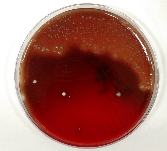
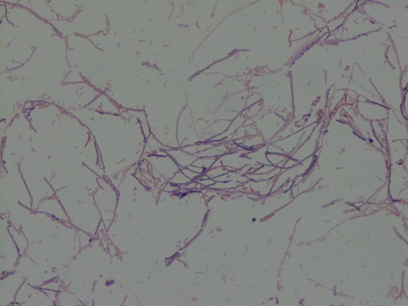
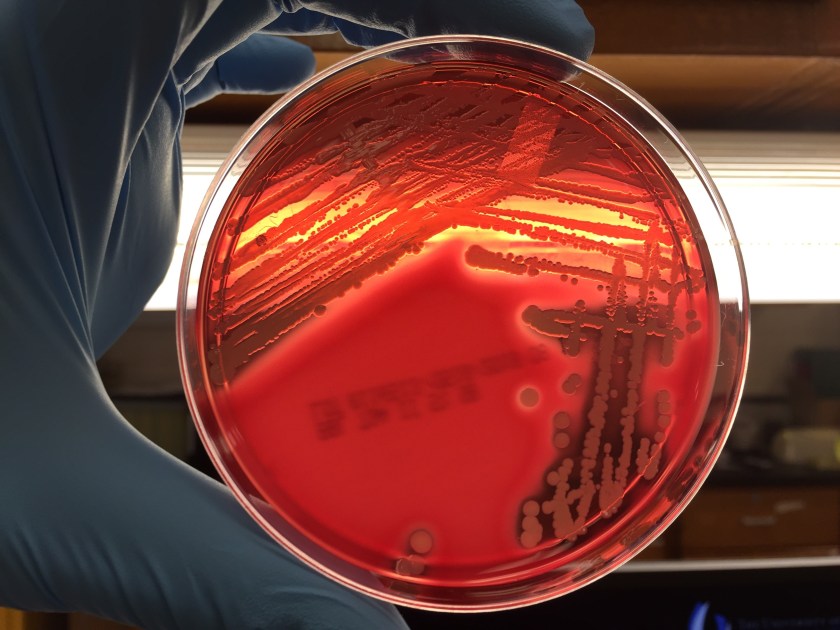
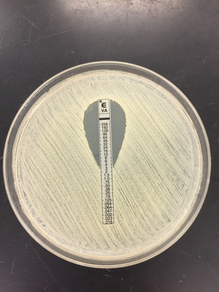
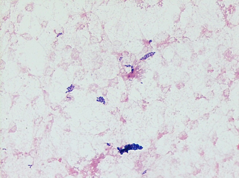
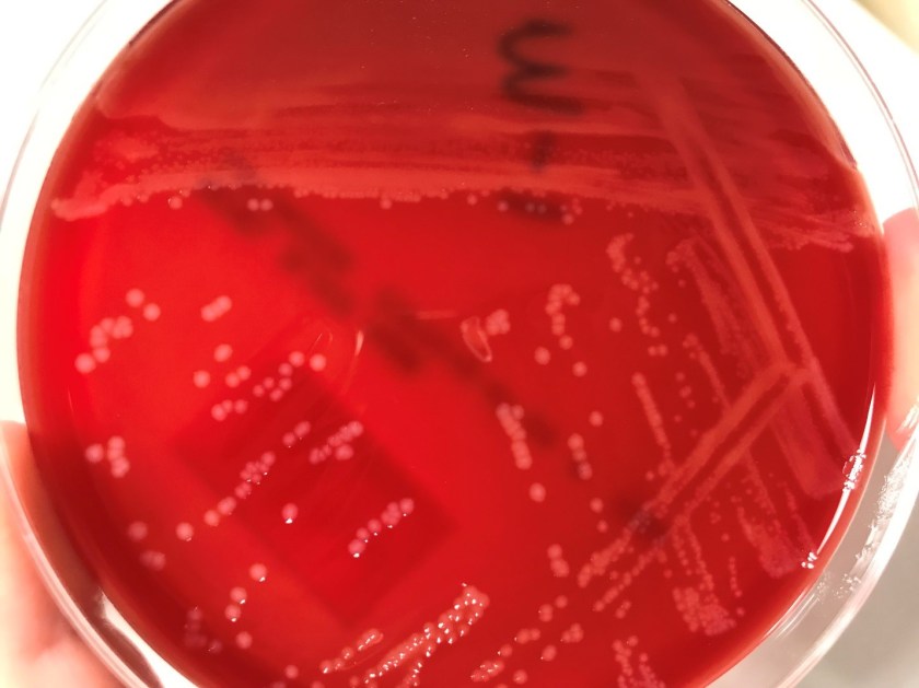
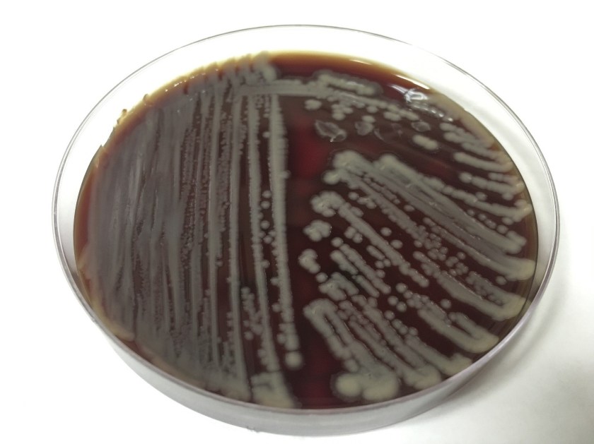
The primary gram smear of the swab specimen was interpreted as no bacteria or polys seen. Routine culture media including blood, chocolate, MacConkey, and CNA agar were inoculated and incubated aerobically. Following incubation, the blood agar showed few gram positive cocci consistent with usual skin flora and few single morphology of medium to large sized gray colonies without hemolysis. On the MacConkey agar, few single morphology non-lactose fermenting colonies were identified. The gray colonies identified on the blood agar gram stained as gram negative bacilli with unremarkable morphology. An oxidase test was performed and the bacteria was found to be oxidase positive. The key biochemical and physiologic characteristics of the isolate included: good growth on thiosulfate citrate bile salts and sucrose (TCBS) agar with yellow colonies, good growth in 6% NaCl nutrient broth, and no growth in 0% NaCl nutrient broth. The organism was identified by matrix-assisted laser desorption/ionization time of flight (MALDI-TOF) as Vibrio alginolyticus.

vibalg
Image 1. Blood agar isolate of medium sized gray colonies.
vibalg2
Image 2. MacConkey agar with non-lactose fermenting colonies.

Discussion

Vibrio spp. are water organisms commonly found in marine or brackish water environments. These organisms are gram negative bacilli which classically have “comma” shaped morphology on gram smear, though this is not an absolute. On sheep blood agar, these organisms are variably beta hemolytic medium to large gray colonies and on MacConkey agar are non-lactose fermenting (with the exception of Vibrio vulnificus)Vibrio spp. are oxidase positive, ferment glucose, and readily grow on most isolation media with growth being enhanced with the addition of 1% NaCl to the media. The growth characteristics on media containing different concentrations of NaCl can be used in differentiating the different Vibrio spp. Thiosulfate Citrate Bile Salts and Sucrose (TCBS) agar is both selective and differential for Vibrio spp. with sucrose fermentation being detected as yellow colony formation.

Vibrio angiolyticus typically causes extraintestinal infections, with wound infections and otitis externa being the most frequent. Transmission frequently occurs via traumatic aquatic injuries in contaminated water. Vibrio angiolyticus rarely causes intestinal disease and is isolated in less than 5% of stool cultures in patients with Vibrio associated diarrhea. Growth characteristics of Vibrio alginolyticus include yellow colonies on TCBS due to its ability to ferment sucrose and good growth on 6% NaCl and no growth on 0% NaCl. Additional key biochemical characteristics of Vibrio alginolyticus include oxidase positivity, nitrite positivity, negative for myo-Inositol fermentation, negative for arginine dihydrolase, positive for lysine decarboxylase, and variable positivity for ornithine decarboxylase. Most wound infections due to Vibrio alginolyticus are non-severe, and most mild infections will clear without antibiotic therapy.

References

  1. Procop GW, Koneman EW. Koneman’s Color Atlas and Textbook of Diagnostic Microbiology, North American Edition. LWW; 2016.
  2. Morris, J., Calderwodd, S., and Bloom, A. Minor Vibrio and Vibrio-like species associated with human disease. In: UpToDate, Post, TW (Ed), UpToDate, Waltham, MA, 2017.


-Justin Rueckert, DO is a 3nd year anatomic and clinical pathology resident at the University of Vermont Medical Center.

Wojewoda-small

-Christi Wojewoda, MD, is the Director of Clinical Microbiology at the University of Vermont Medical Center and an Associate Professor at the University of Vermont.

Gram Stain Examination – Beyond Infectious Organisms

Case History

A 72 year old female with past medical history of stage IV ovarian adenocarcinoma treated with chemotherapy and interval debulking surgery, presented to emergency room with a one week history of confusion and worsening balance.

CT scan of the head showed new communicating hydrocephalus.  A magnetic resonance imaging couldn’t be performed initially because of patient’s uncontrolled agitation.  Lumbar puncture (LP) was performed.  Following this procedure the patient’s mental status showed some improvement and therefore neurosurgery team decided to insert ventriculoperitoneal (VP) shunt to treat her hydrocephalus and prevent recurrence of seizures.

It was Friday afternoon when a microbiology technologist brought the patient’s cerebrospinal fluid (CSF) gram stain to be reviewed.  It was confirmed that no inflammatory cells and organisms were present.  However, cells in the background looked very atypical (Image 1a, b).

CSF1
Image 1:  Gram stain of CSF showing atypical epithelial cells at (a) 40x and (b) 100x with oil.
CSF2
Image 1b.

Discussion

The gram stain is used to provide preliminary information about the microorganism present in the specimen.  Gram stain differentiates bacteria into two fundamental varieties of cells.  Bacteria that retain the initial crystal violet stain (purple) are said to be “Gram-positive,” whereas those that are decolorized and stain red with carbol fuchsin (or safranin) are said to be “Gram-negative” (1).  An adequate examination of a gram-stained smear includes observing numerous representative fields and the fields containing neutrophils yield the most information (2).  Gram stain provides information about number of bacteria present, gram reaction and shape of the bacteria.  In background we can also see epithelial cells and inflammatory cells.  However, it’s a good practice to also appreciate and investigate any odd looking findings.

To investigate further, we visited the hematology laboratory to view their CSF slide to determine if these cells were a processing artifact.  After it was confirmed that hematopathology saw the same atypical cells, a cytopathologist was requested to review the gram stain since patient’s CSF cytology specimen was to be processed after the weekend.  Cytopathologist favored our suspicion and decided to process the cytology specimen late in the day on Friday and it was confirmed that those atypical cells were consistent with the metastatic adenocarcinoma.

Neurosurgery team was immediately contacted to reconsider insertion of the VP shunt as the shunt would drain fluid from the CSF into the peritoneal cavity and thus there was concern for transferring of malignant cells from central nervous system into abdomen/pelvis. However, after consulting oncology team it was later decided to proceed with the procedure since patient’s primary tumor originated in abdomen/pelvis and current intraabdominal tumor burden was not significant as compared to the symptoms driven by CNS involvement. Proceeding with this procedure was considered to be palliative and the best course of action to improve the patient’s quality of life.

References

  1. Beveridge TJ. Use of the gram stain in microbiology. Biotech Histochem.2001 May;76(3):111-8.
  2. Barenfanger J, Drake C. Interpretation of gram stains for the nonmicrobiologist. 2001 July;32(7):368–375.

KM

-Kiran Manjee, MD, is a 1st year anatomic and clinical pathology resident at University of Chicago (NorthShore).

-Erin McElvania, PhD, D(ABMM), is the Director of Clinical Microbiology NorthShore University Health System in Evanston, Illinois. Follow Dr. McElvania on twitter @E-McElvania. 


Hematopathology Case Study: A 56 Year Old Man with Sinus Congestion and Axillary Adenopathy

Case History

A 56 year old male presented to his PCP complaining of sinus congestion, rhinorrhea, night sweats, decreased appetite and fevers of up to 101-102 every evening. Hematologic evaluation revealed a neutropenia and a lymphopenia. An infectious disease work up was negative. His LDH was elevated. Physical examination reveals an enlarged left axillary lymph node. An excisional biopsy was performed.

Biopsy Findings

Figure 1.jpg

Figure 2.jpg

H&E stained sections demonstrate an enlarged node with effaced architecture and scattered residual follicles with small, mature cells. There is a proliferation of intermediate to large, to very large, atypical and highly pleomorphic cells many of which demonstrate bizarre forms, irregular nuclear morphology and acidophilic nucleoli. The lymphoma cells are noted to focally traverse through adipose tissue. Occasional hallmark cells are appreciated.

To further characterize the infiltrate, immunohistochemical stains were performed and interpreted with appropriate controls. The lymphoma cells were diffusely positive for CD45 (LCA), CD43, and CD30 (membranous and Golgi) with a Ki-67 of 80-90%. These cells were negative for CD20, PAX-5, CD3, CD4, CD8 (mostly), CD5, D10, BCl-2, BCl-6 and ALK1.

The morphologic features and immunophenotype of the cells was diagnostic of anaplastic large cell lymphoma, ALK negative.

Discussion

Anaplastic Large Cell Lymphoma (ALCL), ALK-negative (ALK-) is defined as a CD30+ T-cell neoplasm that morphologically resembles ALK-positive ALCL, but lacks ALK protein expression. It most commonly affects adults (aged 40-65 years), and has a slight male preponderance with a male-to-female ratio of 1.5:1. T. Most patients present with advanced disease (stage III-IV), lymphadenopathy and B symptoms. The most common differential diagnosis is ALK-positive ALCL.

The molecular deciphering of ALCL began in the 1990s with the discovery of a recurrent t(2;5) (p23;q35) translocation fusing the ALK gene and the nucleophosmin gene generating a NPM-ALK fusion protein, as well as other ALK translocations resulting in a high ALK kinase activity. This triggers the major oncogenic pathway in ALK-positive ALCL. Pharmacologic therapy has been developed to target ALK, and has shown efficacy. Thus, compared with ALK-negative cases, ALK-positive occurs in younger patients and has a better prognosis. ALK-negative ALCL also tends to involve both lymph nodes and extranodal tissues, although extranodal sites are less commonly involved than in ALK+ ALCL.

The other differential diagnoses of ALK- ALCL includes, primary cutaneous ALCL (C-ALCL), other subtypes of CD30+ T-cell or B-cell lymphoma with anaplastic features and classic Hodgkin Lymphoma. If a single lymph node or cutaneous cases are suggestive of ALK- ALCL, C-ALCL needs to be considered. Any cases that involve the gastrointestinal tract need to be distinguished from CD30+ enteropathy-associated and other intestinal T-cell lymphomas.

Molecular analysis of ALK- ALCL shows characteristic strong expression of CD30, in equal intensity in all the cells. Loss of T-cell markers is frequently seen, however, more than half of all cases express one or more T-cell markers. CD2 and CD3 are more commonly expressed than CD5, and CD43 is almost always expressed. CD4+ is frequently positive, while CD8+ is rare. Many cases also express cytotoxic markers TIA1, granzyme B, and/or perforin.

The genetic profile in ALK-negative ALCL has been found to be pretty heterogenous. Most notably, activating mutations of JAK1 and/or STAT3 have been shown to lead to activation of the JAK/STAT3 pathway. Chromosomal rearrangements of DUSP22 (i.e. chromosomal rearrangements in or near the DUSP22-IRF4 locus on 6p25.3) occur in 30% of the cases, and rearrangements of TP63 occur in about 8% of cases. Neither of the rearrangements have been reported in ALK+ ALCL.

From a prognostic standpoint, studies have shown that the rearrangements have effects on the survival rate. TP63-rearranged cases were shown to have an unfavorable prognosis worse than ALK- ALCL with neither rearrangement, while DUSP22-rearranged cases were shown to have favorable outcomes similar to ALK-positive ALCLs.

References

  1. Gaulard P, de Leval L. ALK-negative anaplastic large-cell lymphoma. 2016 Jan 14;127(2):175-7.
  1. Edgardo R. Parrilla Castellar et al., ALK-negative anaplastic large cell lymphoma is a genetically heterogeneous disease with widely disparate clinical outcomes Blood. 2014 Aug 28; 124(9): 1473–1480.


Bradon Zelman

-Brandon Zelman is 4th year medical student at the Philadelphia College of Osteopathic Medicine and an aspiring pathologist. You can follow Brandon on Twitter @ZelmanBrandon.

Mirza-small

-Kamran M. Mirza, MD PhD is an Assistant Professor of Pathology and Medical Director of Molecular Pathology at Loyola University Medical Center. He was a top 5 honoree in ASCP’s Forty Under 40 2017. Follow Dr. Mirza on twitter @kmirza.

Microbiology Case Study: A 64 Year Old Man with Metastatic Colon Cancer

Case History

A 64 year old man with metastatic colon cancer and a history of recent motor vehicle collision with polytrauma presented from a rehabilitation facility with fever up to 105 degrees Fahrenheit. Two months prior to admission he was hospitalized for the motor vehicle collision in which he sustained orthopedic injuries to multiple extremities. In addition to external fixation of several injuries, he returned to the operating room on multiple occasions for additional incision and drainage of a wrist wound which demonstrated gross purulence with cultures growing Enterococcus, Prevotella, and an extended spectrum beta-lactamase-producing Morganella morganii. Antimicrobial regimen initially consisted of surgically placed antibiotic beads and broad-spectrum therapy with vancomycin, piperacillin-tazobactam, and then meropenem. The patient was eventually transitioned to an oral antibiotic regimen consisting of linezolid and ciprofloxacin for an anticipated course of six weeks and he was discharged to a rehabilitation facility.

After several weeks at the rehabilitation facility, the patient became febrile and was admitted for workup of his fever. Initially the fever was of uncertain origin with malignancy (rectal cancer with metastasis to the liver), drug fever (linezolid), and wound site infection on the differential. Linezolid was discontinued, daptomycin initiated, and ciprofloxacin maintained. Fever persisted and ciprofloxacin was discontinued as another possible source of drug fever. Ertapenem was initiated. Initially, prior wounds and surgical sites appeared well-healing. Blood cultures all yielded no growth. However, on day five of this hospitalization, purulent drainage was noted from the site of a left leg surgical wound. Arthrocentesis yielded 0.5 mL of bloody fluid which was sent for cell count, differential, and culture.

Laboratory Findings

Initial Gram stain showed few polymorphonuclear leukocytes with no bacteria seen. Cell count was unable to be performed due to viscosity of the specimen but differential showed 80% neutrophils. There was no growth on aerobic blood, chocolate, or MacConkey agars. Anaerobic Schaedler (non-selective) agar grew 1-2 mm brownish colonies (Image 1). Gram stain of this isolate revealed gram variable bacilli forming long filaments (Image 2). The isolate was identified using MALDI-TOF MS (Vitek) as Clostridium ramosum.

cloran1
Image 1. 1-2 mm brownish colonies on anaerobic Schaedler agar
cloran2
Image 2. Gram stain showing gram variable bacilli in filamentous chains

The patient was taken to the operating room for incision and drainage of the left knee with two additional samples sent for culture which grew Clostridium ramosum.

Discussion

Clostridium species are anaerobic, spore-forming, gram positive bacilli. C. ramosum is non-motile and is normally found in the human colon and the environment. One study identified C. ramosum in the feces of 83% of sampled adults. Former names include Eubacterium filamentosum, Ramibacterium ramosum, Actinomyces ramosus, and Eubacterium ramosum. Figure 2 demonstrates a notable characteristic of C. ramosum, i.e. its variable appearance on Gram stain. The morphology here may be described as gram negative or “over-decolorized”, though gram positive bacilli are clearly seen forming many of the filaments. Its terminal endospores are often difficult to identify on Gram stain and this is true of Figure 2. These characteristics on Gram stain have historically made identification difficult, though use of MALDI-TOF MS facilitated identification in our case. Biochemically, C. ramosum ferments glucose and hydrolyzes esculin; it is negative for lecithinase and lipase.

C. ramosum possesses an IgA protease though it is not commonly pathogenic. When it is pathogenic, the spectrum of disease overlaps with that of other anaerobes and includes deep-seated abscesses, e.g. intra-abdominal abscess secondary to trauma. Osteomyelitis and primary bacteremia are also possible, particularly in immunocompromised patients. Otitis media in children is another possible clinical scenario.

Septic arthritis due to Clostridium ramosum

A 2016 case report described two cases of septic arthritis due to C. ramosum. In one case, a patient with rheumatoid arthritis on methotrexate and prednisone and history of revision knee arthroplasty eight years prior presented with knee swelling. Synovial fluid aspirate was consistent with an infectious process; the prosthesis was removed but synovial and intraoperative cultures were negative. Antimicrobial therapy with linezolid and ciprofloxacin was administered for six weeks with clinical improvement. Two weeks after discontinuation of antibiotics the patient became febrile. Blood cultures were negative but culture of synovial fluid grew C. ramosum. The patient required multiple operations due to joint destruction and was ultimately managed with intravenous penicillin and clindamycin with transition to oral metronidazole for three months of therapy. The second case of C. ramosum septic arthritis presented in this report was ultimately managed with surgical debridement and amoxicillin-clavulanate. Both cases presented in patients with immunocompromising comorbidities and the course of their septic arthritis was chronic, recurring, and destructive but non-fatal with both patients dying from other causes.

These clinical and laboratory characteristics are consistent with the case of C. ramosum septic arthritis identified at our institution. The case of septic arthritis presented here involved an immunocompromised host (malignancy) with history of trauma, foreign body placement (external fixator), and long-term antibiotic therapy. This patient’s wound required debridement in the operating room on three occasions. Once clinically stable, the patient was discharged to a subacute rehabilitation facility and continued on ertapenem with amoxicillin for an expected duration of six weeks with the plan to switch to amoxicillin-clavulanate and ciprofloxacin for suppressive therapy.

References

  1. Forrester JD, Spain DA. Clostridium ramosum bacteremia: case report and literature review. Surg Infect (Larchmt). 2014 Jun;15(3):343-6. doi: 10.1089/sur.2012.240. Epub 2013 Nov 27. Review. PubMed PMID: 24283763.
  2. García-Jiménez A, Prim N, Crusi X, Benito N. Septic arthritis due to Clostridium ramosum. Semin Arthritis Rheum. 2016 Apr;45(5):617-20. doi: 10.1016/j.semarthrit.2015.09.009. Epub 2015 Oct 1. PubMed PMID: 26546506.
  3. Procop, Gary W et al. Koneman’s Color Atlas and Textbook of Diagnostic Microbiology. Seventh ed., 2017.


-Benjamin F. Smith is a Pathology Student Fellow at University of Vermont Medical Center.

Wojewoda-small

-Christi Wojewoda, MD, is the Director of Clinical Microbiology at the University of Vermont Medical Center and an Associate Professor at the University of Vermont.

Microbiology Case Study: A 37 Year Old Man with Endocarditis

Case History 

A 37 year old African American male was transferred from an outside hospital due to mitral valve endocarditis. His past medical history was significant for diabetes mellitus type II and end stage renal disease requiring long-term dialysis. Four months prior, he was bacteremic with methicillin resistant Staphylococcus aureus and received IV vancomycin therapy. At admission, his temperature was 102.1°F and labs revealed a white blood cell count of 25.3 TH/cm2 with 95% neutrophils and a left shift, a normocytic anemia, and a creatinine of 5.39 mg/dL. Physical exam revealed a severe mitral valve regurgitation. Various imaging modalities showed vegetations on the mitral valve, complete occlusion of the distal infrarenal abdominal aorta, several subacute infarcts in the brain, multiple sites of osteomyelitis of the spine, and a pelvic bone abscess. After collecting initial blood cultures, vancomycin and cefepime were started.

Laboratory Identification

visa1.jpg
Image 1. Large, whitish-yellow colonies grew from a positive blood culture bottle that showed gram positive cocci in clusters. These features are consistent with Staphylococcus aureus
visa2.jpg
Image 2. Susceptibility testing of the isolate revealed a vancomycin Etest minimum inhibitory concentration (MIC) of 3 µg/ml.

Multiple blood cultures were positive and Staphylococcus aureus was identified by MALDI-TOF mass spectrometry (Image 1). Susceptibility testing of the isolate showed resistance to cefoxitin and oxacillin, consistent with methicillin resistant S. aureus (MRSA). Due to a vancomycin MIC of 2 ug/ml by broth microdilution, an Etest was set up and results showed an MIC of 3 ug/ml (Image 2). Rounded up to next doubling dilution, this resulted in a MIC of 4 ug/dl, which was concerning for a vancomycin intermediate S. aureus (VISA). Repeat identification and susceptibility testing confirmed these findings. For this reason, the isolate was sent to the department of health for confirmatory testing.

Discussion

 Vancomycin is the first line agent for treating infections caused by methicillin resistant S. aureus (MRSA). When MRSA isolates show reduced susceptibility results to vancomycin, they are classified as vancomycin intermediate S. aureus (VISA) or vancomycin resistant S. aureus (VRSA). This phenomenon is concerning, as it leaves clinicians with relatively few therapeutic options. Broad spectrum antibiotics such as daptomycin, linezolid, or 5th generation cephalosporins (ceftaroline & ceftobiprobe) are potential treatment selections in these cases.

The Clinical and Laboratory Standards Institute (CLSI) has set the following MIC breakpoints for vancomycin in relation to S. aureus: ≤2 ug/ml susceptible, 4-8 ug/ml intermediate, and ≥16 resistant. If elevated vancomycin MICs are encountered in the laboratory, the isolate should be checked for purity, the organism identification should be confirmed, and susceptibility testing repeated. The laboratory should notify the state health department and hospital infection prevention team if a VISA or VRSA is suspected. Further testing of the isolate by the health department and/or CDC is required. Appropriate infection control measures, such as wearing gowns & gloves and adherence to hand hygiene, should be taken to decrease the spread of VISA/VRSA, as treatment options are limited.

While not fully understood, the reduced vancomycin susceptibility in VISA isolates is thought to be due to an abnormally thickened peptidoglycan cell wall that makes it more difficult for vancomycin to reach the cell membrane and inhibit cell growth. On the other hand, VRSA isolates most commonly acquire the vanA vancomycin resistance gene from Enterococcus faecium, which confers high-level vancomycin resistance. Usually those with VRSA infections previously have been infected with both VRE and MRSA. This co-infection allows for the vanA gene to be transferred by a plasmid or transposon from the VRE to the MRSA isolate, resulting in a S. aureus isolate that is now resistant to vancomycin.

In the case of our patient, further testing by the health department showed his MRSA isolate had a vancomycin MIC of 2 ug/ml by broth microdilution (susceptible) and an MIC 3 ug/ml by Etest. It was noted these results were within a single doubling dilution. As broth micro dilution is the reference method, his isolate was considered susceptible to vancomycin. However, the elevated MICs by both methods suggest the isolate is developing reduced susceptibility to vancomycin. Due to septic emboli in various organs, he was not a surgical candidate and was managed medically with ceftaroline and linezolid.

JK.jpg

-Jaswinder Kaur, MD, is a fourth year Anatomic and Clinical Pathology chief resident at the University of Mississippi Medical Center. 

Stempak

-Lisa Stempak, MD, is an Assistant Professor of Pathology at the University of Mississippi Medical Center in Jackson, MS. She is certified by the American Board of Pathology in Anatomic and Clinical Pathology as well as Medical Microbiology. She is the Director of Clinical Pathology as well as the Microbiology and Serology Laboratories.  Her interests include infectious disease histology, process and quality improvement, and resident education.

Hematopathology Case Study: A 63 Year Old Man with Fatigue

Case history

A 63 year old male presented with extreme fatigue and weakness of unknown duration. Physical examination revealed scattered petechiae and mildly decreased muscle strength. His past medical history included a one year history of cough that had recently improved. Laboratory investigation demonstrated severe anemia and thrombocytopenia with a mild leukopenia.

Review of the peripheral blood smear showed smudge cells, circulating neutrophils with Döhle bodies and toxic granulation. CT scan of the chest showed upper/anterior mediastinal lymphadenopathy without hilar lymphadenopathy.

A biopsy of the bone marrow was performed.

Microscopic Findings

hod1.jpg


hod 2.jpg

The bone core biopsy revealed a hypercellular marrow for the patient’s age with a pronounced lymphohistiocytic infiltrate involving 30-40% of the biopsied marrow space. Interspersed along the infiltrate were large, atypical lymphoid cells with pleomorphic nuclei and prominent nucleoli. The marrow aspirate smear reveals progressive trilineage hematopoiesis with scattered hemophagocytic histiocytes.

Immunophenotype

hod3.jpg

The large atypical lymphoid cells were positive for CD30 and EBER, while being dimly positive for PAX5 and negative for CD20.

Diagnosis

The detection of mononuclear Hodgkin cells staining for CD30 along with a characteristic reactive infiltrate, together with dim PAX-5 staining, positive EBER, and negative CD20 is sufficient to diagnose involvement of a secondary site by Hodgkin lymphoma. The lymphoma was associated with a secondary hemophagocytic lymphohistiocytosis.

Discussion

Hodgkin lymphoma (HL) is a B-cell derived monoclonal lymphoid neoplasm. HL has a bimodal age distribution, with teenagers or patients in their early 20s and patients older than 55 years having the highest incidence. Although the typical presentation is with peripheral lymph node involvement, extranodal sites may be involved by either direct invasion or hematogenous dissemination. These sites include the spleen, liver, lung and bone marrow. About one third of patients have constitutional symptoms such as high fevers, night sweats, and weight loss.

Two broader forms of Hodgkin lymphoma exist: Classic Hodgkin lymphoma (CHL) and the less common nodular lymphocyte-predominant Hodgkin lymphoma (NLPHL). NLPHL tends to preserve the entire B-cell transcriptional phenotype, while the neoplastic cells in CHL fail to do so.

CHL is composed of mononuclear Hodgkin cells and multinucleated Reed-Sternberg cells surrounded by an infiltrate of non-neoplastic reactive cells that might encompass small lymphocytes, plasma cells, eosinophils, neutrophils, and histiocytes. Fibrosis may also be present in the form of bands or may be more diffusely spread. The four histological subtypes: nodular sclerosis CHL, lymphocyte-rich CHL, mixed cellularity CHL, and lymphocyte-depleted CHL are based on the composition and characteristic of the reactive infiltrate, and the cytological features of the neoplastic cells.

The classic Reed-Sternberg cell is binucleated, with prominent eosinophilic nucleoli, often referred to as having an “owl’s eye” appearance. However many neoplastic cells are not of the typical Reed-Sternberg variant, and can be mononuclear, termed Hodgkin cells, or cells with more condensed cytoplasm and pyknotic reddish nuclei known as mummified cells.

Hodgkin/Reed-Sternberg cells (HRS) in Classic Hodgkin Lymphoma fail to preserve their B-cell traits, and this is reflected by their immunophenotype. The majority of cases are negative for CD45, and although CD20 may be expressed, it is usually present only on a minority of the neoplastic cells and stain with varied intensity. The HRS cells stain with PAX5 with a lower intensity than the surrounding reactive cells, making them easily detectable. The HRS cell stains positive for CD30 and CD15 in nearly all cases. Both of them stain the membrane with accentuation around the Golgi apparatus. EBV associated Hodgkin Lymphoma will stain positive with EBER, detecting EBV-encoding small RNA.

Bone marrow involvement is rare, ~5-10% of cases, and suggest vascular dissemination of the disease. Bone marrow trephine biopsies are commonly performed in the staging of patients with newly diagnosed CHL which guides the further treatment and gives us information about prognosis. Involvement of the bone marrow represents stage IV disease (advanced stage) in the Ann Arbor staging classification and patients with advanced stage disease typically receive a more prolonged course of chemotherapy. The 5-year survival rate of stage IV Hodgkin lymphoma is ~65%,  a much worse prognosis when compared with stage I, stage II, and stage III with ~90%, ~90%, and ~80% 5-year survival rates respectively.

References

  1. Stein H, Pileri SA, Weiss LM, et al. Hodgkin Lymphomas. In Swerdlow SH, Campo E, Harris NL, Jaffe ES, Pileri SA, Stein H, Thiele J, editors: WHO classification of tumours of haematopoietic and lymphoid tissues, revised ed 4, Lyon, France, 2017, IARC Press, pp 423-464
  2. Ansell SM. Hodgkin Lymphoma: Diagnosis and Treatment. Mayo Clin Proc. 2015 Nov;90(11):1574-83.
  3. Howell SJ, Grey M, Chang J, Morgenstern GR, Cowan RA, Deakin DP, Radford JA. The value of bone marrow examination in the staging of Hodgkin’s lymphoma: a review of 955 cases seen in a regional cancer centre. Br J Haematol. 2002 Nov;119(2):408-11.
  4. Clarke C, O’Malley C, Glaser S. Hodgkin lymphoma. In: Ries LAG, Young JL, Keel GE, Eisner MP, Lin YD, Horner M-J, eds. SEER Survival Monograph: Cancer Survival Among Adults: U.S. SEER Program, 1988-2001, Patient and Tumor Characteristics. National Cancer Institute, SEER Program, NIH Pub. No. 07-6215, Bethesda, MD, 2007.


Hans.jpg

-Hans Magne is a 6th- year medical student at Poznan University of Medical Sciences. Follow Hans on Twitter @HHamnvag

Mirza-small

-Kamran M. Mirza, MD PhD is an Assistant Professor of Pathology and Medical Director of Molecular Pathology at Loyola University Medical Center. He was a top 5 honoree in ASCP’s Forty Under 40 2017. Follow Dr. Mirza on twitter @kmirza.

Hematopathology Case Study: The Case of Lymphocytosis

Case History

53 year old female was found to have leukocytosis upon a wellness examination. A CBC was performed and found a WBC of 34.0, HgB 13.2, and Plt of 263,000. The WBC differential consisted of 80% Lymphocytes and 12% neutrophils. The patient states that she is feeling well, no fever, chills, or night sweats. She denies any adenopathy. Flow Cytometry was recommended as well as morphologic review along with Cytogenetics and FISH (fluorescence in-situ hybridization).

Lab Identification

CBC

WBC               33.68  [103/uL] NEUT             4.17 [103/uL]      12.3 [%]
RBC                4.54  [106/uL] LYMPH          26.91 [103/uL]    79.9 [%]
HGB               13.2   [g/dL] MONO           2.04 [103/uL]      6.1  [%]
HCT               40.0   [%] EO                  0.39 [103/uL]      1.2  [%]
MCV               88.1   [fL] BASO             0.11 [103/uL]      0.2  [%]
MCH               29.1   [pg] IG                    0.06 [103/uL]      0.2  [%]
MCHC            33.0   [g/dL] NRBC             0.00 [103/uL]      0.0  [%]
RDW-CV         14.3   [%]
PLT                263   [103/uL]
MPV                9.4   [fL]

Morphologic Review

CLL1.jpg
Image 1. 40x magnification showing lymphocytosis.
CLL2.jpg
Image 2. 60x magnification showing lymphocytosis with occasional smudge cells.

Flow Cytometry: Population of Interest – Abnormal Lymphocytes

cll3.JPG

cll4.JPG

cll5.JPG

Cytogenetics: 46,XX[20] Normal Female Karyotype

It is not unusual to observe a normal karyotype in CLL due to the limited number of abnormal cells and/or low spontaneous proliferative activity of the malignant cells. Fluorescence in situ hybridization studies may identify cytogenetic abnormalities of prognostic significance in interphase nuclei not observed in the metaphase cells analyzed. In cases of CLL molecular profiling may be performed to aid in predicting course of the disease. If clinically indicated these studies may be considered. Standard cytogenetic analysis may not detect subtle submicroscopic rearrangements and may not include metaphases from abnormal cell populations with low mitotic rates or present in low levels.

Metaphases Counted: 20 Metaphases Analyzed: 20
Metaphases Karyotyped: 2 Culture Type: 48EB, 72IL2/DSP30
Banding Technique: GTG Banding Resolution: 400

Fluorescence in-situ Hybridization: Abnormal – 13q14 deletion present

Fluorescence in situ hybridization (FISH) analysis was performed using a specific set of probes for Chronic Lymphocytic Leukemia (CLL). This study revealed a 13q14 deletion. Counts for all other probes were within the normal reference range. This finding represents an ABNORMAL result. Deletion of 13q14 is the most common deletion in CLL being reported in 10-20% of cases by conventional cytogenetics and up to 64% of cases by FISH analysis. When present as a sole abnormality this deletion is associated with a good prognosis and a median survival longer than CLL patients with a normal karyotype.

Del(6q) Not Detected
Del(11q)(ATM) Not Detected
Trisomy 12 Not Detected
Del(13q)/-13 DETECTED
t(11;14) Not Detected
Del(17p)(TP53) Not Detected

Discussion

Chronic Lymphocytic Leukemia is a neoplasm of about 5 cases per 100,000 people with a median age of around 70 years old. The neoplasm is composed of monotypic mature B-cells that typical express CD5. Other immunophenotypic characteristics of the leukemic B-cells include CD19, CD20, CD22, and CD79b with dim surface expression of one of the immunoglobulin light chains, Kappa or Lambda. These cells typically express CD200 which helps differentiate the leukemia from Mantle Cell Lymphoma/Leukemia. Patients found to have a mutated IGHV genes typically have a better prognosis than those with an unmutated genes. Expression of ZAP70, CD38, or CD49d is also associated with an adverse prognosis. Complex karyotypes also trend towards a poor outcome. Adverse predictive factors include rapid lymphocyte proliferations in the blood, typically doubling in < 12 months.

Monoclonal B-cell Lymphocytosis is typically characterized as a monoclonal
BCell count of <5 X10⁹/L in the peripheral blood. Monoclonal B-cell Lymphocytosis with a Chronic Lymphocytic Leukemia-type phenotype is the most common which accounts for about 75% of all cases. It has been reported that virtually all Chronic Lymphocytic Leukemias are preceded by Monoclonal B-cell Lymphocytosis, although not all MBLs progress to CLL.

References

  1. Dohner H, et al. N Engl J Med 2000; 343:1910-6.
  2. Hamblin TJ.Best Practice & Research Clinical Haematology. 2007; 20(3):455 – 68.
  3. Swerdlow SH, Campo E, Harris NL, Jaffe ES, Pileri SA, Stein H. WHO classification of tumours of haematopoietic and lymphoid tissues, fourth edition. Lyon, France: IARC; 2017
  4. Nowakowski GS, et al. Br J Hematol. 2005; 130:36 – 42.
  5. Atlas of Genetics and Cytogenetics in Oncology and Hematology http://atlasgeneticsoncology.org/


Troy-Krieger-small

Troy G. Krieger, MS, MLS(ASCP)CMSCYMCMQLSCMCLS(MT) graduated from Montana State University Billings with a BS in Biology, Medical Laboratory Science option. He received a NAACLS Certificate and clinical training from the University of North Dakota in Grand Forks, ND, where he also received his Master’s degree. He is a Medical Laboratory Scientist / Flow Cytometrist at Yellowstone Pathology Institute, Inc in Billings, MT and his interests include Hematology, Immunopathology, and Flow Cytometry.

Microbiology Case Study: 64 Year Old Male with Pleuritic Chest Pain and Fevers

Case History

The patient is a 64 year old male with a history of diabetes mellitus and hypertension who presented as a transfer from an outside hospital with a 2 week history of chest pain and pressure, as well as recurrent fevers, rigors, and soaking sweats, and an echocardiogram concerning for a pericardial effusion. He was also found to have markedly elevated CRP, and mildly elevated troponins, was diagnosed with pericarditis, and was started on colchicine. He continued to have fevers, and developed diarrhea and was transferred for elevation of care. C. difficile PCR was negative, and since the onset of diarrhea coincided with the initiation of colchicine, that was determined to be the cause. Blood cultures on arrival grew a Gram positive rod and a transesophageal echocardiogram was done which again showed pericardial thickening with small effusion, and fluid with fibrinous appearance. There was no evidence of valvular vegetation. At this point the patient was started on IV meropenam as he is allergic to penicillin’s and sulfa drugs. The pericarditis seemed to improve with colchicine so a non-infectious process was favored and a repeat ANA was recommended when he has recovered from his current infection.

Laboratory Identification

listermono1
Image 1. Gram Stain of blood culture showing gram positive palisading rods.
listermono2
Image 2. Gray-white colonies with soft β-hemolysis on blood agar.

Blood cultures grew grey-white colonies that are gram positive, catalase positive rods with soft beta-hemolysis on the blood agar plate, and tumbling motility under light microscopy. CAMP testing would be positive with Staphylococcus aureus. This was identified as Listeria monocytogenes by the MALDI. 

Discussion

Listeria monocytogenes is a gram positive rod that can be found in the soil, water, sewage, vegetation, and as part of the fecal flora of animals. It is facultative intracellular pathogen that is able to invade and survive in human cells including macrophages (1). They possess a surface protein called internalin that interacts with E-cadherin on human cells resulting in endocytosis (1). Once within the cell the bacteria can produce listeriolysin O and other phospholipases which allow it to escape from the phagosome before it fuses with the lysosome, which prevents intracellular killing of the bacteria (2). L. monocytogenes is a common contaminant of food products as it can form biofilms on the food surfaces. Listeria also has the ability to grow a 4°C so it can continue to grow on refrigerated foods (1). Foods such as raw milk, raw vegetables, fish, poultry, and fresh and processed meats are the highest risk for contamination.

Ingestion during pregnancy can result in a flu like illness, occasionally with vaginal discharge, diarrhea, and urinary tract symptoms (1). Infection during pregnancy is particularly dangerous as occult bacteremia with transplacental transmission may occur (2). Infection in utero may result in premature labor and birth of an infected or stillborn fetus. Prognosis is highly dependent on the gestational age at infection.

Non- pregnant adults can also become infected by Listeria. The most common results of ingestion of contaminated food in immunocompetent patients is a transient asymptomatic carrier state, and can be excreted in the feces. Less commonly, febrile gastroenteritis can occur. Immunocompromised patients or those with underlying malignancy tend to present with acute sepsis, meningitis, or meningoencephalitis.  Focal infections such as cutaneous infection, abscesses, arthritis, peritonitis, liver/splenic abscess, cholecystitis, artificial joint/graft infections, osteomyelitis, and myo- and endocarditis can be seen and typically occur in immunocompromised patients as a results of hematogenous spread.1 Treatments includes ampicillin with or without an aminoglycoside. Occasional resistance to tetracyclines has been reported.2

Regarding the patient’s Listeria bacteremia, the patient reported no exposures to the common carriers of Listeria. It is possible that is was translocated from his gut during his diarrhea illness or could have been the cause of his diarrhea, although blood cultures at the outside hospital were negative.

References

  1. Winn, Washington C., et al. Color Atlas and Textbook of Diagnostic Microbiology. Lippincott Williams & Wilkins, 2006.
  2. Tille, Patricia M. Bailey & Scotts Diagnostic Microbiology. 13th ed., Elsevier, 2014.


-Casey Rankins, DO, is a 1st year Anatomic and Clinical Pathology resident at the University of Vermont Medical Center.

Wojewoda-small

-Christi Wojewoda, MD, is the Director of Clinical Microbiology at the University of Vermont Medical Center and an Associate Professor at the University of Vermont.

Chemisty Case Study: Heat-Insoluble Cryoglobulin

Case History

A 50 year old female was admitted for acute renal failure on CKD stage IV, present with gross hematuria, anemia (due to blood loss) and hypertension. The patient has a significant history of unresolved cryoglobulinemic vasculitis initially diagnosed in 2016 and has been treated by several rounds of rituximab. Other medical histories include Sjogren’s syndrome, MGUS with monoclonal IgM Kappa, coagulopathy (protein S deficiency, on anticoagulant), hyperviscosity, myalgia, deep vein thrombosis, leg edema with superficial ulcer, pulmonary embolus and membranoproliferative glomerulonephritis (MPGN). Kidney biopsy revealed intraglomerular hyaline thrombi consistent with cryoglobulinemic glomerulopathy, interstitial fibrosis tubular atrophy, arterial sclerosis, suggestive thrombotic microangiopathy. Immunohistochemistry was positive for C3, IgM, Kappa, Lambda and CD68. Bone marrow biopsy shown dyserythropoesis without malignancy. Blood testing shown negative hepatitis panel and undetectable C4.

We observed unusual cryoprecipitate test results from this patient: gelatinous appearance precipitate which accounts for more than 40% of volume was observed in both plasma and serum and cannot be cleared at 37C° after several hours of incubation. Further testing shown incubation at 56°C for 30min cleared up the serum but not the plasma. After checking the test history, we found that there was a similar situation for the patient’s cryoprecipitate test a few months back earlier in 2018, and was reported negative for cryoglobulins due to the heat-insoluble nature of the precipitate. Patient was transfused for anemia. No plasmapheresis was done. Due to the patient’s incomplete response to rituximab, Cytoxan was also added to help improve the symptoms.

Cryoprecipitate

  • Definition: Cryoprecipitates (or cryoproteins) are blood proteins that form precipitates or gels at temperatures lower than 37°C and typically re-dissolve after warming up to 37°C. There are two types:
  • Cryoglobulin (CG): precipitate from both serum and plasma; either immunoglobulins or a mixture of immunoglobulins and complement components
  • Cryofibrinogen (CF): precipitate from plasma only; typically composed of a mixture of fibrinogen, fibrin, fibronectin, and fibrin split products
  • Lab Testing done in our hospital:
    1. Blood are collected in two pre-warmed tubes (one serum, one EDTA plasma) and kept in warm water (37°C) until the serum tube clots.
    2. The plasma and serum are extracted at room temperature, and then stored in refrigerator for 72 hours.
  • If cryoprotein is present, a precipitate or gel will be seen. An aliquot of the serum is rewarmed at 37°C to verify the cryo-nature.
  1. The precipitate as a percentage of the original serum volume is measured in an ESR tube to determine the cryocrit.
  2. Immunofixation is ordered per pathologist to identify the immunoglobulin compositions of the cryoglobulin.

Cryoglobulinemia

  • Classification

Strictly speaking, cryoglobulinemia refers to the presence of cryoglobulin (CG) in a patient’s serum, which could be either asymptomatic or present with apparent clinic syndromes (i.e. cryoglobulinemic vasculitis). Cryoglobulinemia can be classified into three types (see table below [1]), with mixed cryoglobulinemia (type II and type III) representing 80% of the cases.

cryo1

  • Clinical Manifestations

Type I cryoglobulinemia is frequently asymptomatic, while mixed cryoglobulinemia manifests clinically by a classical triad of purpura, weakness and arthralgias, as well as some other conditions including MPGN, chronic hepatitis, peripheral neuropathy, lymphoma, Raynaud’s, Sjogren’s syndrome, etc.

The presence of heat-insoluble cryoglobulins is rare, and its pathogenesis is poorly understood. On the other side, it may indicate sever clinical consequence as seen in our case and some others as mentioned above.

Reference

  1. Mixed Cryoglobulinemia, Ferri, C; Orphanet Journal of Rare Diseases 2008, 3:25

 Further reading

  1. Essential type II cryoglobulinemia with cryoglobulin-occlusive MPGN and MGUS (Clin Chim Acta. 2009 Aug;406(1-2):170-3):79 y.o. female admitted due to edema and renal failure, cryoglobulin re-dissolved at 56°C, composed of monoclonal IgG-Kappa and polyclonal IgM.
  1. HCV associated thrombotic microangiopathy and cryoglobulin-occlusive MPGN (Am J Med Sci. 2013 Oct;346(4):345-8):57 y.o. female, cryoglobulin re-dissolved at 47°C, composed of monoclonal IgM-Kappa and polyclonal IgG. Symptoms only partially resolved upon treatment of plasmapheresis, corticosteroids and antiviral therapy of peginterferon plus ribavirin.
  1. Essential type I cryoglobulinemia with massive cryoglobulin-occlusive glomerulonephritis (Am J Kidney Dis. 1995 Oct;26(4):654-7):54 y.o. male progressed to ESRD prior to the detection of cryoglobulin. Cryoglobulin with white gelatinous appearance re-dissolved at 54°C, composed of monoclonal IgG-Kappa.
  1. Primary Sjogren’s syndrome with type II cryoglobulinemia and mesangiocapillary glomerulonephritis (Nephrol Dial Transplant. 2000 Jun;15(6):917-8):82 y.o. patient with IgM-MGUS, negative BM, deposition of IgG, IgM and C3 on kidney biopsy, decreased complement levels, negative HCVAb, HBsAb, HBsAg. cryoglobulin re-dissolved at 47°C, composed of monoclonal IgM-Kappa and polyclonal IgG-Kappa.


Huang

-Rongrong Huang, PhD is a first year clinical chemistry fellow at Houston Methodist Hospital. Her interests include general clinical chemistry, genetic biochemistry and applications of mass spectrometry in clinical laboratories.

Xin-small

-Xin Yi, PhD, DABCC, FACB, is a board-certified clinical chemist, currently serving as the Co-director of Clinical Chemistry at Houston Methodist Hospital in Houston, TX and an Assistant Professor of Clinical Pathology and Laboratory Medicine at Weill Cornell Medical College.

Microbiology Case study: 48 Year Old Male with Multiple Injuries Following a Motorcycle Accident

Case History

The patient is a 48 year old male who presented after a motorcycle crash where he slide sideways into a ditch. He did not lose consciousness. At presentation he complained of pain across the abdomen, right wrist and left shoulder. He also had shortness of breath and chest wall pain. He denied hitting his head and was helmeted. He underwent exploratory laparotomy with repair of traumatic diaphragmatic hernia and left chest tube placement with post-operative course complicated by significant leukocytosis and bandemia on post-operative day 5 which triggered CT re-imaging of the chest/abdomen/pelvis. This study demonstrated intraperitoneal free air that prompted repeat exploratory laparotomy with subtotal colectomy with the abdomen open and wound vac in place. On post-operative day 9 the patient had an increasing white count that prompted return to OR with bowel staple line demonstrating leak, which prompted a small bowel resection, and ileostomy formation. After leaving the OR, the patient experienced worsening septic shock. Aggressive antibiotic therapy and IV fluid resuscitation was continued. Blood cultures taken on post-operative day 9 grew a large Gram positive rod. Growth of this organism was seen both aerobic and anaerobically.

Laboratory Identification

closter1
Image 1. Gram stain of the anaerobic blood culture bottle showing gram positive/variable rods.


closter2.jpg
Image 2. Aerobic blood agar plate showing small clear colonies.

Blood cultures showed box shaped gram positive/ variable rods that were growing on blood agar plates both aerobe and anaerobically. It was motile, indole and catalase negative, and esculin positive, and was identified by the MALDI to be Clostridium tertium.

Discussion

Clostridium tertium is an aerotolerant gram positive rod that is widely distributed in the soil and can also be found the GI tract of animals and humans (1). C. tertium is non-toxin producing and produces terminal spores in anaerobic conditions (2). Infection with C. tertium is uncommon though it has been increasing in frequency as a cause of bacteremia, especially in patients with neutropenia, the immunocompromised, those with hematologic malignancy, those with inflammatory bowel disease, and in people with abdominal trauma or who have undergone abdominal surgery. Less commonly, C. tertium can be isolated in spontaneous bacterial peritonitis, enterocolitis, meningitis, septic arthritis, necrotizing fasciitis, post-traumatic brain abscess, and complicated pneumonia in mono- or polymicrobial infections (1). Additionally there has been a link between C. tertium infection and attempted suicide with the herbicides containing Glyphosate, as it causes GI toxicity which alters the gut environment (2). As with Clostridium difficile, the use of broad-spectrum antibiotics such as third-generation cephalosporins might predispose to intestinal colonization with C. tertium (1).

Increases in the diagnosis of C. tertium may be related to better diagnostic equipment such as the MALDI as it can easily be confused with a facultative anaerobic Bacillus or Lactobacillus species due to its gram variable appearance and ability to grow in aerobic conditions. A distinguishing feature between Bacillus species and C. tertium is negatively of the catalase reaction as Bacillus should be positive. Lactobacillus can still be a challenge as they are also catalase negative.

The treatment of C. tertium infection is complicated due to resistance to various antibiotics, including various beta-lactam antibiotics (such as third- and fourth-generation cephalosporins), clindamycin, daptomycin, and cotrimoxazole. Older reports state resistance to metronidazole, but this has not been confirmed in more recent publications. Available data indicate sensitivity to vancomycin, carbapenems, and quinolones (1)(3).

References

  1. Salvador F, Porte L, Durán, L, Marcotti A, Pérez J, Thompson L, Noriega LM, Lois V, Weitzel T. Breakthrough bacteremia due to Clostridium tertium in a patient with neutropenic fever, and identification by MALDI-TOF mass spectrometry. International Journal of Infection Disease. 2013;17:11 (1062-1063). https://doi.org/10.1016/j.ijid.2013.03.005
  2. You M-J, Shin G-W, Lee C-S. Clostridium tertium Bacteremia in a Patient with Glyphosate Ingestion. The American Journal of Case Reports. 2015;16:4-7. doi:10.12659/AJCR.891287.
  3. Miller D, Brazer S, Murdoch D, Reller LB, Corey, GR. Significance of Clostridium tertium Bacteremia in Neutropenic and Nonneutropenic Patients: Review of 32 Cases. Clinical Infectious Diseases. 2001; 32:975–8


-Casey Rankins, DO, is a 1st year Anatomic and Clinical Pathology resident at the University of Vermont Medical Center.

Wojewoda-small

-Christi Wojewoda, MD, is the Director of Clinical Microbiology at the University of Vermont Medical Center and an Associate Professor at the University of Vermont.