Hematology Case Study: Is it Pelger-Huët anomaly or Pseudo Pelger-Huët?

A 73 year old African American female had a CBC ordered as part of routine pre-op testing before knee surgery. The order for a CBC/auto differential and was run on our Sysmex XN-3000. CBC results were unremarkable, with the exception of a decreased platelet count. However, the instrument flagged “Suspect, Left shift?” and a slide was made for review. The CBC results are shown in Table 1 below.

Table 1. CBC results on 73 year old female.

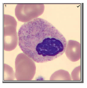
Pelger-Huët anomaly (PHA), is a term familiar to medical laboratory professionals, but mostly from textbook images. PHA is considered to be rare, affecting about 1 in 6000 people. PHA has been found in persons of all ethnic groups and equally in men and women. The characteristic, morphologically abnormal neutrophils were first described by Dutch hematologist Pelger in 1928. He described neutrophils with dumbbell shaped, bi-lobed nuclei. The term ‘pince-nez’ has also been used to describe this spectacle shaped appearance. Pelger also noted that, in addition to hyposegmentation, there is an overly coarse clumping of nuclear chromatin. In 1931, Huët, a Dutch pediatrician, identified this anomaly as an inherited condition.

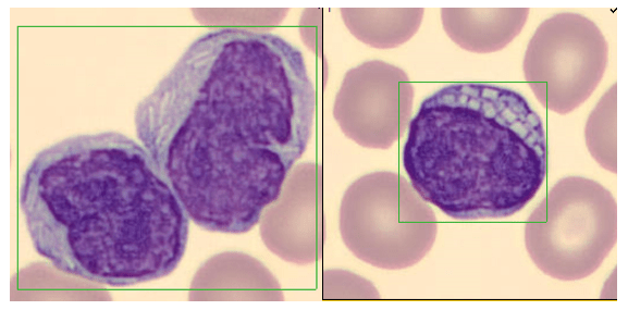
Pelger-Huët anomaly is an autosomal dominant disorder caused by a mutation in the lamina B receptor (LBR) gene on band 1q42. This defect is responsible for the abnormal routing of the heterochromatin and nuclear lamins, proteins that control the shape of the nuclear membrane.2 Because of this mutation, nuclear differentiation is impaired, resulting in white blood cells with fewer lobes or segments. In classic inherited PHA, cells are the size of mature neutrophils and have very clumped nuclear chromatin. About 60-90% of these neutrophils are bi-lobed either with a thin filament between the lobes, or without the filament. About 10-40% of total neutrophils in PHA have a single, non-lobulated nucleus. Occasional normal neutrophils with three-lobed nuclei may be seen.1 Despite their appearance, Pelger-Huët cells are considered mature cells, function normally and therefore can fight infection. It is considered a benign condition; affected individuals are healthy and no treatment is necessary for PHA.

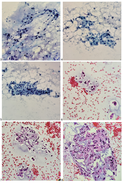
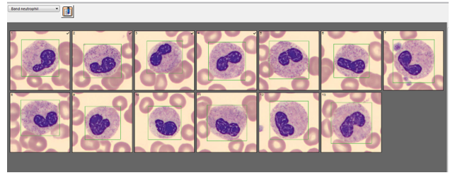
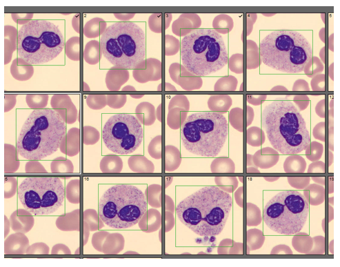
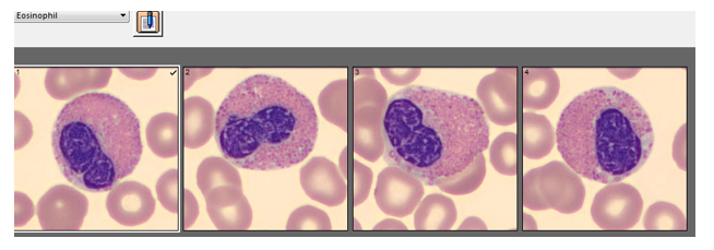
Automated instruments may flag a left shift when they detect these Pelger-Huët cells. In this patient, the analyzer flagged a left shift and a slide was made and sent to CellaVision. The CellaVision pre-classified the Pelger-Huët cells as neutrophils, bands, and myelocytes. All of the neutrophil images were either bi-lobed or non-lobed forms. None of the neutrophils had more than 2 lobes. Eosinophils also had poorly differentiated nuclei. Cell images from this patient can be seen in Images 1-4.

Image 1. Images from CellaVision of bi-lobed “pince-nez” neutrophils with thin filament
Image 2. Non-terminally differentiated neutrophils pre-classified as bands on CellaVision. Bilobed variant without the thin filament.
Image 3. Non-lobed neutrophils with extremely coarse clumping of nuclear chromatin.
Image 4. Eosinophils in Pelger-Huët Anomaly.

If PHA is considered benign, with no clinical implications, why is it important to note these cells on a differential report? This slide was referred to our pathologist for a review. The patient had several previous CBC orders, but no differentials in our LIS. The pathologist reviewed the slide and, based on 100% of these neutrophils being affected, he reported “Pelger-Huët cells present. The presence of non-familial Pelger-Huët anomaly has been associated with medication effect, chronic infections and clonal myeloid neoplasms.” Thus, the importance of reporting this anomaly if seen on a slide. If the instrument flags a left shift, this is typically associated with infection. If these cells are misclassified as bands and immature granulocytes, with no mention of the morphology, there would be a false increase in bands reported and the patient may be unnecessarily worked up for sepsis.

An additional reason for reporting the presence of Pelger-Huët cells is that pelgeroid cells are also seen in a separate anomaly, called acquired or pseudo-Pelger-Huët anomaly (PPHA). PPHA is not inherited and can develop with acute or chronic myelogenous leukemia and in myelodysplastic syndrome. A type of PPHA may also be associated with infections or medications. Certain chemotherapy drugs, immunosuppressive drugs used after organ transplants, and even ibuprofen have been recognized as triggers for PPHA. PPHA caused by medications is typically transient and resolves after discontinuation of the drug. To add to causes, most recently, there have been studies published that report PPHA in COVID-19 patients.3

With several different causes of PHA/PPHA, a differential diagnosis is important. Is this a benign inherited condition, a drug reaction that will self-resolve after therapy is stopped, or something more serious? If Pelger-Huët cell are reported, it is important for the provider to correlate this finding with patient symptoms, treatments and history. There was no medication history and little other medical history in our case patient’s chart, and no mention of inherited PHA. The patient had also been tested for COVID-19 with her pre-op testing and was COVID negative. On initial identification of Pelger-Huët, a benign diagnosis that needs no treatment or work up would be the best outcome, so an attempt could be made to determine if the patient has inherited PHA. If other family members are known to have this anomaly, this would be the likely diagnosis as PHA is autosomal dominant. Family members can also easily be screened with CBC and manual differential. Molecular techniques are available to confirm PHA but are not routinely used. In the absence of this anomaly in other family members, it would need to be determined if the patient was on any medications that can cause pelgeroid cells. Inherited PHA and drug induced PPHA should be ruled out first because PPHA can also be predicative of possible development of CML or MDS. Considering this cause first could lead to unnecessary testing that might include a bone marrow aspirate and biopsy. Additionally, the entire clinical picture should be reviewed because in PPHA associated with myeloproliferative disorders there is usually accompanying anemia and thrombocytopenia and the % of pelgeroid cells tends to be lower.

Today most clinical laboratories have instruments that do automated differentials, and we encourage physicians to order these because they are very accurate and count thousands of cells compared to the 100 cells counted by a tech on a manual differential. Automated differentials are desirable for consistency and to improve turnaround times. Yet, it is important to know when a slide needs to be reviewed under the scope or with CellaVision. If a patient presents with a normal WBC and a left shift on the auto diff with no apparent reason, pictures can reveal important clinical information. Awareness of different causes of PHA/PPHA can relieve anxiety in patients and prevent extensive, unnecessary testing and invasive procedures.

References

  1. https://emedicine.medscape.com/article/957277-followup updated 8/4/2020
  2. Ayan MS, Abdelrahman AA, Khanal N, Elsallabi OS, Birch NC. Case of acquired or pseudo-Pelger-Huët anomaly. Oxf Med Case Reports. 2015;2015(4):248-250. Published 2015 Apr 1. doi:10.1093/omcr/omv025
  3. Alia Nazarullah, MD; Christine Liang, MD; Andrew Villarreal, MLS; Russell A. Higgins, MD; Daniel D. Mais, MD. Am J Clin Peripheral Blood Examination Findings in SARS-CoV-2 Infection . Pathol. 2020;154(3):319-329. 

-Becky Socha, MS, MLS(ASCP)CM BB CM graduated from Merrimack College in N. Andover, Massachusetts with a BS in Medical Technology and completed her MS in Clinical Laboratory Sciences at the University of Massachusetts, Lowell. She has worked as a Medical Technologist for over 30 years. She’s worked in all areas of the clinical laboratory, but has a special interest in Hematology and Blood Banking. When she’s not busy being a mad scientist, she can be found outside riding her bicycle.

Cytology Case Study: Strike a Chord

Every FNA ROSE attended where the patient is conscious and attentive can be tricky to navigate. You have to remain cognizant of your word choice, your demeanor, and the delivery of your adequacy statement to the clinician. The patient is already in a heightened state of awareness because he or she is about to be probed with a needle (or six!) for a test that is likely to rule out a benign or malignant process. I prefer to go into my biopsies with some sort of clinical picture and as many details as I can retain – is there a previous history of cancer? Where is the lesion located? Is it a single mass or are there multiple lesions? What does the radiologic imaging suggest? Are there any elevated serum tumor markers? I need to be able to walk the walk and talk the talk. However, there are rare instances when cytotechs are asked to rush down to an unscheduled add-on biopsy where we have yet to research the impression documented in the patient’s medical record. In those situations, I ask the clinician (typically an interventional radiologist) all the questions I can think of while still emulating some form of confidence to the patient.

I entered the procedure room and greeted the radiologist, radiology fellow, tech, nurse, and the patient, a 57-year-old male who was prone and alert on the table. I jotted down notes during the timeout and pulled the radiologist aside to ask, “does the patient have a history of cancer?” In this case, the answer was “they have a soft tissue tumor in the left gluteus, which is what we’re biopsying.” “Alright, let’s get those differentials rolling – sarcoma; after my hibernoma experience – a lipomatous tumor; or could it be a carcinoma (because yes, I’ve seen a lung adenocarcinoma metastasize to the gluteal muscle before)? Hmm… what else? What other mesenchymal tumors could originate here… or metastasize here?” My brainstorming balloon was popped by the radiologist asking if I was ready for the first needle pass. I replied, “Yes, of course!” I glance over at the patient and smile, trying to assure him AND myself that I’ll be able to give him a definitive answer to his puzzle.

Here’s what I visualized under the microscope after I stained the first air-dried smear in our Diff-Quik solutions.

Images 1-2. Left gluteal FNA, DQ-stained smears.

My inner monologue became: “Well, it’s not a sarcoma or a carcinoma. It doesn’t look malignant.  Not quite a hibernoma. What is with that myxoid matrix? It’s not mucinous or serous, so… what is it…? It’s granular! Wait. Those nuclei. They’re so… what’s the word? It’s definitely representative of the lesion. Regardless, it’s adequate!” I turned away from my microscope to face the team – “The sample is adequate. May I have a few more passes for my cell block, and will you collect core biopsies, too? “Yes and yes,” the radiologist replied. I smiled again at the patient, and he mouthed, “thank you.” “Phew, mission accomplished,” I thought. “Now what the heck are those hallmark cells called mixed in with a majority of epithelioid cells arranged in chords?”

I climb the stairs up to the lab and do a quick Google search. “DUH! Physaliphorous cells!” These cells have a distinct feature where the nucleus is centrally located but is also scalloped by cytoplasmic vacuoles. There weren’t as many physaliphorous (physaliferous) cells as I had hoped to appreciate. Some of the cells looked lipoblastic in nature with larger vacuoles displacing the nuclei to the periphery, almost signet ring in nature, many were cuboidal. But that was it… those cells! Now, imagine the scene in Finding Nemo where Nemo struggles to tell his classmates he lives in an anemone. That was my garbled attempt at pronouncing “physaliphorous” to the attending pathologist when sharing my interpretation. She looked at me like I was saying anything other than the word I was trying to reproduce. I cannot blame her; I still turn beet-red at the memory. But I was convinced that a chordoma was the tumor I presented to her.

After I processed my FNA, I examined the patient’s electronic health record to see if he had any previously biopsied neoplasms on file, and much to my surprise, the patient was diagnosed with a primary chordoma of the sacrum and treated with en bloc resection and radiation in 2013. Mutation analysis was performed on the resection of this chordoma, which exhibited a homozygous loss of CDK2NA (p16). The patient had one recurrence at an outside facility in 2015 and transferred his care to our institution for follow-up. Now, the patient presented with this gluteal metastasis and soon thereafter, a paraspinal metastasis. As the patient’s chordoma did not completely respond to radiation, the clinical care team turned to the tyrosine kinase inhibitor, Gleevec, which was discontinued due to disease progression. The patient’s regimen then went on to include sunitinib, which was also discontinued due to disease progression, palbociclib, then nivolumab, followed by radiation to the thoracic spine, sorafenib, and now is on a clinical trial for patients with advanced refractory cancers.

When I turned in my Diff-Quik & Pap-stained slides and the cell block H&E sections with a diagnosis of chordoma the next day, the attending cytopathologist paged through one of our cytology texts to a tidbit on chordomas before signing out the case. She reviewed the section with me. Other than the unique physaliphorous cells, it turns out a diagnosis of chordoma is fairly rare, as it is the only malignancy derived from the notochord, typically occurring at either end of the axial skeleton.1 Metastasis of these tumors is also rare, so this case of widespread metastatic disease was even more intriguing to me.

Images 3-8. Left gluteal FNA . Images 3-5, Pap-stained smears; 6-8, H&E cell block sections.

References

  1. Cibas, E. S., & Ducatman, B. S. (2009). Cytology: Diagnostic Principles and Clinical Correlates, Expert Consult – Online and Print (3rd ed.). Saunders.

-Taryn Waraksa, MS, SCT(ASCP)CM, CT(IAC), has worked as a cytotechnologist at Fox Chase Cancer Center, in Philadelphia, Pennsylvania, since earning her master’s degree from Thomas Jefferson University in 2014. She is an ASCP board-certified Specialist in Cytotechnology with an additional certification by the International Academy of Cytology (IAC). She is also a 2020 ASCP 40 Under Forty Honoree.

Microbiology Case Study: A Middle-Aged Woman with Forearm Pain

Case History

A middle-aged female was evaluated for left forearm pain and erythema following a cat bite one-day prior, and was prescribed trimethoprim-sulfamethoxazole for management in the outpatient setting.  She subsequently presented for follow-up where she was noted to have a 3 x 4 cm raised, red, indurated lesion of left arm without any discharge (Image 1).  MRI demonstrated a 6.5 x 2.3 x 2.3 cm abscess within the distal ulnar soft tissues with surrounding cellulitis.  As her pain and erythema were progressively worsening, she was admitted for surgical management.   

Upon admission, a bedside incision found purulent drainage which grew mixed anaerobic gram negative rods.  Blood cultures collected at this same time were negative and remained so for the duration of her hospital course.  Empiric antibiotic therapy was initiated with piperacillin-tazobactam, and the patient underwent formal surgical incision and drainage.  Intraoperative findings were notable for abscess, diffuse and severe tendinopathy, and a thick inflammatory rind surrounding the associated neurovascular bundle.  Intraoperative cultures were obtained and sent to the microbiology laboratory.  The patient’s postoperative course was uneventful, and she was discharged with plans to complete a two week course of amoxicillin-clavulanate.  Follow-up clinic visits demonstrated successful recovery, with a well-healed incision and normal grip strength and range of motion.

Laboratory Identification

Bacterial culture of abscess material collected intraoperatively grew smooth, mucoid colonies on chocolate and blood agars with less than 24 hours of incubation at 35°C in CO2 (Image 2, bacterial isolate). Growth was notably absent on MacConkey agar. Gram stain of the colony revealed tiny, gram negative coccobacilli (Image 2). Biochemical testing determined this organism to be indole, oxidase, and catalase positive.  The organism was definitively identified as Pasteurella multocida by MALDI-TOF MS.

Image 1. Arm lesion prior to incision and drainage.
Image 2. P. multocida growth on Blood and Chocolate agars. Gram stain from a colony revealed small, gram negative coccobacilli (far right).

Discussion

Members of the genus Pasteurella are small, Gram-negative coccobacilli which are able to readily grow on Sheep’s blood agar and chocolate agar, but will typically not grow on MacConkey media.  Infection with these organisms is usually considered to be a zoonosis, with both wild and domestic animals serving as reservoirs.  In animal hosts they can be part of the endogenous flora or pathogens.  P. multocida is the most common member of the genus associated with human infections, which has now been divided into multiple taxonomic subspecies through the use of more discriminatory molecular methods.  Biochemically, P. multocida is positive for catalase, oxidase, indole, and nitrate reduction.

Animal bite wounds are often polymicrobial and contain mixtures of both aerobic and anaerobic organisms.  These organisms can be reflective of the oral flora of the biting animal, or of endogenous skin flora of the bite victim.1  While 80-90% of bites per year can be attributed to dogs, an estimated 400,000 cat bites (5-10% of the total) occur in the United States annually.2  While dog bites often manifest as localized crush injuries with contusions and/or lacerations, a majority of such wounds are accessible to irrigation and cleaning which leads to a relatively low infection rate (5-10%).  By contrast, cat bites are often deep, localized puncture wounds which provide excellent environments for the growth of both aerobic and anaerobic bacteria.  While feline bite wounds may appear less severe after cursory inspection, these wounds can be considerably more difficult to clean, resulting in overall infection rates up to 50%.3 

Management of bite wounds includes cleansing, irrigation and debridement.  Importantly, antimicrobial therapy should include coverage for both aerobic and anaerobic bacteria.4  In this case, amoxicillin-clavulanate was utilized with good results, and provides coverage for the most common oral aerobes and anaerobes encountered in animal bite wounds.  Amoxicillin-clavulanate also has activity against beta-lactamase producing bacteria such as Prevotella sp. and Porphyromonas sp. which are oral anaerobes of dogs, cats, and humans.  The use of macrolides should be avoided due to variable activity against Pasteurella multocida.4  As in this case, bite wounds most frequently are encountered on the upper extremities, and Pasteurella sp. is one of the most common isolates recovered from bites from both cats and dogs (50% of dog bites, and 75% of cat bites).2

References

1. Abrahamian FM, Goldstein EJC. 2011. Microbiology of Animal Bite Wound Infections. Clinical Microbiology Reviews 24:231.

2. Bula-Rudas FJ, Olcott JL. 2018. Human and Animal Bites. Pediatrics in Review 39:490.

3. Kannikeswaran N, Kamat D. 2008. Mammalian Bites. Clinical Pediatrics 48:145-148.

4. Stevens DL, Bisno AL, Chambers HF, Dellinger EP, Goldstein EJC, Gorbach SL, Hirschmann JV, Kaplan SL, Montoya JG, Wade JC. 2014. Practice Guidelines for the Diagnosis and Management of Skin and Soft Tissue Infections: 2014 Update by the Infectious Diseases Society of America. Clinical Infectious Diseases 59:e10-e52.

 -Francesca Lee, MD, is an associate professor in the Departments of Pathology and Internal Medicine (Infectious Diseases) at UT Southwestern Medical Center.

-Huy Dao, MLS(ASCP)CM graduated from the University of Minnesota and has worked for eight years as medical technologist for eight years.  He is interested in clinical mycology and bacteriology.

-Andrew Clark, PhD, D(ABMM) is an Assistant Professor at UT Southwestern Medical Center in the Department of Pathology, and Associate Director of the Clements University Hospital microbiology laboratory. He completed a CPEP-accredited postdoctoral fellowship in Medical and Public Health Microbiology at National Institutes of Health, and is interested in antimicrobial susceptibility and anaerobe pathophysiology.

Microbiology Case Study: 83 Year Old Female with a Perisplenic Abscess

Case History

An 83 year old female with a past medical history of breast cancer, multiple strokes, dysphagia, hypertension and gastroesophageal reflux disease (GERD) presented to an outside hospital with altered mental status, metabolic encephalopathy, decreased appetite, acute kidney injury, and E. coli/Proteus urinary tract infection (UTI). There, she was diagnosed with a perforated gastric ulcer, which was repaired, with a gastrostomy (G) tube in place. The patient later developed a fever and an aspiration pneumonia, which was treated with ampicillin/sulbactam. A follow up imaging revealed a new gastric perforation along the fundus of the stomach with perisplenic fluid collection, along with a pleural effusion with possible communication with the fluid collection in the stomach. Due to her complex medical history, an additional intervention was not pursued and the family agreed to comfort measures, and the patient was discharged home.

The family presented to our emergency department the same day of discharge, as the patient had not been eating and the family needed assistance in using the G tube. In-house computed tomography (CT) of the abdomen/pelvis again showed an organizing collection near the spleen (Image 1). A medium-sized left pleural effusion with left lower lobe collapse due to the communication with the perforation was observed on CT. The patient received IV fluids and a dose of vancomycin and zosyn in the ED. A vascular and interventional radiology (VIR) consult was recommended for potential drainage of the perisplenic abscess and left pleural effusion.

Image 1. CT of the abdomen demonstrating an organizing collection (black circle, approximately 7 cm x 3 cm) posterior/superior to the spleen near the site of the prior gastric perforation concerning for an abscess.
Image 2. Small gram negative rods on a Gram stain of Burkholderia cenocepacia from a subculture.
Image 3. Culture morphology of Burkholderia cenocepacia on a blood agar plate after overnight incubation: smooth colonies are present (upon prolonged incubation, the colonies turned yellowish color – not shown in image).

VIR drained the perisplenic abscess, which was sent to the microbiology lab for aerobic & anaerobic cultures. The Gram stain revealed many white blood cells (WBC’s) and rare yeast. The culture grew 3+ Burkholderia cepacia complex (Burkholderia cenocepacia) and 3+ Candida glabrata. The Gram stain and colony of the subculture of B. cepacia on a blood agar plate are shown in Images 2 and 3. C. glabrata was also isolated from the urine culture. Susceptibility testing of B. cepacia showed that it was sensitive to both meropenem and trimethoprim-sulfamethoxazole. Vancomycin and zosyn were stopped and the patient was switched to IV trimethoprim sulfamethoxazole for B. cepacia and IV micafungin for C. glabrata.

Due to the recurrent perforation, the G tube could not be used; a jejunostomy (J) tube was placed instead. Feeds were successfully started with the J tube. Trimethoprim-sulfamethoxazole was also given via the J-tube. A follow-up endoscopy showed a normal esophagus, the known perforation in the gastric fundus, and erythematous duodenopathy at the level of the duodenal bulb, the remainder of the duodenum was normal. The patient’s clinical status improved and she was discharged home with the support of home health services.

Discussion

We present an uncommon presentation of Burkholderia cenocepacia, a member of B. cepacia complex, in perisplenic abscess fluid. B. cepacia complex consists of at least 17 closely related species. They are rod-shaped, aerobic, motile Gram-negative bacteria. B. cepacia complex has been well characterized as opportunistic pathogens, particularly in patients with cystic fibrosis and chronic granulomatous disease (CGD). However, they can also infect immunocompetent patients and have been reported to cause endocarditis (specifically in IV drug abusers), pneumonitis, UTIs, osteomyelitis, dermatitis, and other wound infections. In the United States, B. multivorans and B. cenocepacia together account for approximately 80% of B. cepacia complex infections. Burkholderia have been isolated on contaminated hospital equipment and even disinfectants. They present a large problem in nosocomial infections due largely to their ability to survive in aqueous environments.1 They are soil-dwelling bacteria commonly found on plant roots. They are of environmental interest secondary to their antifungal and antinematodal properties as well as the ability to degrade many toxic compounds in agriculture (pesticides, herbicides, preservatives).2 Clinically important Burkholderia species outside of the B. cepacia complex include B. pseudomallei, the causative agent of melioidosis, and B. mallei, the causative agent of glanders.1

Rare case reports have previously documented B. cepacia isolated from splenic abscesses/infections. Most B. cepacia splenic infections occurred secondary to pneumonia or multi-organ involvements in CGD patients. 3, 4, 5 However, one report indicated the B. cepacia-mediated multiple splenic abscesses, in the setting of malignancy and diabetes. 6 While the splenic abscesses in the context of meliodosis, either due to B. pesudomallei or B. mallei infection, have been reported, 7 B. cepacia-mediated splenic infections are rarely encountered.

B. cepcacia complex has intrinsic resistance toseveral antibiotics including penicillins, amoxicillin-clavulanate, ertapenem, polymixin B, Colistin, and fosfomycin.8 B. cepacia complex possesses an inducible β-lactamase, encoded by the gene penA, which can hydrolyze penicillin and use it as a source of carbon. In one study involving 40 bloodstream isolates of B. cepacia in patients without cystic fibrosis, 93% of the isolates were susceptible to ceftazadime and 95% of isolates were susceptible to trimethoprim-sulfamethoxazole.9 Following discussion with our infectious disease colleagues, we believe that the B. cepacia isolate from our patient was likely a nosocomial infection from possible contamination of her G-tube in combination with the gastric perforation.

References

  1. Jorgensen, J. H., Pfaller, M. A., & Carroll, K. C. (2015). Manual of clinical microbiology. Washington, DC, DC: ASM Press.
  2. Kenyon College Department of Biology. (2011, April 22). Burkholderia cepacia. Retrieved September 21, 2020, from https://microbewiki.kenyon.edu/index.php/MicrobeWiki
  3. Clegg HW, Ephros M, Newburger PE. Pseudomonas cepacia pneumonia in chronic granulomatous disease. Pediatr Infect Dis. 1986 Jan-Feb;5(1):111. PMID: 3945563.
  4. Sirinavin, Sayomporn MD*; Techasaensiri, Chonnamet MD*; Pakakasama, Samart MD*; Vorachit, Malai DSc; Pornkul, Rattanaporn MD; Wacharasin, Rames MD Hemophagocytic Syndrome and Burkholderia cepacia Splenic Microabscesses in a Child With Chronic Granulomatous Disease, The Pediatric Infectious Disease Journal: September 2004 – Volume 23 – Issue 9 – p 882-884 doi: 10.1097/01.inf.0000137565.23501.03
  5. Bottone EJ, Douglas SD, Rausen AR, Keusch GT. Association of Pseudomonas cepacia with chronic granulomatous disease. J Clin Microbiol. 1975 May;1(5):425-8. doi: 10.1128/JCM.1.5.425-428.1975. PMID: 1176612; PMCID: PMC275137.
  6. Jayawardena, M. N., Chandrasiri, N. S., Wijekoon, S., Madanayake, P., Corea, E., Ranasinghe, D. D., & Lamahewage, N. D. (2017). Burkholderia cepacia; an unusual cause of multiple splenic abscesses : A case report. Sri Lankan Journal of Infectious Diseases, 7(2), 123. doi:10.4038/sljid.v7i2.8146
  7. Chen, H., Hu, Z., Fang, Y., Lu, X., Li, L., Li, Y, Mao, X, Qian, L. (2018). Splenic abscess caused by Burkholderia pseudomallei. Medicine, 97(26). doi:10.1097/md.0000000000011208
  8. Patel, J. B., Weinstein, M. P., Eliopoulos, G.M., Jenkins, S.G., Lewis, J.S., Limbago, B., Mathers, A., Mazzulli, T., Patel, R., Richter, S.S., Satlin, M., Swenson, J.M., Traczewski, M.M., Turnidge, J.D. & Zimmer, B.L. (2017). Performance standards for antimicrobial susceptibility testing. Wayne, PA: Clinical and Laboratory Standards Institute.
  9. Bressler A.M., Kaye K.S., LiPuma, J.J., Alexander, B.D., Moore, C.M., Reller, L.B. & Woods, C.W. Risk factors for Burkholderia cepacia complex bacteremia among intensive care unit patients without cystic fibrosis: A case-control study. Infect Control Hosp Epidemiol 2007; 28(8):951-8 doi : http://dx.doi.org/10.1086/519177

-J. Stephen Stalls, MD is a PGY-II pathology resident at the East Carolina University Department of Pathology and Laboratory Medicine. He plans to pursue hematopathology and molecular pathology fellowships, but also greatly enjoys his time in the microbiology lab. Outside of work, he enjoys playing the drums and going to concerts.

-Phyu Thwe, PhD, MLS(ASCP)CM is Technical Consultant/Technical Director of Clinical Microbiology Laboratory at Vidant Medical Center, Greenville, NC.

Case Study Hematology: The Mouse Strikes again! Lymphocytes with Intracytoplasmic Inclusions

If you read my last blog, you heard the about the story “if You Give a Mouse a Cookie” by Laura Numeroff.4 The curious little mouse has a mind that never rests. As his mind wanders and hops from one thing to another, he keeps discovering more things to check out along the way. Medical laboratory lcientists are a lot like this. We’re a curious bunch, and, in investigations, one thing often leads to the next. Well folks, the mouse has struck again! We were given another cookie in the form of these beautiful cells.

Image 1. Lymphocytes with intracytoplasmic inclusions.

These cells were found by my coworker Liz Marr, MLS(ASCP), and the adventure began! First, we wanted to know what those were, and then we needed to find out more about them, and then, mostly, I wanted to know why in almost 40 years of working in and teaching hematology that I have never before seen this!

The story begins with our case history. We received a CBC from a 71 year old female with a 4 year history of untreated chronic lymphocytic leukemia/ small lymphocytic lymphoma(CLL/SLL). The patient’s recent history included a myocardial infarction(MI) 5 months prior. The patient was found to have leukocytosis (WBC 25.38 x 10³/μL) and absolute lymphocytosis (18.25 x 10³/μL) with normal hemoglobin and hematocrit (Hgb 13.4 g/dL, Hct 40.8%) and normal platelet count (272 x 103/μL). The differential had 71.9% lymphocytes with many abnormal forms noted. The slide was sent for a pathology review. The pathologist reported “Atypical lymphocytosis consistent with patient’s known chronic lymphocytic leukemia/small lymphocytic lymphoma (CLL/SLL) Filament-like inclusions are present in the cytoplasm which has been previously reported in patients with CLL.”

Image 2. Lymphocytes with crystalline-like inclusions.

A curious tech can’t stop at just that description. If you tell me they are filament-like inclusions, I will have all kinds of questions. What are these filaments made of? Are they crystals, or something else? How common are these? Are these diagnostic of CLL? Are these only seen in CLL? What is their significance? And, of course, the most puzzling question, why have I never seen these before??

CLL is a form of non-Hodgkin lymphoma and is the most common leukemia in the Western world. It is generally a leukemia of older age with a median age at diagnosis around 67-72. The disease is widely variable, with some patients asymptomatic and requiring no treatment for many years, while others have a more rapidly progressive course of disease requiring treatment. About 60% of patients are diagnosed before they exhibit any symptoms. CLL and SLL are considered to be different manifestations of the same disease. In CLL, the abnormal B lymphocytes are found mostly in the peripheral blood and bone marrow, but in SLL, there is lymph node involvement, with abnormal cells mostly found in the lymph nodes. CLL is diagnosed based on absolute B lymphocyte counts ≥5 x 10⁹/L. Flow cytometry typically reveals a distinctive cell immunophenotype with expression of CD19, CD5, CD23, and Κ/λ; and weak expression of CD20, CD79b, and surface immunoglobulin.1

The most recent flow cytometry report on our patient was from one year ago. An 8 color analysis with CD45/SSC gating was performed by LabCorp. The flow revealed an abnormal cell population representing 56% of total cells. Two monoclonal B cell populations were detected with identical phenotypes except for light chain expression. These cells expressed CD45, CD19, CD20, CD22, CD5, and CD23., CD38-. This phenotype was consistent with her previous diagnosis of CLL/SLL.

A literature search revealed only a few articles about intracytoplasmic inclusions in CLL. Cytoplasmic inclusions in lymphomas are uncommon, but have been noted as vacuoles, crystals, and pseudocrystals. These crystalline inclusions represent immunoglobulin(Ig) heavy and light chain that precipitate in the cytoplasm. Using electron microscopy it has been found that theses Ig deposits localize in the rough endoplasmic reticulum (RER).5 When surface Ig can be demonstrated on the B lymphocytes, it has been found to be same as Ig in the inclusions.6

In two published studies that describe these crystal like inclusions, photographs are very similar to the ones we found on our patient.3,5 It is interesting to note that, in these two studies, neither of the subjects was a known CLL patient. The inclusions were noted in the patients’ cells and the peripheral blood was subsequently sent for flow. Phenotypes reported confirmed monoclonal B-cells representing a large percentage of cells. Huang reported monoclonal B-cells which expressed CD45, CD19, CD20, CD22, CD79b, CD5, CD23, CD148 and CD200(hi), with partial expression lambda, and negative for FMC7, CD10, CD11c, CD49d, CD103, CD38, CD25, CD160, IgM, CD81, kappa and Ki67.3 In the Ramlal case study, phenotype was CD5, CD19, CD20, CD23, positive, CD10, FMC7 negative.5 On the basis of flow, along with the CBC results, the patients were diagnosed with CLL.

Of course, while researching this, the little mouse in me kept asking questions and finding more questions to ask. One question that I still had questions about was if these inclusions have any prognostic value. In three recent studies3,5,6 it was indicated that these inclusions can be used to help with diagnosis, but are not prognostic for course of disease. Rodriguez followed a patient with asymptomatic Rai stage 0 CLL. This patient consistently had inclusions noted in lymphocytes for 9 years before any progression of disease was noted.6 In the medical field even if one study reports no prognostic significance, this opinion could change in the future with more studies. Could these crystalline inclusions be used to forecast time to first treatment (TFT) or overall survival?(OS). So far, because of the rarity of these cytoplasmic inclusions, there is no evidence of prognostic value. As well, the mechanism related to their formation and their role in CLL is yet to be determined.

Our case study patient and the various reports found in literature had common flow cytometry immunophenotypes. Patients were all either previously diagnosed with CLL or lymphocytic lymphoma, or were diagnosed at the time of the findings of these inclusions. While these crystalline inclusions alone are not considered diagnostic for CLL, their recognition can be used to assist in a prompt diagnosis of a lymphoproliferative disease. And they are so pretty! What medical laboratory scientist doesn’t love pretty cells? Be like that mouse. Be curious, keep your eyes open, and be on the lookout for these interesting cells in CLL patients, but, more importantly, in patients with lymphocytosis without a known diagnosis of a lymphoproliferative disorder.

References

  1. AJMC, January 7, 2019
  2. Chronic Lymphocytic Leukemia: An Overview of Diagnosis, Prognosis, and Treatment
  3. Huang, Y., Zhang, L. Intracellular rod-like crystals in chronic lymphocyte leukemia. Int J Hematol 112, 267 (2020). https://doi.org/10.1007/s12185-020-02933-7
  4. Numeroff, Laura If You Give a Mouse a Cookie. 1986
  5. Ramlal, B, DiGiuseppe, JA. Intracytoplasmic crystalline inclusions in chronic lymphocytic leukemia. Clin Case Rep. 2019; 7: 1460– 1461. https://doi.org/10.1002/ccr3.2250
  6. Cecilia M. Rodríguez, Carmen Stanganelli, Claudio Bussi, Daniela Arroyo, Darío Sastre, Viviana Heller, Pablo Iribarren & Irma Slavutsky (2018)Intracytoplasmic filamentous inclusions and IGHV rearrangements in a patient with chronic lymphocytic leukemia, Leukemia & Lymphoma, 59:5,1239-1243, DOI: 10.1080/10428194.2017.1370549

-Becky Socha, MS, MLS(ASCP)CM BB CM graduated from Merrimack College in N. Andover, Massachusetts with a BS in Medical Technology and completed her MS in Clinical Laboratory Sciences at the University of Massachusetts, Lowell. She has worked as a Medical Technologist for over 30 years. She’s worked in all areas of the clinical laboratory, but has a special interest in Hematology and Blood Banking. When she’s not busy being a mad scientist, she can be found outside riding her bicycle.

Microbiology Case Study: 83 Year Old Male with Bladder Cancer

Case History An 83 year old male with bladder cancer was treated with Mycobacterium bovis Bacillus Calmette-Guérin (BCG), his last treatment occurring 1.5 months prior to presentation. He has a past medical history of chronic obstructive pulmonary disease, hypertension, obstructive sleep apnea, obesity, and diabetes. The patient has been hospitalized four times over the last two months and his symptoms include generalized weakness, malaise, shortness of breath and recurrent fever. He was found to have patchy lung infiltrates and he was diagnosed with pneumonia, COPD exacerbation and symptoms of heart filature. He was treated previously with antibiotics, steroids and fluid management which would temporarily relieve his symptoms. He presents to the hospital again, four days after his last hospital discharge, with generalized weakness, malaise, shortness of breath and recurrent fever. On initial evaluation he was found to be pancytopenic.  

Laboratory Identification

Blood cultures were negative. A bone marrow biopsy was performed for fever of unknown origin and pancytopenia. The biopsy showed non-caseating granulomas which were negative for acid-fast bacilli (AFB) by Ziehl-Neelsen stain and fungal elements by Gomori Methenamine Silver Stain (GMS). A laboratory-develped PCR test for Mycobacterium tuberculosis complex (MTBC) was performed on the bone marrow and was negative. AFB culture of bone marrow was positive for after 30 days of incubation and the organism was confirmed to be acid-fast bacilli by auramine-rhodamine fluorescent dye and Kinyoun stain. A second laboratory-developed test that uses heat shock protein (HSP) 2 and HSP3 to determine species level identification of Mycobacteria identified the organism as M. tuberculosis complex. Due to the patient’s history, further identification was performed at a reference lab using specific oligonucleotides targeting the gyrb DNA sequence polymorphisms which is able to separate different members of the MTBC. The patient’s isolate contained a RD1 deletion which is specific for Mycobacterium bovis bacillus Calmette-Guérin (BCG).

Discussion

Mycobacterium bovis is a slow growing mycobacterium which produces rough, dry colonies on growth solid media. It is one of the species in the MTBC with a natural host of domestic and wild animals. Routine molecular tests will not accurately differentiate between members of the MTBC. For definitive identification of M. bovis, 16S rRNA and gyrB gene sequencing is necessary. Safe handling procedures should be followed prior to molecular testing of MTBC.

Mycobacterium bovis BCG is a live, attenuated strain of Mycobacterium bovis that was created for vaccine and is used in the treatment of superficial bladder cancer. The treatment may cause localized symptoms including hematuria, fever, nausea, and dysuria which are marker of anti-tumor effect. Serious complications occur in <5% of patients with complications including sepsis, pneumonitis, hepatitis, lymphocytic meningitis, bone marrow involvement, and mycotic aneurysms. The cardinal sign of BCG infection is a relapsing fever with drenching night sweats persisting beyond 48 hours. Disseminated infection can occur days to years after the therapy. Clinical suspicion should be high for M. bovis BCG dissemination if there are symptoms and a high grade fever ≥72 hours. Treatment includes a regiment of isoniazid, rifampin and ethambutol. Most isolates of M. bovis are resistant to pyrazinamide.

References

  1. Lamm DL. Efficacy and safety of bacille Calmette-Guérin immunotherapy in superficial bladder cancer. Clin Infect Dis 2000; 31 Suppl 3:S86.
  2. Shelley MD, Court JB, Kynaston H, et al. Intravesical Bacillus Calmette-Guerin in Ta and T1 Bladder Cancer. Cochrane Database Syst Rev 2000; :CD001986.
  3. Richter E, Weizenegger M, Rusch-Gerdes S, Niemann S. Evaluation of Genotype MTBC Assay for Differentiation of Clinical Mycobacterium tuberculosis Complex Isolates. Journal of Clinical Microbiology 2003; 41(6): 2672-2675
  4. UpToDate

-Crystal Bockoven, MD is a 4th year anatomic pathology resident at University of Chicago (NorthShore). Crystal has an interest in and will be doing a fellowship in pediatric and perinatal pathology. In her spare time, she enjoys reading, hiking and biking. 

-Erin McElvania, PhD, D(ABMM), is the Director of Clinical Microbiology NorthShore University Health System in Evanston, Illinois. Follow Dr. McElvania on twitter @E-McElvania. 

Microbiology Case Study: 35 Year Old Male with Chest Pain

Case History

A 35 year old man presented to the Emergency Department (ED) with intermittent chest pain for 3-4 days, abdominal pain, fatigue, and lightheadedness over the same time period. Additionally, his family reported symptoms of progressive malaise for about a month, worse over the last week. In the ED, he was found to have ST elevations in the inferior leads of the electrocardiogram, which can be indicative of a heart attack. He was given 325 mg of aspirin and was emergently taken to the catheterization lab. He was found to have multiple complete occlusions in the distal left anterior descending artery (LAD), posterior descending artery (PDA), and posterior left ventricular artery (PLV). He underwent aspiration thrombectomy and the resulting clots were thought to be emboli; segments were sent to pathology for histopathologic evaluation and to microbiology for culture. There was no evidence of underlying plaque. He was admitted for management of ST-elevation myocardial infarction (STEMI). While in the ED, he was found to have white blood cell count of 23,000 and tachycardia to 110 beats per minute. A transthoracic echocardiogram demonstrated thickened aortic valve leaflets with evidence of leaflet destruction, severe aortic insufficiency, and right coronary cusp perforation which are consistent with endocarditis. Blood cultures were obtained and he was started on broad spectrum antibiotics (Vancomycin and Cefepime).

He has a past medical history significant for previous shoulder abscess with Methicillin-resistant Staphylococcus aureus (MRSA) and intravenous drug use (IVDU) (heroin, last use ~6 days prior to admission).

Computed tomography (CT) of his abdomen and pelvis revealed multiple renal infarctions and a splenic infarction (Image 1). In addition, the CT of the brain showed: “Multifocal scattered supratentorial and infratentorial subarachnoid hemorrhages and findings suggestive of evolving ischemic infarct involving the right inferior frontal gyrus, without evidence of hemorrhagic transformation currently. No midline shift or other complication identified.”

Image 1. Computed tomography of the abdomen demonstrating multiple renal infarctions (left, circled) and a splenic infarction (right, circled).

On hospital day 1 (HD1), both sets of initial blood cultures turned positive with gram positive cocci (GPC) in clusters and thrombectomy cultures were also growing GPC in clusters (Image 2). On HD2, the GPC in the thrombectomy culture was identified as Rothia mucilaginosa. GPC growing in the blood cultures were also Rothia mucilaginosa (Image 2). The patient was continued on Vancomycin. Repeat blood cultures were obtained after catheterization on HD0, and HD2, which were negative. On HD2, the pathology of the initial clots showed “fibrinopurulent debris and fibrin plaques with innumerable cocci in clusters” (Image 3).

Image 2. Microscopic and culture morphology of Rothia mucilaginosa. Left: Gram stain from a blood culture demonstrating groups of Gram-positive cocci in small clusters (1000x magfication, oil immersion). Right: Blood agar plate with mucoid light pink-gray colonies.
Image 3. Hematoxylin and eosin stained slide of formalin fixed paraffin embedded tissue of the thrombus removed during the initial emergent catheterization procedure. Sections demonstrate fibrinous material with entrapped white cells and innumerable cocci. Top: 100x magnification; Bottom: 400x magnification.

On HD3, the patient developed 10/10 chest pain with troponin elevation and T-wave inversion. He was taken back to the catheterization lab for another procedure where he was found to have recurrent, complete occlusion of the PDA with unsuccessful recanalization due to the dense thrombus. On HD6, he developed tamponade physiology due to a large pericardial effusion that was drained. Cultures of the pericardial fluid were negative. Given the recurrent embolization events, the patient was transferred to another hospital to undergo aortic valve replacement surgery and coronary artery bypass graft surgery. Cultures taken at the time of the valve replacement surgery were negative and the valve tissue was not sent for pathologic evaluation.  

Discussion

We present an uncommon case of extensive Rothia mucilaginosa sepsis with septic emboli and endocarditis. Rothia mucilaginosa has experienced the scientific name-change game over the last several decades. It was first identified as Micrococcus mucilaginosus, then became Stomatococcus mucilaginosus, was also known as Staphylococcus salivarius before finally arriving to today’s name of Rothia mucilaginosa.1,2 R. mucilaginosa is a normal inhabitant of the oropharynx and is often associated with dental caries.3 R. mucilaginosa can cause invasive infections, typically in patients with compromised immune systems, disrupted mucosal barriers or injection drug use.4

R. mucilaginosa is a facultatively anaerobic, gram positive, non-fastidious coccus that is coagulase negative but with variable catalase positivity. Colony morphology is usually white to gray nonhemolytic colonies with a mucoid appearance. Although the variable catalase reaction may point toward a Streptococcus spp., the Gram stain morphology of clusters helps to rule it out. Although not all strains are mucoid, the classic colony morphology is wet and is due to polysaccharide capsule.

The organism is generally susceptible to antibiotics designed to target gram positive bacteria including, penicillin, ampicillin, cefotaxime, rifampin and vancomycin.4 It is important to note that R. mucilaginosa is not predictably susceptible to clindamycin, trimethoprim-sulfamethoxazole or ciprofloxacin.5 The patient presented in this case received intravenous vancomycin in part due to the extensive disease on presentation, but also because he was at risk for methicillin-resistant Staphylococcus aureus (MRSA) sepsis and had a previously documented abscess from MRSA.

References

  1. Bergan T, Kocur M. 1982. Stomatococcus mucilaginosus gen. nov., sp.nov., ep. Rev., a member of the family Micrococcaceae. Int. J. Syst. Bacteriol. 32:374-377
  2. Collins MD, Hutson RA, Baverud V, Falsen E. 2000. Characterization of a Rothia-like organism from a mouse: description of Rothia nasimurium sp.nov. and reclassification of Stomatococcus mucilaginosus as Rothia mucilaginosa comb.nov. Int. J. Syst. Evol. Microbiol. 3:1247-1251.
  3. Trivedi MN, Malhotra P. Rothia prosthetic knee joint infection. 2015. J. Microbiol. Immunol. Infect. 48(4):453-455.
  4. Bruminhent J, Tokarczyk MJ, Jungkind D, DeSimone JA. Rothia mucilaginosa Prosthetic Device Infections: A Case of Prosthetic Valve Endocarditis. J. Clin. Microbiol. 5;15:1629-1632.
  5. Kaasch AJ, Saxler G, Seifert H. 2011. Septic arthritis due to Rothia mucilaginosa. Infection. 39:81-82.

-Doreen Palsgrove, MD is a board certified Anatomic and Clinical Pathologist who joined the faculty at UT Southwestern as an Assistant Professor in 2019. She specializes in head and neck and genitourinary pathology. 

Dominick Cavuoti, DO is a professor of AP and CP at UT Southwestern, specializing in infectious disease pathology, cytology and medical microbiology.

-Clare McCormick-Baw, MD, PhD is an Assistant Professor of Clinical Microbiology at UT Southwestern in Dallas, Texas. She has a passion for teaching about laboratory medicine in general and the best uses of the microbiology lab in particular.

Microbiology Case Study: An 80 Year Old Man with Dyspnea, Fatigue, and Weight Loss

Case History

An 80 year old male was seen by his cardiologist for approximately one month of dyspnea, fatigue, and weight loss. Past medical history was significant for aortic stenosis requiring placement of a bioprosthetic valve and multivessel coronary artery disease 13 years prior. He underwent cardiac catheterization and echocardiography that revealed severe bioprosthetic valve stenosis. The patient was in the process of evaluation for a prosthetic valve replacement when he presented to the emergency room for rapid decline of the previously noted symptoms. Exam upon hospital admission was notable for cardiac murmur, lower extremity edema, mild leukocytosis, and anemia. He had normal dentition and no skin lesions. A pre-operative TEE confirmed severe aortic prosthetic valve stenosis, restricted leaflet motion, thrombus on all three leaflets, and thickening of the periannular aortic root and ascending aorta. Subsequent cardiac CT was concerning for either pseudoaneurysm or paravalvular leak suggestive of an infectious or inflammatory process.

Due to the persistent, mild leukocytosis, blood cultures were obtained on the second day of admission. On hospital day 3, one set of blood cultures flagged positive with Gram-variable rods in the aerobic bottle (Image 1). The patient was empirically started on vancomycin and piperacillin/tazobactam. Repeat blood cultures were obtained on hospital days 4 and 7, both again positive for Gram-variable rods within 2 days of collection. The infectious diseases consult team suspected subacute bacterial endocarditis and changed therapy to ceftriaxone.On hospital day 9, the patient underwent a redo sternotomy for aortic valve replacement and aortic root repair. Intraoperative findings included a large amount of phlegmon on the aortic leaflets, near circumferential aortic annulus tissue destruction and abscess cavity. Culture of the intraoperative specimens was negative for bacterial growth. The anatomic pathology findings revealed fibrinoid vegetations and acute inflammation and reparative changes. The patient was subsequently discharged home in stable condition 20 days after his admission. Interval outpatient clinic visits demonstrate that he is recovering well, including a return to baseline levels of endurance and function.

Laboratory Identification

Gram stain of the positive blood cultures revealed pleomorphic gram variable rods which were arranged in clusters, pairs, short chains, and characteristic rosette patterns (Image 1 and inset). Pinpoint, opaque colonies were visible on blood and chocolate agars after 48-72 hours of incubation at 35°C in CO2 (Image 2). No growth was observed on MacConkey agar. The colonies were catalase-negative, and oxidase- and indole-positive. The recovered organism was definitively identified by MALDI-TOF MS as Cardiobacterium hominis.

Image 1. Gram stain from the positive aerobic blood culture bottles exhibiting gram variable rods (1000X magnification, oil immersion). Organisms were visualized in characteristic “rosette” patterns. Image inset is a magnified view of the rosette arrangement from another field.
Image 2. Growth on blood agar following 48 hours incubation at 35°C in 5% CO2. Small, white, pinpoint colonies were observed on blood and chocolate agars.

Discussion

In 1962, four cases of infective endocarditis (IE) due to a Pasteurella-like organism belonging to CDC Group-IID were reported. Two years later, this group of organisms was reclassified as Cardiobacterium in recognition of their ability to cause endocarditis. Two species, Cardiobacterium hominis and Cardiobacterium valvarum, have been reported to cause IE, with the former being the etiological agent in a vast majority of cases.1 There is a strong association between C. hominis bacteremia and IE, as the organism is rarely recovered from blood cultures outside of this setting. Most cases of C. hominis endocarditis involve the aortic valve, particularly in the presence of pre-existing abnormalities or when a prosthetic valve is in place.2 C. hominis is a member of the normal flora of the nose and throat of ~70% of individuals (1), and endocarditis can be caused by periodontitis or dental procedures without prophylaxis.3

C. hominis is a member of the HACEK group of organisms which also include Haemophilus spp., Aggregatibacter spp., Eikenella corrodens, and Kingella kingae. HACEK organisms exhibit similar manifestations of disease, prognosis, and epidemiology. While over 80% of cases of IE are caused by Gram-positive bacteria (notably staphylococci and oral streptococci), Gram-negative IE is far less frequent, with a majority of cases caused by HACEK organisms (1-3% of all IE cases).4 In general, IE caused by HACEK organisms has an excellent prognosis, but delays in diagnosis and associated complications can lead to poorer outcomes.2 Susceptibility testing of C. hominis is difficult to perform due to its nutritional requirements. Most strains are susceptible to fluoroquinolones, rifampin, tetracycline, and beta-lactams. As beta-lactamase producing isolates have been reported, the current American Heart Association Guidelines recommend the use of a 4-6 week course of ceftriaxone for treatment of HACEK IE; fluoroquinolones may be used in cases where patients cannot tolerate cephalosporin therapy.5

Historically, prolonged blood culture incubation for the recovery of HACEK group organisms has been recommended due to their fastidious nature and slow growth rate. However, modern automated blood culture systems utilize enriched media which readily support their growth and facilitate recovery within a standard 5-day incubation period (average of 3.4 days incubation).6 Additional studies have demonstrated that prolonged incubation times do not significantly enhance the recovery of HACEK organisms and are of little clinical value.7 This case demonstrates many hallmarks of a characteristic description of a HACEK bacterial endocarditis: 1) the patient had a prosthetic valve as a pre-existing risk factor, 2) the subacute presentation caused a delay in recognition of an infectious etiology as contributing to his clinical decline, 3) C. hominis grew in less than 5 days in our automated blood culture system without prolonged incubation, 4) blood culture Gram stain findings were consistent with the MALDI identification of a HACEK group member, and 5) the patient was treated with ceftriaxone and with surgical intervention and has recovered successfully.

References

  1. Malani AN, Aronoff DM, Bradley SF, Kauffman CA.2006. Cardiobacterium hominis endocarditis: two cases and a review of the literature. European Journal of Clinical Microbiology and Infectious Diseases 25:587-595.
  2. Sharara SL, Tayyar R, Kanafani ZA, Kanj SS.2016. HACEK endocarditis: a review. Expert Review of Anti-infective Therapy 14:539-545.
  3. Steinberg JP, Burd EM. 2015. 238 – Other Gram-Negative and Gram-Variable Bacilli, p 2667-2683.e4. In Bennett JE, Dolin R, Blaser MJ (ed), Mandell, Douglas, and Bennett’s Principles and Practice of Infectious Diseases (Eighth Edition) doi:https://doi.org/10.1016/B978-1-4557-4801-3.00238-1. Elsevier, Philadelphia, PA.
  4. Revest M, Egmann G, Cattoir V, Tattevin P.2016. HACEK endocarditis: state-of-the-art. Expert Review of Anti-infective Therapy 14:523-530.
  5. Baddour Larry M, Wilson Walter R, Bayer Arnold S, Fowler Vance G, Tleyjeh Imad M, Rybak Michael J, Barsic B, Lockhart Peter B, Gewitz Michael H, Levison Matthew E, Bolger Ann F, Steckelberg James M, Baltimore Robert S, Fink Anne M, O’Gara P, Taubert Kathryn A.2015. Infective Endocarditis in Adults: Diagnosis, Antimicrobial Therapy, and Management of Complications. Circulation 132:1435-1486.
  6. Petti CA, Bhally HS, Weinstein MP, Joho K, Wakefield T, Reller LB, Carroll KC.2006. Utility of extended blood culture incubation for isolation of Haemophilus, Actinobacillus, Cardiobacterium, Eikenella, and Kingella organisms: a retrospective multicenter evaluation. Journal of clinical microbiology 44:257-259.
  7. Weinstein MP.2005. Emerging Data Indicating that Extended Incubation of Blood Cultures Has Little Clinical Value. Clinical Infectious Diseases 41:1681-1682.

-Francesca Lee, MD, is an associate professor in the Departments of Pathology and Internal Medicine (Infectious Diseases) at UT Southwestern Medical Center. She serves as Medical Director of the microbiology laboratory and pre-analytical services at Clements University Hospital.

-Julia Sweetnam, MLS(ASCP)CM has worked for six years as medical technologist in the microbiology laboratory at Clements University Hospital. She is interested in antimicrobial susceptibility testing and diagnostic bacteriology.

-Andrew Clark, PhD, D(ABMM) is an Assistant Professor at UT Southwestern Medical Center in the Department of Pathology, and Associate Director of the Clements University Hospital microbiology laboratory. He completed a CPEP-accredited postdoctoral fellowship in Medical and Public Health Microbiology at National Institutes of Health, and is interested in antimicrobial susceptibility and anaerobe pathophysiology.

Microbiology Case Study: 40 Year Old Male with A Diabetic Foot Ulcer

Clinical Presentation and History

The patient is a 40 year old male with a past medical history of type 2 diabetes mellitus with significant neuropathy and hypertension with a past surgical history of right metatarsal osteomyelitis. He presents to hospital with fever, right ear pain, headache, two episodes of diarrhea and redness and blistering to the right 3rd metatarsal. Upon examination he was noted to have a 1 cm ulceration on the right 3rd toe on the dorsal aspect associated with redness and edema. He was therefore assessed as having diabetic foot ulcer with possible osteomyelitis for which blood cultures were performed.

Laboratory Identification

Gram stains performed on the positive blood culture broth showed gram negative rods (Image 1). In our institution initial positive blood cultures are tested by the Verigene System (Luminex Corp., Austin, TX), which allows for rapid identification of common bacterial pathogens causing blood stream infections (Escherichia coli, Klebsiella oxytoca, Klebsiella pneumonia, Pseudomonas aeruginosa, Acinetobacter spp., Citrobacter spp., Enterobacter spp., and Proteus spp.) along with detection of several resistance genes (CTX-M, IMP, KPC, NDM, OXA, VIM) within ~ 3 hours. In this case, no targets on the Verigene panel were detected. Simultaneously, the specimen was plated onto blood, chocolate and MacConkey agars where the organism grew robustly on all three plates (Image 2). The MacConkey agar showed the organism to be a non-lactose fermenter. Once the organism adequately grew on these agar plates, final species identification was performed on the automated MALDI-TOF instrument which showed Salmonella species. To appropriately type the organism, Salmonella latex agglutination testing was performed which identified Salmonella species Group B (Non-typhoidal). Of note, multiple blood cultures from this patient were positive for Salmonella species, Group B.

Image 1. Gram stain of blood culture broth containing gram negative rods.
Image 2. Growth of the organism on chocolate, 5% sheep blood, and MacConkey agars.

Discussion

Salmonella is a gram negative, flagellated facultative anaerobic, non-lactose fermenting bacilli. The taxonomy and nomenclature of salmonella organisms are quite complex however the most widely used classification scheme is the Kauffman-White which is updated yearly by the WHO. Currently, members of the 7 Salmonella subspecies can be serotyped into one of more than 2500 serotypes (serovars) according to antigenically diverse surface structures: somatic O antigens (the carbohydrate component of lipopolysaccharide [LPS]) and flagellar (H) antigens.

Nontyphoidal salmonellae are a major cause of diarrhea worldwide. In the United States, non-typhoidal salmonellosis is one of the leading causes of foodborne disease. Salmonella enteritidis and Salmonella typhimurium are among the most frequently isolated organisms. Salmonella is most commonly associated with ingestion of contaminated poultry, eggs, and milk products. Salmonella gastroenteritis typically occur within 8 to 72 hours following exposure, however lower bacterial doses can prolong the incubation period. Although Salmonella typically causes diarrheal diseases including gastroenteritis and enteric fever, however there are rare instances where hematogenous involvement leads to bacteremia, osteomyelitis or endovascular infections.

In this case the source of Salmonella-related bacteremia is still a mystery. The presumed source was osteomyelitis, but the patient’s subsequent toe amputation revealed minimal osteomyelitis and rare fungal organisms.

References

  1. Procop, Gary W. et al (2017). Koneman’s Color Atlas and Textbook of Diagnostic Microbiology. 7th edition. Philadelphia, PA.
  2. Hohmann, Elizabeth L. (2018). Nontyphoidal salmonella: Gastrointestinal infection and carriage. Uptodate.com. Retrieved on November 14, 2019. https://www-uptodate.com/contents/nontyphoidal-salmonella-gastrointestinal-infection-and-carriage

-Anna-Lee Clarke-Brodber, MD is a 3rd year AP/CP resident at University of Chicago (NorthShore). Academically, Anna-Lee has a particular interest in Cytopathology. In her spare time she enjoys hanging out with her family.

-Erin McElvania, PhD, D(ABMM), is the Director of Clinical Microbiology NorthShore University Health System in Evanston, Illinois. Follow Dr. McElvania on twitter @E-McElvania. 

Microbiology Case Study: A 24 year old with Sore Throat and Difficulty Breathing

Case History

A 24 year old male with a past medical history of recurrent streptococcal pharyngitis presents to the emergency department with a sore throat and dyspnea. His symptoms began three days prior and included left-sided upper neck and lower jaw pain and odynophagia. The patient’s evaluation demonstrated tachycardia, cervical lymphadenopathy, and a small left tonsillar abscess. Labs were significant for an elevated WBC count but blood cultures, Group A streptococcal and mononucleosis screens were negative. The patient was admitted for pain management and treated with a combination of IV ampicillin/sulbactam (amp/sulb) and steroids. He improved with treatment and was discharged the following day on oral amoxicillin/clavulanic acid (amox/clav). Nine days later, the patient re-presented with similar complaints. The tonsillar abscess had increased in size to 2cm. Labs were significant for leukocytosis and a now positive Group A streptococcal screen. 2mL of pus was aspirated from the lesion but no cultures were ordered. The patient’s status again improved, and he was discharged home again on oral amox/clav. The patient returned the following day and was placed on IV amp/sulb and admitted for imaging and symptom management. A neck CT with contrast revealed a now 3cm tonsillar abscess with reactive cervical lymphadenopathy (Image 1). A throat culture was collected; however, no beta-hemolytic streptococci were recovered after 48 hours of incubation. Incision and drainage of the abscess was performed at bedside, recovering an additional 10 mL of purulence that was sent to the microbiology laboratory for aerobic and anaerobic culture. The patient improved on IV amp/sulb and was switched to high dose amox/clav on day 15.  

Laboratory Identification

Gram stain of the aspirated purulence revealed many WBCs and a mixture of gram positive rods and cocci (Image 2). The aerobic culture grew a heavy amount of tiny, weakly beta-hemolytic colonies on blood agar. Smears of these colonies revealed Gram-positive coryneform rods. Biochemical testing determined the growth to be catalase-negative and MALDI-TOF MS definitively identified the organism as Arcanobacterium haemolyticum. The anaerobic culture grew oral flora.

Image 1. Computed tomography of the neck in a 24 year old male who presents with difficulty breathing. Area of large tonsillar abscess (yellow circle).
Image 2. Gram stain demonstrating small, pleomorphic gram positive rods in a background of neutrophils and Gram-positive cocci in pairs or short chains. (1000x magnification, oil immersion)
Image 3. A. haemolyticum isolate after 48 hours of incubation. The weak beta-hemolysis was not readily apparent using room (reflected) light. Placing the plate on a lightbox revealed beta-hemolysis.
Image 4. Streptococcus agalactiae exhibiting synergetic hemolysis with a beta-lysin producing strain of S. aureus (CAMP reaction, top). A. haemolyticum inhibits hemolysis by S. aureus in a CAMP-test set up (CAMP inhibition, middle). A. haemolyticum exhibits synergistic hemolysis with S. agalactiae. (Reverse CAMP, bottom).

Discussion

A. haemolyticum is an infrequently isolated  gram positive rod which is an etiologic agent of non-streptococcal pharyngitis diagnosed predominantly in adolescents or young adults. The diagnosis of A. haemolyticum can be challenging because itis often clinically indistinguishable from cases caused by beta-hemolytic streptococci. Most patients exhibit some degree of cervical lymphadenopathy, and a scarlatiniform rash can be present in up to 50% of cases. From a laboratory perspective, A. haemolyticum is slowly growing and weakly beta hemolytic after 24-48 hours on media containing sheep blood (including SBA and Strep Selective agars routinely used for screening throat cultures). The beta-hemolytic activity of A. haemoltyicum is attributed to expression of arcanolysin, a cholesterol-dependent cytolysin. Interestingly, arcanolysin more robustly binds to rabbit and human erythrocytes than those from sheep,1 which may explain the organism’s weak beta hemolysis on routine media.  In this setting, the organism can be missed or dismissed as commensal flora without careful observation. Conversely, if beta-hemolysis is observed, the colony morphology and catalase non-reactivity can lead to misidentification as beta-hemolytic streptococci in the absence of a Gram stain or other determinative methods (i.e. MALDI-TOF MS).

The beta hemolysis of this patient’s A. haemolyticum isolate is difficult to appreciate in reflected (room) light, and was best observed after 48 hours using transduced light from a light box (Image 3). A. haemolyticum displays CAMP inhibition due to the production of phospholipase D which inhibits the hemolytic activity of beta-lysin produced by S. aureus (Image 4) and is reverse-CAMP positive when perpendicular to Group B streptococci which can aid in identification.2

Erythromycin is the drug of choice for treatment of A. haemolyticum, further highlighting the need for definitive identification of this organism in settings of pharyngitis. The use of penicillin for treatment of A. haemolyticum pharyngitis can result in treatment failure, possibly due to invasion of host cells, thus establishing a reservoir,3 or due to a penicillin-tolerant phenotype.4 It is unclear in this case if source control or decreased susceptibility necessitated the multiple courses of antibiotics utilized. Fortunately, the patient’s symptoms resolved on high dose amoxicillin/clavulanic acid following thorough incision and drainage. He subsequently returned for an outpatient tonsillectomy.

References

  1. Jost BH, Lucas EA, Billington SJ, Ratner AJ, McGee DJ. 2011. Arcanolysin is a cholesterol-dependent cytolysin of the human pathogen Arcanobacterium haemolyticum. BMC Microbiology 11:239.
  2. Kang H, Park G, Kim H, Chang K. 2016. Haemolytic differential identification of Arcanobacterium haemolyticum isolated from a patient with diabetic foot ulcers. JMM Case Reports.
  3. Österlund A. 1995. Are Penicillin Treatment Failures in Arcanobacterium haemolyticum Pharyngotonsillitis Caused by Intracellularly Residing Bacteria? Scandinavian Journal of Infectious Diseases 27:131-134.
  4. Nyman M, Danek G, Thore M. 1990. Penicillin Tolerance in Arcanobacterium haemolyticum. The Journal of Infectious Diseases 161:261-265.

-Andrew Clark, PhD, D(ABMM) is an Assistant Professor at UT Southwestern in the Department of Pathology and Associate Director of the Clements University Hospital microbiology laboratory. He completed a CPEP-accredited postdoctoral fellowship in Medical and Public Health Microbiology at National Institutes of Health, and is interested in antimicrobial susceptibility and anaerobe pathophysiology.

-Clare McCormick-Baw, MD, PhD is an Assistant Professor of Clinical Microbiology at UT Southwestern in Dallas, Texas. She has a passion for teaching about laboratory medicine in general and the best uses of the microbiology lab in particular.