I have worked in hematology for many years, and there are certain things that never fail to excite technologists. Working in New Hampshire, it was always exciting to sickle cells or malaria, something common to some, but not common in our patient population. I now work in Baltimore, and see sickle cells nearly every day, and we come across malaria not too infrequently, but we still share good examples and save them for training. When we see something different or unusual, we always share the finding. Cells may need to be sent to the pathologists for a pathology review, and we always check back to see the pathologist’s identification and comments. Medical Technologists by nature are a curious bunch, and we always want to see ‘cool’ things. I wrote a blog two years ago about the only patient I have ever seen with Trypanosoma (Hematology Case Study: The Race to Save a 48 Year Old Man from a Rare Disease). Last month I wrote about Blue-green cytoplasmic inclusions (COVID-19 Patients with “Green Crystals of …” STOP! Please Don’t Call Them That). So, when I saw something else ‘cool’ and different on a peripheral smear, and then saw it AGAIN, on another patient, and saw other techs here in the US and in other countries were also mentioning these, because it’s my nature, I got curious.
When I write these blogs, I often feel a little bit like the mouse in the children’s story “If You Give a Mouse a Cookie”, by Laura Joffe Numeroff. It’s about an adorable little mouse who asks for a cookie, and then decides he needs a glass of milk to go with it, and then he needs a straw, and it goes on and on, in a circle, back to the beginning. Maybe it’s that the mouse is a little ADD, but I like to believe that he’s just creative and curious. I start with an idea, and often go off on many tangents before a blog is finished and comes back to where I started.. When I started writing this, it was because I saw an interesting cell, and I started exploring, and found that others had seen them, too. Then I started looking through my textbooks for references and information, and searched for recent research or studies, and then I wanted to find out more… just like that mouse.
There are some things that we learn about in school and we may see on CAP surveys, but no matter where you work, they are still rarely seen, so are a novelty. Mott cells are one of these things. I have a collection of Hematology texts from grad school and years of teaching Hematology. Several of these don’t even mention Mott cells, but, when they do, it’s barely a sentence in a discussion of plasma cells. I happen to have a very old copy of Abbott Laboratories “The Morphology of Human Blood Cells” . The one with the red cover, from 1975. The term Mott cell does not appear in this manual, but they do show pictures and describe “Plasma cells with globular bodies (Grape, Berry or Morula cells)”, and describe these globules as “Russell bodies”.1 So some of us who have been working in the field for many years may remember Russell bodies and Morula cells, or Grape cells, even if the term Mott cell is not familiar. Regardless of what we or textbooks call them, they tend to trigger a memory because the images are so unique.
So, again, I’m a bit like that mouse and getting distracted with the background. Why am I writing this blog? In recent months I have seen cells identified as plasmacytoid lymphocytes and Mott cells in several hospitalized patients. I have heard reports of these cells in other facilities as well. So, like a good medical technologist, I got curious about Mott cells. What are they, and what is their significance? And why are we seeing more of these now?
Mott Cells are named after surgeon F.W. Mott. In the 1890’s, William Russell first observed these cells with grape like globular inclusions, but did not recognize what the inclusions were or their significance. Russell examined the cytoplasmic globular inclusions and assumed that these cells were fungi. Ten years later, Mott described cells he called morular cells. He recognized that these cells were plasma cells and the inclusions were indicative of chronic inflammation. Thus, today we refer to these cells as Mott cells, morular cells or grape cells, and the inclusions as Russell bodies.2
Hematology texts describe Mott cells as morphologic variations of plasma cells packed with globules called Russell bodies. We know that plasma cells produce immunoglobulin. When the plasma cells produce excessive amounts of immunoglobulin, and there is defective immunoglobulin secretion, it accumulates in the endoplasmic reticulum and golgi complex of the cells, forming Russell bodies. Russell bodies are eosinophilic, but in the staining process the globulin may dissolve and they therefore appear to be clear vacuoles in the cell under the microscope. Thus, a plasma cell with cytoplasm packed with these Ig inclusions is called a Mott cell.
Mott recognized that these atypical plasma cells were present in inflammation. Plasma cells are not typically seen on peripheral blood smears and constitute less than 4% of the cells in a normal bone marrow. Yet, on occasion, we can see plasma cells, including Mott cells, on peripheral blood smears in both malignant and non-malignant conditions. Mott cells are associated with stress conditions occurring in a number of conditions including chronic inflammation, autoimmune diseases, lymphomas, multiple myeloma, and Wiskott–Aldrich syndrome.3
So, why are we seeing an increased mention of Mott cells now? We seem to be seeing these on patients testing positive for SARS-CoV-2. I have seen cells on patients at my facility that resemble Mott cells. I belong to a Hematology Interest group and over the past few months I have seen several people post pictures of Mott cells, cells with Russell bodies, and plasmacytoid lymphocytes identified on peripheral blood smears of COVID-19 patients. Other techs chimed in with comments that they have also seen these cells recently. I have even seen a comment propose that these cells are indicative of COVID-19 infection.
SARS-CoV-2 definitely causes inflammatory processes and stress conditions in the body, so it makes sense that we may see these cells in COVID-19 positive patients.
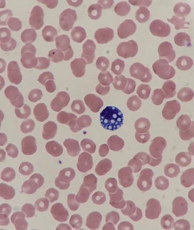
Figure 1 shows a Mott cell on an image from Parkland Medical Center Laboratory, Derry, NH. A Mott cell was identified by pathologist in a male patient who tested negative for COVID-19 at the time the sample was drawn, and subsequently tested positive. Mariana Garza, a Medical Technologist working at Las Palmas Medical Center in El Paso, TX shared a case of a 59 year old diabetic male, diagnosed with COVID-19. The patient’s WBC was 31 x 103/μL. Two Mott cells were identified by pathologist on his differential. So, the curious little mouse in me researched some more.
Several published research papers have studied morphologic changes in peripheral blood cells in COVID-19 patients. As we now know, SARS-CoV-2 affects many organs including the hematopoietic and immune systems. A study in Germany showed that COVID-19 patients exhibited abnormalities in all cell lines; white blood cells, red blood cells and platelets. Increased WBC counts were seen in 41% of samples in their study. Differentials performed on study patients showed lymphocytopenia in 83%, and monocytopenia in 88%. Red blood cell morphology changes were noted. Platelet counts ranged from thrombocytopenia to thrombocytosis, but giant platelets were noted across the board.4
Mott cells are indicative of chronic inflammation and may have significance in association with COVID-1. In the above mentioned study, aberrant lymphocytes were noted in 81% of patients who were SARS-CoV-2 positive, and observable in 86% of the same patients after they tested negative. The paper shows plasmacytoid lymphocytes and Mott cells amongst these aberrant lymphocytes. Moreover, morphologic changes in neutrophils, such as a left shift and pseudo‐Pelger‐Huët anomaly, decreased after virus elimination but changes in lymphocytes, indicators of chronic infection, remained.4
Another study also reported reactive or plasmacytoid lymphocytes and Mott cells observed in peripheral blood.4,5 Researchers at Northwick Park Hospital, UK, presented a case study of a 59 year old male with COVID-19 with a normal WBC and thrombocytosis. His differential revealed lymphocytopenia. His differential also showed lymphoplasmacytoid lymphocytes and Mott cells. In their conclusions it is stated that “In our experience, the lymphocyte features illustrated above are common in blood films of patients presenting to hospital with clinically significant Covid‐19. The observation of plasmacytoid lymphocytes supports a provisional clinical diagnosis of this condition.”5
Can these variant plasma cells, along with other commonly seen morphological changes, be used as part of a diagnostic algorithm for SARS-Cov-2 infection? As we see more COVID-19 patients there will be more, larger studies done and more Mott cells identified. Some disorders, such as Epstein Barr Virus and Dengue Fever are characterized by distinct viral changes in cells. However, since Mott cells can be seen in many conditions, these alone could not be considered diagnostic, but the indications are that these cells, along with the entire differential and morphological patterns, could prove to be a straightforward and easy to perform supplementary diagnostic tool. More, larger studies need to be done. It was concluded in the German study, that this pattern of morphologic changes in cells could be further investigated and validated with a larger blinded study, and that this information could lead to the development of a morphologic COVID‐19 scoring system.4 In the meantime, keep an eye out for Mott cells. These should not be ignored and should be in some way noted because they may be of future diagnostic use. That’s all or now, folks! Something to dig deeper into in another blog! The mouse strikes again!
Many thanks to Nikki O’Donnell, MLT, Parkland Medical Center, Derry, NH and Mariana Garza, MT, Las Palmas Medical Center in El Paso, TX for sharing their case studies and photos.
Becky Socha MS, MLS(ASCP)CMBB
References
- Diggs, LAW, Sturm, D, Bell,A. The Morphology of Human Blood Cells, Third edition. Abbott Laboratories. 1975.
- ManasaRavath CJ, Noopur Kulkarni, et al. Mott cells- at a glance. International Journal of Contemporary Mudeical Research 2017;4(1):43-44.
- Bavle RM. Bizzare plasma cell – mott cell. J Oral Maxillofac Pathol. 2013;17(1):2-3.doi: 10.4103/0973-029X.110682.
- Luke, F, Orso, E, et al. Coronavirus disease 2019 induces multi‐lineage, morphologic changes in peripheral blood cells:eJHaem. 2020;1–8.
- Foldes D, Hinton R, Arami S, Bain BJ. Plasmacytoid lymphocytes in SARS-CoV-2 infection (Covid-19). Am J Hematol. 2020;1–2. https://doi.org/10.1002/ajh.
- Numeroff, Laura. If You Give a Mouse a Cookie, 1985.

-Becky Socha, MS, MLS(ASCP)CM BB CM graduated from Merrimack College in N. Andover, Massachusetts with a BS in Medical Technology and completed her MS in Clinical Laboratory Sciences at the University of Massachusetts, Lowell. She has worked as a Medical Technologist for over 30 years. She’s worked in all areas of the clinical laboratory, but has a special interest in Hematology and Blood Banking. When she’s not busy being a mad scientist, she can be found outside riding her bicycle.

Love this blog story! I remember the Abbott red blood cell manuals, they were great …will be in the look 👀 for the mott cells!!
Wonderful article. Glad to know the Abbott Morphology of cells book is still useful! Ann Bell who coauthored the book was my Hematology instructor many years ago!
I still use this book with all our MLS students rotating through Hematology! Great resource
Becky you might be interested in…Cotter , P. F. and M. Bakst, 2017. A comparison of Mott cell morphology of three avian species. II. – Bad behavior by plasmacytes? Poultry Science 96(2):325-331 http://dx.doi.org/10.3382/ps/pew288
PS I taught biology at Merrimack for 3 years
Paul Cotter