Microbiology Case Study: 3-year-old Female with Facial Wound


Case History

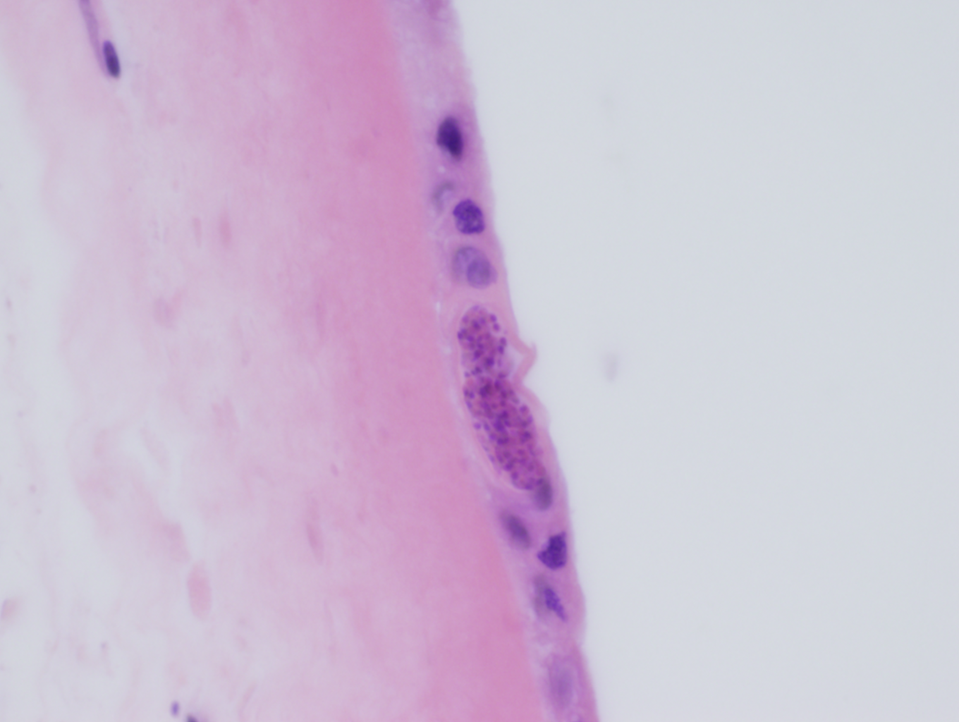
A 3-year-old female with no significant past medical history presented to the emergency department 1 day following a provoked dog bite to the right cheek. At home, the bite area was cleaned, but it subsequently developed progressive erythema, swelling, and purulent discharge. Review of systems was otherwise negative. Both the patient and dog were up-to-date on vaccinations. On exam, the patient displayed a 1 cm area of induration with surrounding erythema and actively draining whitish fluid.

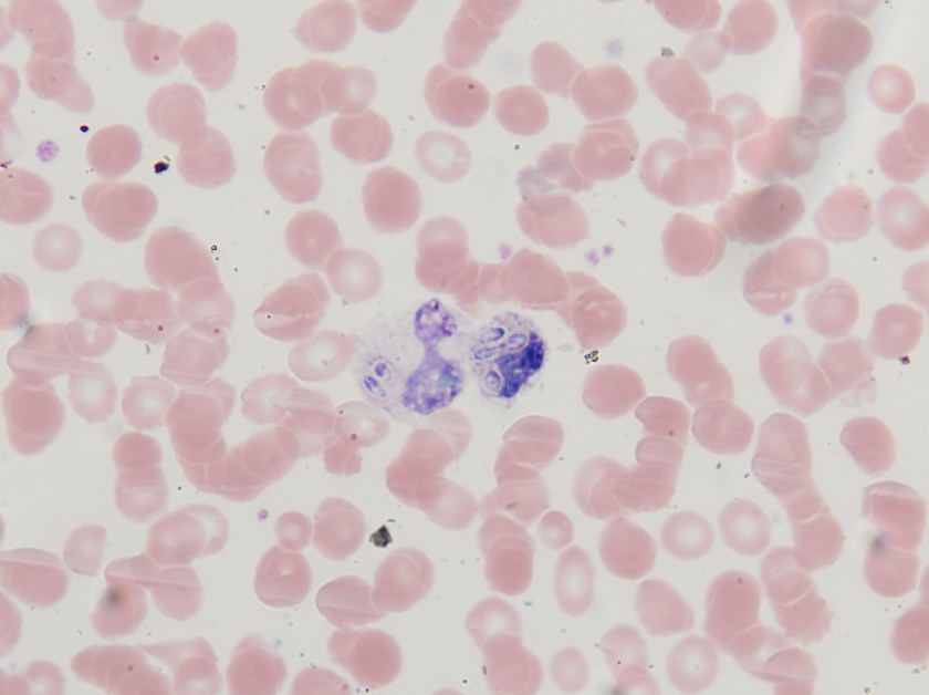
Fluid from the draining wound was sent to the microbiology laboratory for Gram stain and culture. On Gram stain, rare gram-negative rods were identified, as well as few polymorphonuclear leukocytes.  The following organism was recovered from culture the next day.

past1

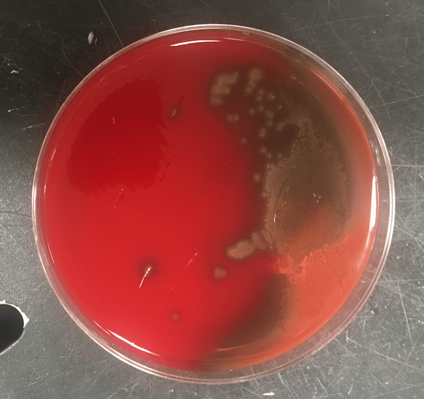
Figure 1: Growth on blood, chocolate and MacConkey agars. Note one larger and one smaller colony morphology growing on blood and chocolate agars.  The organism did not grow on MacConkey agar.

past2

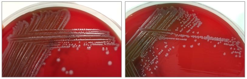
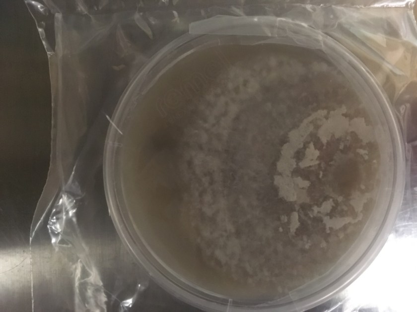
Figure 2. Subcultures of the two distinct colony types. The larger colonies (left) appeared more mucoid than the smaller colonies (right).

past3

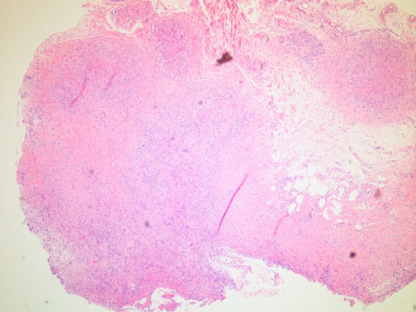
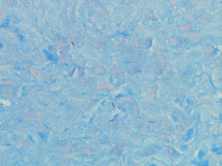
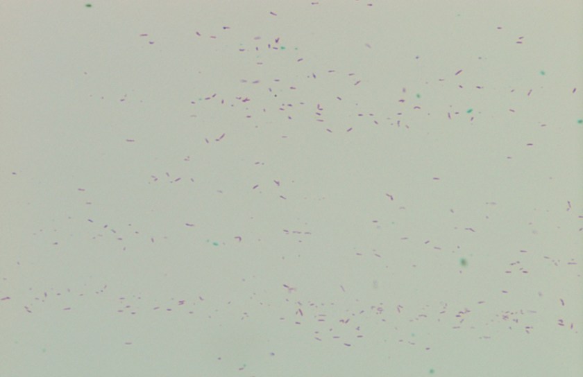
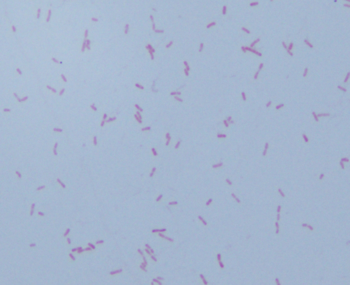
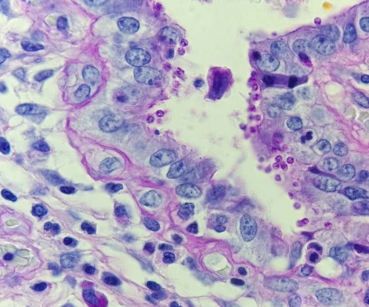
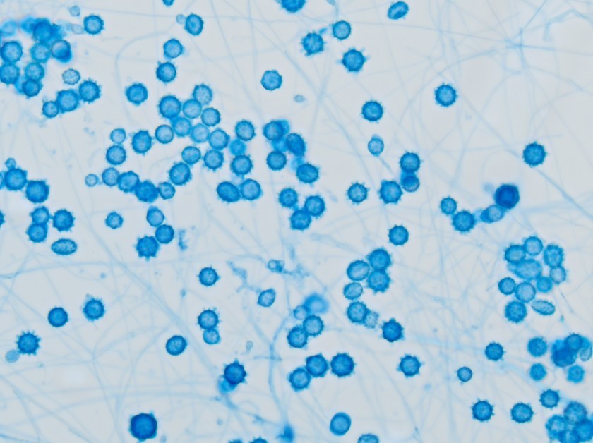
Figure 3. Gram stains of the two colony types revealed a small, Gram-negative coccobacilli identified as Pasteurella multocida (right) and a larger, pleomorphic Gram-negative rod identified as Pasteurella canis (left).

Laboratory Work-Up

The specimen was cultured on 5% sheep blood, chocolate, and MacConkey agars. Two colony morphologies emerged on the blood and chocolate agar plates (Figure 1). The organism did not grow on MacConkey agar. Representatives of each colony morphology were subcultured onto 5% sheep blood agar with growth shown in Figure 2. One colony is larger and mucoid (Figure 2, left) while the second is smaller and non-mucoid (Figure 2, right).

MALDI-TOF (Matrix Assisted Laser Desorption/Ionization Time-of-Flight) identified two different species of Pasteurella. Colony Gram stains show P. multocida as small Gram-negative coccobacilli (Figure 3, right) and P. canis as pleomorphic Gram-negative rod (Figure 3, left).

Discussion

The Pasteurella spp. are non-motile, facultatively anaerobic, Gram-negative coccobacilli found in the respiratory tracts of nonhuman mammals, most notably cats and dogs. By current classification the genus includes P. multocida (with 3 subspecies), P. dagmatis, P. canis, and P. stomatis, with P. multocida being the most common human pathogen and species recovered from animals (>70% carriage in cats, >40% in dogs) [1]. The foremost human infection is bite or scratch wound with cellulitis, with rapid development of erythema, swelling, and purulent drainage as observed in this case. Licking may also transmit the bacteria.  Possible associated findings include fever and regional lymphadenopathy. More serious potential infections include osteomyelitis, septic joint, endocarditis, bacteremia, sepsis, and meningitis. Systemic illness typically requires immunocompromise (classically liver disease). The organisms are generally penicillin-sensitive [2].

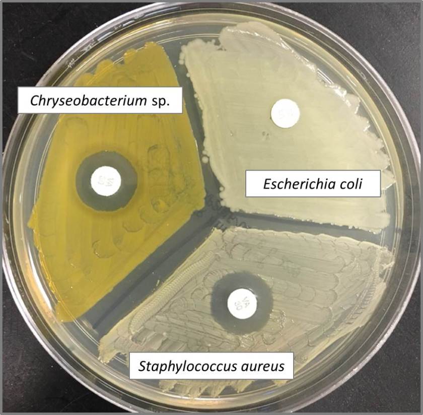
Key identification features of Pasteurella spp. include oxidase positivity and failure to grow on MacConkey agar, both differentiating Pasteurella from the Enterobacteriaceae. Other common features include and catalase and indole positivity. Unlike other certain fastidious gram negative bacteria (e.g., Haemophilus), Pasteurella grow independently on blood agar without the requirement for hemin or NAD. Of note, Capnocytophaga spp. (also associated with dog bites) also grows on blood and chocolate but not MAC. However, Capnocytophaga require a CO2-enriched environment and the Gram stain is notably different as they are long, slender Gram-negative rods [1].

This case was notable for a dual Pasteurella infection, an uncommon but previously reported phenomenon [3]. The organisms differed by colony morphology, with P. multocida appearing larger and mucoid (reflecting capsule production) on the blood agar, and P. canis appearing smaller, grey, and non-mucoid. Capsule is a key virulence factor of P. multocida and tends to be associated with more severe infections [4].

The patient was treated with a 10 day course of amoxicillin-clavulanate and has completely recovered.

References

  1. Procop, G. W., Church, D. L., Hall, G. S., Janda, W. M., Koneman, E. W., Schreckenberger, P. C., & Woods, G. L. (2016). Koneman’s Color Atlas and Textbook of Diagnostic Microbiology (7th ed.). Philadelphia: Wolters Kluwer.
  2. Graevenitz, A., & Zbinden, R. (n.d.). Actinobacillus, Capnocytophaga, Eikenella, Kingella, Pasteurella, and Other Fastidious or Rarely Encountered Gram-Negative Rods. In Manual of Clinical Microbiology, 10th Edition (pp. 574–587). American Society of Microbiology.
  3. Holst, E., Rollof, J., Larsson, L., & Nielsen, J. P. (1992). Characterization and distribution of Pasteurella species recovered from infected humans. Journal of Clinical Microbiology, 30(11), 2984–7.
  4. Harper, M., Boyce, J. D., & Adler, B. (2006). Pasteurella multocida pathogenesis: 125 years after Pasteur. FEMS Microbiology Letters, 265(1), 1–10.


-William Phipps, M.D., 1st year Anatomic and Clinical Pathology Resident, UT Southwestern Medical Center

-Erin McElvania TeKippe, Ph.D., D(ABMM), is the Director of Clinical Microbiology at Children’s Medical Center in Dallas Texas and an Assistant Professor of Pathology and Pediatrics at University of Texas Southwestern Medical Center.

Microbiology Case Study: A 68 Year Old with Refractory Pneumonia

Case History

After failing to improve on outpatient treatment for community acquired pneumonia, a 68 year-old New England man was admitted to the hospital for refractory pneumonia. Despite the initiation of triple antibiotic therapy, he continued to spike fevers and his respiratory status progressively declined. Notably, the patient reported recent travel to Arizona.

Laboratory Identification

This sputum sample was received from an outside hospital with no accompanying clinical history. After a couple days, the sample grew wooly, white-beige colonies on both potato flake and mycobiotic agars. A cellophane tape test performed on day 6 revealed narrow septate hyphae with alternating arthrocondia and empty cells. No conidiophores were present.


cocci1
Sputum fungal culture on Potato Flake Agar
cocci2
Sputum fungal culture on Mycobiotic agar
cocci3
Cellophane tape test


While the overall morphology was most consistent with Malbranchea species, rare slightly swollen arthrocondia were identified prompting increased handling precautions and further investigation into the patient’s clinical course at the referring hospital. After several more days of growth, a repeat cellophane tape test (Figure 3) demonstrated thick-walled, barrel-shaped arthrocondia alternating with empty cells as is characteristic of Coccidioides species.

The organism was then confirmed as Coccidioides immitis/posadasii by PCR.

Discussion

Coccidioides immitis and Coccidioides posadasii are pathogenic soil fungi with limited geographic distribution. Coccidioides immitis is found primarily in California’s San Joaquin Valley, Arizona, and Mexico while Coccidioides posadasii is slightly more widespread throughout arid regions of the Americas. Although genetically distinct, the two species are clinically and morphologically identical.

Coccidioides species cause Coccidioidomycosis, also known as Valley fever, which typically presents as a primary pulmonary infection about 1-4 weeks after exposure. While the majority of infected individuals will be entirely asymptomatic, 40% of cases result in a mild, self-limiting influenza-like illness with fever, sore throat, cough, headache, pleuritic chest pain, and occasionally a maculopapular rash on the trunk and limbs.  The fatigue and arthralgia associated with disease may persist for months after resolution of the pulmonary infection and is therefore sometimes referred to as “desert rheumatism”.

As with many infectious diseases, immunocompromised patients are at greater risk for developing more severe forms of the disease including extra-pulmonary manifestations.  Other risk factors for disseminated disease include high inoculum exposure, chronic illness, and primary infection during pregnancy.  While most infections will resolve without treatment, an extended course of azole therapy is recommended in these more complicated cases.

Since Coccidioidomycosis has relatively nonspecific symptoms, obtaining a history of exposure is often the key to the initiation of appropriate laboratory identification. Serologic testing for Coccidioides is available and is often the method of choice in the ambulatory setting. While highly specific, these tests are not very sensitive due to delayed formation and rapid clearance of the targeted antibodies. Therefore, a negative test result should not be used to exclude a Coccidioides infection especially early in the course of the disease.

In tissues and body fluids, Coccidioides is identified as round, thick-walled spherules (10-80µm) containing multiple endospores (2-5µm). When cultured at both 25°C and 37°C, Coccidioides forms wooly, white-grey colonies which may turn brown as they mature. The coarse hyphae are hyaline and septate with alternating thick-walled, barrel-shaped arthroconidia and empty cells. Although colonies usually form within three to five days, the distinctive arthroconidia may take up to 2 weeks to fully mature. Due to this delayed maturation, Coccidioides is often initially misidentified as its non-pathogenic look-a-like Malbranchea.  Confirmatory testing by PCR may be performed on both bodily fluids and paraffin embedded tissue.

Coccidioides was once considered a “select agent” with the potential to pose a severe threat to human health but advancements in diagnostic techniques and antifungal medications resulted in the loss of that status in 2012. However, since inhalation of even just 10 of its highly infectious arthroconidia may result in disease, Coccidioides is still a major source of laboratory-acquired infections. Technologists should use increased biosafety precautions whenever handling specimens suspicious for Coccidioides.


-Elaine Amoresano, MD, is a 2nd year anatomic and clinical pathology resident at the University of Vermont Medical Center.

Wojewoda-small

-Christi Wojewoda, MD, is the Director of Clinical Microbiology at the University of Vermont Medical Center and an Assistant Professor at the University of Vermont.


Microbiology Case Study: A 68 Year Old Man with Lung Cancer

A 68-year-old gentleman with a 50 pack year smoking history, COPD, and poorly differentiated lung cancer diagnosed after asymptomatic lung cancer screening, and status-post lobectomy presented to the pulmonology clinic for a one year follow-up. At this visit, the patient endorsed ongoing mild shortness of breath when climbing stairs and non-productive cough, but no fevers or chills. He underwent chest CT, which was notable for an enlarging right paratracheal LN as compared to a CT from six months prior, and RML collapse thought secondary to either mucous plugging versus tumor involvement of the bronchus. After a discussion with his physician, the patient agreed to undergo a flexible bronchoscopy with bronchoalveolar lavage (BAL) and lymph node biopsy instead of waiting six months for a repeat CT.


streppseudo1
Gram Stain showing Gram positive cocci in pairs.
streppseudo2.png
Growth on chocolate agar.
streppseudo3.png
Alpha hemolytic colonies on sheep blood agar.

Lab Identification

Gram smear of BAL fluid revealed moderate polys, few squamous epithelial cells, and moderate gram positive cocci. Colony morphology was notable for small, smooth-edged colonies displaying alpha hemolysis, that were bile insoluble. The organism was determined to be Streptococcus pseudopneumoniae by MALDI-TOF.

Discussion

Streptococcus pseudopneumoniae is a novel species of Streptococcus that belongs to the mitis group first described in 2004 by Arbique in an analysis of lower respiratory tract samples. Gram smear reveals GPCs without a capsule, and culture shows tiny, pinpoint, smooth-edged colonies with alpha-hemolysis. It may also resemble a smaller version of typical pneumococcal “checkers,” with raised edges and a depressed center. Species identification has been challenging given the greater than 99% homology of the 16S rRNA gene among S. mitis, S. oralis, S. pneumoniae, and S. pseudopneumoniae (however DNA-DNA similarity of the entire genome is < 60%). It was initially characterized phenotypically, based on its bile insolubility and variable optochin susceptibility (resistance to optochin in the presence of 5% CO2, and susceptibility at ambient atmosphere). The latter is thought to be due to a novel mutation in the F0F1 ATP Synthase subunit c.

The incidence of S. pseudopneumoniae is believed to be low, but it may be underreported due to labs identifying it as an atypical pneumococci. Furthermore, the pathologic significance is poorly understood. Jensen et al. performed molecular mapping of tonsillar crypt microbiota in healthy subjects and patients with recurrent tonsillitis, and found that S. pseudopneumoniae was present in all samples. Multiple retrospective studies looking at patients with S. pseudopneumoniae in their sputum compared to matched controls found that it was more likely to be present in patients with COPD (Keith et al.) and pneumonia (Laurens et al.). It has also been isolated from blood, ascitic fluid, and conjunctival samples. One case report from 2014 described a case of bleb-related endophthalmitis in a 63 year old man who had undergone glaucoma surgery 46 years prior.

A real-time PCR assay specific for S. pseudopneumoniae has been developed by Sistek et al. based on a mutation in the recA gene; however, it has yet to be validated.

As S. pseudopneumoniae belongs to the viridans group, susceptibility breakpoints for determining MIC are based on this group rather than S. pneumoniae. It is generally susceptible to vancomycin, carbapenems, and quinolones, and resistant to macrolides and tetracycline.


References

Janda et al. “The Genus Streptococcus Part II: New Species and Pathogens in the “Miscellaneous” Streptococci and “Viridans” Streptococci Groups.” Clinical Microbiology Newsletter Vol. 36, No. 21 November 1, 2014

Kawakami et al. “Late-onset bleb-related endophthalmitis caused by Streptococcus pseudopneumoniae.” International Ophthalmology (2014) 34:643–646 DOI 10.1007/s10792-013-9835-2


-Sean Bullis is a 4th year medical student at the University of Vermont College of Medicine.

Wojewoda-small

-Christi Wojewoda, MD, is the Director of Clinical Microbiology at the University of Vermont Medical Center and an Assistant Professor at the University of Vermont.

Microbiology Case Study: A 53 Year Old Male with Fatigue, Night Sweats, and Right Knee Swelling

Case History

A 53 year old male presents to the emergency department with complaints of fatigue, night sweats, dyspnea, dry cough, and right knee swelling. He has multiple skin lesions including violaceous papules on his medial thigh, subcutaneous nodules on bilateral lower legs, and ulcerations on his right lateral leg. His past medical history is significant for psoriatic arthritis. Previously, he has taken adalimumab (Humira), etanercept (Enbrel), and golimumab (Simponi), but his current treatment regimen consists of methotrexate and prednisone. His recent travel history is significant for scuba diving in Thailand and honeymooning in the Caribbean. Dermatology was consulted and a punch biopsy was performed close to an ulcer on his right lateral leg and sent to surgical pathology. An additional biopsy specimen was sent to microbiology for bacterial, fungal and mycobacterial cultures.


Laboratory Identification

mycohem1
Figure 1. Histology sections of the punch biopsy near the patient’s right lateral leg ulcer. The epidermis and dermis appeared unremarkable. However, a non-caseating granuloma is seen in the deep subcutaneous tissue (H&E, 40x).
mycohem2
Figure 2. Special stain highlighting the occasional acid fast organisms (Kinyoun, 1000x oil immersion).
mycohem3
Figure 3. Smear from a positive mycobacteria growth indicator tube (MGIT) incubated at 32°C showing abundant acid fast bacilli (Ziehl-Neelsen, 500x oil immersion).

Microscopic examination of the punch biopsy skin specimen revealed a non-caseating granuloma in the deep subcutaneous tissue, with no involvement of the overlying dermis and epidermis (Figure 1). A Kinyoun stain of the tissue showed that the granuloma contained occasional acid-fast bacilli (Figure 2). The bacterial and fungal cultures sent to microbiology were negative. Portions of the specimen used to set up the mycobacterial cultures were incubated in MGITs at 32 and 37°C because the specimen source was skin. The 32°C tube, which was supplemented with hemin, gave a positive signal after 3 weeks of incubation. The Ziehl-Neelsen stain from this tube revealed numerous acid fast bacilli (Figure 3). DNA Gen-Probe analysis was negative for Mycobacterium tuberculosis complex and M. avium-intracellulare (MAI) complex. The organism was identified as M. haemophilum by pyrosequencing.

Discussion

Mycobacterium haemophilum was first identified in 1978 from an Israeli patient with Hodgkin lymphoma. It has a known predilection for infecting the skin and subcutaneous tissue in immunocompromised patients, especially those with lymphopenia as a result of acquired immune deficiency syndrome (AIDS), allogeneic bone marrow transplantation, and those on immunosuppressant therapies for rheumatologic conditions. The clinical presentation frequently consists of painful subcutaneous nodules and ulcers that can progress to abscesses and draining fistulas. Bone and joint infections have also been reported, which manifest as arthritis, tenosynovitis, and osteomyelitis. AIDS patients in particular are known to present with disseminated disease, with multiple cutaneous lesions, mainly involving the extremities. Relatively little is known about this infection and the optimal treatment is not standardized, but combinations of three or four of the following drugs have been used successfully: isoniazid, rifamycins, ciprofloxacin, amikacin, doxycycline, and clarithromycin.

Unlike the majority of mycobacteria, M. haemophilum does not grow well in culture at 37°C. Rather, it prefers lower temperatures, ideally between 28–32°C. This characteristic is shared by several other mycobacterial species that also characteristically infect the skin, including M. marinum, M. chelonae, M. abscessus, and M. ulcerans. A unique feature of M. haemophilum among the mycobacteria is that it requires hemin (X factor) to survive and will only grow in media enriched with this nutrient. Similar to Haemophilus influenzae, M. haemophilum can be cultured on chocolate agar, as well as on Middlebrook 7H10 agar incubated with an “X-factor strip” and on Lowenstein-Jensen medium containing 2% ammonium citrate.

Typically, colonies grow after 2-4 weeks of incubation at 32°C and have either a rough or smooth appearance. M. haemophilum is a non-photochromogen according to the Runyon classification system, and its colonies are buff colored and do not produce pigment in either light or dark conditions. M. haemophilum is chemically inert by traditional biochemical mycobacterial tests, with the exception of pyrazinamidase production. As illustrated by this case, DNA probe analysis is helpful with regard to the mycobacterial species it excludes, but not for speciation of less common organisms. At the present time, DNA probes exist only for M. tuberculosis complex, MAI complex, M. kansasii, and M. gordonae.

In the case of our patient, the species level identification was determined by pyrosequencing. He was treated with an extended course of 3 agents: rifabutin, clarithromycin, and moxifloxacin with good response.


VN

Vikas Nath, MD, is a 4th year resident in Anatomic and Clinical Pathology at the University of Mississippi Medical Center in Jackson, MS.

Stempak

-Lisa Stempak, MD, is an Assistant Professor of Pathology at the University of Mississippi Medical Center in Jackson, MS. She is certified by the American Board of Pathology in Anatomic and Clinical Pathology as well as in Medical Microbiology. Currently, she oversees testing performed in both the chemistry and microbiology laboratories.  Her interests include infectious disease histology, process and quality improvement, and resident education.

Microbiology Case Study: A 34 Year Old Pregnant Woman with Evidence of Fetal Anomalies

Case History:

A 34 year old G3P3 woman presents with PPROM at 30+4 weeks gestational age. Her pregnancy had been complicated by fetal hydrops and intrauterine growth restriction with evidence of multiple fetal anomalies and placenta previa.

Prenatal infectious disease testing was significant for:

  • CMV IgG+/IgM+
  • Toxoplasmosis IgG+/IgM+
  • Parvovirus B19 IgM-/IgG+
  • HIV screen negative (ELISA)
  • HSV IgM-
  • syphilis screen negative (RPR)
  • rubella immune (IgG+)
  • negative serologies for Hepatitis A, B and C
  • Testing for VZV was not performed

Clinically, the fetal hydrops and IUGR were thought to be due to congenital CMV. She underwent caesarian section, and a fetus was delivered with APGAR 0/1/1/2/1, with eventual fetal demise at two hours of life. The placenta was sent to the laboratory for surgical pathology examination. The mother declined fetal autopsy.

Laboratory Work-Up:

Surgical pathology received a singleton placenta (13 x 13 x 4 cm) with attached umbilical cord and fetal membranes. The placental disc weighed 346 grams (<10th percentile for gestational age). Otherwise, the placental disc, umbilical cord and fetal membranes were negative for any gross abnormalities.

Routine microscopic sections demonstrated round to elongate cysts within the amnion of the fetal membranes (Figures 1a & 1b) and within the Wharton’s jelly of the umbilical cord (Figure 2). These cysts measured approximately 50 microns in diameter and had a thin, translucent cyst wall. Within the cysts were innumerable small round “dot-like” forms which could best be appreciated by focusing up and down through the plane of the section.

Tissue gram stain (Brown & Brenn) was negative for significant bacterial infiltrate, and immunohistochemistry for CMV was negative for CMV inclusions.

Figure 1a: Amnion of the fetal membrane: Round to elongate cysts measuring approximately 50 microns in diameter. There are innumerable small round “dot-like” structures within the cysts (H&E, 600X).
Figure 1b: Amnion of the fetal membrane: Round to elongate cysts measuring approximately 50 microns in diameter. There are innumerable small round “dot-like” structures within the cysts (H&E, 600X).
Figure 2: Wharton’s jelly of the umbilical cord: Round cyst measuring approximately 50 microns in diameter. There are innumerable small round “dot-like” structures within the cyst (H&E, 600X).

Discussion:

The histologic features are diagnostic of congenital Toxoplasmosis. The case was sent to the reference laboratory, where immunohistochemical staining for Toxoplasmosis demonstrated positive staining within the tissue cysts.

Toxoplasmosis is caused by the protozoa Toxoplasma gondii, a member of the protozoan subgroup coccidia, which also includes the GI pathogens Cryptosporidium, Isospora and Cyclospora. Cats of the family Felidae (including but not limited to domestic cats) are the only definitive host, while virtually any mammal can serve as an intermediate host. Humans can become incidentally infected in which case they act as “incidental” intermediate hosts.

The life cycle of Toxoplasma involves sexual reproduction in the definitive host (cats), as well as asexual reproduction in the intermediate host. Toxoplasma is “facultatively heteroxenous,” in that reproduction in the intermediate host is not necessary for completion of the life cycle. Unsporulated oocysts are shed in the cat feces and become infective after 1-5 days. Cats may ingest the infective oocysts, leading to sexual reproduction and completion of the life cycle within the intestinal epithelium. Alternatively, intermediate hosts such as rodents or birds ingest infective oocysts and subsequently develop infective tissue cysts. If the intermediate host is eaten by a cat, the infective tissue cysts are ingested, leading to sexual reproduction in the cat and completion of the life cycle.

The life cycle within the intermediate host involves two morphologically distinct stages, the tachyzoite and the bradyzoite. When infective oocysts are ingested by an intermediate host, they transform into tachyzoites, which are able to invade the intestinal epithelium and then widely distribute throughout the body. Tachyzoites are crescent-shaped, non-encysted and measure from 3-7 microns in length by 2-4 microns in diameter. They migrate preferentially to the muscle and neural tissues, where they eventually develop into tissue cysts, which are known as bradyzoites. Bradyzoites are much larger than tachyzoites (approximately 50 microns), are round to elongate and contain numerous “dot-like” parasitic forms encased within a thin cyst wall.  Tachyzoites are eventually cleared following acute infection, but the intermediate host remains chronically infected with bradyzoites. If the host becomes immunocompromised, the bradyzoites differentiate into tachyzoites, which then recirculate through the body leading to reactivation of latent disease.

It is estimated that 10-20% of the U.S. population is chronically infected with Toxoplasma. Humans can become infected through one of five mechanisms: (1) ingestion of infective oocysts, either from cat feces or from infected water or other environmental sources, (2) ingestion of infective tissue cysts in undercooked meat, (3) vertical transmission to the fetus from a mother acutely infected with Toxoplasma, (4) through organ transplantation and (5) through blood transfusion. Epidemiologically, it is not clear whether the majority of infections occur through ingestion of infective oocysts or whether tissue cysts in undercooked meat are the major source of infection.

Vertical transmission from mother to fetus requires a first-time exposure during pregnancy. In primary/acute infection, tachyzoites widely disseminate and are able to invade the developing fetal tissues. By contrast pregnant women who are chronically infected with Toxoplasma harbor only tissue cysts (bradyzoites) and will not transmit infection to the fetus.

Acute infection is self-limited and usually asymptomatic, however some patients may have mild flu-like symptoms. A smaller subset of patients present with moderate to severe acute infection which can mimic mononucleosis: fever, sore throat, myalgias and cervical lymphadenopathy. Biopsy of inflamed lymph nodes reveals the classic histologic triad of follicular hyperplasia, monocytoid B-cell hyperplasia and epithelioid histiocytic aggregates. Once acute infection has passed, chronic infection is usually asymptomatic, unless the host becomes immunocompromised, in which case reactivation of latent disease can occur.

Treatment for immunocompetent patients in not indicated as acute infections are self-limited and chronic infection is asymptomatic. Immunosuppressed patients with CD4 counts <100 cells/mm3 should receive Toxoplasma prophylaxis with trimethoprim-sulfamethoxazole (TMP-SMX). Reactivation of latent disease can occur in immunosuppressed patients who are not taking prophylaxis, in which case first line treatment includes combination therapy with sulfadiazine and pyrimethamine.


-Javier De Luca-Johnson, MD is a 3rd year anatomic and clinical pathology resident at the University of Vermont Medical Center.

Wojewoda-small

-Christi Wojewoda, MD, is the Director of Clinical Microbiology at the University of Vermont Medical Center and an Assistant Professor at the University of Vermont.

Microbiology Case Study: A 2 Year Old Ventilator-Dependent Child with Acute Desaturations

Case History

A 2 year old female with a past medical history of spinal muscular atrophy type 1, trach/ventilator dependency presents to the hospital with complaints of desaturations at home. She has been hospitalized four times within the past year with an additional two visits to the emergency department. Patient has history of growing Pseudomonas aeruginosa from her lower respiratory tract culture. They began to work up causes of acute desaturations: mucus plug vs. viral illness vs. tracheitis vs. bacterial pneumonia. They sent tracheal aspirate and blood for cultures. The following organism was identified as the predominant organism from the tracheal aspirate.

chryseo1
Colony morphology on 5% sheep blood agar.
chryseo2
Colony Gram stain.


Discussion

Chryseobacterium spp. are non-motile, oxidase positive, indole positive Gram-negative bacilli. While often considered low virulence environmental organisms, Chryseobacterium spp. can be pathogenic in immunocompromised patients and those less than six months of age. Chryseobacterium spp. and closely related bacteria are able to survive in chlorinated water and are intrinsically resistant to many antibiotics. They are often nosocomal pathogens that are selected for in immunocompromised patients who are treated with broad-spectrum antibiotics for long periods of time. Chryseobacterium spp. have been reported to cause blood stream infections, cellulitis, pneumonia, meningitis, pyomyositis and keratitis. The most common setting of Chryseobacterium infections is foreign bodies such as indwelling catheters or prosthetic joint materials. In the adult setting, Chryseobacterium indologenes has been associated with the history of colistin or tigecycline use. Due to its low pathogenicity, Chryseobacterium spp. are generally considered contaminants in otherwise healthy individuals.

Chryseobacterium spp. have a pair of interesting resistance mechanisms. They inherently have a Class A β-lactamase and a Class B-carbapenemase that function to hydrolyze β-lactamases. Due to these mechanisms, Chryseobacterium spp. are intrinsically resistant to carbapenems and cephalosporins. Common treatment options include: levofloxacin, sulfamethoxazole trimethoprim and piperacillin-tazobactam.

Chryseobacterium spp. have been shown to be susceptible to vancomycin in vitro (Image 3). This is a bizarre trait because they are Gram-negative bacteria, a group which is generally intrinsically resistant to vancomycin. The “susceptible” phenotype is based on Kirby Bauer disk diffusion testing, which was found not to correlate well with vancomycin MIC testing. In vitro studies found Chryseobacterium to be susceptible to vancomycin in 0 to 65% of isolates tested based on Gram-positive NCCLS or CLSI breakpoints. Due to this, vancomycin “susceptibility” can be used to aid in identification of the organism, but would not be an appropriate antibiotic for the treatment of Chryseobacterium spp.

chryseo3
Vancomycin disk diffusion profile for isolates of Chryseobacterium spp., E. coli, and S. aureus.

References

  1. Chou DW, Wu SL, Lee CT, et al. Clinical Characteristics, Antimicrobial susceptibilities and outcomes of patients with Chryseobacterium indologenes bacteremia in an intensive care unit. J Infect Dis. 2011;64:520-524.
  2. Fraser SL and Jorgensen JH. Reappraisal of the antimicobial susceptibilites of Chryseobacterium and Flavobacterium species and methods for reliable susceptibility testing. Antimicrobial agents and chemotherpy. 1997;41(12):2738-2741.
  3. Nemli SA, Demirdal T, Ural S. A Case of healthcare associated pneumonia casued by Chryseobacterium indologenes in an immunocompetent patient. Case reports in infectious disease. 2015;2015:1-3.
  4. Srinivasan G, Muthusamy S, Raveendran V, et al. Unforeseeable presentaiton of Chryseobacterium indologenes infection in a paediatric patient. BMC Res Notes. 2016;9:212-217.


Contributor

-Erin Waehner, Pharm.D. PGY2 Pediatric Pharmacy Resident, Children’s Health, Children’s Medical Center Dallas.

-Erin McElvania TeKippe, Ph.D., D(ABMM), is the Director of Clinical Microbiology at Children’s Medical Center in Dallas Texas and an Assistant Professor of Pathology and Pediatrics at University of Texas Southwestern Medical Center.


CMS Proposes Rule that Promotes Antibiotic Stewardship

In mid-June, CMS proposed a rule that, in part, will help promote antimicrobial stewardship in hospitals. The 60-day comment period is nearing its end, so if you have thoughts on this proposed rule, let them know.

CMS press release


Microbiology Case Study: A 43 Year Old Man with AIDS

A 43-year-old man with AIDS with a complicated medical history, including multiple recent hospitalizations for CMV esophagitis, recurrent Clostridium difficile, multiple Klebsiella urinary tract infections and pyelonephritis, presented with right upper quadrant pain and profuse watery diarrhea. The ensuing work-up results in a diagnosis of cholecystitis and the patient underwent cholecystectomy. Subsequently, he developed an infected biloma necessitating the placement of a biliary drain. Photomicrographs of his cholecystectomy specimen are shown below.

crypto1
H&E stain of the gallbladder (20x).
crypto2
H&E stain of the gallbladder (100x).
crypto3
H&E stain of the gallbladder (100x).


Laboratory Identification

The diagnosis of cryptosporidiosis was made by H&E section. In a background of acute and chronic inflammation, numerous spherical organisms were identified lining the epithelial surface. Special stains, including PAS and mucin, were performed. The PAS was positive and the mucin stain negative in the organisms. The organisms measured between 2-5 microns. Important differential diagnoses include Cyclospora cayatanesis, which is a bit larger in size (8 microns), Isospora belli, which is much larger (20 microns) and Microsporidia, which is smaller (1-4 microns). Of note, microsporidia appears to be within the apical cytoplasm of mucosal epithelial cells on histology.

While a stool sample was not submitted to the microbiology laboratory in this case, there are several characteristic features of cryptosporidia that should be noted. Importantly, the detection of the oocysts by traditional O&P methods can be very difficult because they can look like yeast which is normal bowel flora. If a clinician suspects cryptosporidiosis, a modified acid fast stain should be performed as cryptosporidia oocyts are weakly acid fast. They also have the ability to autofluorescence when exposed to UV light. More sensitive techniques being used in laboratories today include direct fluorescent antibody [DFA], and/or enzyme immunoassays (ELISAs) for detection of Cryptosporidium sp. antigens.


Discussion

Cryptospoidiosis usually causes watery diarrhea, abdominal pain, nausea, vomiting, fever and weight loss. In a patient with an intact immune system, symptoms usually last 1-2 weeks, and eventually resolve without medical therapy. In an immunocomprimised host such as our patient, however, ingestion can result in severe and even life-threatening disease. Furthermore, use of usual antimicrobial drugs is generally ineffective in this patient population, making them particularly vulnerable.

Cryptosporidium belongs to the taxonomic group Protozoa, phylum Apicomplexa. This group of organisms includes many other important parasites such as Toxoplasma, Plasmodium, Babesia, Cyclospora, and Isopora. The Apicomplexan group is significant for the fact that they possess an apical structure enabling their penetration of host cells. Interestingly, Cryptosporidium is considered to be a eukaryotic intracellular, but extracytoplasmic pathogen as it resides in the columnar brush border of epithelial cells. This particular niche is thought to allow the parasite to evade the immune surveillance while being able to simultaneously take advantage of the solutes transported across the host microvillus membrane. Of the many Cryptosporidium species that exist, Cryptosporidium parvum and Cryptosporidium hominis are the most important in human disease (cryptosporidiosis).

Excretion of thick-walled sporulated oocysts by an infected host results in transmission of the disease through contaminated water sources, such as drinking supplies and recreational water sources. Occasionally contaminated food sources and zoonotic transmission may occur. Importantly, the oocysts are infective upon excretion, thus permitting direct and immediate fecal-oral transmission. Following ingestion, the oocyst excyts in the stomach and then undergo two stages of reproduction within the gastrointestinal tract, an asexual followed by a sexual phase referred to as merogony. Merogony is important in the pathogenicity of many Apicomplexa organisms as it results in massive DNA replication and the creation of hundreds of daughter cells simultaneously.

Cryptosporidia is an important public health hazard because of its resistance to chlorine. Chlorine is often used in water treatment facilities and public water sources as a disinfectant as it kills important pathogens such as Salmonella, Campylobacter, and norovirus. It is ineffective, however, against the thick walled cysts of cryptosporidia. UV light or a filtration system is needed to eradicate cryptosporidium from a contaminated water source.

References

  • CDC website, accessed 2/8/2016 http://www.cdc.gov/parasites/crypto/diagnosis.html
  • Forbes, B, Sahm, D, Wessfeld, A, Bailey & Scott’s Diagnostic Microbiology, 12th ed. 2007.
  • Chalmers, RM, Cryptosporidium: From Laboratory Diagnosis to Surveillance and Outbreaks. Parasite, 2008, 15, 372–378.


-Jessica Crothers, MD is a 4th year anatomic and clinical pathology resident at the University of Vermont Medical Center.

Wojewoda-small

-Christi Wojewoda, MD, is the Director of Clinical Microbiology at the University of Vermont Medical Center and an Assistant Professor at the University of Vermont.

Potential Antimicrobial Therapy Hiding in Plain Sight

Yesterday, Nature published a paper that might help in the fight against MRSA. In a nutshell, German researchers discovered that Staphylococcus lugdunensis–a common bacteria in commensal flora–produces a compound that reduces colonization with MRSA.

From the abstract:

“Notably, human nasal colonization by S. lugdunensis was associated with a significantly reduced S. aureus carriage rate, suggesting that lugdunin or lugdunin-producing commensal bacteria could be valuable for preventing staphylococcal infections.”


Microbiology Case Study: A 61 Year Old Male with Productive Cough and Altered Mental Status

Case History

A 61 year old African American male presents to the emergency department with complaints of a productive cough, dyspnea and altered mental status. His past medical history is significant for HIV and currently he is non-compliant with his anti-retroviral medications. On arrival, he is found to be hypoglycemic (glucose 49 mg/dL) and tachycardic (heart rate between 160-180 beats/min). He lives in a group home and they report decreased oral intake for several days but he denies fever, chills, chest pain or abdominal pain. He is a tobacco smoker and admits to previous illicit drug use. On physical exam, he is lethargic and respiratory auscultation reveals coarse lung sounds, bilaterally. A chest x-ray shows bilateral interstitial and airspace opacities suggestive of an infectious process. His CD4 count is markedly decreased at 3 cells/cmm. Peripheral blood and bronchoalveolar lavage (BAL) fluid are sent to the hematology and cytology laboratories for microscopic examination. Blood and BAL specimens were also transported to the microbiology lab for bacterial, fungal and mycobacterial cultures.

Laboratory Identification

histo1
Figure 1. Peripheral blood smear highlighting small, intracellular yeast forms with narrow based budding (Giemsa stain, 1000x oil immersion).
histo2
Figure 2. Fluid from a bronchoalveolar lavage showing macrophages filled with numerous small yeast forms that have an “acorn-like” appearance (Giesma stain, 1000x oil immersion).
histo3
Figure 3. White colonies with a fine, cottony texture growing on Mycosel agar after 21 days of incubation at 25°C.
histo4
Figure 4. Numerous tuberculate, thick-walled macroconidia with septate hyphae in the background (Lactophenol cotton blue stain, 400x).

Both the peripheral blood smear and BAL showed intracellular, small, ovoid yeast cells with narrow based budding (Figures 1 & 2). The yeast forms measured between 2-4 µm in diameter. The characteristic “acorn-like” appearance of the yeast cells surrounded by a thin halo is the result of staining fixation. The blood and the BAL cultures grew white colonies with a fine, cottony texture after incubation for 21 days at 25°C (Figure 3). The mold form grew on Sabouraud dextrose, SAB with chloramphenicol and Mycosel agars. Microscopic morphology of a lactophenol cotton blue prep illustrated septate hyphae bearing round to pear-shaped microconidia as well as tuberculate, thick-walled macroconidia, which measured between 8-15 µm in size (Figure 4).  The dimorphic mold was confirmed to be Histoplasma capsulatum by DNA probe testing. The patient also had a positive Histoplasma urinary antigen and fungitell was found to be >500 pg/ml.

Discussion

Histoplasma capsulatum is a thermally dimorphic fungus and the most common endemic mycosis in North America. In the United States, the disease is most prevalent in areas surrounding the Mississippi and Ohio River valleys. Inhalation of conidia occurs as a result of environmental exposure to soil contaminated with bird dropping or exploring caves and other dwelling inhabited by bats. Pulmonary infections are the most frequent manifestation of disease; however, disseminated infection can occur in individuals with underlying cell-mediated immunological defects, including those with HIV, transplant recipients, and individuals receiving tumor necrosis factor alpha inhibitors for rheumatologic conditions. Other extra-pulmonary sites from which H. capsulatum has been isolated include the skin, liver, spleen, central nervous system and bone marrow.

In the environment and when cultured in the laboratory at 25°C, H. capsulatum is a filamentous mold and exhibits both pear shaped microconidia (2-5 µm) and thick walled macroconidia that display characteristic tubercles or projections on their surface (8-15 µm). The yeast phase occurs in tissue and at temperatures above 35°C. The yeast phase is characterized as small oval budding cells, 2-4 µm in diameter and are  often found in clusters within macrophages. Historically, mold to yeast culture conversion was used to confirm the diagnosis, but with the advent of more rapid DNA probe technologies, this has been discontinued. Other rapid tests routinely utilized include a urine test to detect the Histoplasma antigen.

H. capsulatum var. duboisii, which is endemic in central and western Africa, is also implicated in causing disease in humans. It can be distinguished from H. capsulatum var. capsulatum due to its larger diameter in tissue where the yeast form of H. capsulatum var. duboisii measures between 8-15 µm in diameter as opposed to 2-4 µm for var. capsulatum. Caution is recommended, however, due to the yeast forms of the two variants being the same size when grown in culture.

Amphotericin B is the antifungal agent used to treat disseminated histoplasmosis infections.  In cases of less severe disease, itraconazole is effective and commonly utilized. In the case of our patient, he received 14 days of amphotericin B infusion as an inpatient and was then transitioned to oral itraconazole upon discharge.


JK

-Joy King, MD, is a third year Anatomic and Clinical Pathology resident at the University of Mississippi Medical Center. 

Stempak

-Lisa Stempak, MD, is an Assistant Professor of Pathology at the University of Mississippi Medical Center in Jackson, MS. She is certified by the American Board of Pathology in Anatomic and Clinical Pathology as well as Medical Microbiology. Currently, she oversees testing performed in both the Chemistry and Microbiology Laboratories.  Her interests include infectious disease histology, process and quality improvement and resident education.