A Cording Too: “Cording” in Clinical Isolates of Mycobacterium

A cornerstone of good clinical microbiology laboratory practice is to look for visual clues in how organisms grow in culture. This can help quickly signal to the laboratorian that a particularly meaningful pathogen is present.

For example, a wound culture where an anaerobic blood agar plate is showing a double zone of hemolysis? The tech should immediately think that Clostridium perfrigens may be present. An abscess culture growing a ridiculously mucoid colony of a lactose-fermenting Gram negative rod? Hyper-mucoid Klebsiella pneumoniae is a hypervirulent strain associated with abscess formation. Training and experience in what to look for in cultures is one of the fascinating and exciting (and potentially daunting) aspects of clinical microbiology!

But crucially, sometimes the textbook visual association is NOT as specific a finding as we may believe! One such example: Mycobacterium species and cording.

Cording and M. tuberculosis

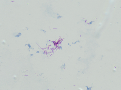
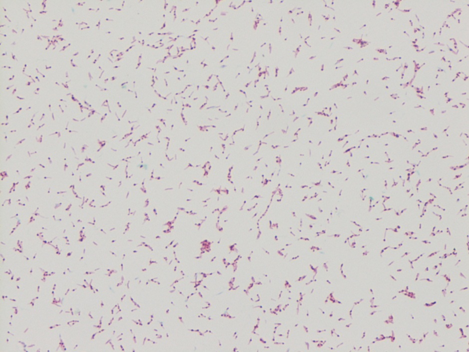
“Cording” is term used to describe twisting, serpentine appearance of Mycobacterium in liquid broth culture. And, at least in many places, it is taught to budding microbiologists and infectious disease clinicians as a hallmark characteristic of Mycobacterium tuberculosis (Mtb).

How long have people been observing Mtb cording in liquid culture? From very beginning!

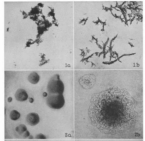
Robert Koch, originator of the famous Koch’s Postulates, used Mtb (now recognized to be just one member of the M. tuberculosis complex) to demonstrate that disease was caused by discrete organisms originating from one host and infecting another, what we accept as germ theory. In his original description of Mtb, Koch wrote that the bacilli would “ordinarily form small groups of cells which are pressed together and arranged in bundles.”

Cording phenotype is distinct from “clumping.” With cording, bacilli lay in tightly packed parallel strands, and not clumped together showing random bacilli orientation.

The phenomenon of cording is distinct to organisms cultured in liquid media like Middlebrook 7H9 broth. But in 1947 Middlebrook (where Middlebrook 7H9 broth gets its name), Dubos and Pierce published a key paper showing that cording Mtb strains also grew as rough colonies on solid agar and that this phenotype was associated with virulent and not avirulent Mtb strains.

Figures from 1947 Middlebrook et al., showing avirulent Mtb clumping (1a) and smooth colonies (2a) and virulent Mtb with cording (1b) and rough colonies (2b).

Only a few years later in 1950, the actual cell wall component, the so-called “cording factor,” that causes these growth characteristics was extracted and identified. Trehalose-6,6-dimycolate (TDM), a major glycolipid from the cell walls of virulent strains of Mtb.

Further work suggested that, not only was this TDM glycolipid the cause of the unique cording appearance and rough colonies in in vitro culture conditions, but that directly was a virulence factor: studies in animal models showed that TDM not only directly allowed Mtb to avoid phagocytosis from macrophages by virtue of forming large clusters of bacilli, but it also directly prevented macrophage intracellular killing mechanisms (see Hunter, 2006).

The association between cording and clinical isolates of Mtb was seen as a very sensitive and specific finding. Such a robust feature, in fact, that a number of publications from clinical microbiology labs specifically to cording as a reliable method to quickly identify Mtb (see McCarter 1998) or at least to select which isolates should be further identified by more specific methods(see Nelson, 1998).

Cording in non-tuberculosis Mycobacteria

Though there was a strong the association between cording and Mtb, scientists had known for years that other Mycobacterium species, outside the M. tuberculosis complex, also expressed TDM. But beginning with a case report in 2008, an isolate of Mycobacterium marinum was seen to have a biofilm with “cording morphology.” Other reports of cording in M. marinum followed (see Staropoli 2008).

Importantly, cording in non-tuberculosis Mycobacterium species is 1) actual cording and 2) visually indistinguishable from cording seen in Mtb.

In my laboratory, (Spokane, Washington, USA) we regularly see isolates of Mycobacterium abscessus, a rapid growing mycobacterium species, with clear cording morphology. These twisting structure are clearly cords, not just clumps.

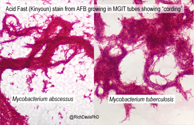
Researchers have since identified not just different levels of TDM in isolates of M. abscessus but also that those levels correlate to the known rough or smooth appearance of these isolates (see Llorens-Fons, 2017). It is interesting to consider that more virulent isolates of M. abscessus are likely to be rough, cording-type growers, similar to what is seen with Mtb.

Beyond what I hope was an interesting look into the history of Mycobacterium identification, and a great chance to show some beautiful AFB cording, it’s a good reminder that in our lab culture (pun definitely intended!) visual clues to organism identification may not fit what you were first taught.

References

  1. Middlebrook G, Dubos RJ, Pierce C. VIRULENCE AND MORPHOLOGICAL CHARACTERISTICS OF MAMMALIAN TUBERCLE BACILLI. J Exp Med. 1947 Jul 31;86(2):175-84. doi: 10.1084/jem.86.2.175. PMID: 19871665; PMCID: PMC2135722.
  2. Hunter RL, Olsen MR, Jagannath C, Actor JK. Multiple roles of cord factor in the pathogenesis of primary, secondary, and cavitary tuberculosis, including a revised description of the pathology of secondary disease. Ann Clin Lab Sci. 2006 Autumn;36(4):371-86. PMID: 17127724.
  3. McCarter YS, Ratkiewicz IN, Robinson A. Cord formation in BACTEC medium is a reliable, rapid method for presumptive identification of Mycobacterium tuberculosis complex. J Clin Microbiol. 1998 Sep;36(9):2769-71. doi: 10.1128/JCM.36.9.2769-2771.1998. PMID: 9705435; PMCID: PMC105205.
  4. Nelson SM, Cartwright CP. Comparison of algorithms for selective use of nucleic-acid probes for identification of Mycobacterium tuberculosis from BACTEC 12B bottles. Diagn Microbiol Infect Dis. 1998 Aug;31(4):537-41. doi: 10.1016/s0732-8893(98)00049-2. PMID: 9764392.
  5. Hall-Stoodley L, Brun OS, Polshyna G, Barker LP. Mycobacterium marinum biofilm formation reveals cording morphology. FEMS Microbiol Lett. 2006 Apr;257(1):43-9. doi: 10.1111/j.1574-6968.2006.00143.x. PMID: 16553830.
  6. Staropoli JF, Branda JA. Cord formation in a clinical isolate of Mycobacterium marinum. J Clin Microbiol. 2008 Aug;46(8):2814-6. doi: 10.1128/JCM.00197-08. Epub 2008 Jun 25. PMID: 18579723; PMCID: PMC2519507.
  7. Llorens-Fons M, Pérez-Trujillo M, Julián E, Brambilla C, Alcaide F, Byrd TF, Luquin M. Trehalose Polyphleates, External Cell Wall Lipids in Mycobacterium abscessus, Are Associated with the Formation of Clumps with Cording Morphology, Which Have Been Associated with Virulence. Front Microbiol. 2017 Jul 25;8:1402. doi: 10.3389/fmicb.2017.01402. PMID: 28790995; PMCID: PMC5524727.

-Dr. Richard Davis, PhD, D(ABMM), MLS(ASCP)CM is a clinical microbiologist and regional director of microbiology for Providence Health Care in Eastern Washington. A certified medical laboratory scientist, he received his PhD studying the tropical parasite Leishmania. He transitioned back to laboratory medicine (though he still loves parasites!), and completed a clinical microbiology fellowship at the University of Utah/ARUP Laboratories in Utah before accepting his current position. He is a 2020 ASCP 40 Under Forty Honoree.

Microbiology Case Study: A 62 Year Old Male with Productive Cough

Case History

62 year old male from Louisiana with a medical history of COPD presents with fever, productive cough, weight loss, and a red nodule on the left hand. Chest x-ray shows interstitial and lobar infiltrates. Patient reports no recent travel history. Mycobacterial culture of the sputum is positive for an organism. MALDI-ToF of the sputum culture confirms the result.

Lab Identification

mycokans1
Image 1. Kinyoun stained long bacilli with a banded, ladder like pattern isolated from sputum culture.

mycokans2
Image 2. Bright lemon yellow colonies growing on LJ slant after exposure to light on the right compared with unpigmented control colonies on the left.

mycokans3
Image 3. Bright yellow colonies growing on 7H11 media after exposure to light on the right compared with unpigmented control colonies on the left.

Mycobacterium kansasii grows on 7H11 media and Lowenstein-Jensen (LJ) slants. The colonies appear smooth or rough and unpigmented when isolated in the dark. Upon exposure to light, colonies turn bright lemon yellow due to enhanced b-carotene production, which makes M. kansasii a photochromogen. M. kansasii stains positive with acid-fast stains. On Kinyoun stain, it shows long bacilli with a banded, ladder like pattern. M. kansasii is positive for nitrate reduction, catalase, urease, and tween hydrolysis. It is negative for niacin and pyrazinamindase. The identification of M. kansasii is confirmed by MALDI or DNA probe of an isolate.

Discussion

Mycobacterium kansasii is a slow growing photochromogen, an organism that grows unpigmented colonies in the dark but produces a bright lemon yellow pigment upon exposed to light3. It is also an acid-fast positive long bacillus that causes TB-like chronic pneumonia, which is the second most common non-TB mycobacterial infection after MAC in the AIDS population. There are five genotypes of Mycobacterium kansasii. Genotypes I and II infect humans, with I being the most prevalent. Because environmental sources of M. kansasii are rarely identified, isolation of M. kansasii from a culture is never considered a contaminant.

M. kansasii is usually found in the tap water in cities endemic for the infection. In the U.S., it is most prevalent in the central and southern states including Louisiana, Illinois, Texas, and Florida1. Internationally, it is most prevalent in Israel, Korea, France, Japan, Portugal, with the highest incidence in England, Wales, and among South American gold miners.

The majority of patients with M. kansasii infection have an underlying pulmonary disease such as COPD, bronchiectasis, or TB infection3. The clinical manifestation of M. kansasii infection includes a primarily unilateral cavitary infiltrate in the lungs without pleural effusions. Patients are generally older compared with those infected with TB. They present with productive cough, weight loss, fever, night sweats, and dyspnea. Symptoms are usually less severe but more chronic compared with those of TB pneumonia. M. kansasii can also present as cutaneous lesions similar to sporothrichosis. Lesions include nodules, pustules, red plaques, and ulcers1.

M. kansasii infection is diagnosed by chest X-ray or chest CT, positive respiratory culture, and clinical exclusion of other diagnoses2. The criteria for a positive culture include either two consecutive positive sputum cultures, one positive culture from bronchoscopy specimens, or one positive culture with compatible clinical symptoms2. The treatment of M. kansasii infection depends on the resistance of the organism to rifampin. Rifampin-sensitive organisms are treated with at least three drugs including rifampin, ethambutol, isoniazid, and pyridoxine1. Rifampin-resistant organisms are treated with three drugs including clarithromycin or azithromycin, moxifloxacin, ethambutol, sulfamethoxazole, or streptomycin1. Due to drug interaction, rifampin is contra-indicated among HIV patients taking protease inhibitors and nonnucleoside reverse transcriptase inhibitors. Because rifampin increases the metabolism of these HIV medications, it can lead to HIV drug resistance among these patients.


References

  1. Akram SM, Bhimji SS. Mycobacterium Kansasii. StatPearls. 2018. https://www.ncbi.nlm.nih.gov/books/NBK430906/
  2. Johnston JC, Chiang L, Elwood K. Mycobacterium Microbiol Spectrum. 2017;5(1):TNMI7-0011-2016. doi:10.1128/microbiolspec.TNMI7-0011-2016.
  3. Meraz A, Raheem S. Pulmonary Mycobacterium Kansasii Infection –A Tale of a Right Upper Lobe Cavitary Lesion [abstract]. Journal of Hospital Medicine. 2015; 10 (suppl 2). https://www.shmabstracts.com/abstract/pulmonary-mycobacterium-kansasii-infection-a-tale-of-a-right-upper-lobe-cavitary-lesion/. Accessed March 12, 2018.


-Ting Chen, MD is a 1st year anatomic and clinical pathology resident at the University of Vermont Medical Center.

Wojewoda-small

-Christi Wojewoda, MD, is the Director of Clinical Microbiology at the University of Vermont Medical Center and an Associate Professor at the University of Vermont.

Microbiology Case Study: A 47 Year Old Woman with Three Day History of Fever

Case History

A 47 year old Caucasian female presented to her primary care physician with a three day history of fever (Tmax 102°F), chills and generalized body aches. Her rapid influenza test was negative, but she was treated with oseltamivir for suspected viral infection. Her past medical history was significant for severe mitral regurgitation for which she had had a prosthetic valve replacement two years prior, ischemic cardiomyopathy with recent pacemaker placement one month prior and an undifferentiated connective tissue disease. Her current medications included hydroxychloroquine (Plaquenil) and warfarin. Her symptoms persisted and upon return to clinic, a urinalysis was performed and blood cultures were collected. On physical exam, the pacemaker site was erythematous and tender to palpation. She was started on doxycycline and fluconazole for a presumed urinary tract infection. After 4 days of incubation on the automated instrument, the two aerobic blood cultures bottles were positive and the patient was admitted to the hospital for further workup and therapy.

Laboratory Identification

Microscopic examination from the positive blood culture bottle revealed slender, beaded Gram positive bacilli (Figure 1). No definitive branching was identified. Given the morphology on Gram stain, a Kinyoun stain was performed and revealed red-purple, beaded acid fast bacilli which were consistent with a Mycobacterium spp. (Figure 2). A Mycobacterial Growth Indicator Tube (MGIT), a Lowenstein Jensen slate and blood & chocolate agars were inoculated with specimen. Given that the organism grew after 2 days, a rapidly growing Mycobacterium spp. was suspected (Figure 3). High performance liquid chromatography (HPLC) identified the organism as M. fortuitum.

mycofort1

Figure 1. Gram stain from the positive blood culture bottle showed slender, beaded Gram positive bacilli that were arranged in clumps (100x oil immersion).

mycofort2

Figure 2. Kinyoun stain of the organisms was consistent with acid fast bacilli (100x oil immersion).

mycofort3

Figure 3. Small, off-white colonies grew on chocolate agar after 3 days of incubation at 35°C in a CO2 incubator.

Discussion

M. fortuitum is a common rapid growing mycobacterial species that is ubiquitous in the environment and tap water. Most common infections due to M. fortuitum include post-traumatic or post-surgical wound infections and it can be associated with the insertion of prosthetic devices including heart valves, artificial joints and rods inserted after fractures. Of the rapid grower group (Runyon Group IV), which includes M. chelonae, M. abscessus and M. mucogenicum, it is M. fortuitum that accounts for approximately 60% of localized cutaneous skin infections and prosthetic device infections most frequently.

In the laboratory, M. fortuitum typically grows after two to five days incubation and appear as small, off-white colonies on a variety of different agars. The organism is typically slender, beaded Gram positive bacilli on Gram stain and positive for acid fast bacilli on a Ziehl-Neelsen or Kinyoun stain. As part of a traditional lab work up, M. fortuitum is arylsulfatase positive and is capable of reducing nitrates. Today a variety of methods, including HPLC, pyrosequencing, sequence analysis and matrix assisted laser desorption ionization time of flight mass spectrometry (MALDI-TOF MS), have become routine identification options. Susceptibility testing of isolates from clinically significant sites should be performed by broth microdilution and includes the following antimicrobials: amikacin, cefoxitin, ciprofloxacin, moxifloxacin, clarithromycin, doxycycline, linezolid, imipenem, meropenem, minocycline, trimethoprim-sulfamethoxazole and tobramycin.

In the case of our patient, it was discovered her pacemaker site was infected and upon further questioning it was discovered she wasn’t able to complete her antibiotic course after device placement due to nausea. A transesophageal echocardiogram showed no evidence of infective endocarditis and she was taken to the operating room for removal of the pacemaker and leads. The site was filled with pus and wound cultures obtained during surgery were consistent with M. fortuitum as well. Repeat blood cultures were negative and she was treated with intravenous amikacin and imipenem as well as oral levofloxacin for an anticipated 6-8 weeks before transitioning to oral therapy.


-Debbie Rigney Walley, MD, is a 1st year Anatomic and Clinical Pathology Resident at the University of Mississippi Medical Center.

Stempak

-Lisa Stempak, MD, is an Assistant Professor of Pathology at the University of Mississippi Medical Center in Jackson, MS. She is certified by the American Board of Pathology in Anatomic and Clinical Pathology as well as Medical Microbiology. She is the director of the Microbiology and Serology Laboratories.  Her interests include infectious disease histology, process and quality improvement and resident education. 

Microbiology Case Study: 81 Year Old Man with Prior History of Bladder Cancer

Case history

An 81 year old man with prior history of bladder cancer treated with Bacillus Calmette–Guérin  (BCG) presented with dysuria. Three urine samples were sent for AFB culture.

Laboratory Workup

The urine samples were plated on 7H11 solid media and Middlebrook liquid media. Colonies grew in the liquid media in five days and the solid media in 6 days (Figure 1). The colonies were subcultured to 7H11 solid media and Lowenstein-Jensen (LJ) media.  At this time, the colonies were probed for Mycobacterium tuberculosis complex, M. avium complex and M. gordonae, all of which were negative.  The organism grew on all of the subcultured media within one day. This fulfilled the criteria for rapidly- growing Mycobacterium species given the organism had grown in less than 7 days on subculture from solid media to solid media.  The specimen was sent out to our reference laboratory for further speciation and was identified as Mycobacterium chelonae.

chel1

Figure 1: 7H11 media with non-pigmented white colonies.


Discussion

Rapidly growing mycobacteria include many species, but the main clinically relevant species are M. fortuitum, M. chelonae, and M. abscessus. These organisms are widely distributed in nature and can survive nutritional deprivation and extreme temperatures. They have been isolated from soil, dust, natural surfaces, water, wild animals and domestic animals.  Risk factors for infection include patients with immunosuppression, organ transplant and autoimmune disorders. Immunocompetent patients are also at risk if they have had trauma or invasive medical procedures.  M. chelonae may cause a spectrum of human disease. The most common manifestations are cutaneous infection, osteomyelitis and catheter infections. Nosocomial outbreaks of M. chelonae have been reported and linked to various water sources, including water-based solutions, distilled water, tap water and ice.  Rapidly growing mycobacterium are generally resistant to the classic antituberculosis drugs (rifampin, ethambutol and isoniazid). M. chelonae is usually sensitive to aminoglycosides, however treatment should be determined by antibiotic susceptibility testing.  In our patient, we had expected the colonies to be M. bovis because of the patient’s history of BCG treatment which is a live attenuated strain of M. bovis. Cystitis induce by M. chelonae is a rare clinical manifestation. We believed this is a true infection, as opposed a contaminated patient sample, given the patient’s symptoms in conjunction with all three urine samples being positive for M. chelonae.


-Jill Miller, MD is a 4th year anatomic and clinical pathology resident at the University of Vermont Medical Center.

Wojewoda-small

-Christi Wojewoda, MD, is the Director of Clinical Microbiology at the University of Vermont Medical Center and an Assistant Professor at the University of Vermont.

Microbiology Case Study: A 46 Year Old Man Newly Diagnosed with HIV

Clinical Case

A 46 year old male with history of anal HPV with AIN II and anal fissure status-post sphincterectomy and fissurectomy, tobacco and cannabis use, and recent shingles outbreak presented with 1 year of diarrhea, fevers, chills and weight loss and a 2 week history of congestion and productive cough. He was found to have diffuse ground glass opacities with large cysts of the lungs on CT scan, and after admission was found to be positive for HIV with concern for AIDS. He received a bronchoscopy on that showed Pneumocystis jiroveci pneumonia. He was treated and clinically improved over several days when HAART therapy was initiated. Shortly afterwards he became neutropenic with his ANC as low as 150. The initial BAL fluid and stool became positive for acid fast bacilli.

Laboratory Identification

On Löwenstein-Jensen media, the organisms show small, flat, translucent, smooth colonies. They are slow-growers readily detected by acid fast and Kinyoun staining. In broth, the organisms do not show clustering or “cording.”


mac1
Gram stain, from Middlebrook 7H11 agar

mac2
Kinyoun stain, from Middlebrook 7H11 agar

mac3
Kinyoun stain, from Middlebrook 7H11 agar

mac4
Growth on Löwenstein-Jensen media

Discussion

Mycobacterium avium complex (MAC) is the most common nontuberculous mycobacterium (NTM) species causing human disease in the United States and is ubiquitous in the environment. MAC refers to infection caused by one of two slowly-growing NTM species, M. avium and M. intracellulare.

The pathogenesis of MAC lung disease is poorly understood. Infection is most likely acquired via ingestion or inhalation of aerosols from the environment inoculating a mucosal surface. Soon after inhalation or ingestion of MAC organisms, the infection disseminates lymphohematogenously. The bacteria are taken up by mononuclear phagocytic cells throughout the body, seeding other organs and tissues. Unlike M. tuberculosis, there is no convincing evidence demonstrating human-to-human transmission of MAC.

Disseminated NTM disease occurred in 5.5% of AIDS cases reported to the Centers for Disease Control and Prevention (CDC) from 1981 to 1987. This dropped to 4% after 1996, and is now at a rate of less than 1% per year. The dramatic decline in the disseminated disease is attributed to the use of effective prophylaxis with clarithromycin and azithromycin, as well as the advent of potent antiretroviral therapy. However, as in our case, patients with low CD4 cell counts remain at risk. Blood cultures for MAC isolation should be obtained before prophylaxis is initiated if there is any suspicion of clinical disease; the treatment regimen is different if blood cultures are positive (ie, the patient has active disease).

Traditional methods of speciating mycobacterial isolates were based upon growth characteristics on solid media and subsequent biochemical tests, requiring additional weeks for subcultures. Now we have commercially available highly-accurate nucleic acid probes that can identify MAC isolates within one day of growth. Other techniques such as 16S ribosomal DNA sequencing, Matrix-assisted laser desorption/ionization time-of-flight mass spectrometry, PCR-restriction length polymorphism analysis (PRA), and high-performance liquid chromatography (HPLC) are also available.

Susceptibility testing of MAC is difficult and controversial compared with M. tuberculosis. Exceptions to this are macrolides and amikacin, for which the MICs have been shown to correlate clinically with in vivo response. Additionally, clarithromycin resistance can be detected by a mutation in the 23S ribosomal macrolide binding site.


-Thomas Rogers, DO is a 3rd year anatomic and clinical pathology resident at the University of Vermont Medical Center.

Wojewoda-small

-Christi Wojewoda, MD, is the Director of Clinical Microbiology at the University of Vermont Medical Center and an Assistant Professor at the University of Vermont.


Microbiology Case Study: A 53 Year Old Male with Fatigue, Night Sweats, and Right Knee Swelling

Case History

A 53 year old male presents to the emergency department with complaints of fatigue, night sweats, dyspnea, dry cough, and right knee swelling. He has multiple skin lesions including violaceous papules on his medial thigh, subcutaneous nodules on bilateral lower legs, and ulcerations on his right lateral leg. His past medical history is significant for psoriatic arthritis. Previously, he has taken adalimumab (Humira), etanercept (Enbrel), and golimumab (Simponi), but his current treatment regimen consists of methotrexate and prednisone. His recent travel history is significant for scuba diving in Thailand and honeymooning in the Caribbean. Dermatology was consulted and a punch biopsy was performed close to an ulcer on his right lateral leg and sent to surgical pathology. An additional biopsy specimen was sent to microbiology for bacterial, fungal and mycobacterial cultures.


Laboratory Identification

mycohem1
Figure 1. Histology sections of the punch biopsy near the patient’s right lateral leg ulcer. The epidermis and dermis appeared unremarkable. However, a non-caseating granuloma is seen in the deep subcutaneous tissue (H&E, 40x).

mycohem2
Figure 2. Special stain highlighting the occasional acid fast organisms (Kinyoun, 1000x oil immersion).

mycohem3
Figure 3. Smear from a positive mycobacteria growth indicator tube (MGIT) incubated at 32°C showing abundant acid fast bacilli (Ziehl-Neelsen, 500x oil immersion).

Microscopic examination of the punch biopsy skin specimen revealed a non-caseating granuloma in the deep subcutaneous tissue, with no involvement of the overlying dermis and epidermis (Figure 1). A Kinyoun stain of the tissue showed that the granuloma contained occasional acid-fast bacilli (Figure 2). The bacterial and fungal cultures sent to microbiology were negative. Portions of the specimen used to set up the mycobacterial cultures were incubated in MGITs at 32 and 37°C because the specimen source was skin. The 32°C tube, which was supplemented with hemin, gave a positive signal after 3 weeks of incubation. The Ziehl-Neelsen stain from this tube revealed numerous acid fast bacilli (Figure 3). DNA Gen-Probe analysis was negative for Mycobacterium tuberculosis complex and M. avium-intracellulare (MAI) complex. The organism was identified as M. haemophilum by pyrosequencing.

Discussion

Mycobacterium haemophilum was first identified in 1978 from an Israeli patient with Hodgkin lymphoma. It has a known predilection for infecting the skin and subcutaneous tissue in immunocompromised patients, especially those with lymphopenia as a result of acquired immune deficiency syndrome (AIDS), allogeneic bone marrow transplantation, and those on immunosuppressant therapies for rheumatologic conditions. The clinical presentation frequently consists of painful subcutaneous nodules and ulcers that can progress to abscesses and draining fistulas. Bone and joint infections have also been reported, which manifest as arthritis, tenosynovitis, and osteomyelitis. AIDS patients in particular are known to present with disseminated disease, with multiple cutaneous lesions, mainly involving the extremities. Relatively little is known about this infection and the optimal treatment is not standardized, but combinations of three or four of the following drugs have been used successfully: isoniazid, rifamycins, ciprofloxacin, amikacin, doxycycline, and clarithromycin.

Unlike the majority of mycobacteria, M. haemophilum does not grow well in culture at 37°C. Rather, it prefers lower temperatures, ideally between 28–32°C. This characteristic is shared by several other mycobacterial species that also characteristically infect the skin, including M. marinum, M. chelonae, M. abscessus, and M. ulcerans. A unique feature of M. haemophilum among the mycobacteria is that it requires hemin (X factor) to survive and will only grow in media enriched with this nutrient. Similar to Haemophilus influenzae, M. haemophilum can be cultured on chocolate agar, as well as on Middlebrook 7H10 agar incubated with an “X-factor strip” and on Lowenstein-Jensen medium containing 2% ammonium citrate.

Typically, colonies grow after 2-4 weeks of incubation at 32°C and have either a rough or smooth appearance. M. haemophilum is a non-photochromogen according to the Runyon classification system, and its colonies are buff colored and do not produce pigment in either light or dark conditions. M. haemophilum is chemically inert by traditional biochemical mycobacterial tests, with the exception of pyrazinamidase production. As illustrated by this case, DNA probe analysis is helpful with regard to the mycobacterial species it excludes, but not for speciation of less common organisms. At the present time, DNA probes exist only for M. tuberculosis complex, MAI complex, M. kansasii, and M. gordonae.

In the case of our patient, the species level identification was determined by pyrosequencing. He was treated with an extended course of 3 agents: rifabutin, clarithromycin, and moxifloxacin with good response.


VN

Vikas Nath, MD, is a 4th year resident in Anatomic and Clinical Pathology at the University of Mississippi Medical Center in Jackson, MS.

Stempak

-Lisa Stempak, MD, is an Assistant Professor of Pathology at the University of Mississippi Medical Center in Jackson, MS. She is certified by the American Board of Pathology in Anatomic and Clinical Pathology as well as in Medical Microbiology. Currently, she oversees testing performed in both the chemistry and microbiology laboratories.  Her interests include infectious disease histology, process and quality improvement, and resident education.

Microbiology Case Study: 77 Year Old Male with Asthma

A 77 year old male with history of asthma, atrial fibrillation, and recurrent respiratory distress when visiting Vermont presented to the ED with progressive dyspnea and wheezing for the past 4 days. Two days prior, he required a “breathing treatment” at his PCP. One day ago, he saw his PCP and was prescribed prednisone and azithromycin. He denies cough, fevers, or chills. He used his albuterol and Advair inhalers which barely helped. He was found to be in Afib with RVR to the 160s, a respiratory rate in the 40s, and an oxygen saturation of 70%.

Kinyoun stain revealing broad rods with cross-barring.

Colony growth on Lowenstein-Jensen medium.


Lab Identification

The organism was auramine fluorescent stain positive from the broth. The AFB culture bottle was sub-cultured to agar based medium and Lowenstein-Jensen medium, which yielded small yellow colonies. Kinyoun stain revealed broad rods with cross-barring. The organisms produced a yellow pigment when exposed to light, and a nucleic acid probe for Mycobacterium kansasii was positive.


Discussion

Mycobacterium kansasii was discovered in 1953 by Buhler and Pollack. It is an acid fast bacillus that produces yellow pigment when exposed to light (photochromogen). The bacilli are thick, long and cross-barred and have been described as ladder-like. It is prevalent in the Midwest and Southeast, and is the second most common cause of nontuberculous mycobacteria disease in patients with AIDS. Mycobacterium kansasii manifests as lung disease that clinically appears similar to tuberculosis. It can also cause local disease of the skin and subcutaneous tissue, as well as lymphadenitis and disseminated disease. Symptoms are more severe in immunocompromised hosts. Mycobacterium kansasii is generally acquired via either aspiration or local inoculation from the environment, with little evidence to support person to person transmission.


-Mustafa Mohammed, MD is a 2nd year anatomic and clinical pathology resident at the University of Vermont Medical Center.

Wojewoda-small

-Christi Wojewoda, MD, is the Director of Clinical Microbiology at the University of Vermont Medical Center and an Assistant Professor at the University of Vermont.

Vinegar to the Rescue

Can a common pantry staple kill bacteria? Possibly, according a paper recently published in mBio. Researchers mixed acetic acid–the main ingredient in white vinegar–with suspensions of bacterial cultures and found that a exposure times as little as 20 minutes reduced the viable bacterial population by 710. The researchers then performed the same experiment, this time swapping out hydrochloric acid for the vinegar; they noted no bactericidal effect. Mycobacterium tuberculosis required a longer kill time (30 minutes vs. 20 minutes) to reach a 810 reduction in population.

These results suggest that vinegar could be used as a cheap-yet-effective disinfectant in resource-poor laboratory settings.


I’ll Have Pasteurized Milk, Please

Recently officials determined the cause of death of a young mother in Las Vegas: tuberculosis. The family Mycobacteriaceae contains several pathogenic species, including the most famous, M. tuberculosis. While none of the articles I read mention the species, they do mention the patient consumed unpasteurized dairy products, which leads me to believe she died of the zoonotic organism M. bovis.

Since these organisms are recovered infrequently, clinical microbiologists should brush up on the basics of these organisms on occasion. The CDC has some general information on Tuberculosis; the illustrious contributors at Wikipedia go a bit more in depth. The best sources for information are reference textbooks such as the Manual of Microbiology. It’s important to remember that mycobacteria can infect any region of the body, not just the respiratory system, so it’s important to keep an open mind. It’s also helpful to know that some species are rapid growers and may present on blood agar in a routine culture.

-Kelly Swails