Microbiology Case Study: A 5 Month Old with Redness in her Left Eye

Case History

A 5 month old girl was brought to her pediatrician by her mother for redness of the medial canthus of her left eye. There was some associated thick discharge as well. The mother mentioned that her daughter had excessive tearing from the left eye since birth. The pediatrician diagnosed dacryocystitis, and prescribed a course of oral cefdinir. However, the swelling and redness continued to worsen, and the infant began to have low-grade fevers. She was sent to pediatric ophthalmology for a consult, and admitted to the hospital on IV clindamycin. The following day, she was taken to the operating room, where they opened the obstructed nasolacrimal duct and sent the contents for culture.

Laboratory Work-Up

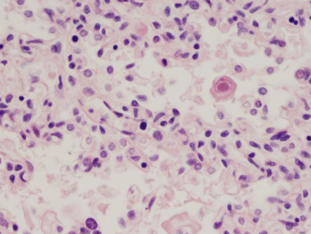
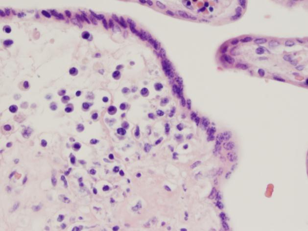
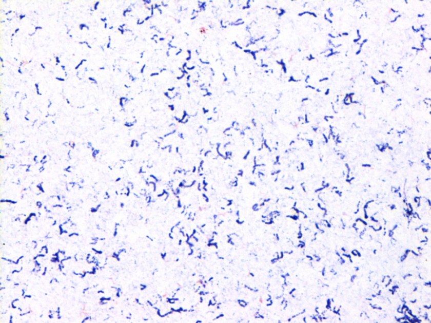
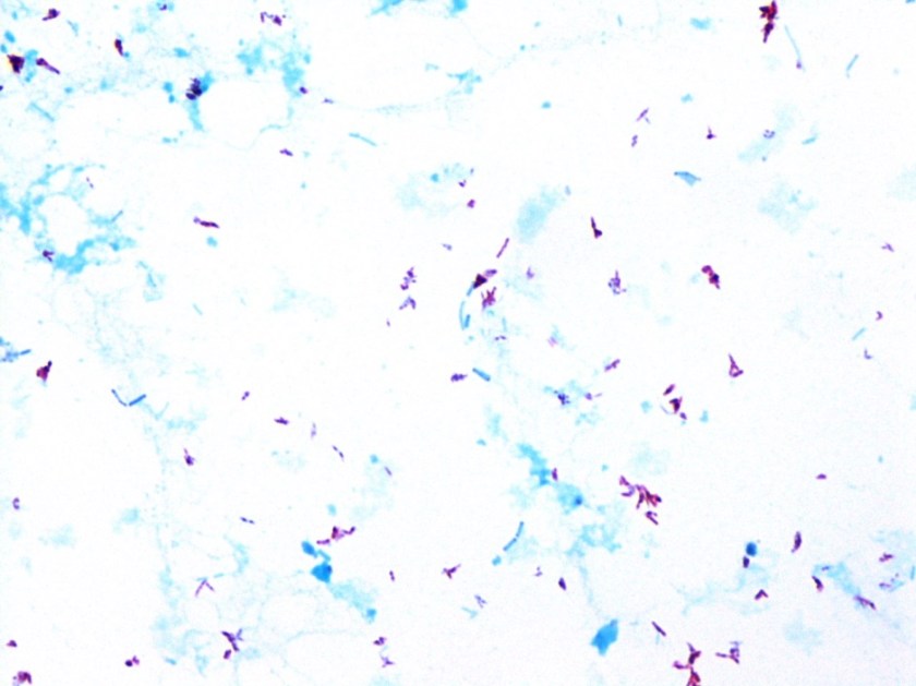
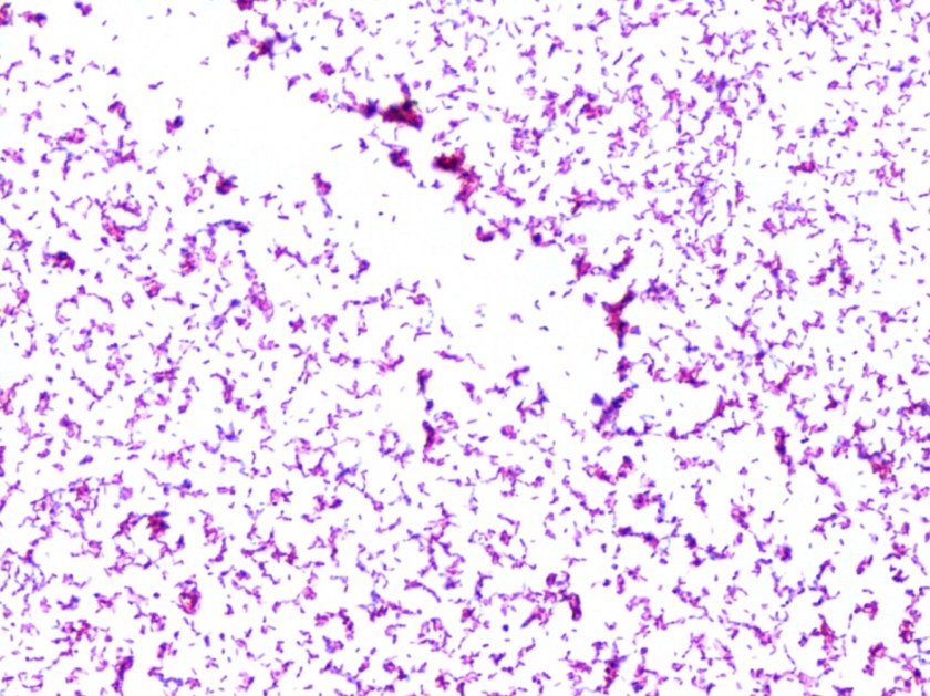
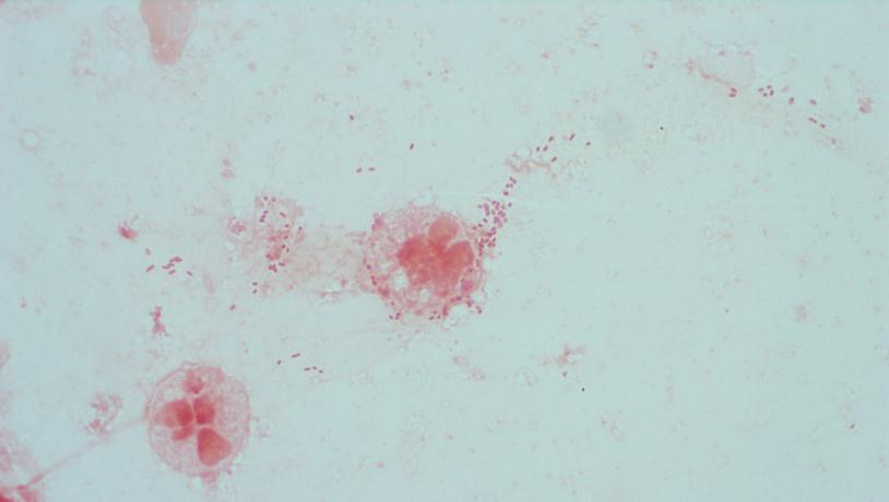
Gram stain and smear: No neutrophils, no bacteria seen.

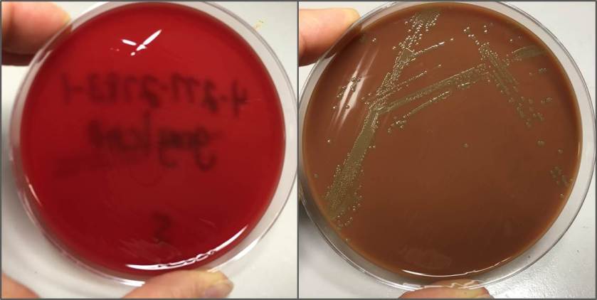
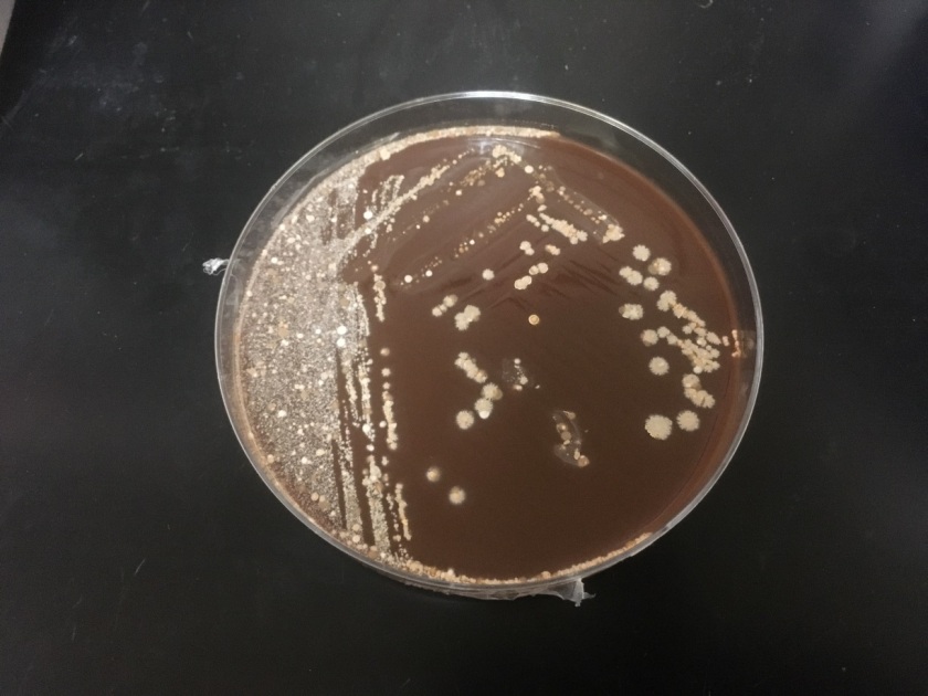
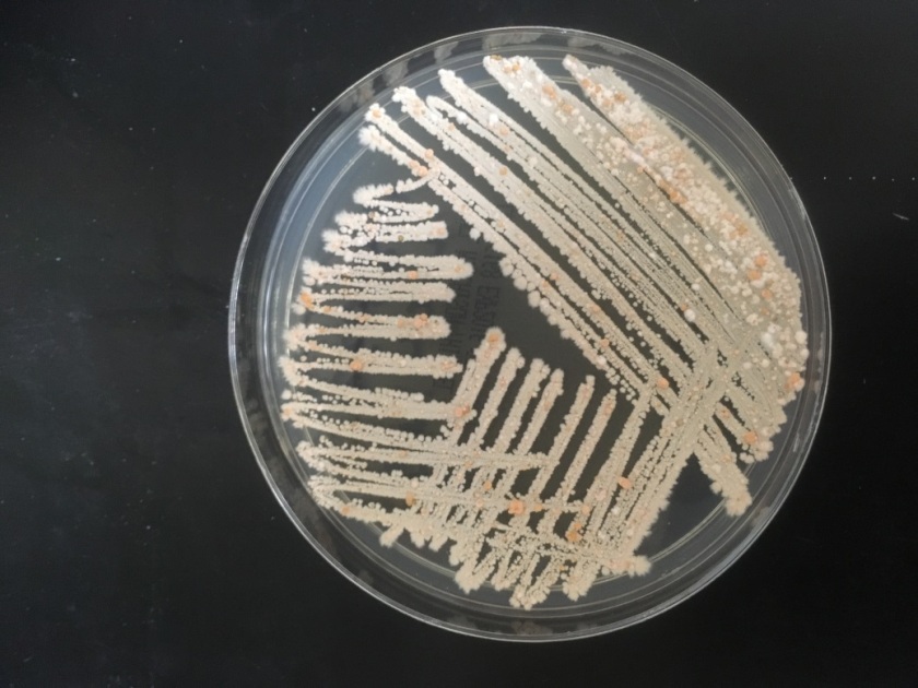
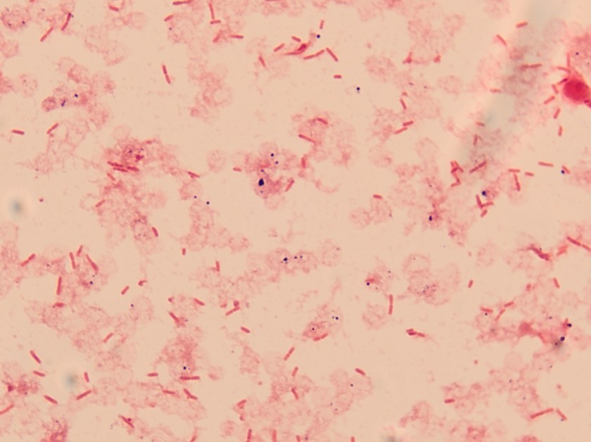
Routine bacterial culture showed growth of small, smooth and translucent colonies on the chocolate agar only. The colonies had a distinct wet-mouse odor.

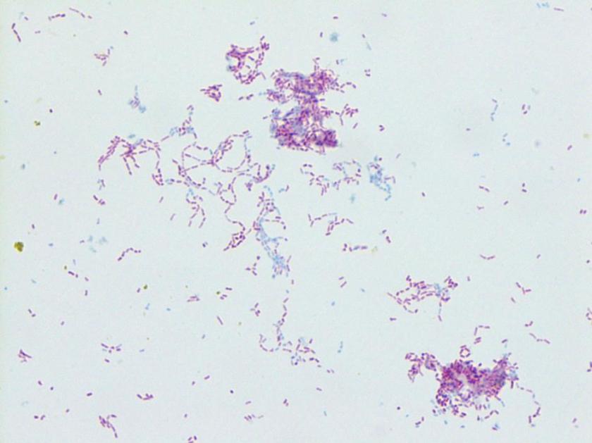
Repeat review of the gram stain showed moderate neutrophils, with gram negative coccibacilli both extracellular and intracellular.

Hflu1
Chocolate plate demonstrating growth of smooth, translucent colonies.
Hflu2
Thick, uninterpretable region of gram stain.
Hflu3
Repeat review of gram stain showed gram negative coccobacilli, with intraleukocytic organisms.

Discussion

The gram stain and colony morphology described above are consistent with Haemophilus influenzae. Identification of the organism was confirmed by MALDI-TOF.

Congenital nasolacrimal duct obstruction is a common problem, affecting up to 6% of newborns. The vast majority of cases will resolve without treatment by the time the child is 6 months old. However, complications can arise, include acute or chronic dacryocystitis. Acute dacryocystitis causes swelling, warmth, and erythema with or without purulent discharge. The organisms most commonly implicated are alpha-hemolytic streptococci, Staphylococcus epidermidis, and Staphylococcus aureus. In chronic dacryocystitis, there is purulent drainage from the eye but no other signs or symptoms. The most common organisms isolated in these cases are Streptococcus pneumonia, Haemophilus influenzae, Pseudomonas aeruginosa, and viridans group Streptococci.  Acute dacryocystitis usually requires systemic antibiotics, while chronic can be treated with topical antibiotics. In this case, the infant had clinical features of acute dacryocystitis, but infection with an organism more typically associated with chronic dacryocystitis.

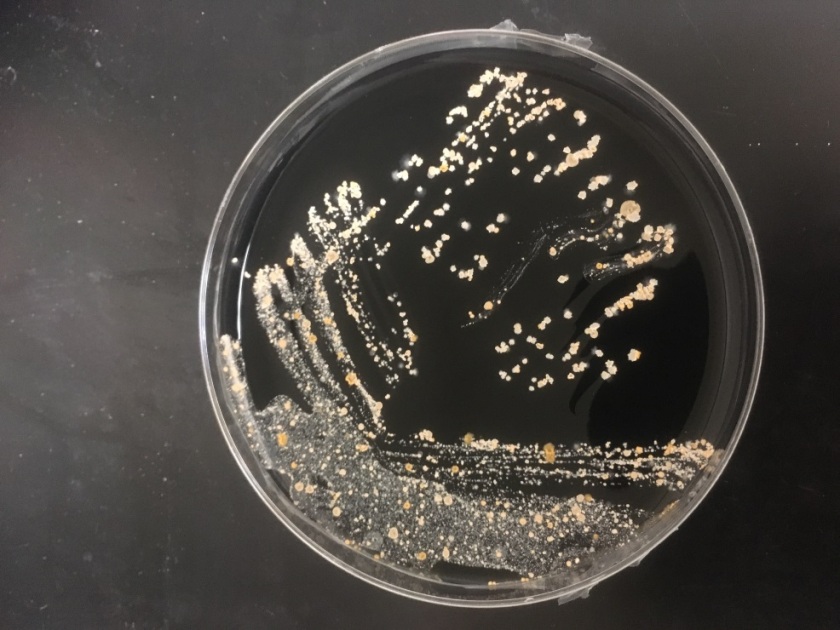
H. influenzae is a gram negative coccobacillus, which grows only on chocolate agar due to its requirement for factors V and X. However, H. influenzae can grow on blood agar if it is growing around an organism that hemolyzes the red blood cells in the media, releasing factor V (i.e. Staphylococcus aureus), a phenomenon known as satelliting.

As illustrated by this case, reviewing gram smears inconsistent with the final culture is an important aspect of quality assurance in the microbiology laboratory. This allows the opportunity to provide continuous feedback to technologists on their technique, and lets us keep track of any trends or common mistakes that may be occurring. In this instance, on review of the gram smear it appears the original reader examined only the very thick portions of the smear, which were uninterpretable. However, by moving out to the edges, neutrophils and bacteria were clearly visible. Feedback was provided to the original reader. The provider was immediately called and notified that a corrected report would be issued; the patient was switched to oral Augmentin based on these results.

-Alison Krywanczyk, MD is a 3rd year anatomic and clinical pathology resident at the University of Vermont Medical Center.

Wojewoda-small

-Christi Wojewoda, MD, is the Director of Clinical Microbiology at the University of Vermont Medical Center and an Assistant Professor at the University of Vermont.

Microbiology Case Study: 15 Year Old with Bacteremia

Case History

A 15-year-old male patient with acute myeloid leukemia (AML) had a central line placed for chemotherapy and subsequently developed symptoms of fever, abdominal pain, and diarrhea a few weeks later. He was treated with metronidazole for intra-abdominal infection and experienced improvement in diarrhea and abdominal pain, however his fever remained. Blood cultures were drawn from his central line and were positive.

gran1

Figure 1. Gram stain of the positive blood culture showing Gram-variable cocci in pairs and chains

gran2

Figure 2. Growth profile on organism on 5% sheep blood and chocolate agars

gran3

Figure 3. Colony Gram stain demonstrating pleomorphic, Gram-variable cocci in pairs and short chains

Laboratory diagnosis

Gram stain of the blood specimen revealed Gram-variable cocci in pairs and chains (Figure 1). The specimen was cultured on 5% sheep blood, chocolate blood, MacConkey, and Columbia colistin nalidix agar (CNA) agars. The next day, growth of small, round, grey colonies was seen only on the chocolate agar (Figure 2). Catalase test was negative. MALDI-TOF (Matrix Assisted Laser Desorption/Ionization, Time-of-Flight) identified Granulicatella adiacens with a 2.267 match score.

Discussion

The Granulicatella genus is a former member of the Abiotrophia genus, previously known as nutritionally variant or satelliting streptococci. The Granulicatella genera consists of G. adiacens and G. elegans (formerly A. adiacens and A. elegans respectively); of the Abiotrophia genus, only A. defectiva remains. These organisms are normal flora of the oral cavity, upper respiratory tract, gastrointestinal tract, and genitourinary tract. Granulicatella endocarditis is an uncommon, but well-documented phenomenon of both native and prosthetic valves and accounts for 5-6% of all streptococcal endocarditis. Other potential complications include ocular, central nervous system, musculoskeletal infections, bacteremia, pneumonia, scrotal abscess, septic arthritis, peritoneal dialysis-associated peritonitis, and breast-implant associated infections.

The Granulicatella spp. are catalase-negative, oxidase-negative, facultative anaerobic, gram-positive coccobacillus arranged in pairs and chains; although pleomorphism may occur (Figure 3, colony Gram stain). Notably, Granulicatella will grow on chocolate blood agar, but not 5% sheep blood agar or CNA agar because it requires Pyridoxine or vitamin B6 for growth. In addition to chocolate agar, Granulicatella can grow on Brucella agar with 5% horse blood and in thioglyconate broth. Granulicatella and other nutritionally variant streptococci exhibit satelliting behavior.  Satellite testing is performed on a media that supports no or little growth of Granulicatella (e.g. sheep blood agar). A single streak of Staphyloccous aureus across an area of the media inoculated with Granulicatella is incubated at 35°C in a high CO2 atmosphere. Granulicatella will grow only in the vicinity of S. aureus growth.  An alternate test for satelliting involves supplementation of media with pyridoxine hydrochloride, to allow growth of Granulicatella.

Biochemical reactions include: pyrrolidonyl arylamidase production (PYR) positive, leucine aminopeptidase production (LAP) positive, 6.5% NaCl negative, and bile esculin negative. Species identification is accomplished by arginine hydrolysis (ARG) and beta-glucuronidase (BGUR) activity testing. G. elegans is ARG-positive, while G. adiacens and Abitrophia are both negative. G. adiacens is further identified by a positive BGUR analysis, and Abiotrophia is again negative.

Abiotrophia and Granulicatella have shown variable susceptibility to both penicillin and aminoglycoside antibiotics. There is documented resistance to clindamycin, tetracycline, erythromycin, and ciprofloxacin, but not to rifampin or vancomycin. Current recommendations are to treat similarly as for enterococcal endocarditis using a combination therapy of a beta-lactam antibiotic with an aminoglycoside antibiotic, such as penicillin plus gentamycin. Unfortunately, relapse rates appear high despite appropriate treatment.

The likely source of this patient’s Granulicatella bacteremia is bacterial translocation from the gut in the setting of an immunocompromised state. The work up for a central line source of the bacteremia is still currently in progress, and echocardiogram was negative for vegetation.  He is currently being treated with vancomycin for bacteremia, and cefepime and metronidazole for intra-abdominal infection.

References

  1. Ruoff, K. Aerococcus, Abiotrophia, and other aerobic catalase-negative, gram-positive cocci. Manual of Clinical Microbiology, 10th Edition(pp. 365–376). American Society of Microbiology.
  2. Procop, G. W., Church, D. L., Hall, G. S., Janda, W. M., Koneman, E. W., Schreckenberger, P. C., & Woods, G. L. (2016). Koneman’s Color Atlas and Textbook of Diagnostic Microbiology(7th ed.). Philadelphia: Wolters Kluwer.
  3. Cargill, J., Scott, K., Gascoyne-Binzi, D., Sandoe, J. “Granulicatella infections: diagnosis and management.” Journal of Medical Microbiology 16 (2012): 755-761.


-Melinda Flores, MD, is a 1st year clinical and anatomic pathology resident at the University of Texas Southwestern Medical Center, Dallas, Texas.

-Erin McElvania TeKippe, PhD, D(ABMM), is the Director of Clinical Microbiology at Children’s Medical Center in Dallas Texas and an Assistant Professor of Pathology and Pediatrics at University of Texas Southwestern Medical Center.


Candida auris: An Emerging Pathogen

Clinical laboratory professionals, microbiologists, and pathologists need to be aware of an emerging fungal pathogen. According to the CDC, Candida auris is a threat because:

  • It’s multi-drug resistant
  • It’s often incorrectly identified by common laboratory techniques and analyzers
  • It causes outbreaks in the healthcare setting.

Here are a few resources if you’d like to read more.


Microbiology Case Study: A Newborn Baby in Respiratory Distress

Case History

A 29 year old G2P1 woman presented in labor at 39+2 weeks gestational age. Her pregnancy had been previously uncomplicated. Prenatal infectious disease testing showed that she was negative for HIV and Hepatitis C, but that she was positive for Group B Streptococcus. No test results were available for rubella, VZV, toxoplasmosis, or syphilis.

A term male infant was born shortly afterwards by spontaneous vaginal delivery; the mother received less than 4 hours of antibiotics.  The baby was noted to be covered in petechiae, and in a moderate amount of respiratory distress. A CBC showed thrombocytopenia to 23 K/cmm. The baby was transported emergently to the neonatal intensive care unit, where platelet transfusions were given. Blood cultures were drawn. The baby was started empirically on ampicillin/gentamycin, and the following day, once platelet counts were improved, a lumbar puncture was performed. The cell counts in the CSF were unremarkable. A cranial ultrasound showed scattered bilateral parenchymal calcifications, mineralized vasculature of the lenticulostriate arteries, and a subependymal cyst. Urine PCR testing was positive for CMV.

Laboratory Work-up

  • Bacterial Culture and Smear, CSF: No neutrophils, no bacteria. No growth.
  • CSF Viral PCR: Negative for HSV and VZV.
  • Urine CMV PCR: Positive.
  • The placenta was not sent for pathologic examination.

CMV1

Cranial ultrasound demonstrating scattered parenchymal calcifications.

CMV2
Photomicrograph of a lung from a 20 week gestation fetus demonstrating the characteristic “Owl’s Eye” inclusion of CMV.
CMV3
Photomicrograph of the placenta from the same case as B. The chronic villitis with plasma cells seen here is a sign of CMV infection.


Discussion

Cytomegalovirus is one of the classic “TORCH” infections. TORCH is an acronym for a group of pathogens that can cause in-utero or intrapartum infections:

  •                 T= Toxoplasmosis
  •                 O= other (syphilis, VZV, parvovirus)
  •                 R= rubella
  •                 C= CMV
  •                 H= HSV

Although these infections share several common signs and symptoms, there are clinically suggestive findings that can help target testing. The combination of thrombocytopenia and intracranial calcifications in this infant raised strong suspicion for congenital CMV. CMV is a member of the herpesvirus family.  It is a double-stranded DNA virus with both a viral capsid and envelope. While most babies born with congenital CMV are asymptomatic (~90%), congenital CMV infection is the main etiology of non-hereditary sensorineural hearing loss. This occurs in up to 50% of symptomatic infants and in 10-15% of asymptomatic infants. Symptomatic infants may be small for gestational age, and can be afflicted by thrombocytopenia, petechiae, intracranial calcifications, chorioretinitis, hepatosplenomegaly, microcephaly, and jaundice. While toxoplasmosis can also cause intracranial calcifications, it does not typically cause thrombocytopenia. Congenital HSV can cause thrombocytopenia, but is not associated with intracranial calcifications.

CMV infection during pregnancy is most often acquired by contact with young children. CMV has the ability to remain latent in the host, and become reactivated at a later time, so pregnancies can be affected by either primary infection or by reactivation of the virus. The risk of vertical transmission is much higher with primary CMV infection (32%) than with recurrent infection (1.4%). Although the rate of vertical transmission increases if the infection occurs later in pregnancy, infections acquired in early pregnancy are more likely to cause symptomatic disease. Treatment for the baby is generally supportive, with antivirals generally used only in symptomatic disease (their utility in asymptomatic infection is debated).

An audiology screen and ophthalmologic exam were both normal in the infant presented here. Oral valgancyclovir was started in addition to other supportive measures.


-Alison Krywanczyk, MD is a 3rd year anatomic and clinical pathology resident at the University of Vermont Medical Center.

Wojewoda-small

-Christi Wojewoda, MD, is the Director of Clinical Microbiology at the University of Vermont Medical Center and an Assistant Professor at the University of Vermont.

Minimal Inhibitory Concentrations and Antimicrobial Dosing: How are they related?

Microbiology laboratories use clinical breakpoints to categorize microorganisms as susceptible, intermediate, or resistant. These breakpoints help guide the selection of antimicrobial therapy having a high likelihood of achieving therapeutic success in patients. One in vitro marker of antimicrobial activity is the minimal inhibitory concentration (MIC), the lowest concentration of antibiotic that prevents visible growth of a standard bacterial inoculum. National committees, such as the Clinical Laboratories Standards Institute (CLSI), Food and Drug Administration (FDA), and The European Committee on Antimicrobial Susceptibility Testing (EUCAST) define clinical practice breakpoint MIC values for each bacterial genus. These defined values are determined using wild type value distributions in relation to what serum drug levels are achievable with standard antimicrobial dosing.

All drugs have individual pharmacokinetic properties such as absorption, volume of distribution, and rate of elimination. These factors contribute to what concentration of drug will be achieved at a certain site of infection. A good index of overall antibiotic exposure in a patient is the serum area-under-the-curve (AUC), which is influenced directly by the drug dose and clearance. Antibiotics also have pharmacodynamic properties, which relate to the drug’s effect on the microorganism over time. There are two main groups of pharmacodynamic characteristics seen with antimicrobial agents: time-dependent bactericidal action (Figure 1) and concentration-dependent bactericidal action (Figure 2). The clinical efficacy of an antibiotic is related to the relationship between pharmacokinetic/pharmacodynamic (PK/PD) parameters of a drug and the MIC of the specific organism. Bacterial strains with an increase in MIC may exhibit relative resistance by in vitro laboratory standards, but because there is no increase in the PK/PD parameters, the increased MIC can sometimes be overcome by altering dosing regimens to optimize the drug concentrations achieved.

For example, β-lactam antibiotics exhibit time-dependent killing activity, so dosing regimens which maximize duration of exposure to drug concentrations above the MIC of the organism are particularly effective for treating bacteria with this this class of antibiotics. Prolonged infusion times and smaller fractions of total daily doses given more frequently are two strategies through which this can be achieved. For drugs exhibiting concentration-dependent killing, such as aminoglycosides, dosing regimens can be optimized by giving a higher dose in order to achieve higher peak concentrations. The pharmacokinetic properties of drug can also be used to overcome elevated MICs for some organisms depending on the site of the infection. A good example of this would be urinary tract infections. Antibiotics that achieve high concentrations in the urine, such as aminoglycosides, can be used to successfully treat organisms with elevated MICs. Therefore, while healthcare providers utilize breakpoint MIC values to select antimicrobial regimens, understanding characteristics of an antimicrobial, including PK/PD parameters and tissue distribution, along with taking into account the site of infection and the MIC of the infecting organism, can provide the opportunity for optimization of antimicrobial dosing strategies.

fig1

Figure 1. For antibiotics which confer time-dependent antimicrobial activity, microbial killing is optimized when the concentration of antibiotic is above the MIC for as long of a time period as possible.

fig2

Figure 2. For antibiotics which confer concentration-dependent antimicrobial activity, microbial killing is optimized when a high peak concentration of antimicrobial is achieved.


References:

  1. Mouton JW, Brown DFJ, Apfalter P et al. The role of pharmacokinetics/pharmacodynamics in setting clinical MIC breakpoints: the EUCAST approach. Clin Microbiol Infect. 2012;18:E37-E45.
  2. Levison ME, Levison JH. Pharmacokinetics and pharmacodynamics of antibacterial agents. Infect Dis Clin North Am. 2009;23(4):791-vii.
  3. MacGowen AP. Role of pharmacokinetics and pharmacodynamics: does the dose matter? CID. 2001;33(suppl 3):S238-239.
  4. Martinez MN, Papich MG, Drusano GL. Dosing regimen matters: the importance of early intervention and rapid attainment of the pharmacokinetic/pharmacodynamic target. Antimicrob Agents Chemother. 2012;56(6):2795-2805.


burns

-Alaina Burns, PharmD, is a PGY-2 Pediatric Pharmacy Resident at Children’s Health, Children’s Medical Center in Dallas, Texas.

Microbiology Case Study: A 60 Year Old Woman with Increasingly Frequent Asthma Attacks

Case History

After experiencing increasingly frequent asthma attacks and multiple episodes of pneumonia within the last two years, a 60 year-old woman with a longstanding history of allergic asthma presents to a pulmonologist complaining of increased shortness of breath and cough. The patient reports a history of abnormal lung infiltrates for which she was previously treated with a three month course of azithromycin. A repeat chest CT shows diffusely scattered, nodular, ground-glass opacities which have increased in number since her last CT two year prior. A bronchoalveolar lavage is performed and specimen is sent for bacterial, fungal, and AFB cultures as well as a respiratory virus PCR panel.

nocar1
BAL on Chocolate agar
nocar2
BAL on 7H11 agar
nocar3
BAL on BCYE agar
nocar4
BAL with Modified Kinyoun stain


Laboratory Identification

The bacterial and fungal cultures do not grow any pathogens and the respiratory virus panel is negative. The AFB culture, however, grows beaded, Gram-Positive bacilli which are Auramine/Rhodamine negative and Modified Kinyoun positive. The organism grows well on 7H11, Chocolate, and Buffered Charcoal Yeast Extract (BCYE) agars forming irregular, chalky, white-pink colonies.

The organism is confirmed as Nocardia nova by molecular methods.

Discussion

Nocardia nova is a ubiquitous soil bacteria and one of several Nocardia species known to cause disease in humans. When contracted through traumatic inoculation, Nocardia may cause cutaneous diseases such as a mycetoma, superficial abscesses, or cellulitis. More commonly, however, Nocardia is contracted via inhalation and presents as a chronic, slowly progressive pulmonary infection with cough, shortness of breath, and fever. Complicated pulmonary infections may result in pleural effusions, empyema, pericarditis, chest wall abscesses, or dissemination to the brain and other deep organs. Due to low virulence, Nocardia primarily affects only the immunocompromised but those with pre-existing pulmonary disease are also susceptible to infection.

Nocardia is identified in the laboratory as an aerobic filamentous, beaded, Gram-Positive bacilli demonstrating right-angled branching. Nocardia is also weakly acid-fast and is usually identified by a Modified Kinyoun stain. While Nocardia grows within 3-5 days on blood and chocolate agar, it is often isolated on mycobacterial media or BCYE plates where it forms chalky white to faintly pigmented colonies. Accurate identification and speciation of Nocardia currently requires the use of molecular methods (primarily 16S ribosomal RNA gene sequencing). While many infections are successfully treated with a sulfonamide for 6 months to 1 year, the CDC recommends performing speciation and anti-microbial susceptibility testing on every clinical isolate due to species specific susceptibility profiles and multi-drug resistant strains. Nocardia farcinica, for example, is resistant to many antibiotics including 3rd generation cephalosporins.


-Elaine Amoresano, MD, is a 2nd year anatomic and clinical pathology resident at the University of Vermont Medical Center.

Wojewoda-small

-Christi Wojewoda, MD, is the Director of Clinical Microbiology at the University of Vermont Medical Center and an Assistant Professor at the University of Vermont.


Microbiology Case Study: 12 Year Old with Abdominal Pain

Case presentation

A 12-year-old female is seen in gastroenterology clinic following 2 weeks of abdominal pain. She is an otherwise healthy child with no significant past medical history. Her abdominal pain was diffuse, but it has worsened in the past 5 days and is now localized to the left upper quadrant and is sharp in nature. The pain was severe enough to prevent her from attending school last week. She was evaluated for appendicitis, which was ruled out. The patient was admitted for further management including an upper and lower endoscopy. During the endoscopy procedure, small, mobile worms were visualized in the ascending colon. Two worms were collected and removed for identification (Figure 1).

pin1

Figure 1. (A) Small, threadlike worms measuring 5-10 mm. Note pointed posterior tail. (B) Haematoxylin and eosin (H&E) stained cross section of the worm.

pin2.png

Figure 2. Cross section of the worms shows (A) anterior with cephalic inflations of the cuticle (arrows) and (B) a long pointed tail.

pin3

Figure 3. Internal eggs are 50-60 µm x 20-30 µm in size. They are elongated, flattened on one side, and have a thick colorless shell.


Discussion

The worms were identified as Enterobius vermicularis or pinworm. E. vermicularis is a nematode or round worm. Adult worms are 2.5 mm x 0.1 mm (males) and 8-13mm x 0.3-0.5mm (females). Both male and female worms have cephalic inflations of the cuticle at their anterior end (Figure 2A, arrows). Males have a wide blunt posterior tail while females have a long, pointed tail (Figure 1, 2B). Our worms were females as internal eggs were found in both worms. The eggs of E. vermicularis are 50-60 µm x 20-30 µm in size. They are elongated, flattened on one side, and have a thick colorless shell (Figure 3).

E. vermicularis infection is very common in preschool and school aged children as well as families and caregivers of infected children. Transmission occurs through the fecal-oral route. Embryonated eggs are ingested and travel to the small intestine. Adult worms reside in the colon. Gravid females migrate to the anus and deposit eggs onto the perianal area during the night. A single female can deposit as many as 10,000 fertilized eggs. Larvae within the eggs develop and become infective as quickly as 4-6 hours after they are deposited. The entire life cycle from ingestion until eggs are laid by a gravid female in the perianal area is 1-2 months. Perianal scratching and autoinfection are common as well as infection from contaminated fomites such as bedding, clothes, and shared toys.

The most common method of E. vermicularis detection is the Tape Prep method. Briefly, transparent (unfrosted) tape is used to touch the perianal region, after which the tape is placed on a glass slide for microscopic examination. The best time to detect E. vermicularis is 2-3 hours after the patient has gone to sleep due to the nocturnal cycle of the gravid females. Because E. vermicularis does not enter the stool stream, ova and parasite examination often fails to detect the parasite and is not recommended.

Discovery of E. vermicularis in our patient was an unexpected finding, as our patient had no perianal itching. Asymptomatic detection of E. vermicularis has been described in the past, so this finding is not unique. The patient was given a dose of albendazole and will receive another in two weeks, as the drug has reduced effectiveness at killing the eggs or larval stages of development. Her abdominal pain was attributed to overuse of nonsteroidal anti-inflammatory drugs (NSAIDS) and she is being monitored by gastroenterology outpatient clinic.


References

  1. Ash and Orihel’s Atlas of Human Parasitology, 5th
  2. Red Book 2015 Report of the Committee on Infectious Diseases, 30th


I would like to thank the staff of the Children’s Medical Center Histology Laboratory for sharing my enthusiasm for parasites as well as sectioning, staining, and taking images of the worms for educational purposes.


-Erin McElvania TeKippe, PhD, D(ABMM), is the Director of Clinical Microbiology at Children’s Medical Center in Dallas Texas and an Assistant Professor of Pathology and Pediatrics at University of Texas Southwestern Medical Center.


Microbiology Case Study: A 46 Year Old Man Newly Diagnosed with HIV

Clinical Case

A 46 year old male with history of anal HPV with AIN II and anal fissure status-post sphincterectomy and fissurectomy, tobacco and cannabis use, and recent shingles outbreak presented with 1 year of diarrhea, fevers, chills and weight loss and a 2 week history of congestion and productive cough. He was found to have diffuse ground glass opacities with large cysts of the lungs on CT scan, and after admission was found to be positive for HIV with concern for AIDS. He received a bronchoscopy on that showed Pneumocystis jiroveci pneumonia. He was treated and clinically improved over several days when HAART therapy was initiated. Shortly afterwards he became neutropenic with his ANC as low as 150. The initial BAL fluid and stool became positive for acid fast bacilli.

Laboratory Identification

On Löwenstein-Jensen media, the organisms show small, flat, translucent, smooth colonies. They are slow-growers readily detected by acid fast and Kinyoun staining. In broth, the organisms do not show clustering or “cording.”


mac1
Gram stain, from Middlebrook 7H11 agar
mac2
Kinyoun stain, from Middlebrook 7H11 agar
mac3
Kinyoun stain, from Middlebrook 7H11 agar
mac4
Growth on Löwenstein-Jensen media

Discussion

Mycobacterium avium complex (MAC) is the most common nontuberculous mycobacterium (NTM) species causing human disease in the United States and is ubiquitous in the environment. MAC refers to infection caused by one of two slowly-growing NTM species, M. avium and M. intracellulare.

The pathogenesis of MAC lung disease is poorly understood. Infection is most likely acquired via ingestion or inhalation of aerosols from the environment inoculating a mucosal surface. Soon after inhalation or ingestion of MAC organisms, the infection disseminates lymphohematogenously. The bacteria are taken up by mononuclear phagocytic cells throughout the body, seeding other organs and tissues. Unlike M. tuberculosis, there is no convincing evidence demonstrating human-to-human transmission of MAC.

Disseminated NTM disease occurred in 5.5% of AIDS cases reported to the Centers for Disease Control and Prevention (CDC) from 1981 to 1987. This dropped to 4% after 1996, and is now at a rate of less than 1% per year. The dramatic decline in the disseminated disease is attributed to the use of effective prophylaxis with clarithromycin and azithromycin, as well as the advent of potent antiretroviral therapy. However, as in our case, patients with low CD4 cell counts remain at risk. Blood cultures for MAC isolation should be obtained before prophylaxis is initiated if there is any suspicion of clinical disease; the treatment regimen is different if blood cultures are positive (ie, the patient has active disease).

Traditional methods of speciating mycobacterial isolates were based upon growth characteristics on solid media and subsequent biochemical tests, requiring additional weeks for subcultures. Now we have commercially available highly-accurate nucleic acid probes that can identify MAC isolates within one day of growth. Other techniques such as 16S ribosomal DNA sequencing, Matrix-assisted laser desorption/ionization time-of-flight mass spectrometry, PCR-restriction length polymorphism analysis (PRA), and high-performance liquid chromatography (HPLC) are also available.

Susceptibility testing of MAC is difficult and controversial compared with M. tuberculosis. Exceptions to this are macrolides and amikacin, for which the MICs have been shown to correlate clinically with in vivo response. Additionally, clarithromycin resistance can be detected by a mutation in the 23S ribosomal macrolide binding site.


-Thomas Rogers, DO is a 3rd year anatomic and clinical pathology resident at the University of Vermont Medical Center.

Wojewoda-small

-Christi Wojewoda, MD, is the Director of Clinical Microbiology at the University of Vermont Medical Center and an Assistant Professor at the University of Vermont.


Microbiology Case Study: 37 Year Old Male with Cyclic Fever and Severe Headache

Case History

A 37 year old Indian male presents to the emergency department with complaints of a cyclic fever (102-103°F), chills, fatigue and a severe headache. He denies nausea, vomiting or diarrheal symptoms. His travel history is significant for a recent 2 month vacation to India and he is concerned he has malaria, given his symptoms. He reports no sick contacts or suspicious ingestions during his trip and his other family members are well. On physical exam, he is ill appearing, with dry mucous membranes but has no aversion to light or neck pain. Blood work revealed a normal white blood cell count and elevated liver enzymes (ALT 383 U/L, AST 282 U/L). Blood and CSF were collected and sent to the microbiology laboratory for Gram stain and culture. Additional tests for influenza, viral hepatitis and malaria were performed.

Laboratory Identification

salm1

Figure 1. Gram stain from a positive blood culture illustrating large Gram negative rods (100x, oil immersion).

salm2

Figure 2. Small, lactose negative colonies growing on MacConkey agar after 24 hours incubation in CO2 at 35°C.

salm3

Figure 3. Green colonies growing on Hektoen enteric agar after 24 hours incubation in O2 at 35°C.

Within a day of collection, multiple blood cultures were positive for a large Gram negative rods (Figure 1). The organism grew after 24 hours incubation in CO2 at 35°C on blood, chocolate and MacConkey agars and was lactose negative (Figure 2). The identification by MALDI-TOF was Salmonella group. The isolate was then sent to the public health department for additional testing via molecular typing methods. The diagnosis of Salmonella serotype Typhi was confirmed. Testing for influenza, viral hepatitis and malaria were all negative.

Discussion

Salmonella serotype Typhi is a motile, Gram negative rod that is a member of Enterobacteriaceae family.  Typhoid fever is the cause of serious bloodstream infections in developing countries and the great majority of cases in the United States are identified after recent travel. Clinically, it presents with high fever and headaches, in the absence of gastrointestinal manifestations and causes a more severe illness than other Salmonella serotypes. The infecting organism, of which humans are the only known reservoir, is transmitted by food or drink contaminated with feces or person to person contact and has a low infectious dose. Healthy carriers that are able to asymptomatically shed the bacteria have been documented.

Salmonella serotype Typhi is more commonly isolated from blood rather than fecal specimens and grows well on a variety of media including blood, chocolate and Hektoen enteric agar (Figure 3). The characteristic reaction on a triple sugar iron (TSI) slate is alkaline/acid (K/A, only glucose fermented) with a small moustache of H2S production at the site of inoculation and no gas production. In addition, a positive lysine decarboxylase reaction helps to distinguish Typhi from non-typhoidal Salmonella subspecies I members. MALDI-TOF mass spectrometry is successfully able to identify the isolate as Salmonella spp., but additional testing must be performed to determine the particular serotypes.

Traditionally, serotyping of the O (somatic), H (flagellar) and Vi (capsular) antigens and applying results to the Kauffmann-White scheme is useful in confirming the diagnosis of Salmonella and defining the serotype name. In the case of Salmonella serotype Typhi, the somatic antigen groups as D1 and the Vi antigen is present. The Vi antigen (heat labile) sometimes masks the identity of the O antigen (heat stable). In these cases, heating the bacterial suspension in boiling water for 15 minutes and repeating the O antigen serotyping yields the correct O antigen. Given the expansion in molecular testing, methods based on identifying the genes responsible for the serotype are gaining favor.

Due to the high mortality rate in untreated cases of typhoid fever, treatment with antibiotics is necessary and given the increasing levels of resistance reported, particularly to ciprofloxacin, susceptibility testing should be performed in all cases of Salmonella serotype Typhi. It is recommended that ampicillin, a fluoroquinolone, trimethoprim-sulfamethoxazole and a 3rd generation cephalosporin be reported for all typhoid isolates based on the most current M100S-26 CLSI guidelines. Also, in the case of S. Typhi, reporting susceptibilities to azithromycin is encouraged (MIC ≤16 µg/mL S, ≥32 µg/mL R, interpretative criteria based on MIC distribution data).

In the case of our patient, he was started on ceftriaxone and continued to receive this IV antibiotic for seven days after blood cultures became negative. Susceptibility testing showed intermediate results for ciprofloxacin by disk diffusion and azithromycin was found to be susceptible with an MIC of ≤16 µg/mL. The patient has an uncomplicated hospital course and made a complete recovery.


Stempak

-Lisa Stempak, MD, is an Assistant Professor of Pathology at the University of Mississippi Medical Center in Jackson, MS. She is certified by the American Board of Pathology in Anatomic and Clinical Pathology as well as Medical Microbiology. Currently, she oversees testing performed in both the Chemistry and Microbiology Laboratories.  Her interests include infectious disease histology, process and quality improvement and resident education. 

Antimicrobial Stewardship Down Under

If you’re an infectious disease/antimicrobial stewardship/microbiology geek, then the Australian blog AIMED is relevant to your interests. AIMED focuses on practical antimicrobial prescribing issues of relevance to hospital and community prescribers. It is supported by a local brains trust of General Practitioners, Pharmacologists, Pharmacists, Microbiologists and Infectious Disease Physicians. It also provides internet access to key Hunter New England resources for medical staff including guides to local antibiograms, infection control resources and personnel.

For those who don’t know, AIMED is an acronym for five principles that guide patient treatment with antimicrobials:

  • Antimicrobial selection and dosage
  • Indication for antimicrobial treatment
  • Microbiological assessment
  • Evaluate patient at 48-72 hours
  • Duration should be specified

If you’d like to learn more, check out their blog.