Microbiology Case Study: A 27 Year Old Male with Unintended Weight Loss

Case History

A 27 year old African American male presented to the emergency department with confusion and abdominal pain. His past medical history was significant for a 100 pound unintended weight loss and oral candidiasis which prompted a recent diagnosis of HIV. He was prescribed anti-retroviral therapy and antibiotic prophylaxis with which he reported compliance. Currently, he had no fever or chills. An abdominal CT scan showed an enlarged liver & spleen, generalized lymphadenopathy and a small amount of fluid. Significant lab work included anemia with a platelet count of 18,000 TH/cm2, absolute CD4 100 cells/cm2 (reference range: 506-3142 cells/ cm2) and a HIV viral load of 4,871 vc/mL. Given the concern for an infectious process, the infectious disease service was consulted and the patient underwent a thorough infectious work up including lumbar puncture, was started on board spectrum antibiotics and antifungals and was placed in airborne isolation until Mycobacterium tuberculosis could be ruled out.

Laboratory Identification

mac1
Image 1. Direct smear from a stool specimen showed beaded acid fast bacilli (Kinyoun stain, 1000x oil immersion).
mac2
Image 2. Direct smear from a stool specimen showed beaded acid fast bacilli (Kinyoun stain, 1000x oil immersion).

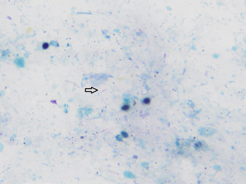
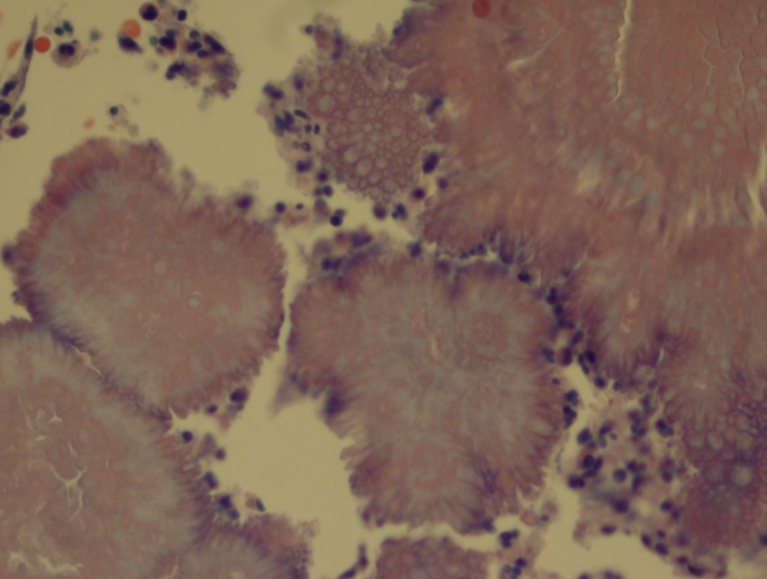
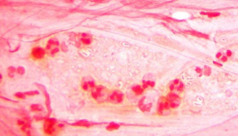
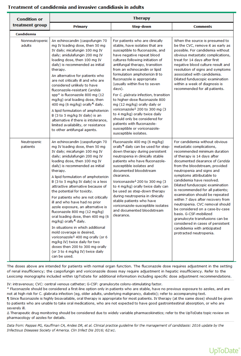
Initial diagnostic testing for bacterial, fungal and viral pathogens was negative. Three concentrated sputum AFB smears as well as a TB PCR were negative. The quantiferon gold TB test was also negative. The physician additionally ordered AFB blood and stool cultures. The direct smear from the stool specimen showed rare, beaded acid fast bacilli in a background of bacteria and yeast normally present in the stool via Kinyoun stain (Images 1 & 2). The specimen was sent to the department of health for additional work up. There was growth after 21 days incubation and Mycobacterium avium complex was identified by high performance liquid chromatography (HPLC).

Discussion

Mycobacterium avium complex (MAC) is a slow growing nontuberculous mycobacteria (NTM) frequently involved in human disease. Historically, it was classified as Runyon group III which are non-chromogens and do not produce pigment regardless of culture conditions. The group encompasses two taxa, M. avium and M. intracellulare. The species M. avium can further be classified into four subspecies: subsp. avium, subsp. silvaticum, subsp. paratuberculosis and subsp. hominissuis. Of interest, M. avium subsp. paratuberculosis can often be seen in association with Crohn’s disease.

In general, MAC organisms have low pathogenicity, but in the setting of those with lung disease (including cystic fibrosis), heavy smokers, immunocompromised patients and those with HIV, it is a well-known cause of disease. Infections with MAC can range from localized mycobacterial lymphadenitis and isolated pulmonary disease to bacteremia with dissemination to almost any organ. The organisms are located in circulating monocytes and further spread most commonly to the lungs, gastrointestinal tract and lymph nodes. In the case of HIV positive patients, MAC is the most common environmental NTM causing disease, especially in those with CD4 counts less than 100 cells/mm3 who are more likely to have disseminated disease.

In order to diagnosis MAC infections, specimens from blood, sputum, lymph nodes and other tissues are preferred. In addition, stool may also be an acceptable alternative in HIV patients if other specimens are negative or unable to be obtained. However, the sensitivity of a direct stool smear is only 32 to 34% making it not a very effective approach to identifying those at risk for disseminated infections. Once the culture has growth, various methods can be used to identify MAC, including phenotypic methods, DNA probe testing, HPLC, pyrosequencing and other forms of PCR & sequencing.

In the case of our patient, he was started on M. tuberculosis therapy: rifabutin, isoniazid, pyrazinamide & ethambutol (RIPE) until TB was ruled out. At that time, he was removed from isolation and switched to a drug regimen that included azithromycin, rifabutin and ethambutol. He showed clinical improvement and his cell counts, renal function and liver enzymes trended to normal ranges.


Stempak

-Lisa Stempak, MD, is an Assistant Professor of Pathology at the University of Mississippi Medical Center in Jackson, MS. She is certified by the American Board of Pathology in Anatomic and Clinical Pathology as well as Medical Microbiology. She is the Director of Clinical Pathology as well as the Microbiology and Serology Laboratories.  Her interests include infectious disease histology, process and quality improvement and resident education.

Microbiology Case Study: An Elderly Woman with a Cough Develops Skin Nodules

Case History

Our patient is an 83 year old female with previous history of arterial hypertension, atrial flutter and chronic obstructive pulmonary disease who presented with dry cough (~2 weeks), fever (102ºF), and cutaneous ulcerated plaques with elevated borders on forearm, foot, leg, and neck. Chest radiographs and chest CT scan showed numerous bilateral nodular consolidations compatible with pneumonia. Additionally, mild leukocytosis (14,200 cells /mm3) and hypohemoglobinemia (10.9 mg/dl) were documented. A skin biopsy was taken from the forearm lesion. Periodic acid–Schiff (PAS) and Grocott-Gomori’s (or Gömöri) methenamine silver (GMS) stains identified rare budding yeast (PAS, Image 1). Acid-fast bacilli (AFB) and Gram stains were negative for mycobacteria and bacteria, respectively.

blasto
Image 1. Budding yeast with a thick double cell wall were identified microscopically surrounded by prominent supurative granulomatous reaction (PAS, 100x).

Discussion

A diagnosis of disseminated blastomycosis was made based broad based budding yeast seen on PAS stain (Image 1).

Blastomycosis infection most commonly affects persons living in the Mississippi and Ohio River valleys, Great Lakes Region of the United States, and southern Canadian provinces. It is a fungal infection that can cause asymptomatic infection, isolated pulmonary disease, or serious and potentially fatal disseminated disease. B. dermatitidis can infect every organ of the body giving great variety of clinical manifestations, which is the reason why it is known as “the great pretender.” More than half of infected patients are asymptomatic. Symptomatic patients generally present with pulmonary symptoms, and the development of disseminated disease after hematogenous spread is common (~25 to 40% of symptomatic cases). The most common extra-pulmonary locations are: skin, bone, genitourinary tract, and central nervous system (CNS). Unlike histoplasmosis, most cases of blastomycosis are seen in immunocompetent patients, although immunocompromised patients may be at higher risk to develop severe forms of the disease.

Blastomyces is a thermally dimorphic fungus that grows as a yeast in the body and as filamentous fungi with septate hyphae in the environment. Recent phylogenic analysis has divided the Blastomyces genus into 2 species, B dermatitidis and B gilchristi . Culture of B. dermatitidis from the environment is extremely difficult, and much of what we know is conjecture from a few documented outbreaks, of which several occurred in wooded areas near waterways. These investigations found that exposure to dust clouds associated with construction or crop harvesting were the only identified risk factors for infection. Blastomycosis infection occurs through aerosolization of conidia from the environment causing respiratory infection or less commonly through direct inoculation into cutaneous abrasions. Once in the host, the conidia transform into yeast. The specific proteins expressed during the yeast phase allow the evasion of phagocytic and CD4+ cells.

Laboratory diagnosis

The most expedient method to diagnose blastomycosis remains examination of stained fluid or tissue specimens. Yeast are 8-15 µm in size with broad based buds of 4-5 µm and have a characteristic refractory double cell wall. Fluid can be stained with 10% potassium hydroxide plus calcofluor white, whereas formalin fixed paraffin embedded tissue samples can be stained with GMS or PAS. B. dermatitidis yeast can be difficult to visualize with Gram or hematoxylin and eosin (H&E) stains, but if found, the characteristic broad-based budding pattern of yeast can lead to presumptive diagnosis before culture and non-culture based diagnostic test results are available.

Culture of B. dermatitidis provides a definitive diagnosis of pulmonary and extra-pulmonary disease. B. dermatitidis grows well on routine fungal media such as Sabouraud dextrose agar, potato dextrose agar, and brain–heart infusion media. The yeast phase is inhibited by media containing cyclohexamide. Culture typically demonstrates growth in as little as 4-7 days. Colonies will initially appear yeast-like, but then develop white cottony aerial mycelium and turn tan with age. Mature growth is achieved around day 14 and the reverse of the colony is a tan color. At 25-30°C, B. dermatitidis forms septate hyphae with round or pear-shaped conidia attached to the hyphae by short or long conidiophores. This gives the characteristic appearance of “lollipops.” Scedosporium spp. and Chrysosporium spp. are common confounders because they make similar structures. Definitive identification of Blastomyces sp. can be made by conversion of the mold phase to the yeast phase by incubation at 37°C. An alternative to conversion is using a DNA probe assay.


References

  1. Medically Important Fungi, 5th edition
  2. Principles and Practices of Infectious Disease, 7th edition

Julio picture

-Julio Diaz-Perez, MD is a 1st year anatomic and clinical pathology resident at University of Chicago (NorthShore).

-Erin McElvania, PhD, D(ABMM), is the Director of Clinical Microbiology NorthShore University Health System in Evanston, Illinois.

Microbiology Case Study: 42 Year Old Female with HPV

Case History

A 42 y/o female G2P2002 patient presented to her Ob/Gyn for Colposcopy for monitoring of persistent High-Risk HPV. She was originally found positive for HPV in 2015, but has had never had a Pap with a squamous intraepithelial lesion, abnormalities on colposcopy, or dysplasia seen on endocervical curettage. Additionally, she endorsed a complaint of vague diffuse pelvic/lower abdominal pain for approximately the last 2 months. She states that the pain is mild and comes and goes and is not associated with anything in particular. She has noticed some clear to gray-white discharge now and then since she first noticed the pain, but nothing that really worried her. Pt denies changes in bowel or bladder habits, denies nausea, fever, or chills. Pt has been in a monogamous relationship with her partner for the last 12 years. She had a Mirana IUD placed 4 years prior, without complication, and has not had menses since placement. Prior to that, the patient had normal, regular cycles. She has 2 children with the same father, both were delivered by spontaneous vaginal delivery without complications. She has mild anxiety and depression for which she is treated, but no other medical problems. There is no surgical history. She has 1-2 glasses of red wine every week, denies tobacco use, and denies illicit drug use.

Pelvic exam revealed a benign appearing cervix that was not painful to touch or motion. There was a clear to white mild discharge that was suspected to be normal vaginal secretions. IUD strings were noted. Colposcopy revealed an easily appreciated transformational zone without any obvious lesions. A routine endocervical curettage (ECC) was performed followed by observed increased clear discharge from the cervical os. ECC was sent for routine pathology:

actinomyces1
Actinomyces, H&E, 20x
actinomyces2
Actinomyces, H&E, 40x

Discussion

Actinomycosis is an infection by a species within the Actinomyces genus, generally seen in dental and other oropharyngeal abscess formations. However, rare occurrences of pelvic Actinomycosis can be seen in women with intrauterine devices in place. Pelvic infections can result in cervicitis and endometritis and progress into abscess formation within the fallopian tubes and the ovaries along with salphigitis. The more profound disease consisting of abscess formation generally presents with fever, specific lower abdominal tenderness, and elevated WBCs, thus can mimic acute appendicitis, ovarian torsion, or ectopic pregnancy (1). The first case reported in the literature was in 1967 (2).

Three main species of Actinomyces have been found to be associated with IUD-associated pelvic infection: A. naeslundii, A. odontolyticus (3), and A. hongkongensis (4). All of these species are obligate to facultative anaerobes, catalase negative, and nitrate reducing. A sub-species group of A. naeslundii, however, can be catalase positive and is CAMP test-positive. All members of A. naeslundii are urease positive while A. odontolyticus and A. hongkongensis are urease negative.

References

  1. Joshi et al. Pelvic Actinomycosis: a Rare Entity Presenting as Tubo-ovarian Abscess. Arch Gynecol Obstet. 2010, 281:305-306.
  2. Brenner et al. Pelvic Actinomycosis in the Presence of an Endocervical Contraceptive Device. Obstet Gynecol. 1967, 29: 71-73.
  3. Woo et al. Diagnosis of Pelvic Actinomycosis by 16S ribosomal RNA Gene Sequencing and its Clinical Significance. Diagnostic Microbiology and Infectious Disease. 2002; 43: 113-118.
  4. Flynn et al. Identification by 16S rRNA Gene Sequencing of an Actinomyces hogkongensis Isolate Recovered from a Patient with Pelvic Actinomycosis. J. Clin. Microbiol. 2013, 51(8):2721. DOI: 10.1128/JCM.00509-13.


-Jeff Covington, MD, PhD, is a 1st year anatomic and clinical pathology resident at the University of Vermont Medical Center.

Wojewoda-small

-Christi Wojewoda, MD, is the Director of Clinical Microbiology at the University of Vermont Medical Center and an Associate Professor at the University of Vermont.


Microbiology Case Study: A 36 Year Old Man with Symptoms of Bowel Obstruction

Case History  

A 36 year old Caucasian male presented to the hospital with symptoms of a bowel obstruction.  His past medical history was significant for a gunshot wound to the abdomen followed by the development of colon cancer with metastasis to the liver. Recently, he had an intestinal stent placed in order to relieve an obstruction from tumoral growth. During the current admission, he was taken to the operating room for a diverting colostomy and two days later developed a fever of 101.1°F and increasing abdominal pain. Due to a concern for sepsis, blood cultures were collected and piperacillin-tazobactam was added to his antibiotic therapy regimen which already included vancomycin and ciprofloxacin.


Laboratory Identification

aero1
Image 1. Gram stain from the blood bottle showing many gram negative rods (100x oil immersion).
aero2
Image 2. The organism grew as mucoid colonies on blood, chocolate and MacConkey agars after 24 hours of incubation at 35°C in 5% CO2.


aero3
Image 3. The organism exhibited beta hemolysis on blood agar when held up to a light source.

The blood culture signaled positive after less than 24 hours on the automated instrument and Gram stain revealed gram negative rods (Image 1). The organism grew as mucoid colonies on blood, chocolate and MacConkey agars after 24 hours of incubation at 35°C in 5% CO2 (Image 2). When the blood agar plate was examined with a light source, the organism clearly illustrated beta hemolysis (Image 3). Rapid biochemical testing was positive for catalase, oxidase and indole. The Vitek II identified the isolate as Aeromonas veronii bv. sobria.

Discussion 

Aeromonas spp. are facultative anaerobic gram negative rods. They are inhabitants of aquatic ecosystems and as such can cause wound infections in people exposed to polluted lakes or brackish water with fresh breaks in their skin. Additionally, gastroenteritis is common with Aeromonas spp. and are often acquired through ingestion of unpurified water or to a lesser extent by consumption of contaminated meats, fresh produce or dairy products. Extraintestinal infections, including sepsis and meningitis, can result by spread from GI sources or wound infections. Interesting, medicinal leeches, used in the post-operative setting to increase blood flow to the surgical site, are colonized with Aeromonas spp. (most commonly Aeromonas veronii bv. sobria) and can result in systemic infections in the patient.

In the laboratory, Aeromonas spp. grow readily from stool, wound and blood sources on commonly used media and exhibits beta hemolysis on blood agar. In addition, Aeromonas spp. will grow on CIN agar (at room temperature as well as incubator temperature) as colonies with a pink center surrounded by a white apron and are indistinguishable from Yersinia spp. Aeromonas spp. is positive for catalase, oxidase and indole by rapid testing. In most cases, identification of Aeromonas spp. to the complex level can be accomplished by biochemical testing (esculin, VP), automated instrumentation or MALDI-TOF mass spectrometry. The three clinically relevant complexes include: A. hydrophila complex, A. caviae complex and A. veronii complex.

With regards to susceptibility testing for Aeromonas spp., the CLSI M45, 3rd edition provides guidelines for the three complexes discussed above. Third or fourth generation cephalosporins, fluoroquinolones and trimethoprim-sulfamethoxazole are recommended as antibiotics for primary testing for isolates from extraintestinal sites. Aeromonas spp. are uniformly resistant to ampicillin, amoxicillin-clavulanate and cefazolin and many strains may possess various inducible beta lactamases.

In the case of our patient, with the laboratory identification of Aeromonas veronii bv. sobria, his gram negative coverage was switched to ciprofloxacin for a 10 day course. His PICC was removed and on further imaging studies he was found to have a large abdominal abscess which required surgical drainage. Subsequent cultures from the abdominal abscess were negative.


Stempak

-Lisa Stempak, MD, is an Assistant Professor of Pathology at the University of Mississippi Medical Center in Jackson, MS. She is certified by the American Board of Pathology in Anatomic and Clinical Pathology as well as Medical Microbiology. She is the director of the Microbiology and Serology Laboratories. Her interests include infectious disease histology, process and quality improvement, and resident education.

Tissue is the Issue: Splitting Specimens, Part 1

When we think about infectious disease (ID) and specific syndromes (culture-negative endocarditis, for example), it can be difficult to know the etiology.1 This is because different microorganisms can cause similar symptoms and depending on the specimen submitted to the laboratory for testing, you may need to split your specimen. It may be that the infection is localized to a small area (valve vegetation) and all you get for processing is a small volume of tissue. Another scenario is that you get a sufficient amount of specimen, but you must split the specimen for culture, multiple send out studies, pathology, etc. Or even worse–a small volume specimen that you need to split for multiple diagnostic tests.

We recently had a case of endocarditis. The patient’s blood cultures were negative, and she was going to have her mitral valve replaced. The ID team requested that we send the tissue for broad-range bacterial and fungal sequencing. We can do that- not a problem.

The Issue

As requested, the specimen (mitral valve vegetation) was split once it was received in the laboratory. Half went into the freezer for sequencing requests which we send to a reference laboratory and the other half was processed for bacterial (including mycobacteria) and fungal cultures.

In our experience, if no organisms are observed, then no DNA is detected. Therefore, it is not beneficial to send tissue for sequencing if we do not observe an organism (or something that looks suspicious for an organism) to begin with. In parts 2 and 3 of this series we will go into greater detail of the workflow for examining tissue for infection, but for now we will focus on the processing piece.

No organisms were observed in the direct smears (Gram, fungal, and acid-fast) and all cultures were negative. Because no organisms were observed, we did not send the tissue for sequencing. However, the patient was not improving and ID insisted that we send the tissue for sequencing anyway. As a last ditch effort we decided to homogenize the frozen tissue to see if by chance organism was present.

Long story short: the Gram stain did not reveal organism, but acridine orange2 did. We cultured the tissue and recovered an organism. Moral of the story: specimen processing can be tricky. It is an inherent issue that we must be aware of. How were we to know in which part of the tissue the organisms were? By definition, this is sampling error at its finest.

The Solution

Moving forward, rather than split the specimen prior to processing we have changed our protocol to homogenize the tissue first, then split the specimen. We believe this will eliminate similar scenarios from happening again in the future.

The Conclusion

All specimens are different in their composition. Unlike body fluids, which are easy to vortex and make homogeneous; tissue is more complex. Whatever the specimen, make sure your protocol(s) reduces sampling error.

References

  1. Subedi SJennings ZChen SC. 2017. Laboratory Approach to the Diagnosis of Culture-Negative Infective Endocarditis. Heart Lung Circ. 26(8):763-771.
  2. Lauer BAReller LBMirrett S. 1981. Comparison of acridine orange and Gram stains for detection of microorganisms in cerebrospinal fluid and other clinical specimens. J Clin Microbiol.14(2):201-5.


Martinez Headshot-small 2017

-Raquel Martinez, PhD, D(ABMM), was named an ASCP 40 Under Forty TOP FIVE honoree for 2017. She is one of two System Directors of Clinical and Molecular Microbiology at Geisinger Health System in Danville, Pennsylvania. Her research interests focus on infectious disease diagnostics, specifically rapid molecular technologies for the detection of bloodstream and respiratory virus infections, and antimicrobial resistance, with the overall goal to improve patient outcomes.

Blood Bank Case Study: Transfusion Transmitted Malaria

Case Study

A 26 year old African American female with sickle cell anemia presented to a New York emergency room with cough, chest pain, fever and shortness of breath. Laboratory results showed an increased white blood cell count, slightly decreased platelet count and a hemoglobin of 6.2 g/dl. Her reticulocyte count was 7%, considerably below her baseline of 13%. Consulting the patient’s medical records revealed history of stroke as a child and subsequent treatment with chronic blood transfusions. She was admitted to the hospital for acute chest syndrome and aplastic crisis and care was transferred to her hematologist. Two units of RBCs were ordered for transfusion.

The blood bank technologists checked the patient’s blood bank history and noted her blood type was A, Rh(D) positive, with a history of a warm autoantibody and anti-E. The current blood bank sample confirmed the patient was blood type A, RH(D) positive with a negative DAT but the antibody screen was positive. Anti-E was identified. Per request of the hematologist, phenotypically similar units were found and the patient was transfused with 2 units of A RH(negative), C/E/K negative, HgS negative, irradiated blood. The patient’s hemoglobin rose to 8g/dl and she was discharged from the hospital 3 days after transfusion.

Ten days after discharge the patient returned to the emergency room with symptoms including aching muscles, fever and chills. A delayed transfusion reaction was suspected. A type and screen was immediately sent to the blood bank. The post transfusion type and screen remained positive for anti-E, DAT was negative. No additional antibodies were identified. However, a CBC sent to the lab at the same time revealed malarial parasites on the peripheral smear. The patient was consulted for a more complete medical history and reported that she had never traveled outside of the country. A pathology review was ordered and the patient was started on treatment for Plasmodium falciparum.

plasfal1

Discussion

Red Blood cell transfusions can be life saving for patients with sickle cells anemia. These patients are frequently transfused by either simple transfusion of red cell units or by exchange transfusion. Because of this, alloimmunization is reported to occur in 20% to 40% of sickle cell patients.1 Blood bank technologists are very diligent in adhering to strict procedures and follow a standard of practice aimed to prevent transfusion reactions. While preventing immune transfusion reactions may be the most forefront in our minds when transfusing the alloimmunized patient, it is important to consider transfusion transmitted diseases as a potential complication of blood transfusions.

Malaria is caused by a red blood cell parasite of any of the Plasmodium species. Mosquito transmitted infection is transmitted to humans through the bite of an infected mosquito. Transfusion-transmitted malaria is an accidental Plasmodium infection caused by a blood transfusion from a malaria infected donor to a recipient.

Donors, especially those from malarial endemic countries who may have partial immunity, may have very low subclinical levels of Plasmodium in their blood for years. Even these very low levels of parasites are sufficient to transmit malaria to a recipient of a blood donation. Though very rare, transfusion-transmitted malaria remains a serious concern for transfusion recipients. These transfusion-transmitted malaria cases can cause high percent parisitemia because the transfused blood releases malarial parasites directly into the recipient’s blood stream.

Blood is considered a medication in the United States, and, as such, is closely regulated by the FDA. Blood banks test a sample of blood from each donation to identify any potential infectious agents. Blood donations in the US are carefully screened for 8 infectious diseases, but malaria remains one infectious disease for which there is no FDA-approved screening test available. For this reason, screening is accomplished solely by donor questioning.2 A donor is deferred from donating if they have had possible exposure to malaria or have had a malarial infection. Deferral is 12 months after travel to an endemic region, and 3 years after living in an endemic region. In addition, a donor is deferred from donating for 3 years after recovering from malaria. It is important, therefore, for careful screening to take place by questionnaire and in person, to make sure that the potential donor understands and responds appropriately to questions concerning travel and past infection.

Malaria was eliminated from the United States in the early 1950’s. Currently, about 1700 cases of malaria are reported in the US each year, almost all of them in recent travelers to endemic areas. From 1963-2015, there have been 97 cases of accidental transfusion-transmitted malaria reported in the United States. The estimated incidence of transfusion-transmitted malaria is less than 1 case in 1 million units.4 Approximately two thirds of these cases could have been prevented if the implicated donors had been deferred according to the above established guidelines.3 While the risk of catching a virus or any other blood-borne infection from a blood transfusion is very low, a blood supply with zero risk of transmitting infectious disease may be unattainable. With that being said, the blood supply in the United Sates today is the safest it has ever been and continues to become safer as screening tests are added and improved. Careful screening of donors according to the recommended exclusion guidelines remains the best way to prevent transfusion-transmitted malaria.

References

  1. LabQ, Clinical laboratory 2014 No.8, Transfusion Medicine. Jeanne E. Hendrickson, MD, Christopher Tormey, MD, Department of Laboratory Medicine, Yale University School of Medicine
  2. Technical Manual, editor Mark K. Fung-18th edition, AABB. 2014. P 201-202
  3. https://www.cdc.gov/malaria/about/facts.html. Accessed April 2018
  4. The New England Journal of Medicine. Transfusion-Transmitted Malaria in the United States from 1963 through 1999. Mary Mungai, MD, Gary Tegtmeier, Ph.D., Mary Chamberland, M.D., M.P.H., June 28, 2001. Accessed April 2018
  5. Malaria Journal. A systematic review of transfusion-transmitted malaria in non-endemic areas. 2018; 17: 36. Published online 2018 Jan 16. doi: 1186/s12936-018-2181-0. Accessed April 2018
  6. http://www.aabb.org/advocacy/regulatorygovernment/donoreligibility/malaria/Pages/default.aspx


Socha-small

-Becky Socha, MS, MLS(ASCP)CM BB CM graduated from Merrimack College in N. Andover, Massachusetts with a BS in Medical Technology and completed her MS in Clinical Laboratory Sciences at the University of Massachusetts, Lowell. She has worked as a Medical Technologist for over 30 years. She’s worked in all areas of the clinical laboratory, but has a special interest in Hematology and Blood Banking. When she’s not busy being a mad scientist, she can be found outside riding her bicycle.

Microbiology Case Study: A 79 Year Old Male with Rheumatic Heart Disease

Case History

The patient is a 79 y/o male with past medical history of rheumatic heart disease, permanent atrial fibrillation, mechanical aortic and mitral valves (2004), status post single chamber pace maker for bradycardia (2010), and prostate adenocarcinoma treated in 2000. He had new MRI compatible pace maker placed on Oct 19, 2017. During follow-up he was noted to have a hematoma over the incision site. He had a revision done on Nov 3, 2017. At that time, the blood from the incision site was sent for culture. 

Laboratory Identification

Gram stain showed moderate amount of polys with no bacteria seen. The isolate was a gram-negative rod that was identified on the MALDI-ToF as Burkholderia multivorans.


burkmult1
Image 1: Semi-mucoid, yellow-grey colonies on Chocolate agar and on Blood agar plates.

Discussion

The Burkholderia genus appears as gram-negative medium-sized straight rods, with the exception being B. mallei which is a coccobacillus. The will grow on blood, chocolate, and MacConkey agar. Oxidative-fermentative-base-polymyxin B-bacitracin-lactose (OFPBL) agar can be used to isolate B. cepacia and Ashdown medium can be used to isolate B. pseudomallei. They are non-lactose fermenters on MacConkey, but B. cepacia can turn into a dark pink to red due to oxidation of lactose after 4-7 days.

B. multivorans is a species within the Burkholderia genus, which are normal to plant, soil, and water, but not normally considered common human flora. Formerly of the Pseudomonas genus, B. cepacia, B. mallei, and B. pseudomallei are the most commonly seen as infections in humans. Further, B. cepacia and B. mallei are not typically human pathogens in a healthy human host. Because of the rarity of this genus to infect humans, their pathogenicity is not well known; but, importantly, they are intrinsically resistant to many antibiotics and can thus be associated with hospital acquired infections.

Of this genus, very little literature is present on B. multivorans specifically, and of the literature that does exist, most of it is in relation to cystic fibrosis patients. Taxonomic advances has shown that B. cepacia complex is a cluster of nine closesly related genomic species or genomorvars (1).  B. multivorans represents genomorvar II. Hospital acquired clinical infections from this complex (but perhaps not specifically from this particular genomorvar) has been seen following catheterization, cystoscopy, heart surgery, and with contaminated ventriculoatrial shunt (2). B. multivorans biochemically is oxidase positive, catalase positive, lipase positive, nitrate-reducing, urease positive, resistant to colistin, and can grow at 42C (3, 4).

A recent comparative genomic study showed that B. multivorans is a highly evolutionarily preserved genome with genomic characteristics from the environment and isolated from cystic fibrosis patients to be similar, and that isolates from different continents are also similar (5). Further, a murine model for pulmonary infections showed that B. multivorans could persist in the host by establishing an intracellular presence within macrophages, which could explain the persistence of this pathogen in cystic fibrosis patients (6). Importantly though, due to the conserved and common genomic structure, there rests a possibility for potential vaccination for cystic fibrosis patients against B. multivorans.

The patient was prescribed a single dose of oral Bactrim and then advised to come into the hospital for admission for IV antibiotics. IV ceftazidime was started with pending blood cultures, which are negative at the time of this documentation.

References:

  1. Coenye T. et al. Taxonomy and identification of the Burkholderia cepacia complex. J Clin Microbiol 2001;39:3427-3436.
  2. Pallent LJ. et al. Pseudomonas cepacia as contaminant and infective age. J Hosp Infect 1983;4:9-13.
  3. Henry DA. et al. Phenotypic methods for determining genomovar status of Burkholderia cepacia complex. J Clin Microbiol 2001;39:1073-1078.
  4. Vandamme P. et al. Occurrence of multiple genomovars of Burkholderia cepacia in patients with cystic fibrosis and proposal of Burkholderia multivorans sp. nov. Int J Syst Bacteriol 1997;47:1188-1200.
  5. Peeters C. et al. Comparative genomics of Burkholderia multivorans, a ubiquitous pathogen with a highly conserved genomic structure. PLoS One. 2017, 21; 12 (4): e0176191.
  6. Chu KK. et al. Persistence of Burkholderia multivorans with the Pulmonary Macrophage in the Murine Lung. Infect Immun 2004; 72 (10): 6142-6147.


-Jeff Covington, MD, PhD, is a 1st year anatomic and clinical pathology resident at the University of Vermont Medical Center.

Wojewoda-small

-Christi Wojewoda, MD, is the Director of Clinical Microbiology at the University of Vermont Medical Center and an Associate Professor at the University of Vermont.

Microbiology Case Study: A Middle-Aged Man with Malaise, Shaking, and Chills

Case history

A middle-aged male presented to the hospital emergency room with the complaints of malaise, shaking and chills for the last two days. He denied any runny nose, cough, abdominal pain, nausea, vomiting, headache or known sick contacts. His past medical history was significant for alcohol use disorder. Imaging of the abdomen revealed an ill-defined region of decreased attenuation in the right lobe of the liver measuring 4.8 x 4.7 x 2.2 cm. The Gram stain of the abscess showed 4+ WBCs (PMNs) and 4+ gram negative rods with a very large capsule surrounding them (Image 1).  The organisms grew very mucoid colonies on 5% sheep blood, chocolate, and MacConkey agars (Image 2).  A string test performed on the mucoid bacterial colonies was >5 mm (Image 3).

kleb1
Image 1. Gram stain of abscess showing 4+ WBCs and 4+ GNR with large capsule.
kleb2
Image 2. Cultures showed mucoid colonies on the chocolate and MacConkey agars.
kleb3
Image 3. A string test was performed on the mucoid colonies and was positive (mucoid capsule “string” > 5mm).

Discussion

The organism was identified as Klebsiella pneumoniae by MALDI-TOF MS.  Based on the mucoid capsule and positive string test, this organism was further identified as hypermucoviscous K. pneumoniae.

Hypermucoviscous K. pneumoniae is a relatively newly recognized hypervirulent variant of K. pneumoniae. It was first described in the Asian Pacific rim and is now increasingly recognized in Western countries. Defining clinical features include serious, life-threatening community-acquired infection in younger healthy hosts, an unusual feature for enteric gram negative bacilli in the non-immunocompromised population. It can cause a variety of diseases including, but not limited to liver abscess, pneumonia, meningitis, osteomyelitis, necrotizing fasciitis and endophthalmitis.

Intestinal colonization, appears to be a critical step leading to infection. It is seen mostly in Asians, raising the issue of a genetic predisposition vs. geospecific strain acquisition.  The increased virulence might be due to the ability to more efficiently acquire iron and perhaps an increase in capsule production, which confers the hypermucoviscous phenotype to the organism. The vehicles for acquisition and subsequent colonization appear to be food and water, person-to-person transmission (e.g., close contacts such as family members or sexual partners) or animal-to-person transmission (e.g., between pets and their owners).

To date, most strains of hypermucoviscous K. pneumoniae have been very susceptible to antimicrobials except ampicillin.  However, in recent literature, propensity for hypermucoviscous Klebsiella pneumoniae to become multi-, extreme or pandrug-resistant, including the acquisition of extended-spectrum β-lactamases (ESBL) and carbapenemases has been reported. Since hypermucoviscous K. pneumoniae strains often cause abscesses, source control is a major aspect of the overall management plan and a need to drain abscesses and closed space infections is essential for optimal outcome.

References

  1. Alyssa S. Shon, Rajinder P.S. Bajwa and Thomas A. Russo; Hypervirulent (hypermucoviscous) Klebsiella pneumonia: A new and dangerous breed; Virulence 4:2, 107–118; February 15, 2013; 2013 Landes Bioscience
  2. Bonnie C Prokesch, Michael TeKippe, Jiwoong Kim, Prithvi Raj, Erin McElvania TeKippe, David E Greenberg; Primary osteomyelitis caused by hypervirulent Klebsiella pneumonia; The Lancet Infectious Diseases , Volume 16 , Issue 9 , e190 – e195


MA

-Muhammad Ahmad, MD is a 2nd year anatomic and clinical pathology resident at University of Chicago (NorthShore) program based at Evanston Hospital, Evanston, IL. His academic interests include breast pathology and cytopathology.

-Erin McElvania, PhD, D(ABMM), is the Director of Clinical Microbiology NorthShore University Health System in Evanston, Illinois.

Microbiology Case Study: A 28 Year Old Female with Cough.

Case History

A 28 y/o female with a past medical history of chronic eosinophilic pneumonia, chronic persistent asthma, and elevated IgE status post Xolair therapy presented with a cough. She is a former smoker and a former IV drug user. She has been having a productive cough since March and has not improved despite multiple courses of antibiotic therapy. She coughs mostly in the morning and describes her sputum as thick and greenish. She does not have any associated fevers and does not feel that her rescue inhalers help much. She was given a course of doxycycline for 10 days, and sputum was sent for culture.

Laboratory Identification

pasmul1
Image 1: Gram stain showed many polys, moderate mixed gram positive and gram negative organisms. Sputum culture was reported out as mixed gram negatives.
pasmul2
Image 2: Chocolate and blood agar plates of the mixed gram positive and gram negative organisms.

One of the gram negative rods was identified by the MALDI-ToF as Pasteurella multocida.

Discussion

The genus Pasteurella consists of multiple identified species with the one most commonly seen in the clinical setting as Pasteurella multocida. As a genus, they are typically gram-negative straight bacilli that are nonmotile, oxidase-positive, catalase-positive, nitrate reducing, and ferment glucose. They will grow on blood and on chocolate agars, but importantly will not grow on MacConkey. Their colony morphology on blood agar is generally convex, smooth, and nonhemolytic.

Infections with Pasteurella are classically associated with animal bites, such as from a dog or cat. However, prior cases in the literature have shown that pulmonary infection with Pasteurella can be associated with other chronic pulmonary diseases such as COPD (1). The choice for using doxycycline is supported in the literature and was specifically discussed in a prior case with improvement (2).

References:

  1. Klein NC. et al. Pasteurella multocida pneumonia. Semin Respir Infect 1997; 12 (1): 54-56.
  2. Bhat S. et al. A case of lower respiratory tract infection with canine-associated Pasteurella canis in a patient with chronic obstructive pulmonary disease. J Clin Diagn Res 2015; 9 (8): DD03-DD04.


-Jeff Covington, MD, PhD, is a 1st year anatomic and clinical pathology resident at the University of Vermont Medical Center.

Wojewoda-small

-Christi Wojewoda, MD, is the Director of Clinical Microbiology at the University of Vermont Medical Center and an Associate Professor at the University of Vermont.

A Candida Comeback?

Hello again everyone! And special thanks to the readers who read, commented, shared, and reached out to me from my last post “A Serious Aside,” talking about physician burnout and health worker suicide. Numerous people had so much to say in support of this topic—and it’s well deserved—sharing their personal stories and relating their own connects, so I truly appreciate it.

This time, how about something different? In the past few months, I’ve been working through my clinical rotations at a major community hospital in New York City, in the Bronx. A CDC-sponsored screensaver image keeps appearing at terminals throughout floors, services, and clinics; and it directly addresses healthcare professionals to monitor hygiene practices to eliminate Candida infections. I’ll have to admit—innocuous stuff—I’ve been seeing health-message PSA-like screensavers at work for years, about a myriad of topics. Who hasn’t seen those? “Keep beds out of the hallways,” “Protect you and your patients from MRSA,” “Make sure lab requisitions are filled out properly…” the list is endless. But having seen my aforementioned screensaver about Candida one too many times, I had to find out what this was about. You might have thought that, since I spent time working in an HIV clinic, this was a simple PSA for those patients otherwise immunocompromised. Right? Nope.

This particular PSA from the CDC warns about Candida auris, a true blue (or pinkish gold, rather) member of everyone’s favorite budding, germ-tube positive, yeast family. C. auris has been in literature for roughly the past decade. Having etiologic origins in southeast Asia and spreading west through the Middle-East, all throughout Africa, and even the UK, this bug has caught the eyes of epidemiologists around the world. Two years ago, the CDC1 and Public health England2 issued warnings about this pathogen, its multi-drug resistance, and its virulence in healthcare-associated infections. Last fall, the NY State Department of Health published their official update for guiding clinicians and laboratory staff.3 In this report, they discussed infection control, prevention, and detection limitations.

canaur1
Image 1. CDC screensaver on hospital computers. Due to increased incidence of reported cases, epidemiologic data suggest prevention measures would benefit patients. C. auris is associated with healthcare-related infections and can live in the environment for an extended period of time.
canaur2
Image 2. NY State Department of Health, Report on C. auris. Informing clinicans and laboratory staff about epidemiology, prevention, detection in the laboratory, and associated implications of limitations and multidrug resistance.

So what’s so scary about C. auris? The two most challenging features of this emerging pathogen are its multi-drug resistance and its relatively difficult identification.

This yeast has been shown to show resistance to many antifungal/antimicrobial agents including fluconazole, voriconazole, amphotericin-B, echinocandins, and even flucytosine. Even more concerning is that nearly half of the C. auris strains collected in research done in Asia, Africa, and South America demonstrated multi-drug resistance patterns to two or more combination therapies. These are most of our first-line standard of care therapies for invasive candidiasis in patients!

canaur3.png
Image 3. UpToDate recommendations summary for candidemia and invasive candida infections.

There are various other recommendations regarding therapies to C. auris specifically, as its potential for resistance are known, but infection control along with empiric therapy seem to be the current standard.

The major risk factors for C. auris infections include the relative status of individual patients: intensive care, acute renal failure, immunocompromised status, localized or systemic infections, and colonization. Simply being hospitalized is an associated risk. On my current service of patients I’m part of a nephrology/medicine team. There are several chronic infection, ESRD, immunocompromised, or otherwise applicable patients to these risk stratifications. No wonder we’ve got those screensavers!

Concerns for identifying C. auris take us back to the lab. Detecting this bug is not as simple as a couple microscopic morphologies and a yeast API strip—sorry to my old mycology professors. C. auris based on chemical tests like these can produce confounding results. Even VITEK identification (unless you’re running Vitek 2 with Biomerieux software) or culture growth can yield non-specifics like C. haemulonii or Saccharomyces cerevisiae. C. auris has a very high salt and temperature tolerance, and with no particular morphologic identifiable features, it remains a challenging identification. It can be grown on dulcitol agar or CHROMagar, but you do not get clear results. What’s the way to get the ID then? Ultimately MALDI-TOF, PCR, and molecular testing is the answer. There are already available C. auris sequences you can obtain for in-house validation if you’re using MALDI already. And when it comes to susceptibility, fear not: as far as I’ve been able to read E-Tests still work.

canaur4.png
Image 4. Definitely not a definitive CHROMagar result.

I was very impressed with MALDI when I was working in Chicago, and a community hospital I was with just finished validating when I left for medical school. I am glad to see it again with this emergent pathogen, and it definitely demonstrates the next wave of instrumentation. Extremely rapid and very accurate.

The variable drug susceptibility, virulence, and ability to thrive in the environment actively threaten those with long inpatient stays. This microorganism is treated with standard precautions and infection control measures. Currently NY leads the nation by far in purported cases of C. auris. So … please wash your hands. A lot. I know I am.

Thanks! See you next time!

References

  1. Centers for Disease Control and Prevention. Clinical Alert to U.S. Healthcare Facilities – Global Emergence of Invasive Infections Caused by the Multidrug-Resistant Yeast Candida auris. https://www.cdc.gov/fungal/diseases/candidiasis/candida-auris-alert.html
  2. Public Health England. Candida auris identified in England. https://www.gov.uk/government/publications/candida-auris-emergence-in-england/candida-auris-identified-in-england
  3. NY State Department of Health https://www.health.ny.gov/diseases/communicable/c_auris/docs/c_auris_update_for_lab_staff.pdf


ckanakisheadshot_small

Constantine E. Kanakis MSc, MLS (ASCP)CM graduated from Loyola University Chicago with a BS in Molecular Biology and Bioethics and then Rush University with an MS in Medical Laboratory Science. He is currently a medical student at the American University of the Caribbean and actively involved with local public health.