The “Safety Eyes” Epiphany

The manager walked into the lab to talk to Joan about the schedule. Joan was working with Mike, an older technologist who never seemed to follow the safety regulations of the laboratory. Joan was glad her manager was coming, because today Mike was wearing mesh sneakers and he was chewing gum! She couldn’t wait to see the manager chewing him out about that. However, the manager walked quickly to Joan, asked her a question and went immediately back to the office. She never said a word to Mike.

There are several things to be said about a scenario like this. First, why doesn’t Joan say something to Mike herself? Why doesn’t she feel empowered to speak up for safety? Has anyone ever taught her how to coach her peers for safety? Second, does the manager realize how much damage was just done to the lab’s safety culture? Intended or not, her ignoring Mike’s behavior is tantamount to permitting it, and therefore promoting it.

I was a laboratory manager before I became a Lab Safety Officer. When I moved to the safety role, I was lucky to have the previous safety officer still on site to provide orientation. As we walked through the labs, my predecessor noticed several safety issues and corrected them. I was very disheartened since I didn’t notice any of them myself. I wondered if I could do the job. Not long after that, I had a “safety epiphany.” I realized I could not see those safety issues because I had not trained myself to see them. I had not yet learned how to use my “Safety Eyes.”

Since then, I have been training lab people that “Safety Eyes” is a super-power that all laboratorians have. It is a latent ability and it must be honed in order for it to be effective. In order to do that, you need to know what to look for- be aware of the lab safety regulations so you can discern between right and wrong when it occurs. Next, you need to practice. That is the most effective development method for “Safety Eyes.” Knowing where to start can be difficult, so it is best to start by focusing on one safety topic at a time.

Begin by looking at Bloodborne Pathogens issues. For example, are people wearing correct PPE? Are waste receptacles properly labeled? Are spill kits in place and not expired? Next, look at chemical hygiene issues. Are secondary containers labeled correctly? Are acids and bases stored near the floor? There are a large variety of safety items under each topic that can be checked visually in the laboratory. Move to fire safety and on to other topics. Choose one area per week to start, and over time you will become proficient in spotting safety issues with your now-powerful “Safety Eyes.”

During the first year of my role as a Lab Safety Officer, I would become angry with the lab managers who didn’t seem to support the safety program- those who would walk through their labs and not see what I considered to be obvious safety issues. Then I had to remember my days as a manager- did I pay attention to those things? No, I didn’t. In part that was because I had so many things on my plate that I was focused elsewhere- just like the manager talking to Joan about the schedule. I also did not have any Safety Eyes training, so I simply wasn’t equipped to see all of the problems.

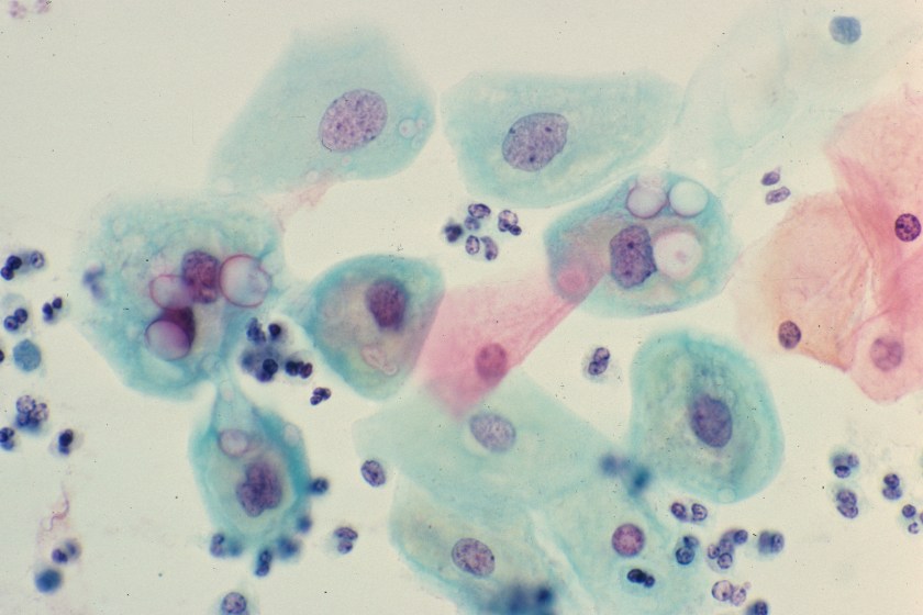
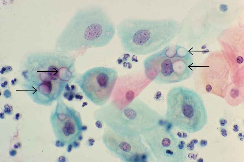
Today, I provide Safety Eyes training to lab managers. I use photographs of safety issues- pictures taken of real issues in the lab. Using these visual aids hones their ability to see the issues the next time they walk through the lab, and it raises safety awareness for everyone. If you lead a laboratory, and if you do not focus on safety, I understand that. However, you should understand that is important to make a change- develop those Safety Eyes and advance your safety culture in a way your staff can appreciate and support.


Scungio 1

-Dan Scungio, MT(ASCP), SLS, CQA (ASQ) has over 25 years experience as a certified medical technologist. Today he is the Laboratory Safety Officer for Sentara Healthcare, a system of seven hospitals and over 20 laboratories and draw sites in the Tidewater area of Virginia. He is also known as Dan the Lab Safety Man, a lab safety consultant, educator, and trainer.

Microbiology Case Study: Young Girl with Community-Acquired Pneumonia

A young girl presented to the emergency department with 6 days of cough, congestion, and intermittent fever up to 102°F. Respiratory viral testing, blood cultures, and a chest x-ray were ordered. The patient was found to be RSV positive and sent home with oral steroids. Two days later she presented the emergency department once again with worsening respiratory symptoms and a positive blood culture with the following organism (Figure 1).

streppneu1
Figure 1. Gram stain demonstrating Gram-positive cocci in pairs and chains.

Our patient developed a Streptococcus pneumoniae superinfection and bacteremia in conjunction with RSV pneumonia.

Identification

S. pneumoniae is a Gram-positive cocci that forms “lancet” shaped pairs on Gram stain (Figure 1). Due to pretreatment with antibiotics, our Gram stain shows some pairs, but many Gram variable chains of cocci as well. S. pneumoniae grows as alpha hemolytic colonies on 5% sheep blood, chocolate, and CAN (colistin nalidixic acid) agar in 12-18 hours, where it forms umbelicated colonies with a characteristic navel-like depression in the middle due to autolysins produced by the bacterium. Some serotypes of S. pneumoniae, primarily serotype 3, have a mucoid phenotype seen in Figure 3. S. pneumoniae is a member of the Streptococcus mitis group, but due to its pathogenic potential it has always been singled out. This is accomplished using two biochemical tests: bile solubility testing with 10% deoxycholate, which dissolves colonies of S. pneumoniae but not those of other Viridans group streptococci, and optochin disc testing, to which S. pneumoniae is sensitive while other Virdians group streptococci are resistant (Figures 2 and 3). Many molecular assays have trouble differentiating S. pneumoniae from S. mitis group due to their similarities on a nucleotide and protein level, so biochemical testing is still a mainstay of organism identification.

streppneu2
Figure 2. Growth of α-hemolytic bacterial colonies on 5% sheep blood agar. Zone around the disc indicates the organism is optochin susceptible.


streppneu3
Figure 3. Growth of mucoid, α-hemolytic bacterial colonies on 5% sheep blood agar. The mucoid colony morphology suggests this isolate is likely serotype 3.

Clinical Significance

S. pneumoniae is known to cause a variety of clinical manifestations in children, from community acquired pneumonia and acute otitis media to bacteremia and meningitis. S. pneumoniae is also a colonizer of the upper respiratory tract; approximately 21% of children in developed countries and 90% of children in developing countries are asymptotically colonized. Due to the high rates of S. pneumoniae colonization in children, direct urine antigen testing is inappropriate, as it cannot distinguish asymptomatic carriage from invasive disease. S. pneumoniae direct antigen detection from CSF has been shown to have < 30% sensitivity and offers no benefit over a routine cytospin Gram stain.

Vaccination in children

Around 2000 the first S. pneumoniae vaccine became available. PCV7 was a heptavalent conjugate vaccine which provided protection from the 7 most common S. pneumoniae serotypes known to cause invasive disease (4, 6B, 9V, 14, 18C, 19F, and 23F). Routine vaccination of children was a huge success which reduced the incidence of invasive pneumococcal disease attributed to vaccine strains by 99%. An indirect benefit of the PCV7 vaccine was that adults >65 years of age saw a 92% decrease in invasive pneumococcal disease caused by PCV7 serotypes, despite not being vaccinated themselves, because of reduced transmission of S. pneumoniae from children to adults. Due to the selective pressure of the vaccine, non-vaccine serotypes of S. pneumoniae such as 19A subsequently became the predominant causes of invasive streptococcal disease. In 2010, a 13-valent pneumococcal conjugate vaccine (PCV13) was FDA approved. It includes all seven S. pneumoniae serotypes contained in PCV7, plus six additional serotypes (1, 3, 5, 6A, 7F, and 19A). PCV13 provides coverage against 2/3 of all serotypes responsible for invasive pneumococcal disease in children under 5 years of age.

Follow up

The patient had an uneventful hospital stay. All subsequent blood cultures were negative and susceptibility testing found the patient’s S. pneumoniae isolate to be susceptible to penicillin, cefotaxime, and clindamycin. The patient and was discharged home after 24 hours of observation with a 7 day course of amoxicillin.


References:

  • Manual of Clinical Microbiology, 11th edition
  • Pediatric Red Book, 2015 Report of the Committee on Infectious Diseases, 30th edition


-Erin McElvania TeKippe, Ph.D., D(ABMM), is the Director of Clinical Microbiology at Children’s Medical Center in Dallas Texas and an Assistant Professor of Pathology and Pediatrics at University of Texas Southwestern Medical Center.

Mycobacterium chimaera Infections in Cardiac Surgery Patients

A group of physicians from the University of Iowa have started a dialogue about Mycobacterium chimaera infections in patients who have undergone cardiac surgery. It seems as though the bacteria finds its way to the patients via a heater-cooler device used during their procedure. If you’re a micro tech or a pathologist and you come across a cardiac patient who has a fever of unknown origin, night sweats, loss of energy, and failure to gain weight, M. chimaera is something to keep in mind.

You can read more about this issue in the original blog post from the Iowa infectious disease doctors, Maryn McKenna’s write up over at Nat Geo, and the CDC guidance paper.

Routine Pap Smear from a 33 Year Old Woman

You are reviewing a routine Papanicoloaou smear from a 33 year-old female. She has no complaints and appears healthy. A representative field from her Pap smear is shown here. What organism is the most likely cause of the morphologic changes seen here?

pap1

  1. Chlamydia trachomatis
  2. Human papillomavirus
  3. Trichomonas vaginalis
  4. Leptothrix
  5. Gardnerella vaginalis


The diagnosis in this case is Chlamydial infection. Genital infection by Chlamydia trachomatis is the most common sexually transmitted disease in the world. In women, chlamydial infection of the cervix is frequently asymptomatic, as it was in this patient. If untreated, however, the infection can lead to pelvic inflammatory disease, infertility and ectopic pregnancy.

Chlamydia trachomatis is a tiny, gram-negative bacterium that exists in two different forms: the elementary body, which is the infectious form, and the reticulate body, which is the replicative form. While Chlamydial organisms are too small to be visible with a gram stain, large, glassy inclusions containing both reticulate bodies and elementary bodies are occasionally clearly visible within cells, as seen in the squamous epithelial cells in this image (see arrows).

pap2


Krafts

-Kristine Krafts, MD, is an Assistant Professor of Pathology at the University of Minnesota School of Medicine and School of Dentistry and the founder of the educational website Pathology Student.

Introns, and Exons, and Splicing … Oh My!

One of the first things students of biology learn is basic molecular theory or, the central dogma of biology. The central dogma of biology states that “DNA makes RNA and RNA makes protein.” We know that the process of converting DNA to protein is not one involving magic, but rather more intricate methods termed, transcription and translation. For a quick review, check out my post A Primer on DNA and the Consequences of Mutation.

As we take a closer look at transcription, we will investigate how DNA is converted into pre-messenger RNA and ultimately into messenger RNA. The manner in which this occurs is termed RNA Splicing. Following splicing, mRNA is transported out of the nucleus to indirectly assist with protein synthesis.

In most eukaryotic genes, coding regions are interrupted by non-coding regions. Coding regions are termed exons, where alternatively non-coding regions are called introns. The coding region made up of exons is also called the open reading frame. During the process of transcription, DNA of an entire gene is translated into RNA, more specifically termed pre-messenger RNA (pre-mRNA). Pre-mRNA includes both exons and introns. Through a process, called RNA splicing, the introns of the gene are removed allowing the exons to be joined forming a continuous coding sequence. The continuous coding sequence is referred to as messenger RNA. mRNA contains a 5’ cap consisting of 7 methyl guanosine, a 3’ polyA tail, and is ready for translation into protein.

EXONS–Coding regions of the DNA that make up the mature messenger RNA

INTRONS–Non-coding regions that are removed from the DNA sequence to form mature messenger RNA

PROMOTER–Found at the 5′ end of the DNA sequence and functions to initiate transcription

RNA Splicing Made Easy

DNA: 5′–xxxxxx11111111111222222222222222222233333–3′

Transcribed to…

Pre-mRNA:5′–11111111111222222222222222222233333–3′

Further processing/splicing …

Mature mRNA: 5′ 7 Me G–111122222222222222233333–AAAAA 3′


Test your Knowledge!

  1. What is the function of the 5’ 7-methyl guanosine cap?
  2. What enzyme is responsible for the addition of the polyA tail to the 3’ end of the mRNA?
  3. What structures of the mRNA are translated into protein?


Answers

  1. The 7-methyl guanosine cap is recognized as a site that signals the start of translation
  2. Polyadenylate polymerase adds the multiple adenines to the 3’ end of an mRNA sequence
  3. Introns are spliced out of the open reading frame and the exons are translated into protein


L Noll Image_small

-LeAnne Noll, BS, MB(ASCP)CM is a molecular technologist at Children’s Hospital of Wisconsin and was recognized as one of ASCP’s Top Five from the 40 Under Forty Program in 2015.

Microbiology Case Study: 45 Year Old Woman with Subarachnoid Hemorrhage

Case History:

A 45 year-old woman was hospitalized for a bilateral subarachnoid hemorrhage with right-sided intraventricular hemorrhage secondary to a basilar artery aneurysm. On hospital day 2, after endovascular coiling of her aneurysm, she developed seizure-like activity. She was found to have new bilateral thalamic and brainstem infarcts with increased hydrocephalus and had an external ventricular drain placed.

She showed some minimal neurological improvements over the next 2 weeks until hospital day 17 when her mental status acutely declined and she developed a fever, leukocytosis, and meningeal signs. 8cc of cloudy, yellow fluid was collected via lumbar puncture. Analysis of the fluid showed pleocytosis (cell count of 54K) with a neutrophil predominance and markedly elevated protein.

CSF Gram stain
CSF Gram stain
CSF Gram stain
CSF Gram stain
CSF aerobic culture on blood agar
CSF aerobic culture on MacConkey

Laboratory Identification:

Initial review of the spinal fluid gram stain showed many polymorphonuclear leukocytes and what appeared to be paired gram negative cocci suspicious for Neisseria meningitidis. However, many organisms lacked the more characteristic “kidney-bean” shape. Further inspection of the gram stain revealed many gram negative organisms which more closely resembled bacilli. Given the patient’s history and clinical course, it was determined the pathological agent was most likely a gram negative coccobacillus. On culture, the bacteria formed smooth, round, opaque colonies on Blood and Chocolate agar and was lactose non-fermenting on MacConkey agar. The bacterial colonies were also oxidase negative.

Mass spectrometry identified the organism as Acinetobacter radioresistens.

Rare gram positive appearing organisms of similar shape were also located on the gram stain. This demonstrates that Acinetobacter is known to occasionally retain the crystal violet stain leading to cases of initial misidentification.

Discussion:

Acinetobacter radioresistens is one of about 30 species of bacteria included in the Acinetobacter genus. Acinetobacter is characterized as a gram negative, aerobic coccobacillus which is non-motile, non-fermentative, and oxidase-negative. It grows well on standard aerobic media and typically forms smooth, round, mucoid colonies at 37°C. Acinetobacter is a water organism which preferentially colonizes aquatic, humid, and tropical environments; perhaps accounting for the increased incidence of Acinetobacter infections between the months of July and October.

While there have been reported cases of community-acquired Acinetobacter pneumonia in Southeast Asia and Australia, in most areas of the world Acinetobacter is known primarily as an agent of nosocomial infections. Studies show that an estimated 33% of healthcare workers are colonized with Acinetobacter and that it is one of the most prevalent bacteria isolated from the white coats of medical students. Despite its ubiquity in hospitals, Acinetobacter infections are relatively rare. Many patients may be colonized with it, but Acinetobacter only usually causes disease in immunocompromised and/or critically-ill patients with long hospitalizations. At particular risk are ventilated patients supported with multiple lines, drains, and catheters. Acinetobacter is reported as the pathological agent in a small percent of ventilator-associated pneumonias, central line-associated bloodstream infections, catheter-associated urinary tract infections, and surgical site infections. It is also recognized as a cause of nosocomial meningitis in neurosurgical patients with external ventricular drains, especially those with a history of intracranial hemorrhage and recent prior antibiotic therapy.

Acinetobacter infections are of particular concern because several species demonstrate resistance to many antimicrobials. Acinetobacter baumanni, the species responsible for the majority of Acinetobacter infections, has demonstrated resistance to 1st-3rd generation cephalosporins, macrolides, penicillins, and aminoglycosides. Because these infections are robust and difficult to treat, patients with Acinetobacter infections have a 25-75% mortality risk depending on the site of their infection and their baseline cardiopulmonary and immune status. Currently, carbapenems are considered the gold standard treatment.

-Elaine Amoresano, MD, is a 1st year anatomic and clinical pathology resident at the University of Vermont Medical Center.


Wojewoda-small

-Christi Wojewoda, MD, is the Director of Clinical Microbiology at the University of Vermont Medical Center and an Assistant Professor at the University of Vermont.


Zika Virus

If you’re an infectious disease geek and you’ve been following the news, you know that the Zika virus is causing a pandemic in Central and South America and is linked to cases of microcephaly in those regions. It’s gotten bad enough fast enough that the CDC has issued travel warnings for pregnant women and women of child-bearing age.

If you’d like to get up-to-date on this outbreak, check out Maryn McKenna’s blog on National Geographic. Also, the CDC has great information for clinicians and laboratory professionals.


Microbiology Case Study: Immunocompromised 65 Year Old Man

Case History:

A 65 year old man presents to the emergency room with acute onset back pain. Of note, the man was diagnosed with Burkitt’s lymphoma two months prior and had recently received a course of chemotherapy. During the workup for his back pain, a chest CT is obtained and reveals a 2 cm pulmonary nodule in the left upper lobe with a surrounding “groundglass halo” highly suspicious for invasive fungal involvement. A fine needle aspiration (FNA) of the nodule is performed and tissue is sent for histopathologic examination as well as bacterial and fungal culture.

Chest CT showing a 2 cm nodule in the left upper lobe
Silver stain of the FNA specimen showing ribbon-like fungal elements with rare septations
Scotch-tape preparation reveals rarely septate hyphae with internodal rhizoids and pyriform sporangia supported by funnel-shaped apophyses.


Laboratory Identification:

One rapidly growing white colony was identified that became grey over time. The colony was a “lid-lifter” that began pushing at the lid after only a few days. Microscopically, the organisms had broad hyphae with single and branching sporangiophores. At the ends of the sporangiophores there were pyriform, or pear-shaped, sporangia sitting atop funnel-shaped apophyses. Rhizoids were found to be internodal, or arising from the hyphae between the sporangiophores. Based on this morphology, the fungal organisms were identified as Lichtheimia corymbifera complex (formally Absidia corymbifera).

Discussion:

Lichtheimia corymbifera is an organism within the phylum Zygomycota and is one of two pathogenic species in the genus Lichtheimia. This organism is known as an uncommon cause of Zygomycosis and is only implicated in approximately 5% of cases. As in most cases of Zygomycosis, exemplified in our patient, Lichtheimia corymbifera most often affects immunocompromised patients. It is ubiquitous in the environment and is associated with decaying plant matter and soil. Disease is caused by inhalation of spores.

Important points for laboratory identification:

Lichtheimia

  • Growth at 35-37°C (capable of growth up to 50°C)
  • Inhibited by media containing cycloheximide
  • Internodal rhizoids
  • Pyriform sporangia
  • Apophysis

Compared to other common Zygomycetes:

Mucor

  • No rhizoids
  • Round sporangia
  • No apophysis

Rhizopus

  • Nodal rhizoids (directly opposite of the sporangiophores)
  • Round sporangia
  • No apophysis


-Britni Bryant, MD is a 2nd year anatomic and clinical pathology resident at the University of Vermont Medical Center.


Wojewoda-small

-Christi Wojewoda, MD, is the Director of Clinical Microbiology at the University of Vermont Medical Center and an Assistant Professor at the University of Vermont.

The Value of Electives (Both Internal and External) During Pathology Residency

It’s been a while since my last blog. I haven’t had as much time and energy as I would like this past year. For now, I’ll just say…appreciate all that your chief residents do because much more time and effort lies beneath the surface than everyone is able to see.

But the topic for this blog is the value of electives during pathology residency. Our programs vary with respect to electives in terms of number and ability to take them externally or not. My previous program had five electives that could be taken internally or externally. However, external electives did not receive salary support and I didn’t take any electives although I could’ve during my two years in Chicago. Since we had a decent number of electives, many residents scheduled them internally during fourth year to have lighter months to study for boards although a handful did utilize them during earlier years for external electives.

My current program has two and we do receive salary support with external electives. For my first, I had an extra month of hematopathology internally because I wanted to see another perspective on one of my chosen subspecialties. Internal electives are good to spend more focused time in an area of subspecialty interest that you may have for fellowship. It also allows for the opportunity to develop deeper relationships with the faculty who will most likely be writing your letters of recommendation for that fellowship. They may also provide you with the opportunity to become more involved in research and/or publications (eg – book chapters, case reports, research articles) with a mentor and these are all helpful in enhancing your fellowship and future job applications and build up your CV.

Currently, I’m on an external elective at the institution where I’ll do both my hematopathology and molecular genetic fellowships. I’m laying the groundwork for molecular hematopathology research now that hopefully results in data analysis over the ensuing months to culminate in an abstract submission for the American Society of Hematology (ASH) which has a deadline only a month after I start fellowship. I also want to use this time away to get to know people at my future program better, prepare for my eventual move here, and study for boards. Hopefully, I’ll also get a sense of the daily work flow as I am also attending signouts and intra- and interdepartmental conferences so that I can manage my time as efficiently as I can from day 1 of fellowship. I really like the culture and people here, but that’s subject matter for a future blog. I also am enjoying the benefits of attending inter-program activities as TMC is the largest medical center in the world with active interaction and collaboration between member hospitals. Not so much in my case since I obtained both my consecutive fellowships last year as a PGY-3, but for many, the value of an early external elective is that it can be seen it as an “audition” rotation to obtain a desired fellowship. You may even be able to ask for an interview before you finish (which saves you time and money). I also have some friends who were offered fellowship spots at the end of their elective rotation because they impressed the fellowship director. Obtaining fellowship positions is pretty competitive and there tends to be fewer spots than there are for residency. And in many cases, positions are not even available if an internal candidate is chosen early (even during their PGY-1) so anything to augment your fellowship application is a plus.

Now that I’ve mentioned external electives, I’d like to give some advice on setting up an external elective. First, start as EARLY as possible! Even a year or more before isn’t too early to ask about getting the ball rolling. Start preparing and updating your CV from your PGY-1 as you’ll need this for both external elective and fellowship applications. Scan and keep a PDF of all your vaccinations and work-related health requirements (eg – PPD/Quantiferon results, flu vaccine, hepatitis B testing, MMR and hepatitis B antibody titers, and N-95 fit testing) as well because its likely you’ll also have to include this in your external elective application.

I initiated the elective rotation request about a half year prior to the actual rotation. And even then, that was not early enough and everything came down to the wire. It’s far more complicated than when we applied for elective rotations as a medical student and takes much more time. Since we are now physicians, you are more than likely required to have at least a medical permit in that state to rotate and this process can take a while. Also the back-and-forth between program coordinators and the respective GME departments can appear glacial at times, and in my case, was the main cause of delay. I hit several delays at obtaining paperwork (especially between Christmas and New Year’s when many staff were off at both programs, my medical school, and the Texas Medical Board where I needed paperwork from). It can be time-consuming to have to make multiple phone calls, and often, the process cannot be completed until its antecedent step has been approved. I know residents who have had to postpone external rotations because paperwork between GME departments (eg – PLAs or malpractice agreements) were not in place in time. Maintaining constant and open communication between all parties involved is a must and sometimes easier said than done the more people that are involved.

In addition to obtaining the state medical permit (which generally requires an application fee; in my case, $135), there may be other requirements that are also time-consuming and financially expensive. You may be required to do pre-employment-type health screening (in my case, a $36 urine drug screen) at your own cost as you are not a true employee. I also had to become ACLS certified (at $120, despite being BLS certified and a former American Red Cross CPR instructor). But since I’m going to be a fellow here, I can get it reimbursed through my GME funds and would have to do it later anyways so I might as well do it now. But if you are not doing an elective at your future fellowship institution, it’s good to find out what items may incur cost in your application for your elective since you are not likely to get reimbursed and so you can decide whether those expenses are acceptable. One way to defray costs is to apply for grants such as the ASCP subspecialty grant which is administered to six residents twice a year (Jan/Aug). In fact, the next deadline is this Friday, Jan 15th! You can find more information on how to apply at http://www.ascp.org/Residents/Resident-Grants.


Chung

-Betty Chung, DO, MPH, MA is a fourth year resident physician at Rutgers – Robert Wood Johnson University Hospital in New Brunswick, NJ.